Article

Activation of Adrenoceptor Alpha-2 (ADRA2A) Promotes Chemosensitization to Carboplatin in Ovarian Cancer Cell Lines

College of Science, Health and Pharmacy, Roosevelt University, 1400 N Roosevelt Blvd, Schaumburg, IL 60173, USA
*
Authors to whom correspondence should be addressed.
These authors contributed equally to this work.
Curr. Issues Mol. Biol. 2023, 45(12), 9566-9578; https://doi.org/10.3390/cimb45120598
Submission received: 30 October 2023 / Revised: 23 November 2023 / Accepted: 26 November 2023 / Published: 28 November 2023
(This article belongs to the Special Issue Advanced Molecular Solutions for Cancer Therapy)

Abstract

:
Recurrence of ovarian cancer (OvCa) following surgery and standard carboplatin/paclitaxel first-line therapy signifies poor median progression-free survival (<24 months) in the majority of patients with OvCa. The current study utilized unbiased high-throughput screening (HTS) to evaluate an FDA-approved compound library for drugs that could be repurposed to improve OvCa sensitivity to carboplatin. The initial screen revealed six compounds with agonistic activity for the adrenoceptor alpha-2a (ADRA2A). These findings were validated in multiple OvCa cell lines (TYKnu, CAOV3, OVCAR8) using three ADRA2A agonists (xylazine, dexmedetomidine, and clonidine) and two independent viability assays. In all the experiments, these compounds enhanced the cytotoxicity of carboplatin treatment. Genetic overexpression of ADRA2A was also sufficient to reduce cell viability and increase carboplatin sensitivity. Taken together, these data indicate that ADRA2A activation may promote chemosensitivity in OvCa, which could be targeted by widely used medications currently indicated for other disease states.

1. Introduction

Ovarian cancer (OvCa) commonly presents at advanced stages and has a poor prognosis, with an overall 5-year survival rate of 50% across all subtypes and stages at diagnosis [1]. The standard of care for high-grade serous ovarian carcinoma (HGSOC), the most prevalent subtype of OvCa, includes cytoreductive debulking surgery and chemotherapy consisting of a platinum agent such as carboplatin (CP) and a taxane such as paclitaxel [2]. Following active transport, CP promotes DNA lesions that include direct platinum–DNA adducts as well as inter- and intra-strand crosslinking that ultimately result in single- and double-strand breaks, inducing apoptosis [3]. In a clinical context, the use of CP/paclitaxel (CarboTaxol) allows for the ablation of unresectable or visually undetectable tumor cells to prevent recurrence of the disease.
While the majority of patients initially respond to CP therapy, the majority will relapse and inevitably succumb to their disease [2,4,5]. Multiple mechanisms have been attributed to CP treatment failure, including dysfunctional transport, compensatory DNA repair, and the upregulation of pro-survival and anti-apoptotic pathways [6]. The use of paclitaxel in combination to impede progression through the M phase of cells that evade apoptosis despite DNA adduct accumulation by carboplatin may be central to its ability to enhance tumor cell ablation following surgery and to improve the initial therapeutic response. Conceivably, drugs targeting other processes that evade apoptosis or promote survival may complement the current therapeutic approaches [7]. In line with this, paclitaxel has been used in conjunction with carboplatin as first-line therapy to improve survival in patients with advanced HGSOC due to its inhibition of microtubule disassembly and subsequent cell cycle arrest to further enhance the apoptosis caused by CP-dependent DNA damage [8]. Emerging molecular targets have been identified that may delay recurrence following first-line treatment in OvCa, such as VEGF (e.g., bevacizumab) and PARP (e.g., olaparib), which have provided new therapeutic strategies to improve progression-free survival [9]. In the case of PARP inhibitors, suppression of nucleotide excision repair may further enhance the efficacy of CP by preventing the removal of intrastrand crosslinking to promote apoptosis [6,10]; however, PARylation is critical in regulating numerous processes involved in DNA repair and replication that may be involved in preventing the proliferation of CP-treated cells [11]. While promising, in both cases of VEGF and PARP inhibitors, it has been observed that treatment failure may still occur due to resistance that develops in response to therapy [12,13]. Thus, it is critical to continue expanding the repertoire of nonredundant treatment options available to clinicians that may facilitate personalized treatment plans to overcome any acquired drug resistance and to prolong patient survival.
To expedite the implementation of novel targetable mechanisms for drug therapies, a drug repurposing paradigm is often used in the drug discovery process for many disease states. This approach allows researchers to proceed more rapidly to clinical trials to determine efficacy and has facilitated the common use of multiple drugs as mainstays in current cancer therapy, such as tamoxifen in breast cancer [14,15]. Many of the drugs repositioned for off-label use in these contexts have been evaluated as a result of retrospective cohort analyses that have identified associations between disease onset or progression and medications taken for other indications [14]. Although these retrospective associations have, in many cases, facilitated preclinical experimentation and the pursuit of clinical trials to determine efficacy, their application in new clinical contexts is not always successful [14]. An alternative strategy for drug repurposing has been through the prospective preclinical assessment of drugs already on the market. In particular, in vitro high-throughput screening (HTS) using curated compound libraries to evaluate thousands of compounds rapidly [16] allows for lead-compound identification and in vivo testing prior to clinical application. Indeed, a recent study using a synthetic lethality HTS approach identified FDA-approved drugs that may improve the response to ATP8B-dependent cisplatin resistances in OvCa [17], indicating the potential use of these compounds (e.g., tranilast) in ATP8B-overexpressing ovarian tumors. Using a similar unbiased strategy, the current study aimed to enhance the cytotoxicity of carboplatin to prevent treatment resistance by utilizing an FDA-approved compound library.

2. Materials and Methods

2.1. Reagents

High-throughput screening (HTS) was performed using the DiscoveryProbe L1021 FDA-approved drug library (APExBIO L1021). Carboplatin (#c2538), xylazine (#x1126), dexmedetomidine (#sml0956), and clonidine (#c7897) were purchased from Sigma-Aldrich.

2.2. Cell Lines

The OvCa cell lines TYKnu, CAOV3, and OVCAR8 were a gift from Dr. Ernst Lengyel and Dr. Iris Romero of the University of Chicago Section of Gynecologic Oncology (Chicago, IL, USA). All cell lines were passaged at least 3 times prior to their use in experiments. Cell lines were banked in liquid nitrogen and were confirmed to be mycoplasma-free by the donating investigators as described previously [18]. Viability was confirmed to be 98% or higher (using trypan exclusions with Countess II, ThermoFisher, Waltham MA, USA) prior to counting and seeding for the HTS as well as the primary and secondary assays. All three cell lines were maintained in RPMI1640 with L-glutamine (VWR) supplemented with 10% FB-Essence (VWR), 1% penicillin/streptomycin (VWR), 1% MEM vitamins (VWR), and 1% nonessential amino acids (VWR).

2.3. High-Throughput Screening (HTS)

The initial high-throughput screening (HTS) was performed using a 96-well format with the DiscoveryProbe L1021 FDA-approved drug library (ApexBio, Houston, TX, USA). The compound library was reconstituted to 2 mM (in DMSO) so that the addition of 5 µL of each drug would yield 50 µM per drug per well. A total of 88 compounds were tested per plate, with 1584 compounds tested. For each plate, internal controls included 4 untreated samples and 4 samples treated only with the IC50 of carboplatin. Due to their relatively high resistance to carboplatin, as indicated by the highest IC50 of the cell lines tested, OVCAR8 cells were used in the pilot HTS assay. Cells were seeded at 3.0 × 10⁴/well and allowed to grow for 24 h prior to treatment with carboplatin (at IC50) ± the L1021 drug library for 48 h. After 48 h of treatment, the MTT assay was performed (as described below) to determine the effect on cell viability. Absorbance measurements were then processed in which the absorbance of each drug with carboplatin was compared to the positive carboplatin control (i.e., the IC50 of carboplatin alone). Any well which was observed to increase the cytotoxicity of carboplatin by 80% or higher was considered a potential lead (marked as red; Figure 1E and Supplementary Table S1). These statistics were then cross-referenced per well per plate to identify compounds of interest for subsequent primary (MTT viability) and secondary (colony formation) confirmatory assays.

2.4. Primary Assay—MTT Viability Assay

The thiazolyl blue tetrazolium bromide (MTT) colorimetric metabolism assay was used to determine cell viability (GoldBio, St. Louis, MO, USA; #T-030). Briefly, cells were seeded at 3.0 × 10⁴/well by dispensing 100 µL of a 3.0 × 10⁵/mL suspension of cells into a 96-well plate (Pipette.com, San Diego, CA, USA) and allowed to adhere for 24 h prior to treatment. Media was aspirated and fresh growth media was added so that the addition of carboplatin ± secondary compounds would result in a final volume of 200 µL. Carboplatin was diluted fresh from 10 mM stocks (never previously thawed) to working stocks to allow for 5 µL of drug to result in the desired concentrations (ranging from 10–5000 µM). In the context of the HTS, only the IC50 calculated for OVCAR8 was used in combination with each drug. In the context of the MTT confirmatory experiments, a dose range was used to reestablish the IC50 per plate (carboplatin alone) and to determine IC50 values when used in combination with each alpha-2 agonist (set to 50 µM and 100 µM per agonist). Multichannel pipettes were used to limit variability between wells. After 48 h of treatment, 12 mM MTT was added to a final concentration of 300 µM and allowed to incubate for 1.5 h (OVCAR8) or 2 h (CAOV3 or TYKnu). After incubation, the plates were decanted and patted dry, and MTT-reduced formazan crystals were suspended in 200 µL DMSO. The absorbance was then measured at 560 nm using SpectraMax ABS (MolecularDevices, San Jose, CA, USA). The absorbance values were exported via Microsoft Excel and analyzed using GraphPad Prism (described below).

2.5. Secondary Assay—Colony-Formation Clonogenicity Assay

As a secondary assay to confirm the impact of each drug combination on cell chemoresistance, a colony formation (clonogenicity) assay was performed. Cells were seeded at 500 (0.5 × 10³; OVCAR8) or 1000 (1.0 × 10³; CAOV3, TYKnu) cells per well of a 6-well plate (VWR, Radnor, PA, USA) and allowed to recover for 24 h. After 24 h, drug treatments were initiated. At days 5 and 7, the drug treatments were refreshed, with a full media change performed on day 5. On day 10, the cells were washed with 1× PBS, fixed using 4% paraformaldehyde for 15 min, washed again with 1× PBS, then stained using crystal violet (Sigma-Aldrich, St. Louis, MO, USA, #61135) for 30 min. The crystal violet was then moved to a waste container, and the cells were rinsed with water until the stain was washed away. Plates were then imaged using ChemiDoc XR+ (Bio-Rad, Hercules, CA, USA) while colonies were counted manually using ImageJ/FIJI (NIH, Bethesda, MD, USA), and statistical analyses were performed using GraphPad Prism.

2.6. Plasmid Transfection

To overexpress adrenoceptor alpha-a2, transfection was performed using pmEGFP-ADRA2A on the pcDNA3.1 backbone (Addgene, Watertown, MA, USA; plasmid #190753). pmEGFP-ADRA2A was a gift from Dave Piston of Washington University. OVCAR8 cells were seeded at 4.0 × 10⁶ per 10 cm plate and allowed to proliferate for 24 h prior to transfection. Cells were transfected using Lipofectamine 2000 (Invitrogen, Waltham, MA, USA) according to the manufacturer’s instructions. At 24 h post transfection, cells were seeded into either 96-well or 6-well plates for the MTT or colony formation assays, respectively. After 48 h post transfection, treatments were initiated as described for the primary and secondary assays. For quantitative real-time PCR, the reaction was scaled down to a 6-well format with 1× reaction per well.

2.7. Quantitative Real-Time Polymerase Chain Reactions

The expression of ADRA2A following transfection was confirmed by quantitative real-time PCR (qRT-PCR). RNA isolation was performed using Trizol (Invitrogen) with chloroform extraction. Complementary DNA (cDNA) synthesis was performed using the High-Capacity cDNA Reverse Transcription Kit (Applied Biosystems, Waltham, MA, USA). qRT-PCR was performed using the POWRUP SYBR Master Mix (Life Technologies, Carlsbad, CA, USA) according to the manufacturer’s instructions. Primers were designed using PrimerBank (NP_000672, #194353969c2), and 25 nmole quantities of DNA oligonucleotides were purchased from IDT (Newark, NJ, USA; primers were prepared as LabReady, normalized to 100 µM in IDTE pH 8.0). Forward primer: AGA AGT GGT ACG TCA TCT CGT; Reverse primer: CGC TTG GCG ATC TGG TAG A.

2.8. TCGA Assessment of ADRA2A Expression in Ovarian Cancer

Expression of ADRA2A in ovarian cancer was determined using the Gene Expression Profiling Interactive Analysis (GEPIA) database (GEPIA.cancer-pku.cn). The “OV” (ovarian cancer) dataset from The Cancer Genome Atlas (TCGA) was selected with a log2FC cutoff of 1 and a p-value cutoff of 0.01. The data are presented on a log scale.

2.9. Statistical Analysis

Data was analyzed using Graphpad Prism 10 (Graphpad, La Jolla, CA, USA) by 1-way ANOVA with the post hoc Tukey t-test or Student’s unpaired t-test as appropriate. For the IC50 calculations, XY data tables and graphs were used. Absorbance measurements from the MTT assays were imported into Prism. The data were then transformed to log(x) and normalized as a percentage within each group. The transformed/normalized data were then modeled using nonlinear regression (dose response: log(inhibitor) with four parameters, with constraints for “top” (100%) and “bottom” (0%)). Representative curve fit and calculated IC50 values are presented for each experiment. All experiments were repeated at least once.

3. Results

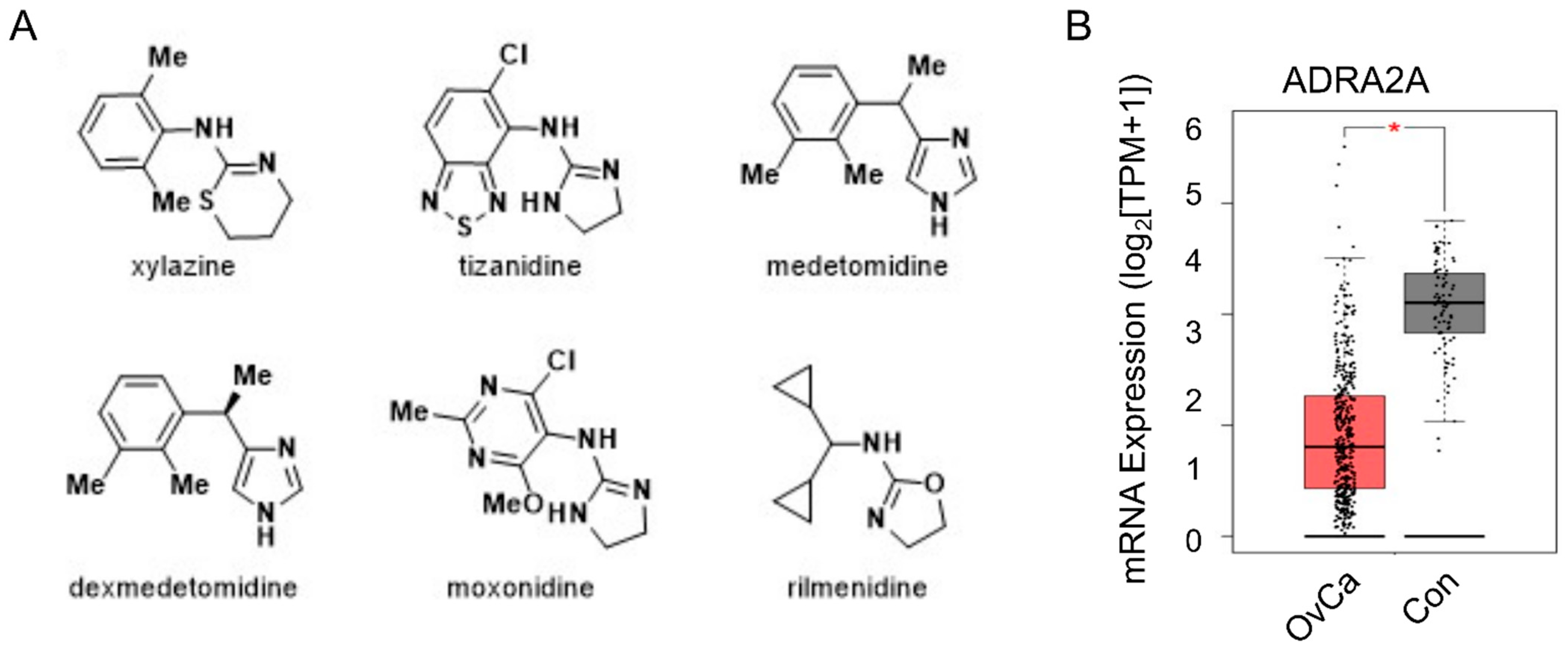
3.1. High-Throughput Screening of an FDA-Approved Drug Library Identifies Adrenoceptor Alpha-2 (ADRA2A) as a Potential Target for Enhancing Carboplatin Treatment

To identify novel compounds that could enhance the cytotoxicity of carboplatin and, thus, possibly reduce resistance to the current standard of care, three cell lines representative of HGSOC were assessed for their potential resistance to carboplatin (CP). Treatment of TYKnu, CAOV3, and OVCAR8 cells with increasing doses of carboplatin for 48 h revealed significant resistance of OVCAR8 cells compared with either CAOV3 or TYKnu, with an IC50 value of 412 µM (Figure 1A) compared with either 316 µM (Figure 1B) or 66 µM (Figure 1C), respectively, as determined by the MTT cell viability assay. OVCAR8 cells were therefore selected for high-throughput screening (HTS) utilizing an FDA-approved drug library (APExBIO L1021). Cells were treated with the relative IC50 of CP (400 µM) ± compound library (50 µM) for 48 h prior to the MTT assay (Figure 1D; example plate analysis shown in Figure 1E). Of the 1651 compounds tested, 278 demonstrated a marked enhancement in cell death that increased the cytotoxicity of CP by more than 80% compared with the carboplatin-only controls (Figure 1F and Supplementary Table S1).
Of the 278 compounds observed to have potential synergy with CP, 4 were identified to be agonists of the ADRA2A receptor, including xylazine, tizanidine, medetomidine, and dexmedetomidine (Figure 2A). Two agents with mixed agonism of ADRA2A and significant I1-imidazoline receptor activity were also identified: moxonidine and rilmenidine (Figure 2A). This trend indicated that ADRA2A could be an important molecular target that may regulate chemosensitivity in OvCa tumor cells. Evaluation of ADRA2A expression in patient tumors from the TCGA database revealed that ADRA2A is significantly repressed in OvCa tumors (Figure 2B), further supporting the hypothesis that inhibition of its signaling may be important in tumor development or progression.

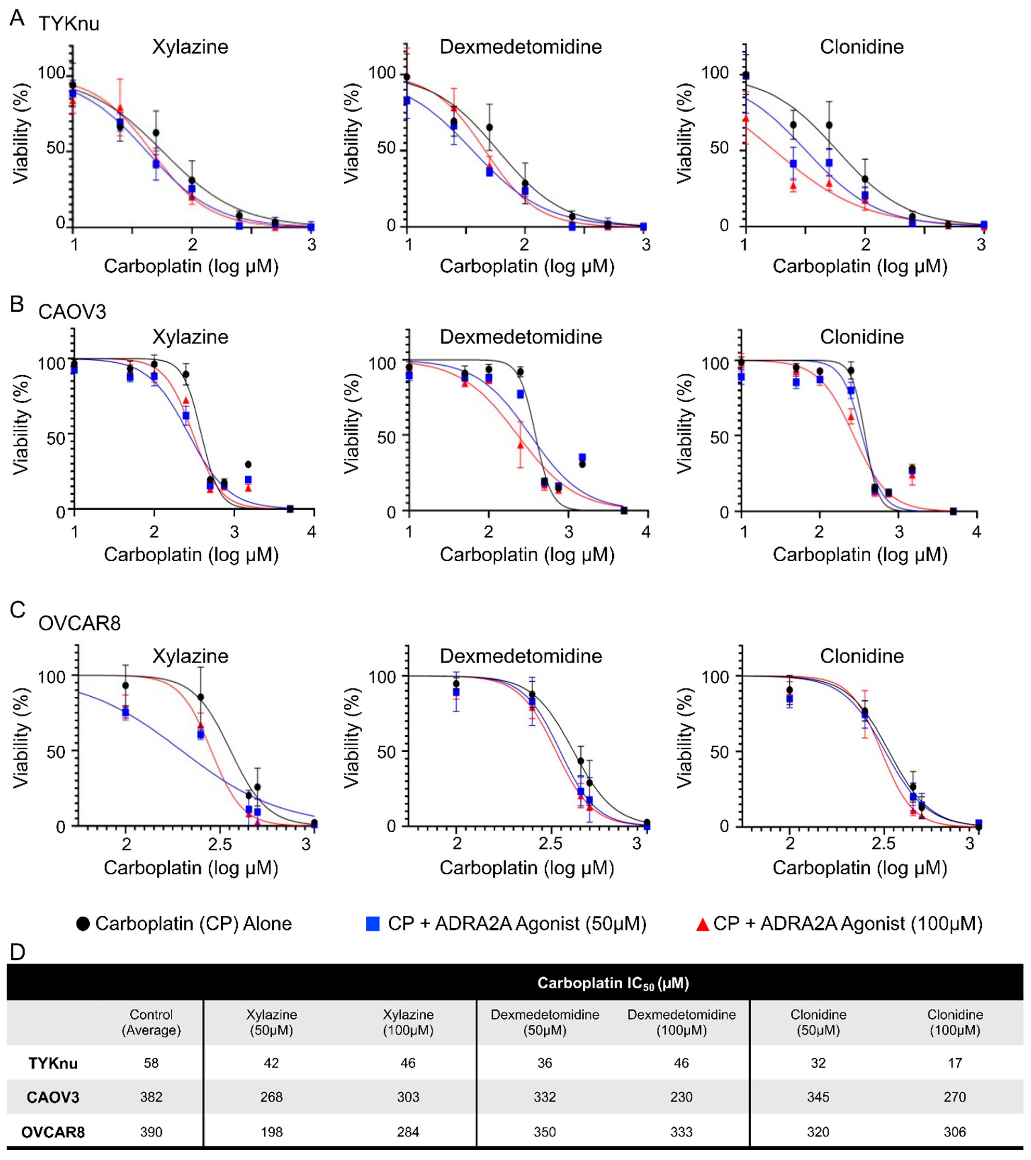
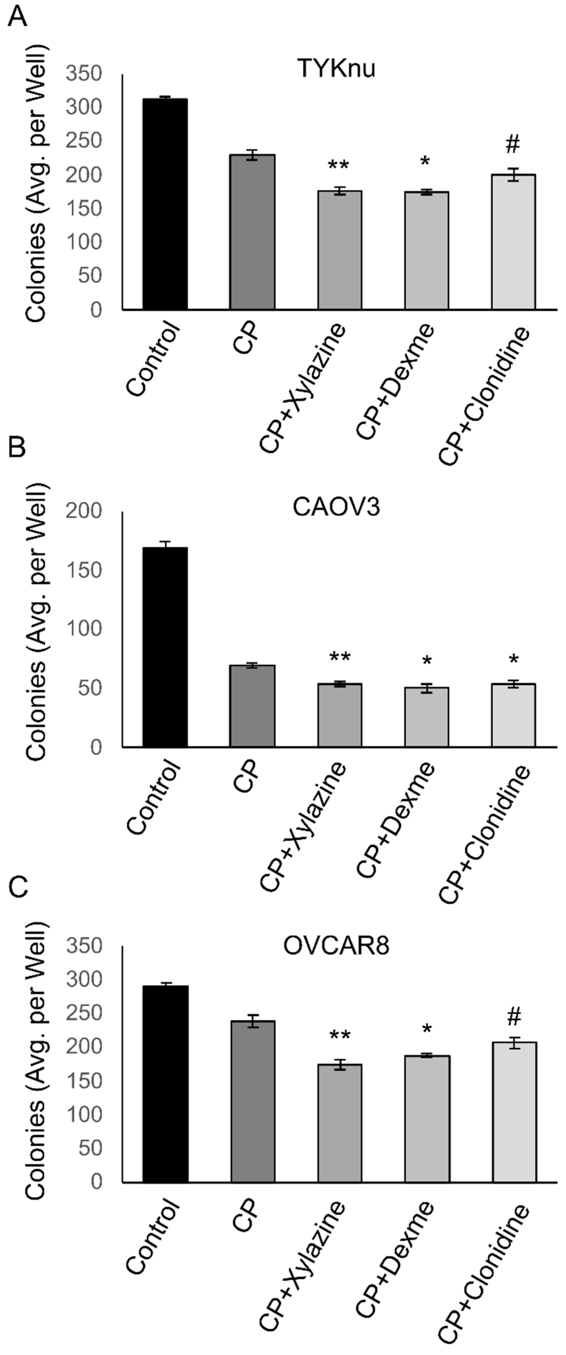
3.2. Activation of ADRA2A Using Pharmacologic and Genetic Manipulation Enhances Carboplatin Toxicity in Viability and Clonogenicity Assays

To validate the findings from the HTS in OVCAR8 cells, TYKnu, CAOV3, and OVCAR8 cells were treated with increasing doses of carboplatin ± the ADRA2A agonists xylazine, dexmedetomidine, or clonidine. While not observed in the HTS, clonidine was chosen due to its widespread clinical use for hypertension, relatively high specificity, and low toxicologic profile. Primary confirmatory MTT assays in these cell lines demonstrated consistent reductions in the IC50 of carboplatin compared with the carboplatin-only groups when 50 or 100 µM of any of the ADRA2A agonists was co-administered (Figure 3A–C). This effect was largely dose-dependent and was consistent across the three cell lines tested (Figure 3D). To determine whether this effect was assay-specific, a secondary colony-formation clonogenicity assay was performed using the approximate IC50 value of CP (1 µM) in combination with each compound. Extended treatment of colonies for 10d with a lower dose (25 µM) of each compound suppressed colony formation, an effect that was particularly significant with xylazine and dexmedetomidine (Figure 4A–C). Clonidine appeared to have weak activity in this assay at the test dose and reached significance in CAOV3 cells (Figure 4B) but did not meet statistical significance in TYKnu (Figure 4A) or OVCAR8 (Figure 4C) cells, despite showing a similar trend [p < 0.09].
To further evaluate our findings using the pharmacologic activation of ADRA2A, genetic upregulation of ADRA2A in the most resistant cell line (OVCAR8) was performed (Figure 5A). Following the ectopic upregulation of ADRA2A, cells were subjected to either MTT or colony formation assays. ADRA2A overexpression in OVCAR8 cells strikingly reduced the IC50 of carboplatin needed in these cells (Figure 5B). Consistently, ADRA2A expression induced an overall inhibition of colony formation in OVCAR8 cells and promoted a further reduction in colonies compared with the carboplatin-only group (Figure 5C). In line with the enhancement in carboplatin sensitivity observed using xylazine, dexmedetomidine, and clonidine, these findings suggest that ADRA2A activation and the consequent inhibition of cyclic AMP (cAMP) production may downregulate processes necessary for tumor cells to escape carboplatin-induced cytotoxicity. Thus, taken together, these data further support the idea that the molecular activity of ADRA2A may suppress the resistance of OvCa tumor cells to carboplatin.

4. Discussion

In summary, an unbiased HTS cytotoxicity assay identified that several modulators of ADRA2A activity could enhance the sensitivity of OvCa cell lines to carboplatin. The initial HTS data was confirmed using the compounds xylazine, dexmedetomidine, and clonidine sourced from a different manufacturer for validation in both primary (MTT viability) and secondary (colony formation) assays. These compounds demonstrated a reduction in the IC50 of CP in all three cell lines tested by the MTT viability assay. When combined with CP, xylazine and dexmedetomidine also showed a marked reduction in the number of colonies formed by all three cell lines. While the use of the three compounds in these assays was largely consistent, clonidine did not reach the same statistical significance compared with xylazine and dexmedetomidine in the colony formation assay in two of the cell lines. This could be in part due to differences in its potency, a difference in specificity for the three α2 subunits (A–C), or its mixed activity on the I1 imidazoline receptor; however, in the case of the latter, dexmedetomidine has also been shown to be pharmacologically active in a nonadrenergic I1 receptor manner [19]. In addition to assessing the impact of α2 B and C subunits on OvCa chemoresistance, further evaluation of the I1 receptor may indicate its usefulness as a target in OvCa and may indicate whether increased specificity for ADRA2A may alter its potential anticancer effects; this may be best accomplished using moxonidine and rilmenidine due to their higher selectivity for I1 compared with clonidine [19]. Regardless, the data presented herein indicate the potential for ADRA2A-modulating antihypertensives (clonidine) or sedative analgesics (dexmedetomidine), which are relatively well tolerated and have well defined pharmacokinetics, to serve as an adjunct to improve the efficacy of carboplatin. This notion was further supported by the genetic upregulation of ADRA2A, which demonstrated a striking sensitization of the highly chemoresistant OVCAR8 cell line to carboplatin.
While the present study did not delineate the exact mechanisms underlying the potential CP sensitization, multiple downstream effectors of ADRA2A have been well described in multiple cancers. ADRA2A belongs to the adrenoceptor alpha-2 family of GPCRs coupled to the inhibitory G-alpha subunit (Gi) that suppresses adenylyl cyclase activity, thereby suppressing the cyclization of AMP to cAMP [20]. cAMP is a key second messenger that impacts multiple intracellular pathways, canonically regulating protein kinase A (PKA) and cAMP-response-element-binding protein (CREB) activity, but also calcium-dependent signaling and mitogenic pathways such as MAPK [21]. While the functional role of cAMP varies by tissue, in OvCa, cAMP signaling has been associated with enhanced proliferation and the suppression of pro-apoptotic signals, frequently associated with the activity of PKA or CREB1 (reviewed in [22]). Notably, PKA activation has been observed to promote platinum resistance in ovarian and other cancers [22,23,24], and CREB1 inhibition potently sensitized OvCa tumor cells to cisplatin [25]. In a recent study, ADRA2A overexpression was observed to suppress proliferation and to promote apoptosis through the repression of PI3K/AKT/mTOR signaling in cervical cancer [26], another signaling axis associated with platinum resistance in several contexts [6]; however, to date, no work has demonstrated the impact of ADRA2A activity in OvCa development or treatment specifically. Further evaluation is needed given the many potential downstream pathways that may be attributed to the effects of ADRA2A activation observed in the current work.
To date, sparse data exist that show the clinical impact of the role of alpha adrenoceptor activity in tumor development and progression. Although antihypertensives, including beta blockers, have not been associated with OvCa patient survival [27], the effects of alpha adrenoceptor activation have yet to be evaluated in this clinical context. As clonidine has been used to mitigate symptoms in OvCa patients following bilateral oophorectomy [28], it may be possible to retrospectively analyze whether the inclusion of this drug may delay the time to recurrence in these patients. To increase the clinical relevancy of our findings, an evaluation of whether ADRA2A activity is relevant in a more physiologic model system that includes components of the OvCa tumor microenvironment (TME) (as performed in a recent study [18]) is required to improve the translatability of these findings, considering that cancer-associated fibroblasts in the TME are thought to significantly contribute to platinum resistance in multiple contexts [29,30]. This may be especially important given recent findings observing that dexmedetomidine induced IL-6 secretion from stromal stellate cells and pro-tumorigenic signaling in hepatocellular cancer cells [31], although whether this is relevant to that distinct TME is to be determined. Moreover, the beta-adrenergic antagonist propranolol has been shown to suppress tumor growth by inhibiting tumor angiogenesis and promoting T-cell recruitment in sarcoma [32], and the nonselective blockade of beta-adrenoceptors in prostate and pancreatic cancer in vivo suppressed tumor growth [33]. Additionally, beta-adrenergic activation has been associated with remodeling of the extracellular matrix to promote invasive phenotypes in breast cancer [34], which may be, in part, due to the potential cAMP-mediated epithelial–mesenchymal transition (EMT) [35,36]; however, whether ADRA2A inhibition of this signaling pathway results in suppression of the EMT is still to be determined. Those results and ours here suggest a complex relationship among adrenergic receptor subtypes and the processes involved in tumor development in a context-dependent manner. Further evaluation of the effects of specific adrenoceptor isoforms in tumor and stromal cells within the TME may provide a clearer understanding of the possible therapeutic value of targeting noradrenergic signaling. Lastly, in vivo data utilizing an orthotopic approach combined with a CarboTaxol regimen will ultimately determine whether the agents identified here would be suitable for inclusion in the therapy for advanced HGSOC.

5. Conclusions

Our HTS findings indicate that a number of currently approved compounds may enhance the chemosensitivity of OvCa cells to CP. Of these, ADRA2A-targeted compounds were highly represented in the HTS. Validation of several of these compounds supports the hypothesis that the activation of ADRA2A may reduce the chemoresistance of OvCa tumor cells and improve the response to CP. These pharmacologic data were in agreement with the effect of genetically upregulating ADRA2A, which resulted in the sensitization of chemoresistant OvCa cells to CP.

Supplementary Materials

The following supporting information can be downloaded at: https://www.mdpi.com/article/10.3390/cimb45120598/s1, Table S1: Viability of OvCa cells in response to carboplatin and the FDA -approved compound library. Percentage of viable cells treated with carboplatin + the FDA library relative to the carboplatin-only control.

Author Contributions

The manuscript was written through contributions from all authors. M.E.O. and P.C.H. led the conceptual and experimental design as well as the development of the methodology. H.A., A.G., D.R., G.R., L.D. and P.C.H. performed and analyzed the experiments, and contributed to the manuscript writing and editing. M.E.O. contributed to the manuscript writing, editing, and revision. M.E.O. and P.C.H. contributed equally to the supervision of the study. All authors have read and agreed to the published version of the manuscript.

Funding

This work was supported, in part, by the American Association of Colleges of Pharmacy (New Investigator Award to PCH) and the Howard Hughes Medical Institute (Inclusive Excellence Grant to Roosevelt University).

Institutional Review Board Statement

Not applicable.

Informed Consent Statement

Not applicable.

Data Availability Statement

Original data is available from the corresponding authors upon request.

Acknowledgments

The authors thank Melissa Hogan and Lawrence A. Potempa for their support in acquisition of funds for the reagents and equipment used to procure data in this manuscript.

Conflicts of Interest

There are no conflict of interest to report.

References

  1. Matulonis, U.A.; Sood, A.K.; Fallowfield, L.; Howitt, B.E.; Sehouli, J.; Karlan, B.Y. Ovarian cancer. Nat. Rev. Dis. Primers 2016, 2, 16061. [Google Scholar] [CrossRef] [PubMed]
  2. Kim, A.; Ueda, Y.; Naka, T.; Enomoto, T. Therapeutic strategies in epithelial ovarian cancer. J. Exp. Clin. Cancer Res. 2012, 31, 14. [Google Scholar] [CrossRef] [PubMed]
  3. Rabik, C.A.; Dolan, M.E. Molecular mechanisms of resistance and toxicity associated with platinating agents. Cancer Treat. Rev. 2007, 33, 9–23. [Google Scholar] [CrossRef] [PubMed]
  4. Pignata, S.; Cecere, S.C.; Du Bois, A.; Harter, P.; Heitz, F. Treatment of recurrent ovarian cancer. Ann. Oncol. 2017, 28 (Suppl. 8), viii51–viii56. [Google Scholar] [CrossRef] [PubMed]
  5. du Bois, A.; Reuss, A.; Pujade-Lauraine, E.; Harter, P.; Ray-Coquard, I.; Pfisterer, J. Role of surgical outcome as prognostic factor in advanced epithelial ovarian cancer: A combined exploratory analysis of 3 prospectively randomized phase 3 multicenter trials: By the Arbeitsgemeinschaft Gynaekologische Onkologie Studiengruppe Ovarialkarzinom (AGO-OVAR) and the Groupe d’Investigateurs Nationaux Pour les Etudes des Cancers de l’Ovaire (GINECO). Cancer 2009, 115, 1234–1244. [Google Scholar] [PubMed]
  6. Zhou, J.; Kang, Y.; Chen, L.; Wang, H.; Liu, J.; Zeng, S.; Yu, L. The Drug-Resistance Mechanisms of Five Platinum-Based Antitumor Agents. Front. Pharmacol. 2020, 11, 343. [Google Scholar] [CrossRef] [PubMed]
  7. Bayat Mokhtari, R.; Homayouni, T.S.; Baluch, N.; Morgatskaya, E.; Kumar, S.; Das, B.; Yeger, H. Combination therapy in combating cancer. Oncotarget 2017, 8, 38022–38043. [Google Scholar] [CrossRef]
  8. Alqahtani, F.Y.; Aleanizy, F.S.; El Tahir, E.; Alkahtani, H.M.; AlQuadeib, B.T. Paclitaxel. Profiles Drug Subst. Excip. Relat. Methodol. 2019, 44, 205–238. [Google Scholar]
  9. Wang, Q.; Peng, H.; Qi, X.; Wu, M.; Zhao, X. Targeted therapies in gynecological cancers: A comprehensive review of clinical evidence. Signal Transduct. Target. Ther. 2020, 5, 137. [Google Scholar] [CrossRef]
  10. Pines, A.; Vrouwe, M.G.; Marteijn, J.A.; Typas, D.; Luijsterburg, M.S.; Cansoy, M.; Hensbergen, P.; Deelder, A.; de Groot, A.; Matsumoto, S.; et al. PARP1 promotes nucleotide excision repair through DDB2 stabilization and recruitment of ALC1. J. Cell Biol. 2012, 199, 235–249. [Google Scholar] [CrossRef]
  11. Ray Chaudhuri, A.; Nussenzweig, A. The multifaceted roles of PARP1 in DNA repair and chromatin remodelling. Nat. Rev. Mol. Cell Biol. 2017, 18, 610–621. [Google Scholar] [CrossRef]
  12. Montemagno, C.; Pages, G. Resistance to Anti-angiogenic Therapies: A Mechanism Depending on the Time of Exposure to the Drugs. Front. Cell Dev. Biol. 2020, 8, 584. [Google Scholar] [CrossRef]
  13. Klotz, D.M.; Wimberger, P. Overcoming PARP inhibitor resistance in ovarian cancer: What are the most promising strategies? Arch. Gynecol. Obstet. 2020, 302, 1087–1102. [Google Scholar] [CrossRef] [PubMed]
  14. Cavalla, D. Using human experience to identify drug repurposing opportunities: Theory and practice. Br. J. Clin. Pharmacol. 2019, 85, 680–689. [Google Scholar] [CrossRef] [PubMed]
  15. Jordan, V.C. 50th anniversary of the first clinical trial with ICI 46,474 (tamoxifen): Then what happened? Endocr. Relat. Cancer 2021, 28, R11–R30. [Google Scholar] [CrossRef] [PubMed]
  16. Ediriweera, M.K.; Tennekoon, K.H.; Samarakoon, S.R. In vitro assays and techniques utilized in anticancer drug discovery. J. Appl. Toxicol. 2019, 39, 38–71. [Google Scholar] [CrossRef] [PubMed]
  17. Mariniello, M.; Petruzzelli, R.; Wanderlingh, L.G.; La Montagna, R.; Carissimo, A.; Pane, F.; Amoresano, A.; Ilyechova, E.Y.; Galagudza, M.M.; Catalano, F.; et al. Synthetic Lethality Screening Identifies FDA-Approved Drugs that Overcome ATP7B-Mediated Tolerance of Tumor Cells to Cisplatin. Cancers 2020, 12, 608. [Google Scholar] [CrossRef] [PubMed]
  18. Kenny, H.A.; Hart, P.C.; Kordylewicz, K.; Lal, M.; Shen, M.; Kara, B.; Chen, Y.J.; Grassl, N.; Alharbi, Y.; Pattnaik, B.R.; et al. The Natural Product beta-Escin Targets Cancer and Stromal Cells of the Tumor Microenvironment to Inhibit Ovarian Cancer Metastasis. Cancers 2021, 13, 3931. [Google Scholar] [CrossRef]
  19. Bousquet, P.; Hudson, A.; García-Sevilla, J.A.; Li, J.-X. Imidazoline Receptor System: The Past, the Present, and the Future. Pharmacol. Rev. 2020, 72, 50–79. [Google Scholar] [CrossRef]
  20. Pettinger, W.A.; Jackson, E.K. α2-Adrenoceptors: Challenges and Opportunities-Enlightenment from the Kidney. Cardiovasc. Ther. 2020, 2020, 2478781. [Google Scholar] [CrossRef]
  21. Ahmed, M.B.; Alghamdi, A.A.A.; Islam, S.U.; Lee, J.-S.; Lee, Y.-S. cAMP Signaling in Cancer: A PKA-CREB and EPAC-Centric Approach. Cells 2022, 11, 2020. [Google Scholar] [CrossRef]
  22. Kilanowska, A.; Ziółkowska, A.; Stasiak, P.; Gibas-Dorna, M. cAMP-Dependent Signaling and Ovarian Cancer. Cells 2022, 11, 3835. [Google Scholar] [CrossRef] [PubMed]
  23. Stewart, D.J. Mechanisms of resistance to cisplatin and carboplatin. Crit. Rev. Oncol. Hematol. 2007, 63, 12–31. [Google Scholar]
  24. Dempke, W.; Voigt, W.; Grothey, A.; Hill, B.T.; Schmoll, H.-J. Cisplatin resistance and oncogenes—A review. Anticancer. Drugs 2000, 11, 225–236. [Google Scholar] [CrossRef] [PubMed]
  25. Dimitrova, N.; Nagaraj, A.B.; Razi, A.; Singh, S.; Kamalakaran, S.; Banerjee, N.; Joseph, P.; Mankovich, A.; Mittal, P.; DiFeo, A.; et al. InFlo: A novel systems biology framework identifies cAMP-CREB1 axis as a key modulator of platinum resistance in ovarian cancer. Oncogene 2017, 36, 2472–2482. [Google Scholar] [CrossRef] [PubMed]
  26. Wang, W.; Guo, X.; Dan, H. alpha2A-Adrenergic Receptor Inhibits the Progression of Cervical Cancer through Blocking PI3K/AKT/mTOR Pathway. Onco Targets Ther. 2020, 13, 10535–10546. [Google Scholar] [CrossRef] [PubMed]
  27. Huang, T.; Townsend, M.K.; Dood, R.L.; Sood, A.K.; Tworoger, S.S. Antihypertensive medication use and ovarian cancer survival. Gynecol. Oncol. 2021, 163, 342–347. [Google Scholar] [CrossRef]
  28. Van Le, L.; McCormack, M. Enhancing Care of the Survivor of Gynecologic Cancer: Managing the Menopause and Radiation Toxicity. Am. Soc. Clin. Oncol. Educ. Book 2016, 35, e270–e275. [Google Scholar] [CrossRef]
  29. Long, X.; Xiong, W.; Zeng, X.; Qi, L.; Cai, Y.; Mo, M.; Jiang, H.; Zhu, B.; Chen, Z.; Li, Y. Cancer-associated fibroblasts promote cisplatin resistance in bladder cancer cells by increasing IGF-1/ERbeta/Bcl-2 signalling. Cell Death Dis. 2019, 10, 375. [Google Scholar] [CrossRef]
  30. Wang, L.; Li, X.; Ren, Y.; Geng, H.; Zhang, Q.; Cao, L.; Meng, Z.; Wu, X.; Xu, M.; Xu, K. Cancer-associated fibroblasts contribute to cisplatin resistance by modulating ANXA3 in lung cancer cells. Cancer Sci. 2019, 110, 1609–1620. [Google Scholar] [CrossRef]
  31. Chen, P.; Luo, X.; Dai, G.; Jiang, Y.; Luo, Y.; Peng, S.; Wang, H.; Xie, P.; Qu, C.; Lin, W.; et al. Dexmedetomidine promotes the progression of hepatocellular carcinoma through hepatic stellate cell activation. Exp. Mol. Med. 2020, 52, 1062–1074. [Google Scholar] [CrossRef] [PubMed]
  32. Fjaestad, K.Y.; Rømer, A.M.; Goitea, V.; Johansen, A.Z.; Thorseth, M.L.; Carretta, M.; Engelholm, L.H.; Grøntved, L.; Junker, N.; Madsen, D.H. Blockade of beta-adrenergic receptors reduces cancer growth and enhances the response to anti-CTLA4 therapy by modulating the tumor microenvironment. Oncogene 2022, 41, 1364–1375. [Google Scholar] [CrossRef] [PubMed]
  33. Zhou, S.; Li, J.; Yu, J.; Wang, Y.; Wang, Z.; He, Z.; Ouyang, D.; Liu, H.; Wang, Y. Tumor microenvironment adrenergic nerves blockade liposomes for cancer therapy. J. Control. Release 2022, 351, 656–666. [Google Scholar] [CrossRef] [PubMed]
  34. Conceicao, F.; Sousa, D.M.; Paredes, J.; Lamghari, M. Sympathetic activity in breast cancer and metastasis: Partners in crime. Bone Res. 2021, 9, 9. [Google Scholar] [CrossRef]
  35. Shaikh, D.; Zhou, Q.; Chen, T.; Ibe, J.C.; Raj, J.U.; Zhou, G. cAMP-dependent protein kinase is essential for hypoxia-mediated epithelial-mesenchymal transition, migration, and invasion in lung cancer cells. Cell Signal 2012, 24, 2396–2406. [Google Scholar] [CrossRef]
  36. Wang, X.; Cui, H.; Lou, Z.; Huang, S.; Ren, Y.; Wang, P.; Weng, G. Cyclic AMP responsive element-binding protein induces metastatic renal cell carcinoma by mediating the expression of matrix metallopeptidase-2/9 and proteins associated with epithelial-mesenchymal transition. Mol. Med. Rep. 2017, 15, 4191–4198. [Google Scholar] [CrossRef]
Figure 1. Determination of carboplatin resistance in OvCa cell lines. Cells were plated and allowed to adhere for 24 h prior to treatments. Cells were treated with 12 escalating doses of carboplatin (0–5000 µM) for 48 h ((A) OVCAR8, (B) CAOV3, and (C) TYKnu cell lines). N = 4 for each data point. (D) Experimental timeline for treating cells with carboplatin at its determined IC50 along with the DiscoveryProbe L1021 FDA-approved drug library (ApexBio) (88 agents per plate at 50 µM final concentration). (E) MTT absorbance was normalized to the carboplatin-only group to determine the impact on cell death. Numbers presented refer to the percentage (%) of carboplatin-only control. Wells marked in red had an absorbance of <20% when divided by the average absorbance of the carboplatin-only controls for each plate. (F) The frequency distribution of compounds + carboplatin binned in 20% increments, with 100% representing having an absorbance equal to the carboplatin-only controls. The number of compounds identified in each category is shown on the right.
Cimb 45 00598 g001
Figure 2. Identification of ADRA2A as a potential therapeutic target in OvCa. (A) High-throughput screening (HTS) of carboplatin + the FDA library identified several alpha-2 adrenoceptor (ADRA2A) agonists that increased cytotoxicity by >80% in combination with carboplatin; these included xylazine, tizanidine, medetomidine, and dexmedetomidine. Drugs with partial ADRA2A agonism, namely moxonidine and rilmenidine, were also identified in the HTS. (B) Gene (mRNA) expression of ADRA2A in TCGA clinical data (GEPIA database) comparing control tissue (gray) to ovarian tumor tissue (pink). * p < 0.01.
Cimb 45 00598 g002
Figure 3. Validation of ADRA2A agonists in enhancing carboplatin-dependent cytotoxicity. Cells were plated and allowed to adhere for 24 h prior to treatments. Cells were treated with escalating doses of carboplatin (0–5000 µM) ± 50 or 100 µM of the indicated ADRA2A agonist for 48 h ((A) TYKnu, (B) CAOV3, (C) OVCAR8). N = 4 for each data point. (D) Carboplatin IC50 values for carboplatin ± ADRA2A agonists (xylazine, dexmedetomidine, clonidine) for each cell line.
Cimb 45 00598 g003
Figure 4. ADRA2A activation further enhances the inhibition of clonogenicity by carboplatin. Cells were allowed to adhere for 24 h prior to treatments. Cells were treated with 1 µM carboplatin ± 25 µM of the indicated ADRA2A agonist for 10 days. ((A) TYKnu, (B) CAOV3, (C) OVCAR8). N = 3 for each condition. * p < 0.05; ** p < 0.01; # p < 0.07. Dexme = dexmedetomidine; CP = carboplatin.
Cimb 45 00598 g004
Figure 5. Overexpression of ADRA2A promotes chemosensitivity to carboplatin. (A) Expression of ADRA2A mRNA at 48 h following the transfection of OVCAR8 cells with pmEGFP-ADRA2A (N = 3). (B) MTT viability assay of OVCAR8 cells ± pmEGFP-ADRA2A treated with an escalating dose curve of carboplatin (0–5000 µM) for 48 h (N = 6). (C) Colony formation assay of OVCAR8 cells ± pmEGFP-ADRA2A [A2] treated with or without carboplatin (1 µM) for 10 days (N = 6). *** p < 0.005.
Cimb 45 00598 g005
Disclaimer/Publisher’s Note: The statements, opinions and data contained in all publications are solely those of the individual author(s) and contributor(s) and not of MDPI and/or the editor(s). MDPI and/or the editor(s) disclaim responsibility for any injury to people or property resulting from any ideas, methods, instructions or products referred to in the content.

