Article

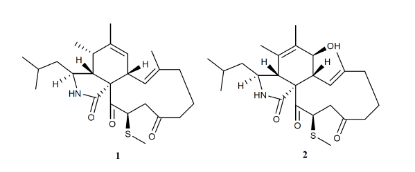
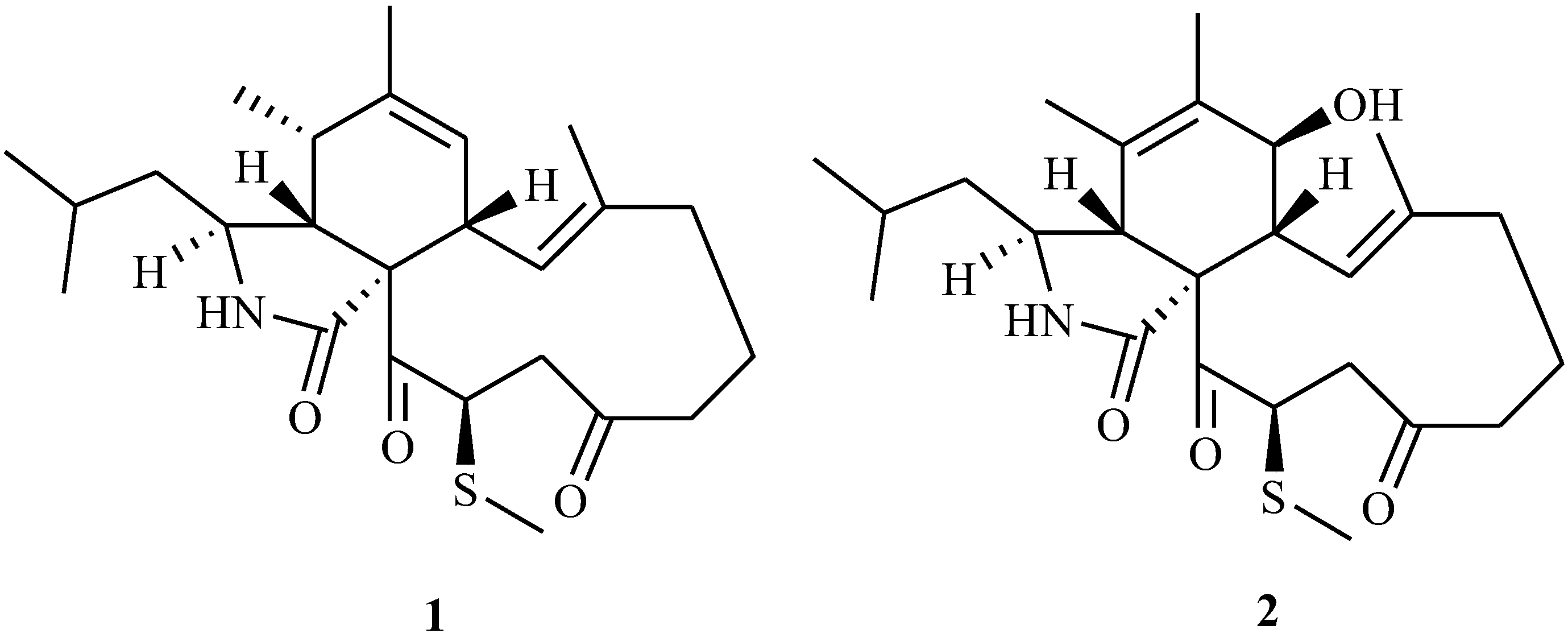
Methylthio-Aspochalasins from a Marine-Derived Fungus Aspergillus sp.

1
Institute of Marine Biology, Ocean College, Zhejiang University, Hangzhou 310058, China
2
CAS Key Laboratory of Tropical Marine Bio-resources and Ecology, South China Sea Institute of Oceanology, Chinese Academy of Sciences, Guangzhou 510301, China
*
Author to whom correspondence should be addressed.
Mar. Drugs 2014, 12(10), 5124-5131; https://doi.org/10.3390/md12105124
Submission received: 16 May 2014 / Revised: 20 September 2014 / Accepted: 22 September 2014 / Published: 30 September 2014

Abstract

:
Two novel aspochalasins, 20-β-methylthio-aspochalsin Q (named as aspochalasin V), (1) and aspochalasin W (2), were isolated from culture broth of Aspergillus sp., which was found in the gut of a marine isopod Ligia oceanica. The structures were determined on the basis of NMR and mass spectral data analysis. This is the first report about methylthio-substituted aspochalasin derivatives. Cytotoxicity against the prostate cancer PC3 cell line and HCT116 cell line was assayed using the MTT method. Apochalasin V showed moderate activity at IC50 values of 30.4 and 39.2 μM, respectively.

Graphical Abstract

1. Introduction

Fungi are important producers of marine natural products and most of them were found from algae and sponge [1]. Actually various microorganisms including fungi were found in the gut of marine isopods [2,3,4,5,6]. These gut symbioses showed some ecological roles such as chemical defense [7]. Therefore gut microbes including fungi may be an interesting source of bioactive marine natural products. In fact, an actinomycete strain IFB-A01, capable of producing new neuraminidase inhibitors, was isolated from the gut of the shrimp Penasus orientalis [8]. In our investigation into new bioactive metabolites of marine gut fungi, one strain (Z-4) was isolated from the gut of the marine isopod Ligia oceanica. In its culture broth two aspochalasins containing unusual methylthiol groups were found. Aspochalasins are a subgroup of cytochalasans, consisting of a macrocylic ring, isoindolone moiety and a 2-methyl-propyl side chain. So far, more than 20 compounds, including aspochalasins A–U and Z [9,10,11,12,13,14], have been reported. These compounds contained almost the same carbon skeleton and their structural diversity included a double bond shift (C5/C6 or C6/C7) in the isoindolone unit together with ketone, hydroxyl, methoxyl and double bond reduction in the macrocylic ring. Aspochalasins showed various bioactivities such as antibiotic [15,16,17], cytotoxicity [13,18], anti-HIV [19], TNF-alpha [20] and melanogenesis [21] inhibitors. Herein, we describe the isolation, structural elucidation, and cytotoxic activity of these two new aspochalasins.

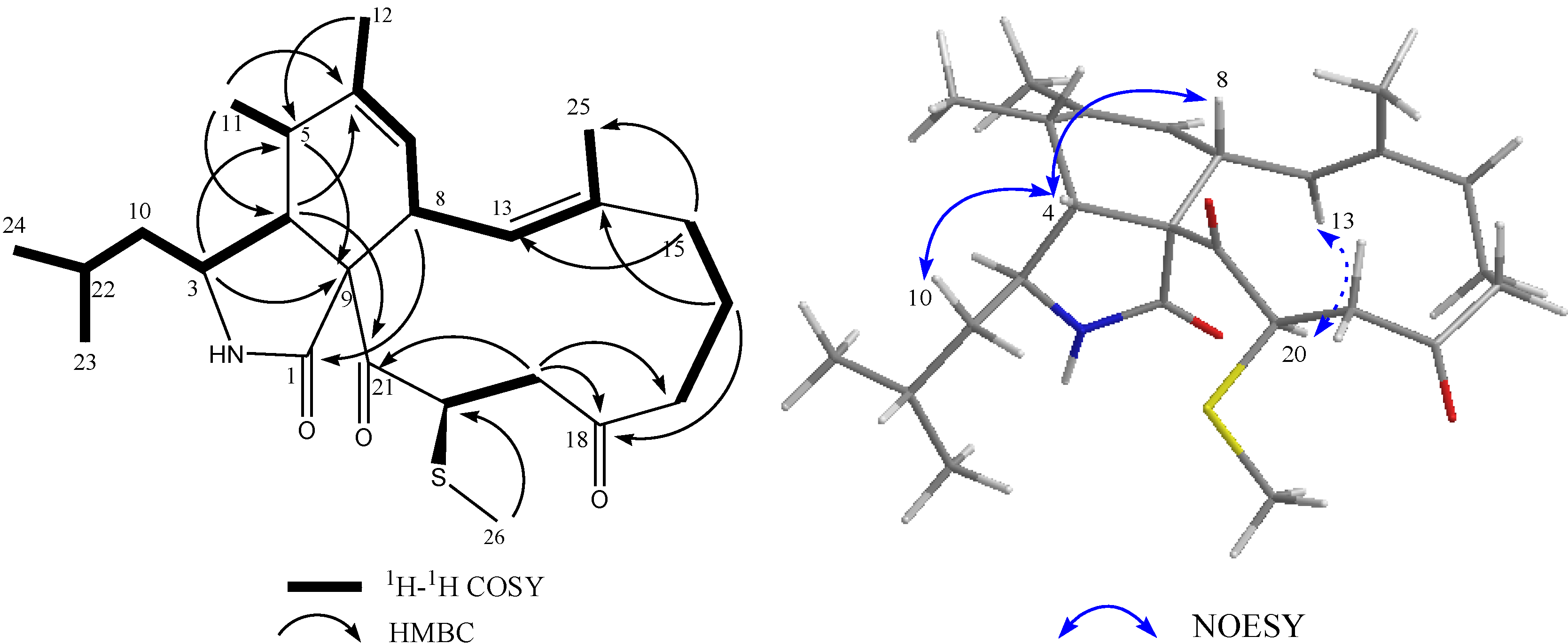
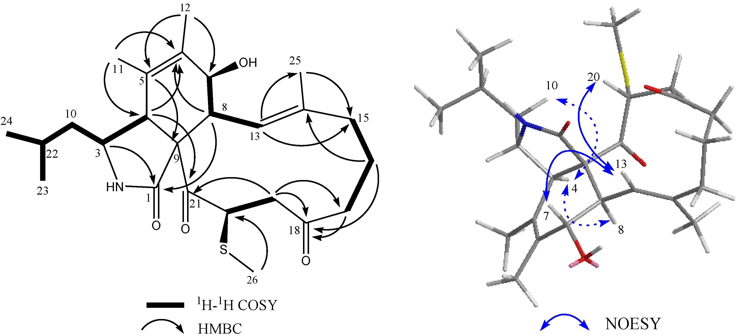
2. Results and Discussion

Compound 1 was isolated as a colorless solid. The ESI-HRMS showed a quasi-molecular ion peak at m/z 432.2572 for [M + H]+, indicating the molecular formula of compound 1 is C25H37NO3S (calcd. 432.2528 for C25H38NO3S) by combination with 1D NMR data. IR spectrum showed obvious peaks at 3350, 1687 and 1700 cm−1. 13C NMR data together with DEPT 135 and DEPT 90 spectra revealed that 1 contained twenty-five carbons, including two ketone carbonyl signals (δC 208.1 and 200.5), one amide carbonyl signal (δC 177.2), four olefinic carbons (δC 141.8, 138.9, 126.5 and 125.2), five aliphatic methylene carbons (δC 49.9, 44.4, 41.9, 40.2 and 19.8), six aliphatic methine carbons (δC 54.1, 52.7, 46.2, 46.0, 37.2 and 25.7), six methyl signals (δC 24.2, 21.9, 20.1, 15.1, 14.0 and 10.8) together with one quaternary carbon (δC 66.1). These data indicated that 1 was an aspochalasin derivative [15], which contained two pairs of double bonds and two ketones. Comparison of 1H NMR data, especially olefinic proton signals (δH 6.29 (d, J = 10.8 Hz) and 5.30 (brs)) and methyl proton signals (δH 1.80 (s), 1.36 (s), 1.28 (d, J = 7.2 Hz), 0.94 (d, J = 6.8 Hz) and 0.92 (d, J = 6.8 Hz)) with those of reported aspochalasin derivatives, indicated that 1 was similar to aspochalasin Q 11 expect for one up-field methyl signal (δH 1.85, δC 10.8). Only one correlation from δH 1.85 to δC 46.0 in the HMBC spectrum means this methyl connected with backbone through S atom. 1H-1H COSY correlations among δH 5.03 (H-20), δH 3.00 (H-19) and δH 2.65 (H-19) together with HMBC correlation from δH 3.00 to δC 40.2 (C-17) revealed that this methylthiol group connected with C-20 of aspochalasin Q (Figure 1). Proton and carbon signals were fully attributed by 2D NMR data analysis (1H-1H COSY, HMQC and HMBC) (Table 1).
Figure 1. Chemical structures of compounds 1 and 2.
Marinedrugs 12 05124 g001
Table 1. NMR data for compounds 1 (methanol-d4) and 2 (CDCl3).
NO.12
13C1H (mult., J in Hz)13C1H (mult., J in Hz)
1177.2 174.7
352.73.17 (m)56.23.25 (m)
454.12.55 (brs)50.33.21 (s)
537.22.55 (brs)127.0
6141.8 131.6
7126.55.30 (brs)69.43.88 (d, 9.6)
846.23.00 (m)49.12.36 (t,10.4)
966.1 59.6
1049.91.21,( m), 1.47 ( m)45.51.22,( m), 1.47 ( m)
1114.01.28 (d, 6.8)17.71.74 (s)
1220.11.80 (s)13.91.74 (s)
13125.26.29 (d, 10.8)121.46.22 (d,10.8)
14138.9 144.7
1541.92.19 (brd, 8.0), 1.96 (td,12.0, 4.0)41.32.19 (m), 2.24 (m)
1619.82.29 (dd, 18.0,8.0 ), 1.56 (m)19.22.32 (m), 2.24 (m)
1740.22.70 (dd, 10.4, 18.6), 2.26 (m)40.02.69 (dd,10.4, 18.0), 2.17 (m)
18208.1 205.1
1944.42.65 (dd,11.6,2.4), 3.00 (m)44.13.08 (t,11.6), 2.57 (dd,11.6,2.0m)
2046.05.03 (dd,12.8,2.4)44.24.77 (dd,12.4,2.0)
21200.5 196.2
2225.71.69 (m)24.81.62 (m)
2321.90.94 (d,6.8)21.50.90 (d,6.8)
2424.20.92 (d,6.8)23.30.89 (d,6.8)
2515.11.36 (brs)15.61.45 (brs)
2610.81.85 (s)10.91.84 (s)
So far, in all natural cytochalasans, the cyclohexane/isoindole ring junction and the macrocyclic ring have been described to have cis- and trans-stereochemistry, respectively [18]. It is reported that this is the absolute configuration of cytochalasans because of the diastereofacial selectivity of the cyclo-addition reaction during the biosynthesis, which assigned the absolute configurations for C-3, C-4, C-8 and C-9 as 3S, 4R, 8R, and 9R, respectively. NOESY correlations between proton signals of δH 2.55 (H-4) and δH 1.21 (H-10β), δH 2.55 (H-4) and δH 3.00 (H-8) in 1 supported this result. Otherwise NOESY spectrum of 1 showed obvious correlation from the proton at δH 5.03 (H-20) to that at δH 6.29 (H-13), by which H-20 was determined in an α-oriention through the model structure of MM2 minimized energy calculation of Chemdraw 3D software (Figure 2). Thus compound 1 was identified as 20-β-methylthio-aspochalasin Q, named as aspochalasin V (Figure 1).
Figure 2. 1H-1H COSY, key HMBC and selected NOESY correlations for 1.
Marinedrugs 12 05124 g002
Compound 2 was obtained as a colorless solid. The ESI-HRMS showed a quasi-molecular ion peaks at m/z 448.2522 and m/z 917.4780 for [M + H]+ and [2M + Na]+, respectively, indicating the molecular formula of compound 2 is C25H37NO4S (calcd. 448.2516 for C25H38NO4S) by combination with 1D NMR data. Similar to those of 1, 1D NMR data of 2 also indicated a methylthio-substituted aspochalasin and 2 contained one oxygen atom more than 1. Different from 1, the presence of one oxygenated aliphatic methine (δH 3.88, d, J = 9.6 Hz, δC 69.4), one olefinic quaternary carbon (δC 131.6) and one singlet methyl proton signals (δH 1.74, s, δC 17.7) together with the absences of one olefinic methine signal (δH 5.30, δC 126.5 in 1), one aliphatic methine (δH 2.55, δC 37.2 in 1) and one doublet methyl (δH 1.28, δC 14.0 in 1) indicated that one double bond at C-6/C-7 in 1 moved to C-5/C-6 and C-7 was oxygenated in 2. Similar backbone was reported in aspochalasin L19. Relative 1H-1H COSY and HMBC correlations (Figure 3) revealed that the methylthiol group was also connected with C-20 in compound 2. One the basis of unambiguous absolute configurations for C-3, C-4, C-8 and C-9 in aspochalasins, the stereochemistry of C-7 was determined as an R-configuration (β-OH) due to the coupling constant (9.6 Hz) between H-7/H-8 and NOESY correlation between H-7/H-13. The orientation of methylthiol group was determined as β through the obvious NOESY cross peak between H-20/H-13 in 2 (Figure 3). Thus absolute structure of compound 2 was determined and named as aspochalasin W (Figure 1). Proton and carbon signals were fully attributed by 2D NMR data analysis (1H-1H COSY, HMQC and HMBC) (Table 1).
Cytochalasans are a major group of fungal metabolites, nearly 100 of which were reported previously. To our best knowledge, this is the first example of methylthio-substituted cytochalasans. Methythio-substitution usually occurs in diketopiperazine derivatives of fungi and is derived from reductive methylation of disulfide linkage [22]. As for the biosynthesis of the methylthiol group in 1 and 2, methanethiol etherification reaction is a possible pathway because of existence of methioninase in Aspergillus sp [23].
Figure 3. 1H-1H COSY, key HMBC and selected NOESY correlations for 2.
Marinedrugs 12 05124 g003
Compound 1 was tested for cytotoxicity against prostate cancer cell line PC3 and the HCT116 cell line through the MTT method and showed moderate growth inhibitory activities at IC50 values of 30.4 and 39.2 μM, respectively. Compound 2 was not subjected to other spectral tests and bioassay because the compound was lost after the NMR and MS data collection. In comparison with reported data [11,18,19], the methylthiol group showed no effect for cytotoxicity of aspochalasins.

3. Experimental

3.1. General Experimental Procedures

NMR spectra were recorded in CDCl3 (ALDRICH, St. Louis, MO, USA) with tetramethylsilane as an internal standard or Methanol-d4 (ALDRICH, St. Louis, MO, USA), using a Bruker DPX 400 MHz NMR spectrometer (Brucker, Ettlingen, Germany). HR-ESIMS data was recorded on an Agilent 6224 TOF LC/MS. Infrared (IR) spectroscopy was performed on a Bruker V-22 spectrometer (Bruker Corporation, Fällanden, Switzerland). UV spectrum was recorded on a HITACHI U-3010 spectrometer (HITACHI, Tokyo, Japan). Optical rotation was measured on a JASCO P-1010 digital polarimeter (JASCO, Tokyo, Japan).

3.2. Fungus Material and Compound Isolation

Marine isopod Ligia oceanica was collected in seaside of Dinghai in Zhoushan, Zhejiang Province of China in December 2011. The gut was isolated and cut into pieces and cultured in 2216E agar media (QingDao Hopebio-Technology Co., Ltd, Qingdao, China) dissolved in artificial seawater. A fungal colony (Z-4) was isolated and determined as Aspergillus sp. by 18S rDNA analysis (see Supplementary Information). The fungus was preserved in China Center for Type Culture Collection (CCTCC No. M2013631). The strain (Z-4) was cultured in one hundred 500-mL Erlenmeyer-flasks containing 200 mL of 2216E liquid media (QingDao Hopebio-Technology Co., Ltd., Qingdao, China) for 4 weeks at room temperature. The culture broth was filtered and extracted with EtOAc to obtain 10 g fungal secondary metabolites extract. Extract was subjected to Silica gel column chromatography eluted in gradient by CH2Cl2–MeOH (100:1–0:100) and separated into 9 fractions. Fraction 4 (CH2Cl2–MeOH 9:1) was separated by silica gel column with cyclohexane–EtOAc gradient elution to obtain 9 subfractions. Subfractions (Z4-8) and (Z4-7) were purified by semi-preparative ODS-HPLC (COSMOSIL PACKED COLUMN, 5C18-MS-II column, 10ID × 250 mm, Nacalai Tesque, Kyoto, Japan) to obtain compounds 1 (30 mg, 58% MeCN-H2O) and 2 (2 mg, 48% MeCN-H2O), respectively.
20-β-methylthio-aspochalsin Q (1): white powder, [α]27D +169.6 (c 5.0, MeOH); UV (MeOH) λmax (log ε): 204 (3.30), 290 (2.15) nm. IR νmax 3350, 1687, 1700 cm−1. 1H and 13C NMR: see Table 1 and Supplementary Information. ESI-HRMS m/z 432.2572 [M + H]+ (calcd. 432.2528 for C25H38NO3S).
Aspochalasin W (2): white powder, 1H and 13C NMR: see Table 1 and Supplementary Information. ESI-HRMS m/z 448.2522 [M + H]+, m/z 917.4780 [2M + Na]+, (calcd. 448.2516 for C25H38NO4S).

3.3. Cytotoxicity against Cancer Cell Lines

The cytotoxicity was measured by 3-(4,5-dimethylthiazol-2-yl)-2,5-diphenyltetrazolium bromide (MTT) assay. Tumor cell lines were seeded in 96-well plates (4 × 10³ per well in 100 μL). After 24 h of incubation in the appropriate medium, cells were treated with different concentrations (100 μM, 50 μM, 25 μM, 12.5 μM, 6.25 μM, 3.125 μM) for another 72 h. Afterwards, MTT solution (5.0 mg/mL in RPMI-1640 Media, Sigma, St. Louis, MO, USA) was added (20 μL/well) and then plates were incubated for another 4 h at 37 °C. The purple formazan crystals were dissolved in 100 μL dimethyl sulfoxide (DMSO). After 5 min, the plates were read on a Multiskan Spectrum (Themo Instruments Inc., Waltham, MA, USA) at 570 nm. The IC50 values were obtained using the software of Dose–Effect Analysis with Microcomputers and were defined as concentration of drug causing 50% inhibition in absorbance compared with control cells. Assays were performed in triplicate in three independent experiments.

4. Conclusions

The chemical investigation of the culture broth of a marine gut fungus Aspergillus sp. led to the isolation of two novel aspochalasin compounds, which contained unusual methylthiol groups at the C-20 position. Their stereochemistry was determined through 2D-NMR data analysis and compared with reported aspochalasins. The result of bioassays indicted that the aspochalasin Compound 1 showed moderate cytotoxicity against PC3 and HCT116 cells.

Supplementary Files

Supplementary File 1

Abbreviations

ESI-HRMS
Electron Spray Ionization-High Resolution Mass Spectrum
DEPT
Distortionless Enhancement by Polarization Transfer
HMBC
Heteronuclear Multiple Bond Correlation
COSY
Correlation Spectroscopy
HMQC
Heteronuclear Multiple Quantum Correlation
NOESY
Nuclear Overhauser Enhancement Spectroscopy
MM2
Minimize Model

Acknowledgments

The research was supported by the National Natural Science Foundation of China (NSFC No. 21102128), Qianjiang Excellence Project of Zhejiang Province (2010R10053) and Union Opening Foundation of the CAS Key Laboratory of Marine-Resources Sustainable Utilization, Chinese Academy of Sciences (LMB121004).

Author Contributions

Jinzhong Xu led the research team and prepared the manuscript. Ying Liu and Shizhi Zhao isolated the metabolites measured various spectra, and determined the structures. Wanjing Ding tested the biological activities. Pinmei Wang isolated and identified the fungus. Xianwen Yang edited the manuscript.

Conflicts of Interest

The authors declare no conflict of interest.

References

  1. Mostafa, E.; Rateb, R.E. Secondary metabolites of fungi from marine habitats. Nat. Prod. Rep. 2011, 28, 290–334. [Google Scholar]
  2. Cafaro, M.J. Baltomyces, a new genus of gut-inhabiting fungus in an isopod. Mycologia 1999, 91, 517–519. [Google Scholar]
  3. White, M.M. Legerioides, a new genus of Harpellales in isopods and other Trichomycetes from New England, USA. Mycologia 1999, 91, 1021–1030. [Google Scholar]
  4. Cafaro, M.J. Gut fungi of isopods: The genus Palavascia. Mycologia 2000, 92, 361–369. [Google Scholar]
  5. Zimmer, M.; Danko, J.P.; Pennings, S.C.; Danford, A.R.; Ziegler, A.; Uglow, R.F.; Carefoot, T.H. Hepatopancreatic endosymbionts in coastal isopods (Crustacea: Isopoda), and their contribution to digestion. Mar. Biol. 2001, 138, 955–963. [Google Scholar]
  6. Roa, J.J.H.; Virella, C.R; Cafaro, M.J. First survey of arthropod gut fungi and associates from Vieques, Puerto Rico. Mycologia 2009, 101, 896–903. [Google Scholar]
  7. Lindquist, N.; Barber, P.H.; Weisz, J.B. Episymbiotic microbes as food and defence for marine isopods: Unique symbioses in a hostile environment. Proc. R. Soc. Lond. Ser. B Biol. Sci. 2005, 272, 1209–1216. [Google Scholar]
  8. Jiao, R.H.; Xu, H.; Cui, J.T.; Ge, H.M.; Tan, R.X. Neuraminidase Inhibitors from marine-derived actinomycete Streptomyces seoulensis. J. Appl. Microbiol. 2013, 114, 1046–1053. [Google Scholar]
  9. Holtzel, A.; Schmid, D.G.; Nicholson, G.J.; Krastel, P.; Zeeck, A.; Gebhardt, K.; Fiedler, H.P.; Jung, G. Aspochalamins A–D and aspochalasin Z produced by the endosymbiotic Fungus Aspergillus niveus LU 9575. II. Structure elucidation. J. Antibiot. (Tokyo) 2004, 57, 715–720. [Google Scholar]
  10. Zheng, C.J.; Shao, C.L.; Wu, L.Y.; Chen, M.; Wang, K.L.; Zhao, D.L.; Sun, X.P.; Chen, G.Y.; Wang, C.Y. Bioactive phenylalanine derivatives and cytochalasins from the soft coral-derived fungus, Aspergillus elegans. Mar Drugs 2013, 11, 2054–2068. [Google Scholar]
  11. Lin, Z.J.; Zhu, T.J.; Wei, H.J.; Zhang, G.J.; Wang, H.; Gu, Q.Q. Spicochalasin A and New Aspochalasins from the Marine-Derived Fungus Spicaria elegans. Eur. J. Org. Chem. 2009, 18, 3045–3051. [Google Scholar]
  12. Tomikawa, T.; Shin-Ya, K.; Seto, H.; Okusa, N.; Kajiura, T.; Hayakawa, Y. Structure of aspochalasin H, a new member of the aspochalasin family. J. Antibiot. (Tokyo) 2002, 55, 666–668. [Google Scholar]
  13. Tomikawa, T.; Shin-Ya, K.; Kinoshita, T.; Miyajima, A.; Seto, H.; Hayakawa, Y. Selective cytotoxicity and stereochemistry of aspochalasin D. J. Antibiot. (Tokyo) 2001, 54, 379–381. [Google Scholar]
  14. Fang, F.; Ui, H.; Shiomi, K.; Masuma, R.; Yamaguchi, Y.; Zhang, C.G.; Zhang, X.W.; Tanaka, Y.; Omura, S. Two new components of the aspochalasins produced by Aspergillus sp. J. Antibiot. (Tokyo) 1997, 50, 919–925. [Google Scholar]
  15. Walter, K.S.; Ernst, K. Metabolites of microorganisms, the aspochalasins A, B, C, and D. Helv. Chim. Acta 1979, 62, 1501–1524. [Google Scholar]
  16. Kohno, J.; Nonaka, N.; Nishio, M.; Ohnuki, T.; Kawano, K.; Okuda, T.; Komatsubara, S. TMC-169, a new antibiotic of the aspochalasin group produced by Aspergillus flavipes. J. Antibiot. (Tokyo) 1999, 52, 575–577. [Google Scholar]
  17. Naruse, N.; Yamamoto, H.; Murata, S.; Sawada, Y.; Fukagawa, Y.; Oki, T. Aspochalasin E, a new antibiotic isolated from a fungus. J Antibiot (Tokyo) 1993, 46, 679–681. [Google Scholar]
  18. Zhou, G.X.; Wijeratne, E.M.; Bigelow, D.; Pierson, L.S., 3rd; VanEtten, H.D.; Gunatilaka, A.A. Aspochalasins I, J, and K: Three new cytotoxic cytochalasans of Aspergillus flavipes from the rhizosphere of Ericameria laricifolia of the Sonoran Desert. J. Nat. Prod. 2004, 67, 328–332. [Google Scholar]
  19. Rochfort, S.; Ford, J.; Ovenden, S.; Wan, S.S.; George, S.; Wildman, H.; Tait, R.M.; Meurer-Grimes, B.; Cox, S.; Coates, J.; Rhodes, D. A novel aspochalasin with HIV-1 integrase inhibitory activity from Aspergillus flavipes. J. Antibiot. (Tokyo) 2005, 58, 279–283. [Google Scholar]
  20. Liu, J.; Hu, Z.; Huang, H.; Zheng, Z.; Xu, Q. Aspochalasin U, a moderate TNF-alpha inhibitor from Aspergillus sp. J. Antibiot. (Tokyo) 2012, 65, 49–52. [Google Scholar]
  21. Choo, S.J.; Yun, B.S.; Ryoo, I.J.; Kim, Y.H.; Bae, K.H.; Yoo, I.D. Aspochalasin I, a melanogenesis inhibitor from Aspergillus sp. J. Microbiol. Biotechnol. 2009, 19, 368–371. [Google Scholar]
  22. Park, H.B.; Kim, Y.J.; Park, J.S.; Yang, H.O.; Lee, K.R.; Kwon, H.C. Glionitrin B, a Cancer Invasion Inhibitory Diketopiperazine Produced by Microbial Coculture. J. Nat. Prod. 2011, 74, 2309–2312. [Google Scholar]
  23. El-Sayed, A.S.A. Purification and Characterization of a New l-Methioninase from Solid Cultures of Aspergillus flavipes. J. Microbiol. 2011, 49, 130–140. [Google Scholar]

