Article

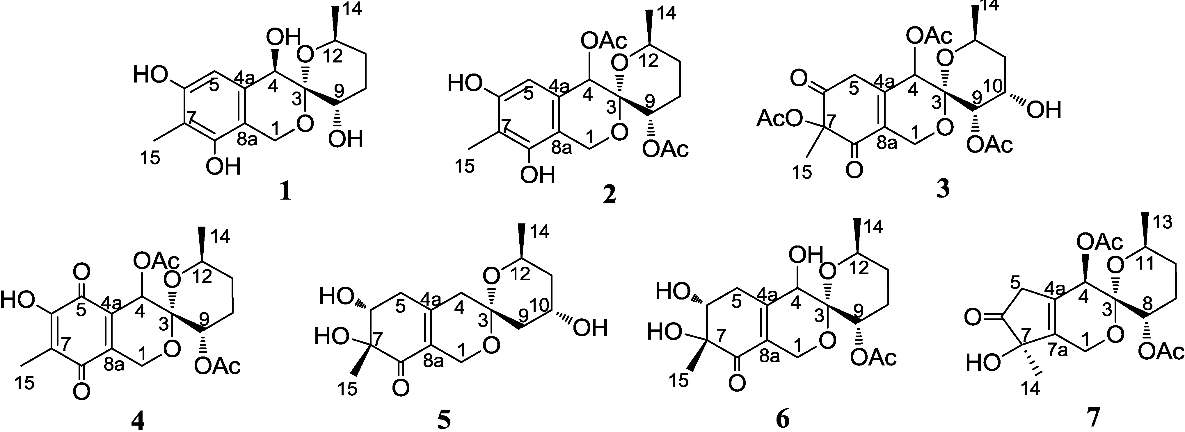
Sargassopenillines A–G, 6,6-Spiroketals from the Alga-Derived Fungi Penicillium thomii and Penicillium lividum

by
Olesya I. Zhuravleva
1,2,
Maria P. Sobolevskaya
1,
Shamil Sh. Afiyatullov
1,*,
Natalya N. Kirichuk
1,
Vladimir A. Denisenko
1,
Pavel S. Dmitrenok
1,
Ekaterina A. Yurchenko
1 and
Sergey A. Dyshlovoy
1,2
1
Elyakov Pacific Institute of Bioorganic Chemistry, Far-Eastern Branch of the Russian Academy of Sciences, Prospect 100-let Vladivostoku 159, Vladivostok 690022, Russia
2
Far Eastern Federal University, Suhanova 8, Vladivostok 690950, Russia
*
Author to whom correspondence should be addressed.
Mar. Drugs 2014, 12(12), 5930-5943; https://doi.org/10.3390/md12125930
Submission received: 24 September 2014 / Revised: 21 November 2014 / Accepted: 2 December 2014 / Published: 9 December 2014

Abstract

:
Seven new 6,6-spiroketals, sargassopenillines A–G (17) were isolated from the alga-derived fungi Penicillium thomii KMM 4645 and Penicillium lividum KMM 4663. The structures of these metabolites were determined by HR-MS and 1D and 2D NMR. The absolute configurations of compounds 1, 5 and 6 were assigned by the modified Mosher’s method and by CD data. Sargassopenilline C (3) inhibited the transcriptional activity of the oncogenic nuclear factor AP-1 with an IC50 value of 15 µM.

1. Introduction

Marine fungi isolated from the surface of marine algae have received great attention as a prolific source of chemically diverse bioactive metabolites [1,2]. As a part of our ongoing search for structurally novel and bioactive metabolites from marine-derived fungi, we have previously isolated ten new austalide meroterpenoids from the strains of Penicillium thomii KMM 4645 and Penicillium lividum KMM 4663 associated with the marine brown alga Sargassum miyabei [3]. Further investigation of metabolites of these fungal strains has now led to the isolation of seven new 6,6-spiroketals, sargassopenillines A–G. We report herein the isolation and structure determination of compounds 17 (Figure 1) and their biological assay results.
Figure 1. Structures of sargassopenillines A–G (17).
Marinedrugs 12 05930 g001

2. Results and Discussion

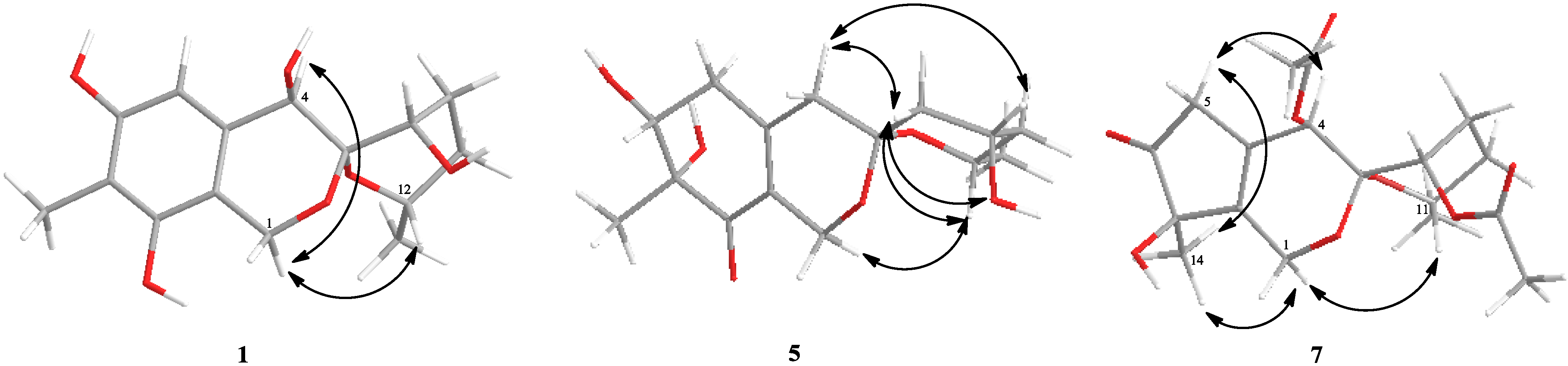
2.1. Structure Elucidation

The fungi were cultured for 21 days on specially modified rice medium [4]. The EtOAc extracts of the mycelia were purified by a combination of silica gel column chromatography and reversed-phase HPLC to yield compounds 1 and 5 from the P. thomii and 27 from the P. lividum as amorphous solids.
The molecular formula of compound 1 was determined as C15H20O6 by a HRESIMS peak at m/z 319.1157 [M + Na]+ and by 13C NMR analyses. A close inspection of 1H and 13C NMR data of 1 (Table 1 and Table 2) by DEPT and HSQC revealed the presence of two methyl (δH 1.08, 2.06, δC 22.1, 9.2) groups, three methylenes (δC 37.3, 40.9 and 60.8) including one oxygen-bearing, three oxygenated methines (δH 3.87, 4.15, 4.19, δC 71.5, 66.1, 64.1) and one olefinic methine (δH 6.36, δC 109.7), five sp2C 113.0, 114.3, 133.5, 152.3 and 156.3) quaternary carbons including two carbons linked to an oxygen atoms and one double oxygenated quaternary carbon (δC 100.6).
The HMBC correlations from methyl singlet (δH 2.06) to oxygenated carbons C-6 (δC 156.3), C-8 (δC 152.3) and C-7 (δC 113.0); from H-5 (δH 6.36) to C-4 (δC 71.5), C-4a (δC 114.3), C-6, C-7, and C-8a (δC 133.54); from H-4 (δH 3.87) to C-8a and double oxygenated C-3 (δC 100.6); and from H2-1 (δH 4.52, 4.74) to C-4a, C-8 and C-3 revealed the connection of C-1 to C-3 through an oxygen atom and indicated the presence of a bicyclic isochroman core in 1 with alcohol functions at C-4, C-6 and C-8 and a methyl group at C-7. The COSY-45 data and HSQC spectra of 1 revealed the connectivity sequences of the protons: (-CH(O)(9)-CH2(10)-CH2(11)-CH(CH3)(12)-). These data and HMBC correlations H-9 (δH 4.15)/C-3, C-10 (δC 37.3), C-11 (δC 40.9); H3-14 (δH 1.08)/C-11, C-12 (δC 64.1), and H-12 (δH 4.19)/C-3 indicated the presence of the 6,6-spiroketal moiety in 1.
Table 1. 1H NMR spectroscopic data (δ, J in Hz) for sargassopenillines A–F (16).
Position1 a2 b3 c4 b5 b6 c
1a: 4.74, d (14.7)a: 4.88, d (15.0) a: 4.70, td (2.8, 16.6) a: 4.73, d (19.7) a: 4.53, dd (1.6, 15.5) a: 4.55, td (2.7, 16.6)
b: 4.52, d (14.7)b: 4.57, d (15.0)b: 4.25, d (16.8)b: 4.35, dd (1.7, 19.7)b: 4.11, mb: 4.05, dd (4.2, 16.6)
43.87, s5.75, s5.31, s5.92, d (1.7)a: 2.40, brd (19.3)3.72, brs
b: 2.22, brd (19.3)
56.36, s6.54, sa: 3.40, td (2.7, 13.9) a: 2.53, dd (5.5, 18.3)a: 3.03, dd (5.6, 18.2)
b: 3.33, d (8.6)b: 2.38, brd (19.3)b: 2.37, m
64.00, dd (5.8, 10.5)4.00, dd (5.7, 10.3)
94.15, t (3.0)5.07, t (2.8)4.99, d (3.0)4.95, t (2.8)a: 1.96, dd (2.1, 14.3)5.02, t (2.9)
b: 1.75, dd (3.6, 14.3)
10a: 2.24, td (2.2, 14.8) a: 2.16, m3.90, brsa: 2.07, m4.10, ma: 2.08, m
b: 1.72, dd (3.8, 14.8)b: 1.84, mb: 1.81, dd (3.2, 14.5)b: 1.89, dd (3.5, 14.6)
11a: 1.74, ma: 1.55, ma: 1.71, dd (2.9, 11.2)a: 1.54, ma: 1.83, dd (2.6, 13.7)a: 1.55, m
b: 1.45, mb: 1.45, mb: 1.74, td (2.9, 14.2)b: 1.44, mb: 1.43, ddd (2.6, 11.9, 13.7)b: 1.46, m
124.19, m3.98, m4.11, m3.78, m4.12, m3.78, m
141.08, d (6.3)1.13, d (6.3)1.23, d (6.3)1.15, d (6.3)1.17, d (6.3)1.14, d (6.3)
152.01, s2.11, s1.57, s1.95, s1.28, s1.27, s
4-OAc1.98, s2.08, s1.97, s
7-OAc2.16, s
9-OAc2.08, s2.05, s2.06, s2.16, s
a Chemical shifts referenced to CD3OD at 500 MHz; b Chemical shifts referenced to CDCl3 at 700 MHz; c Chemical shifts referenced to CDCl3 at 500 MHz.
Table 2. 13C NMR spectroscopic data (δ in ppm) for sargassopenillines A–F (16).
Position1 a2 b3 c4 b5 b6 c
160.858.759.058.157.458.3
3100.696.598.497.196.897.3
471.566.164.659.440.665.9
4a114.3128.7130.2144.2149.5149.2
5109.7108.940.4180.635.933.9
6156.3153.3198.0151.272.372.5
7113.0110.184.7117.477.377.4
8152.3149.7192.2186.0198.9200.2
8a133.5113.1139.8130.7126.6127.9
966.166.265.565.039.166.5
1037.324.265.824.064.724.0
1140.926.834.726.539.226.7
1264.168.463.569.161.668.9
1422.121.320.821.221.221.3
159.27.821.37.717.717.4
4-OAc171.1, 21.2170.5, 20.7168.3, 20.7
7-OAc169.5, 19.9
9-OAc170.3, 21.1169.3, 20.9170.5, 21.4170.7, 21.3
a Chemical shifts referenced to CD3OD at 125 MHz; b Chemical shifts referenced to CDCl3 at 176 MHz; c Chemical shifts referenced to CDCl3 at 125 MHz.
Esterification of 1 with (R)- and (S)-MTPA chloride [5] occurred both at the C-4 and C-9 hydroxy groups to give the (S)-and (R) MTPA esters 1a and 1b, respectively. The observed chemical shift differences Δδ(δSR) (Figure 2) indicated the 4R and 9S configuration. The revealed configuration of the C-9 chiral center in 1 and analysis coupling constants for H-9 (δH 4.15, t, 3.0) and H2-10 (Ha: δH 2.24, td, 2.2, 14.8; Hb: δH 1.72, dd, 3.8, 14.8), that were also calculated using the empirical generalization of the classical Karplus equation [6], showed that the right ring is in the pseudo boat conformation (Figure 3). These data and NOE correlations H-12/H-1b (δH 4.52) (2.957 Å) [7] and H-4/H-1b (4.435 Å) determined the absolute stereostructure of 1 with 3S, 4R, 9S, 12S configurations. Compound 1 was named sargassopenilline A.
Figure 2. The ∆δ (δS−δR) values (in ppm) for the (S)- and (R)-MTPA esters of 1, 5 and 6.
Marinedrugs 12 05930 g002
Figure 3. Chem3D representation of the minimum conformation of 1, 5 and 7 showed observed NOE correlations.
Marinedrugs 12 05930 g003
The molecular formula of 2 was determined to be C19H24O8 by a HRESIMS peak at m/z 403.1366 [M + Na]+ and was in accordance with 13C NMR data (Table 2). The structure of the substituted bicyclic isochroman moiety of 2 was found by extensive NMR spectroscopy (1H and 13C NMR, COSY, HSQC and HMBC) as for sargassopenilline A (1). The HMBC correlations from H-4 (δH 5.75) to 4-Ac (δC 171.1), from 4-Ac (CH3, δH 1.98) to C-4 (δC 66.1), 4-Ac (δC 171.1) and downfield chemical shift of H-4 indicated replacement the alcohol group at C-4 by the acetoxy group. The COSY-45 data and HMBC correlations H-9 (δH 5.07)/C-3 (δC 96.5), 9-Ac (δC 170.3), C-10 (δC 24.2), C-11 (δC 26.8); H3-14 (δH 1.13)/C-11, C-12 (δC 68.4) and H-12 (δH 3.98)/C-3 established the structure of the 9-acetoxy-12-methylpyran ring and indicated the presence of spirocyclic system in 2. The NOE correlation H-12/ H-1b (δH 4.57) indicated a β-orientation for the 14-methyl group and suggested the configuration of C-3 to be S. The small coupling constants of the H-9 signal at δ 5.07 (1H, t, 2.8) and biogenetic relationship between sargassopenilline A and 2 suggested an α-orientation for the 9-acetoxy group. Unfortunately, the correlations observed in the NOESY spectrum did not allow us to unequivocally establish the relative configuration at C-4. Compound 2 was named sargassopenilline B.
The molecular formula of 3 was determined to be C21H26O11 by a HRESIMS peak at m/z 477.1365 [M + Na]+ and was in accordance with 13C NMR data. The 1H and 13C NMR spectra of 3 (Table 1 and Table 2) by DEPT and HSQC indicated the presence of two methyl (δH 1.23, 1.57, δC 20.83, 21.3) and three acetoxy (δH 2.05, δC 20.9, 169.3, δH 2.08, δC 20.7, 170.5, δH 2.16, δC 19.9, 169.5) groups, three methylenes (δC 34.7, 40.4, and 59.0) including one oxygen-bearing, four oxygenated methines (δH 3.90, 4.11, 4.99, 5.31 δC 65.8, 63.5, 65.5, 64.6), two carbonyl functions (δC 192.2 and 198.0), one tetrasubstituted double bond (δC 130.2 and 139.8) and one double oxygenated quaternary carbon (δC 98.4). The HMBC correlations H3-15 (δH 1.57)/C-6 (δC 198.0), C-7 (δC 84.7), C-8 (δC 192.2); H-5a (δH 3.40)/C-4 (δC 64.6), C-4a (δC 130.2), C-6, C-7 and H-1a (δH 4.70)/C-3 (δC 98.4), C-4, C-4a, C-8 and C-8a (δC 139.8) indicated the presence of the bicyclic isochromene core in 3 with carbonyl functions at C-6 and C-8 and a methyl group at C-7. The locations of the acetoxy groups at C-4 and C-7 were evident from the HMBC correlations from 4-Ac (δH 2.08) to C-4 (δC 64.6), 4-Ac (δC 170.5) and from 7-Ac (δH 2.16) to C-7 (δC 84.7), 7-Ac (δC 169.5). The interpretation of the COSY and HSQC data revealed one isolated spin system: (-CH(O)(9)-CH(O)(10)-CH2(11)-CH(CH3)(12)-). These data and the HMBC correlations H-9 (δH 4.99)/C-3 (δC 98.4), 9-Ac (δC 169.3), C-10 (δC 65.8), C-11 (δC 34.7); H-11a (δH 1.74)/C-10; H-11b (δH 1.71)/C-12 (δC 63.5); H3-14 (δH 1.23)/C-3, C-11 and C-12 established the presence of the 9-acetoxy-10-hydroxy-12-methylpyran ring in 3.
The small coupling of the H-10 signal at δ 3.90 (1H, brs) and downfield chemical shift of C-12 (δC 63.5) in comparison with the spectra of 2C 68.4) (γ-effect) indicated that 3 contained a secondary alcohol function with an α-orientation. Furthermore, NOE correlations H-1b (δH 4.25)/H-12 indicated a β-orientation for the 14-methyl group and suggested the configuration of C-3 to be S. We observed a strong NOE correlation between H-9 and H-10 and small coupling of the H-9 signal at δ 4.99 (1H, d, 3.0) proposed to be an α-orientation for the 9-acetoxy group. The configurations at C-4 and C-7 have not been determined. Compound 3 was named sargassopenilline C.
The molecular formula of 4 was determined to be C19H22O9 by a HRESIMS peak at m/z 417.1157 [M + Na]+ and was in accordance with 13C NMR data. The 1H and 13C NMR (Table 1 and Table 2), DEPT and HSQC spectra showed two methyls, three methylenes (one of them oxygenated), three oxymethines, two ketone carbons, four olefinic quaternary carbons, one doubled oxygenated quaternary carbon, one hydroxyl and two acetoxy groups. The HMBC correlations H3-15 (δH 1.95)/C-6 (δC 151.2), C-7 (δC 117.4), C-8 (δC 186.0); 6-OHH 6.95)/C-5 (δC 180.6), C-6, C-7, H-1a (δH 4.73)/C-3 (δC 97.1), C-4a (δC 144.2), C-8, C-8a (δC 130.7) and H-4 (δH 5.92)/C-3, C-4a, C-5 and C-8a indicated the presence of the bicyclic isochromene core in 4 with carbonyl functions at C-5 and C-8 and hydroxyl and methyl groups at C-6 and C-7, respectively. The location of the acetoxy group at C-4 was evident from the HMBC correlations from 4-Ac (δH 1.97) to C-4 (δC 59.4) and 4-Ac (δC 168.3). The structure of the 9-acetoxy-12-methyl pyran ring and the presence 6,6-spiroring system in 4 was found by extensive NMR spectroscopy (1H and 13C NMR, COSY, HSQC and HMBC) as for sargassopenilline B (2). The NOE correlation H-12/ H-1b (δH 4.35) indicated a β-orientation for the 14-methyl group and suggested the configuration of C-3 to be S. The small coupling constants of the H-9 signal at δ 4.95 (1H, t, 2.8) and biogenetic relationship between sargassopenilline A and 4 suggested an α-orientation for the 9-acetoxy group. The configuration at C-4 has not been determined. Compound 4 was named sargassopenilline D.
The molecular formula of 5 was determined to be C15H22O6 by a HRESIMS peak at m/z 299.1483 [M + H]+ and was in accordance with 13C NMR data. The 1H and 13C NMR spectra (Table 1 and Table 2) of 5 indicated the presence of two methyls, five methylenes including one oxygen-bearing, three oxygenated methines and one double oxygenated quaternary carbon. The remaining functionalities, corresponding to the carbon signals at δC 198.9 (C), 149.5 (C) and 126.6 (C), suggested the presence of a carbonyl carbon and one tetrasubstituted double bond. The 1H and 13C data observed for the 3,4,5,6,7,8-hexahydroisochromene core resemble those reported for pestafolide A [8]. The interpretation of the COSY and HSQC data revealed one isolated spin system: (-CH2(9)-CH(O)(10)-CH2(11)-CH(CH3)(12)-). This information and the HMBC correlations H-9a (δH 1.96)/C-3 (δC 96.8), C-11 (δC 39.2); H-9b (δH 1.75)/C-3, C-4 (δC 40.6), C-10 (δC 64.7); H-10 (δH 4.10)/C-3; H3-14 (δH 1.08)/C-11, C-12 (δC 61.6) indicated the presence of the 10-hydroxy-12-methylpyran ring in 5. Thus, the planar structure of 5 was established.
Compound 5 showed the characteristic Cotton effects (CEs) at λ310 +0.29, λ248 −5.70 and λ216 +7.57 in the CD spectra in methanol solution. The two CEs of the high-energy region were in agreement with those for pestafolide A [8] and peneciraistin C [7], supporting the 7R configuration of 5. Esterification of 5 with (R)- and (S)-MTPA chloride occurred both at the C-6 and C-10 hydroxy groups to give the (S)- and (R)-MTPA esters 5a and 5b, respectively. The observed chemical shift differences Δδ(δS − δR) (Figure 2) revealed the 6R and 10S configurations. These data and the NOE correlations, recorded in DMSO-d6 solvent, (Supplementary Figure S44, Figure 3) H-1b (δH 3.93)/7-OHH 5.04), H-12 (δH 4.07); H-1a (δH 4.20)/H-4a (δH 2.35); H-4b (δH 2.08)/H-9b (δH 1.63), H3-14 (δH 1.05) and H-9a (δH 1.77)/10-OHH 4.15), H-12 determined the absolute stereostructure of 5 with 3S, 6R, 10S, and 12S configurations. Compound 5 was named sargassopenilline E.
The molecular formula of 6 was determined to be C17H24O8 by a HRESIMS peak at m/z 357.1548 [M + H]+ and was in accordance with 13C NMR data. The general features of the 1H and 13C NMR spectra (Table 1 and Table 2) of the isochromene core in 6 resembled those of 5 with the exception of the C-4 and C-5 proton and carbon signals. The HMBC correlations H-1a (δH 4.55)/C-3 (δC 97.3), C-4 (δC 65.9), C-4a (δC 149.2), C-8 (δC 200.2) and C-8a (δC 127.9); H-4 (δH 3.72)/C-4a, C-5 (δC 33.9), C-8a; H-5a (δH 3.03)/C-6 (δC 72.5) and C-8a; H-6 (δH 4.00)/C-5, C-7 (δC 77.4), C-8 and C-15 (δC 17.4) indicated the location of the hydroxy group at C-4 and established the structure of a bicyclic isochromene core in 6. The structure of the 9-acetoxy-12-methylpyran ring and the presence 6,6-spiroring system in 6 was found by extensive NMR spectroscopy (1H and 13C NMR, COSY, HSQC and HMBC) as for sargassopenilline B (2).
Compound 6 exhibited a nearly identical CD spectrum in the high-energy region to that of sargassopenilline E (5), which allowed us to determine the 7R configuration of 6. Esterification of 6 with (R)- and (S)-MTPA chloride occurred at the C-6 hydroxy group to give the (S)-and (R) MTPA esters 6a and 6b, respectively. The observed chemical shift differences Δδ(δS − δR) (Figure 2) revealed the 6R configuration. The NOE correlation H-12/H-1b (δH 4.05) indicated a β-orientation for the 14-methyl group and suggested the configuration of C-3 to be S. The small coupling constants of the H-9 signal at δ 5.02 (1H, t, 2.9) and biogenetic relationship between sargassopenilline A and 6 suggested an α-orientation for the 9-acetoxy group. Unfortunately, the correlations observed in the NOESY spectrum could not unequivocally establish the relative configuration at C-4. Compound 6 was named sargassopenilline F.
The molecular formula of compound 7 was determined as C18H24O8 by a HRESIMS peak at m/z 391.1367 [M + Na]+ and by 13C NMR analyses. The 1H and 13C NMR spectra of 7 (Table 3) by DEPT and HSQC indicated the presence of two methyl (δH 1.19, 1.36, δC 21.3, 22.5) and two acetoxy (δH 2.01, 2.03, δC 20.7, 21.0, 170.4, 170.0) groups, four methylenes (δC 24.1, 26.7, 40.1 and 57.5) including one oxygen-bearing, three oxygenated methines (δH 3.85, 5.02, 5.25, δC 68.7, 66.2, 64.3) and one double oxygenated quaternary carbon (δC 97.0). The remaining functionalities, corresponding to the carbon signals at δC 214.4 (C), 141.8 (C) and 127.3 (C), suggested the presence of a ketone function and one tetrasubstituted double bond.
The HMBC correlations H-5b (δH 2.92)/C-4a (δC 127.3), C-6 (δC 214.4), C-7 (δC 77.9) and C-7a (δC 141.8); H3-14 (δH 1.36)/C-6, C-7 and C-7a revealed the presence of 2-hydroxy-2-methylcyclopent-3-enone moiety in 7. Furthermore, HMBC correlations H-1a (δH 4.43)/C-3 (δC 97.0), C-4 (δC 64.3), C-4a, C-7a and H-4 (δH 5.25)/C-3, C-4a, C-5 (δC 40.1), C-7a and 4-Ac (δC 170.4) show that 7 contains an unusual tetrahydrocyclopentapyranone ring system in the molecular structure. The structure of the 9-acetoxy-12-methylpyran ring and the presence 6,6-spiroring system in 7 was found by extensive NMR spectroscopy (1H and 13C NMR, COSY, HSQC and HMBC) as for sargassopenilline B (2). Thus compound 7 is a new spiroketal-containing natural compound and it was named sargassopenilline G.
Table 3. 1H (CDCl3, 700 MHz) and 13C (CDCl3, 176 MHz) NMR spectroscopic data for sargassopenilline G (7).
PositionδCδH (J in Hz)HMBC
157.5a: 4.43, td (3.0, 16.4),3, 4, 4a, 7, 4, 4a, 7a
b: 4.23, d (16.4)
397.0
464.35.25, s4a, 5, 7a, 4-Ac (170.4)
4a127.3
540.1a: 3.02, td (3.3, 22.2),4a, 6, 7a, 4a, 6, 7, 7a
b: 2.92, td (3.1, 22.2)
6214.4
777.9
7a141.8
866.25.02 t (2.8)3, 8-Ac (170.0), 9, 10
924.1a: 2.10, m, b: 1.84, m10, 11, 3, 7a, 10, 11
1026.7a: 1.52, m, b: 1.46, m8, 9, 11, 13, 8, 9, 11
1168.73.85, m3, 9, 13
1321.31.19, d (6.3)3, 10, 11
1422.51.36, s6, 7, 7a
4-Ac170.4, 20.72.01, s4, 4-Ac (170.4)
8-Ac170.0, 21.02.03, s8, 8-Ac (170.0)
The NOE correlation H-11/H-1b (δH 4.23) (2.854 Å) indicated a β-orientation for the 13-methyl group and suggested the configuration of C-3 to be S. The small coupling constants of the H-8 signal at δ 5.02 (1H, t, 2.8) and biogenetic relationship between sargassopenilline A and 7 suggested an α-orientation for the 8-acetoxy group. These data and the observed NOE correlations H3-14/H-1b (δH 4.23), H-5a (δH 3.02) and H-5a/H-4 indicated the relative configuration of 7 (Figure 3).

2.2. Bioassay Results

Sargassopenillines 13 and 7 were assayed for their cytotoxic activity against MDA-MB-231 and JB6 Cl41 cell lines. None of the compounds exhibited cytotoxicity (IC50 < 100 μM).
The effect of compounds 13 and 7 on the basal AP-1-dependent transcriptional activity was also studied using JB6 Cl41 cells stably expressing a luciferase reporter gene controlled by an AP-1-DNA binding sequence [9,10,11,12]. We found that compound 3 is able to inhibit the transcriptional activity of the oncogenic nuclear factor AP-1 with IC50 value of 15 µM after 12 h of treatment.
The sargassopenillines 1, 2, 47 were assayed for their cytotoxic activity against CD-1 mouse splenocytes and membranolytic activity to erythrocytes up to 100 µM. Sargassopenilline E (5) exhibited cytotoxicity against splenocytes with a IC50 value 38 µM. The effects of the compounds 1, 2 and 47 on the functional activity of CD-1 murine peritoneal macrophages were also studied. It was shown that sargassopenillines D and F at a non-toxic concentration (10 µM) inhibit the adhesion of macrophages (30%–40% of inhibition).
In addition, compounds 1 and 5 showed radical scavenging activity against DPPH with IC50 values of 100 and 50 µM, respectively, while others were inactive.

3. Experimental Section

3.1. General Experimental

Optical rotations were measured on a Perkin-Elmer 343 polarimeter. UV spectra were recorded on a Shimadzu UV-1601PC spectrometer in MeOH. CD spectra were measured with a Chirascan-Plus CD Spectrometer (Leatherhead, UK). IR spectra were determined on a Bruker OPUS Vector-22 infrared spectrophotometer in CHCl3. 1H and 13C NMR spectra were recorded in CDCl3, MeOH-d4 and pyridine-d5 on a Bruker Avance-500 and Avance III-700 spectrometers operating at 500.13 MHz and 125.77 MHz and 700.13 and 176.04 MHz, respectively, using TMS as an internal standard. HRESIMS spectra were measured on an Agilent 6510 Q-TOF LC mass spectrometer.
Low-pressure liquid column chromatography was performed using Si gel L (40/100 μm, Sorbpolimer, Russia). Glass plates (4.5 × 6.0 cm) precoated with Si gel (5–17 μm, Sorbfil) were used for thin layer chromatography. Preparative HPLC was carried out on a Beckman-Altex chromatograph, using a Supelco Discovery C-18 (5 μm, 4.6 × 250 mm) column with an RIDK–122 refractometer.
The energy-minimized conformations for 1, 5 and 7 have been determined using crystallographic data (CCDC 940798) for the structure of peniciketal A [13] by the MM2 force field calculation method using ChemBioDraw Ultra 12.0, CambridgeSoft Corporation (Cambridge, MA, USA).

3.2. Fungal Material and Fermentation

The strains of the fungi Penicillium lividum and Penicillium thomii were isolated from superficial mycobiota of the brown alga Sargassum miyabei (Lazurnaya Bay, the Sea of Japan) and were identified on the basis of morphological evaluation by Natalya N. Kirichuk from the G.B. Elyakov Pacific Institute of Bioorganic Chemistry (PIBOC). Strains are stored at the Collection of Marine Microorganisms, PIBOC, Vladivostok, Russia with the codes KMM 4663 and KMM 4645, respectively. The fungi were grown stationary at 22 °C for 21 days in 20 Erlenmeyer flasks (500 mL) (for each strain), each flask containing 20 g of rice, 20 mg of yeast extract, 10 mg of KH2PO4, and 40 mL of natural sea water (Marine Experimental Station of G.B. Elyakov Pacific Institute of Bioorganic Chemistry, Troitsa (Trinity) Bay, Sea of Japan).

3.3. Extraction

At the end of the incubation period, the mycelia and medium were homogenized and extracted with EtOAc (2 L). The extract of each fungus was concentrated to dryness. The residue was dissolved in 20% MeOH–H2O (1 L) and was extracted with n-hexane (0.2 L × 3) and EtOAc (0.2 L × 3). After evaporation of the EtOAc layer, the residual materials (1.5 g, P. thomii and 1.3 g, P. lividum) were passed over silica columns (4 × 20 cm), which were eluted first with n-hexane (1 L) followed by a step gradient from 5% to 100% EtOAc in n-hexane (total volume 7 L). Fractions of 200 mL were collected and combined on the basis of TLC (Si gel, toluene–isopropanol 6:1, v/v).

3.4. Isolation Metabolites from P. thomii

The n-hexane–EtOAc (3:2, 1.5 L) eluate (100 mg) was purified by RP HPLC on a Supelco Discovery C-18 column eluting with MeOH–H2O (40:60) to yield 1 (10 mg). The EtOAc (1.0 L) eluate (60 mg) gave 5 (5 mg) eluting with MeOH–H2O (40:60).

3.5. Isolation Metabolites from P. lividum

The n-hexane–EtOAc (5:1, 1.4 L) eluate (250 mg) was purified by RP HPLC on a Supelco Discovery C-18 column eluting with MeOH–H2O (65:35) to yield 3 (2.1 mg), 4 (5.8 mg) and MeOH–H2O (50:50) to yield 2 (3.4 mg) and 7 (3.5 mg). The EtOAc (1.0 L) eluate (64 mg) gave 5 (3.4 mg) and 6 (4.5 mg) after purification by HPLC (MeOH–H2O, 40:60).

3.6. Physicochemical and Spectroscopic Data of 17

Sargassopenilline A (1): Amorphous solid; [α]20D −45 (c 0.10, MeOH); UV (MeOH) λmax (log ε) 235 (3.22), 282 (3.07) nm; CD (c 0.6 mg/mL, MeOH) λmax (Δε) 233(+0.06), 247 (−0.04), 282 (+0.04), 350 (+0.01) nm; 1H and 13C NMR data, see Table 1 and Table 2; HRESIMS m/z 319.1157 [M + Na]+ (calcd for C15H20O6Na, 319.1152).
Sargassopenilline B (2): Amorphous solid; [α]20D −137 (c 0.09, MeOH); UV (MeOH) λmax (log ε) 219 (3.77), 284 (3.25) nm; CD (c 0.2 mg/mL, MeOH) λmax (Δε) 244 (−0.86), 274 (−0.35), 308 (+0.20), nm; 1H and 13C NMR data, see Table 1 and Table 2; HRESIMS m/z 403.1366 [M+Na]+ (calcd for C19H24O8Na, 403.1363).
Sargassopenilline C (3): Amorphous solid; [α]20D −84 (c 0.17, MeOH); UV (MeOH) λmax (log ε) 208 (4.25), 250 (3.76), 283 (3.82) nm; CD (c 0.025 mg/mL, MeOH) λmax (Δε) 240 (−2.46), 318 (+0.38), 380 (−0.27), nm; IR (CHCl3) νmax 3610, 2928, 2855, 1742, 1690, 1648, 1603, 1456, 1373, 1253, 1164, 1092, 1067 cm−1; 1H and 13C NMR data, see Table 1 and Table 2; HRESIMS m/z 477.1365 [M + Na]+ (calcd for C21H26O11Na, 477.1367).
Sargassopenilline D (4): Amorphous solid; [α]20D −37 (c 0.07, MeOH); UV (MeOH) λmax (log ε) 196 (3.29), 268 (2.91) nm; CD (c 0.17 mg/mL, MeOH) λmax (Δε) 198 (−2.51), 210 (−2.15), 254 (+1.51), 282 (−8.43), 340 (−0.35) nm; 1H and 13C NMR data, see Table 1 and Table 2; HRESIMS m/z 417.1157 [M + Na]+ (calcd for C19H22O9Na, 417.1156), 393.1207 [M − H]+ (calcd for C19H21O9, 393.1191).
Sargassopenilline E (5): Amorphous solid; [α]20D −107 (c 0.16, MeOH); UV (MeOH) λmax (log ε) 242 (3.01) nm; CD (c 0.33 mg/mL, MeOH) λmax (Δε) 216 (+7.57), 248 (−5.07), 310 (+0.29), 350 (−0.05) nm; 1H and 13C NMR data, see Table 1 and Table 2; HRESIMS m/z 321.1296 [M + Na]+ (calcd for C15H22O6Na, 321.1309), 299.1483 [M + H]+ (calcd for C15H23O6, 299.1489).
Sargassopenilline F (6): Amorphous solid; [α]20D −45 (c 0.01, MeOH); UV (MeOH) λmax (log ε) 206 (3.05), 221 (3.02), 247 (2.70), 268 (2.63) nm; CD (c 0.18 mg/mL, MeOH) λmax (Δε) 220 (+4.60), 248 (−6.50), 328 (+0.39) nm; 1H and 13C NMR data, see Table 1 and Table 2; HRESIMS m/z 379.1379 [M + Na]+ (calcd for C17H24O8Na, 379.1363), 357.1548 [M + H]+ (calcd for C17H25O8, 357.1544).
Sargassopenilline G (7): Amorphous solid; [α]20D −158 (c 0.09, MeOH); UV (MeOH) λmax (log ε) 215 (3.69) nm; CD (c 0.2 mg/mL, MeOH) λmax (Δε) 242 (−0.64), 306 (−0.39), 347 (+0.14), nm; 1H and 13C NMR data, see Table 3; HRESIMS m/z 391.1367 [M + Na]+ (calcd for C18H24O8Na, 391.1363).

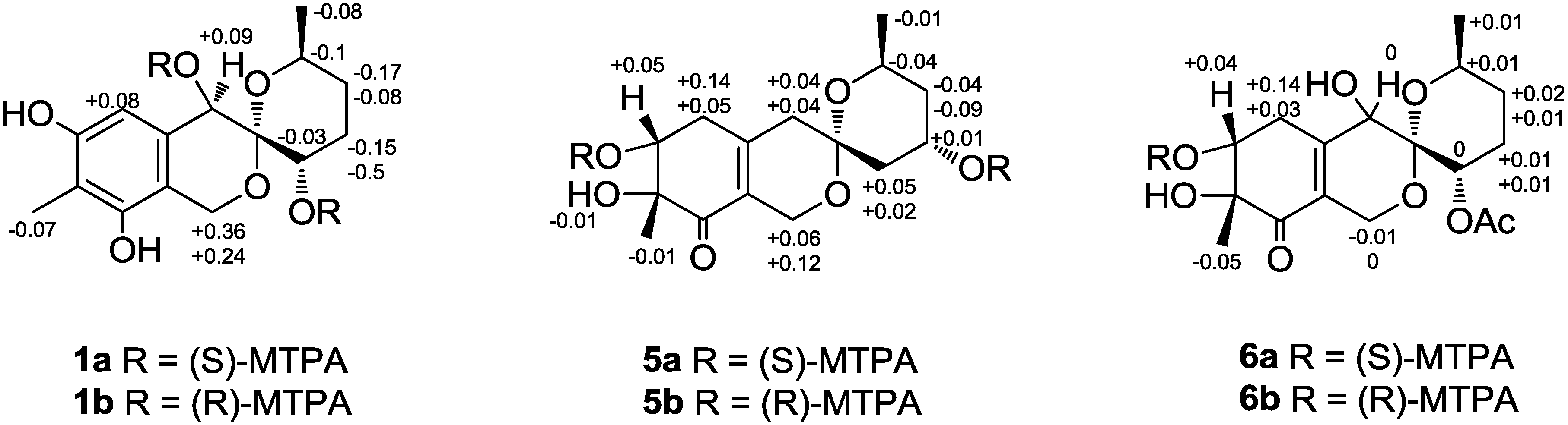
3.7. Preparation of (S)-MTPA and (R)-MTPA Esters of 1

4-Dimethylaminopyridine (a few crystals) and (R)-MTPACl (20 μL) were added to a solution of the 1 (4.0 mg) in pyridine and stirred at room temperature (25 °C) for 24 h. After evaporation of the solvent, the residue was passed through a silica gel column (7% EtOAc–hexane) to generate the (S)-MTPA ester (1a). The (R)-MTPA ester (1b) was prepared in a similar manner using (S)-MTPACl. 1H and COSY data, Supplementary Information S7–S10; ESIMS of 1a m/z 729.39 [M + H]+ and of 1b m/z 729.35 [M + H]+.

3.8. Preparation of (S)-MTPA and (R)-MTPA Esters of 5

4-Dimethylaminopyridine (a few crystals) and (R)-MTPACl (20 μL) were added to a solution of the 5 (4.0 mg) in pyridine and stirred at room temperature (25 °C) for 24 h. After evaporation of the solvent, the residue was passed through a silica gel column (7% EtOAc–hexane) to generate the (S)-MTPA ester (5a). The (R)-MTPA ester (5b) was prepared in a similar manner using (S)-MTPACl. 1H and COSY data, Supplementary Information S35–S38; ESIMS of 5a m/z 731.42 [M + H]+ and of 5b m/z 7315.44 [M + H]+.

3.9. Preparation of (S)-MTPA and (R)-MTPA Esters of 6

4-Dimethylaminopyridine (a few crystals) and (R)-MTPACl (20 μL) were added to a solution of the 6 (3.0 mg) in pyridine and stirred at room temperature (25 °C) for 24 h. After evaporation of the solvent, the residue was passed through a silica gel column (15% EtOAc–hexane) to generate the (S)-MTPA ester (6a). The (R)-MTPA ester (6b) was prepared in a similar manner using (S)-MTPACl. 1H and COSY data, Supplementary Information S51–S54; ESIMS of 6a m/z 573.21 [M + H]+ and of 6b m/z 573.30 [M + H]+.
The spectra of compounds 17 are all given in the Supplementary Information.

3.10. Cytotoxicity Assay

The effect of the compounds on the cells viability was evaluated using the MTS test, which is based on the reduction of MTS into its formazan product by alive cells [14,15,16]. Cytotoxicity towards CD-I mouse splenocytes was determined according to Freshney [17]. Hemolytic activity towards CD-I mouse erythrocytes was determined as previously described [18].

3.11. Determination of the Effects of Compounds on the Basal Transcriptional Activity of AP-1

The effects of the compounds on the basal transcriptional activities of AP-1 were evaluated using the JB6 Cl41 cell line stably expressing a luciferase reporter gene controlled by an AP-1-DNA binding sequence [9]. The experiments were performed as previously reported [16] with slight modifications.

3.12. Macrophage Adhesion Test

Mice were sacrificed by cervical dislocation. Peritoneal macrophages were isolated using standard procedures. For this purpose, 3 mL of PBS (pH 7.4) was immediately injected into the peritoneal cavity and the body intensively palpated for 1–2 min. Then, the peritoneal fluid was aspirated with a syringe and transferred to Petri dishes. Petri dishes with the fluid were incubated at 37 °C for 1–2 h to facilitate attachment of peritoneal macrophages to the dish. Then, a cell monolayer was triply flushed with PBS (pH 7.4) to delete attendant lymphocytes, fibroblasts and erythrocytes. Subsequently, macrophages were removed from the surface of the dishes with a scraper and flow of a saline solution, and then placed on an ice bath until use. The working concentration of cells was usually 1–2 × 10⁶ cells/mL. The number of adhered cells was estimated according to standard method [19] with some modifications.
Twenty milliliters of test compound solutions (at non-toxic concentration 10 μM) and 200 mL of macrophage suspension were added to 96-well flat-bottom plates. After incubation for 2 h at 37 °C, cells were fixed with 70% ethanol solution (100 mL per well) for 15 min at RT. Then, cells were washed with 200 mL of PBS, and a 0.05% solution of Trypan blue (100 mL per well) was added to each well and further incubated at 37 °C for 15 min. Next, media was removed and the cells were gently washed with cold PBS (3 × 200 mL). The cells were then lysed with 50 mL of 1% SDS for at least 4 h and gently triturated. Finally, the SDS/trypan blue solution absorbance was detected spectrophotometrically at 590 nm using a plate reader. The percentage of adhered cells compared to the control level of Trypan blue absorbance was calculated. All samples were assayed in triplicates.

3.13. Radical Scavenging Activity against DPPH

The experiments were performed as previously reported [20]. Ascorbic acid was used as the positive control and showed an IC50 value 21.3 μM.

4. Conclusions

Seven new polyketides, named sargassopenillines A–G (17) have been isolated from the lipophilic extracts of the marine-derived fungi Penicillium thomii and Penicillium lividum. Sargassopenillines A (1) and B (2) are new members of the rare class of natural products that contain an aryl ring fused to the 6,6-spiroketal part [7,21,22]. Notable features of sargassopenillines D (4) and G (7) are the presence of p-benzoquinone and cyclopentenone moieties as their core skeletons. Sargassopenilline C (3) inhibited the transcriptional activity of the oncogenic nuclear factor AP-1 with an IC50 value of 15 µM.

Supplementary Files

Supplementary File 1

Acknowledgments

The study was supported by the program grant from the Russian Foundation for Basic Research (RFBR N 14-04-00910-a and 14-03-31318-mol-a), by the grant of President of Russian Federation MK-6019.2014.4, by the Far Eastern Federal University, project No. 14-08-01-34_u. The isolation and structural elucidation of compounds was supported by the grant Russian Science Foundation No. 14-14-00030.

Author Contributions

O.I. Zhuravleva, M.P. Sobolevskaya and Sh.Sh. Afiyatullov designed the project, performed the isolation and structural elucidation of compounds, analyzed the results and wrote the manuscript. N.N. Kirichuk selected and cultured the fungi. P.S. Dmitrenok and V.A. Denisenko performed the NMR and MS experiments. E.A. Yurchenko and S.A. Dyshlovoy evaluated the cytotoxicity of 17.

Conflicts of Interest

The authors declare no conflict of interest.

References

  1. Blunt, J.W.; Copp, B.R.; Keyzers, R.A.; Munro, M.H.G.; Prinsep, M.R. Marine natural products. Nat. Prod. Rep. 2014, 31, 160–258. [Google Scholar] [CrossRef] [PubMed]
  2. Rateb, M.E.; Ebel, R. Secondary metabolites of fungi from marine habitats. Nat. Prod. Rep. 2011, 28, 290–344. [Google Scholar] [CrossRef] [PubMed]
  3. Zhuravleva, O.I.; Sobolevskaya, M.P.; Leshchenko, E.V.; Kirichuk, N.N.; Denisenko, V.A.; Dmitrenok, P.S.; Dyshlovoy, S.A.; Zakharenko, A.M.; Kim, N.Yu.; Afiyatullov, Sh.Sh. Meroterpenoids from the alga-derived fungi Penicillium thomii Maire and Penicillium lividum Westling. J. Nat. Prod. 2014, 77, 1390–1395. [Google Scholar] [CrossRef] [PubMed]
  4. Afiyatullov, Sh.Sh.; Kuznetsova, T.A.; Isakov, V.V.; Pivkin, M.V.; Prokof’eva, N.G.; Elyakov, G.B. New diterpenic altrosides of the fungus Acremonium stratisporum isolated from sea cucumber. J. Nat. Prod. 2000, 63, 848–850. [Google Scholar] [CrossRef] [PubMed]
  5. Kuzumi, T.; Ooi, T.; Ohkubo, Y.; Yabuuchi, T. The modified Mosher’s method and the sulfoximine method. Bull. Chem. Soc. Jpn. 2006, 79, 965–980. [Google Scholar] [CrossRef]
  6. Haasnoot, C.A.G.; de Leeuw, F.A.A.M.; de Leeuw, H.P.M.; Altona, C. The relationship between proyon-proton NMR coupling-constants and substituent electronegativities: Conformational-analysis of the sugar ring in nucleosides and nucleotides in solution using a generalized Karplus equation. Org. Magn. Reson. 1981, 15, 43–52. [Google Scholar] [CrossRef]
  7. Ma, L.-Y.; Liu, W.-Z.; Shen, L.; Huang, Y.-L.; Rong, X.-G.; Xu, Y.-Y.; Gao, X.-D. Spiroketals, isocumarin, and indoleformic acid derivatives from saline soil derived fungus Penicillium raistrickii. Tetrahedron 2012, 68, 2276–2282. [Google Scholar] [CrossRef]
  8. Ding, G.; Liu, Sh.; Guo, L.; Zhou, Y.; Che, Y. Antifungal metabolites from the plant endophytic fungus Pestalotiopsis foedan. J. Nat. Prod. 2008, 71, 615–618. [Google Scholar] [CrossRef] [PubMed]
  9. Fedorov, S.N.; Shubina, L.K.; Bode, A.M.; Stonik, V.A.; Dong, Z.G. Dactylone inhibits epidermal growth factor-induced transformation and phenotype expression of human cancer cells and induces G(1)-S arrest and apoptosis. Cancer Res. 2007, 67, 5914–5920. [Google Scholar] [CrossRef] [PubMed]
  10. Vesely, P.W.; Staber, P.B.; Hoefler, G.; Kenner, L. Translational regulation mechanisms of AP-1 proteins. Mutut. Res. Rev. Mutat. 2009, 682, 7–12. [Google Scholar] [CrossRef]
  11. Matthews, C.P.; Colburn, N.H.; Young, M.R. AP-1 a target for cancer prevention. Curr. Cancer Drug Targets. 2007, 7, 317–324. [Google Scholar] [CrossRef] [PubMed]
  12. Dong, Z.G.; Birrer, M.J.; Watts, R.G.; Matrisian, L.M.; Colburn, N.H. Blocking of tumor promoter-induced AP-1 activity inhibits induced transformation in JB6 mouse epidermal cells. Proc. Natl. Acad. Sci. USA 1994, 91, 609–613. [Google Scholar] [CrossRef] [PubMed]
  13. Liu, W.Z.; Ma, L.Y.; Liu, D.S.; Huang, Y.L.; Wang, C.H.; Shi, S.S.; Pan, X.H.; Song, X.D.; Zhu, R.X. Peniciketals A–C, new spiroketals from saline soil derived Penicillium raistrichii. Org. Lett. 2014, 16, 90–93. [Google Scholar] [CrossRef] [PubMed]
  14. Barltrop, J.A.; Owen, T.C.; Cory, A.H.; Cory, J.G. 5-(3-carboxymethoxyphenyl)-2-(4,5-dimethylthiazolyl)-3-(4-sulfophenyl)tetrazolium, inner salt (MTS) and related analogs of 3-(4,5-dimethylthiazolyl)-2,5-diphenyltetrazolium bromide (MTT) reducing to purple water-soluble formazans as cell-viability indicators. Bioorg. Med. Chem. Lett. 1991, 1, 611–614. [Google Scholar] [CrossRef]
  15. Dyshlovoy, S.A.; Naeth, I.; Venz, S.; Preukschas, M.; Sievert, H.; Jacobsen, C.; Shubina, L.K.; Gesell Salazar, M.; Scharf, C.; Walther, R.; et al. Proteomic profiling of germ cell cancer cells treated with aaptamine, a marine alkaloid with antiproliferative activity. J. Proteome Res. 2012, 11, 2316–2330. [Google Scholar] [CrossRef] [PubMed]
  16. Dyshlovoy, S.A.; Fedorov, S.N.; Kalinovsky, A.I.; Shubina, L.K.; Bokemeyer, C.; Stonik, V.A.; Honecker, F. Mycalamide A shows cytotoxic properties and prevents EGF-induced neoplastic transformation through inhibition of nuclear factors. Mar. Drugs 2012, 10, 1212–1224. [Google Scholar] [CrossRef] [PubMed]
  17. Freshney, R.I. Culture of Animal Cells: A Manual of Basic Technique and Specialized Applications; Wiley-Liss: New York, NY, USA, 1994. [Google Scholar]
  18. Silchenko, A.S.; Kalinovsky, A.I.; Avilov, S.A.; Andryjaschenko, P.V.; Dmitrenok, P.S.; Yurchenko, E.A.; Dolmatov, I.Y.; Kalinin, V.I.; Stonik, V.A. Structure and biological action of cladolosides B1, B2, C, C1, C2 and D, six new triterpene glycosides from the sea cucumber Cladolabes schmeltzii. Nat. Prod. Comm. 2013, 7, 1527–1534. [Google Scholar]
  19. Uliasz, T.F.; Hewett, S.J. A microtiter trypan blue absorbance assay for the quantitative determination of excitotoxic neuronal injury in cell culture. Neurosci. Methods 2000, 100, 157–163. [Google Scholar] [CrossRef]
  20. Chen, L.; Fang, Y.; Zhu, T.; Gu, Q.; Zhu, W. Gentisyl alcohol derivatives from the marine-derived fungus Penicillium terrestre. J. Nat. Prod. 2008, 71, 66–70. [Google Scholar] [CrossRef] [PubMed]
  21. Li, J.; Li, L.; Si, Y.; Jiang, X.; Guo, L.; Che, Y. Virgatolides A–C, benzanulated spiroketals from the plant endophytic fungus Pestalotiopsis virgatula. Org. Lett. 2011, 13, 2670–2673. [Google Scholar] [CrossRef] [PubMed]
  22. Liu, W.-Z.; Ma, L.-Y.; Liu, D.-Sh.; Huang, Y.-L.; Wang, C.-H.; Shi, Sh.-S.; Pan, X.-H.; Song, X.-D.; Zhu, R.-X. Peniciketals A–C, new spiroketals from saline soil derived Penicillium raistrichii. Org. Lett. 2014, 16, 90–93. [Google Scholar] [CrossRef] [PubMed]

