Article

Rapid and Accurate Identification by Real-Time PCR of Biotoxin-Producing Dinoflagellates from the Family Gymnodiniaceae

1
Cawthron Institute, 98 Halifax Street East, Private Bag 2, Nelson 7042, New Zealand
2
Tasmanian Herbarium, Tasmanian Museum and Art Gallery, Private Bag 4, Hobart, Tasmania 7001, Australia
*
Author to whom correspondence should be addressed.
Mar. Drugs 2014, 12(3), 1361-1376; https://doi.org/10.3390/md12031361
Submission received: 10 October 2013 / Revised: 27 January 2014 / Accepted: 18 February 2014 / Published: 7 March 2014
(This article belongs to the Special Issue Advances and New Perspectives in Marine Biotechnology)

Abstract

:
The identification of toxin-producing dinoflagellates for monitoring programmes and bio-compound discovery requires considerable taxonomic expertise. It can also be difficult to morphologically differentiate toxic and non-toxic species or strains. Various molecular methods have been used for dinoflagellate identification and detection, and this study describes the development of eight real-time polymerase chain reaction (PCR) assays targeting the large subunit ribosomal RNA (LSU rRNA) gene of species from the genera Gymnodinium, Karenia, Karlodinium, and Takayama. Assays proved to be highly specific and sensitive, and the assay for G. catenatum was further developed for quantification in response to a bloom in Manukau Harbour, New Zealand. The assay estimated cell densities from environmental samples as low as 0.07 cells per PCR reaction, which equated to three cells per litre. This assay not only enabled conclusive species identification but also detected the presence of cells below the limit of detection for light microscopy. This study demonstrates the usefulness of real-time PCR as a sensitive and rapid molecular technique for the detection and quantification of micro-algae from environmental samples.

1. Introduction

The dinoflagellate family Gymnodiniaceae includes the genera Gymnodinium, Karenia, Karlodinium, and Takayama [1,2]. Species from the genera Gymnodinium and Karenia have been responsible for fish kills and shellfish contamination events worldwide [3,4] including New Zealand [5,6,7,8]. The only toxic Gymnodinium species, G. catenatum was first recorded along the northwest coastline of New Zealand following the detection of paralytic shellfish poisons (PSP) in shellfish in May 2000. During that bloom event, PSP toxicity reached 4027 μg saxitoxin equivalents/100 g in Greenshell™ mussels (Perna canaliculus) [7]. A number of species in the Karenia genus have been reported to cause severe blooms including K. brevis, K. mikimotoi, K. brevisulcata, K. selliformis, K. longicanalis, and K. digitata [6,9,10,11]. The first recorded major bloom of a Karenia species in New Zealand occurred in 1992/93 along the coast of Northland. At that time 180 cases of illness that fitted the symptoms of neurotoxic shellfish poisoning (NSP) were reported [5,12,13]. The identity of the causative organism was not definitely determined, but later confirmed as K. mikimotoi [13]. Brevetoxins and brevetoxin analogues were detected in shellfish samples causing the total closure of all bivalve industries in New Zealand [12]. In addition to brevetoxins, Karenia species are known to produce gymnodimines [11,14], Karenia brevisulcata toxins (KBTs) and brevisulcatic acids (BSXs) [15], the ichthyotoxic gymnocins A and B [16,17], and haemolytic glycolipids that cause gill damage in fish and have been linked to fish kills in Japan and Norway [11]. Additionally, species from the genera Karlodinium and Takayama have been implicated in fish kills worldwide [18].
Aside from their negative impacts on food safety, the biotoxins and compounds produced by Gymnodiniaceae species are of interest for commercial exploitation and potential medical applications [19]. For example, compounds (karlotoxins) produced by Karlodinium veneficum have been investigated for application as non-toxic cholesterol pharmacophores [20]. Due to their molecular complexity the main method for obtaining these compounds from dinoflagellates is still extraction and purification from laboratory cultures of cells isolated from environmental samples [21] and, in some cases, via contaminated shellfish tissue [22]. Because of the large variability in the type of compounds produced even within a species, accurate identification of biotoxin-producing species from both cultures and environmental samples is crucial.
Routine phytoplankton monitoring of seawater is carried out weekly at approximately 100 sites around New Zealand to inform shellfish harvesters of the potential for toxins in shellfish [23,24]. Analyses are currently carried out at the Cawthron Institute (Nelson, New Zealand), with results expected within 24 h. This monitoring data is critical for shellfish harvesting management decisions in New Zealand. Species in the genus Karenia can be difficult to distinguish from each other under the light microscope [11] and are identified as Karenia cf. mikimotoi for the New Zealand Non-Commercial Marine Biotoxin Monitoring Programme [25]. This term encompasses the following species: K. mikimotoi, K. bidigitata, K. brevisulcata, K. papilionacea, K. selliformis, Karlodinium veneficum and Gymnodinium impudicum. Efforts to differentiate G. catenatum from look-alike, non-toxic species, e.g., G. impudicum, using light microscopy can also be difficult and the morphology of G. catenatum cells are often variable [26].
The rapid and accurate identification of toxin-producing dinoflagellate species is essential to assess the risk of bloom formation that can negatively impact human health, marine ecosystems, and aquaculture activities [27,28,29,30], and to aid with isolation of the valuable bioactive compounds produced by these species [19]. Monitoring programmes typically involve microscopic examination of water samples, which requires considerable taxonomic experience [31]. Additionally, the species of interest may only occur as a minor component of the community and it can be difficult to morphologically differentiate between toxic and non-toxic species or even strains, e.g., the Alexandrium tamarense species complex [32]. In recent years the application of molecular methods for the detection of dinoflagellate species has increased, as these methods are generally rapid, species-specific and do not require specialised expertise [33]. Various molecular methods have been utilised for dinoflagellate detection each with its own advantages and disadvantages [31,33,34], including most commonly: fluorescence in situ hybridisation assay (FISH) [26,35,36], sandwich hybridisation assay (SHA) [26,37,38], and traditional or real-time polymerase chain reaction (PCR) [39,40,41,42,43,44,45,46,47]. Several types of real-time PCR assays with differing levels of specificity have been developed and positive reactions are detected either with a fluorescent reporter probe (e.g., hydrolysis probes, molecular beacons, locked-nucleic acid bases (LNA)) or a double-stranded DNA-binding dye (e.g., SYBR green). More recently, microarray technologies have demonstrated the ability to detect numerous species simultaneously [48,49].
This study describes the development and optimisation of eight real-time PCR assays, all targeting the large subunit ribosomal RNA (LSU rRNA) gene, for the detection of a range of toxic and morphologically similar non-toxic Karenia, Gymnodinium, Karlodinium, and Takayama species to assist with toxic dinoflagellate monitoring programmes [24] as well as chemical and ecological research.

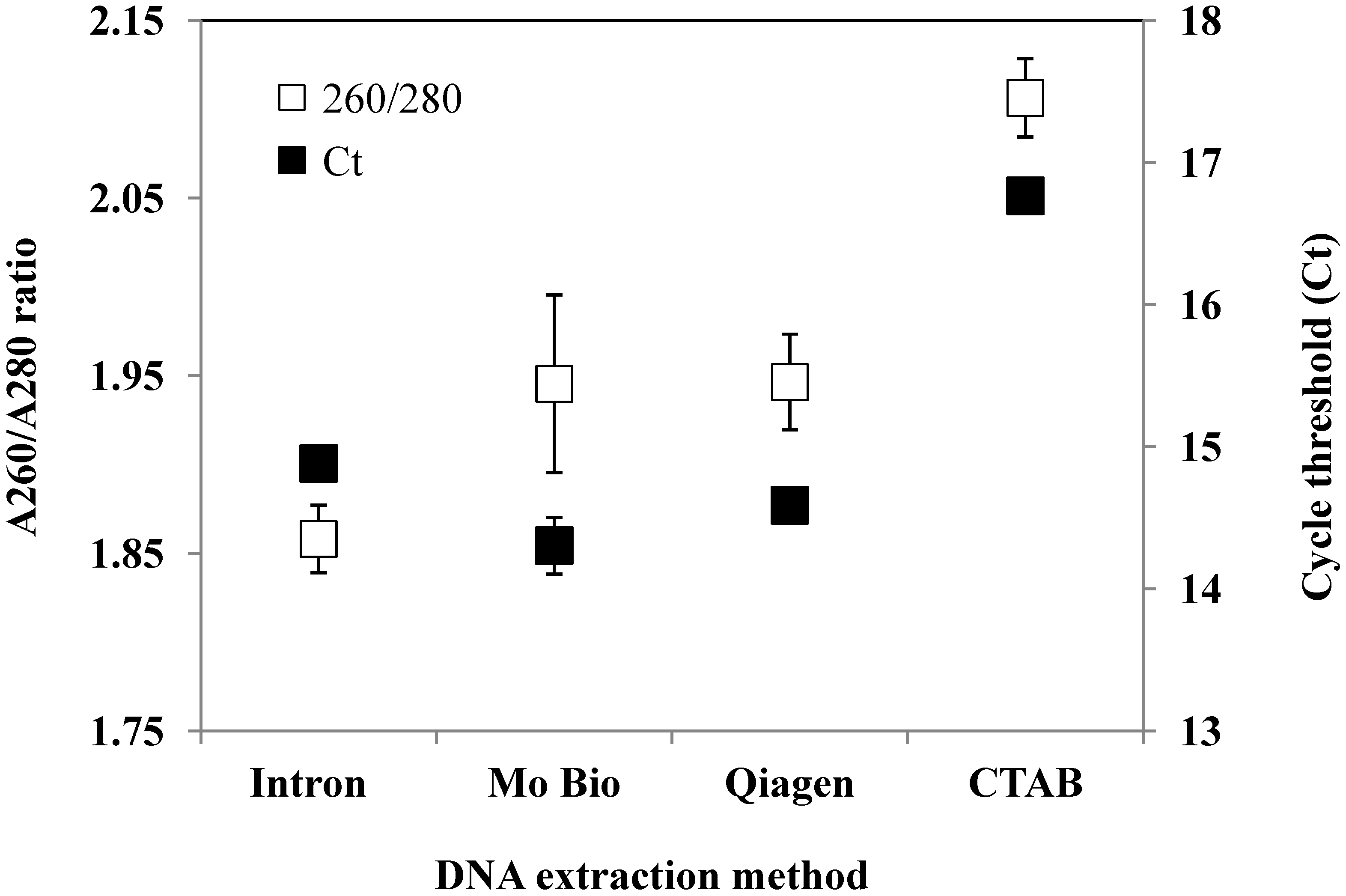
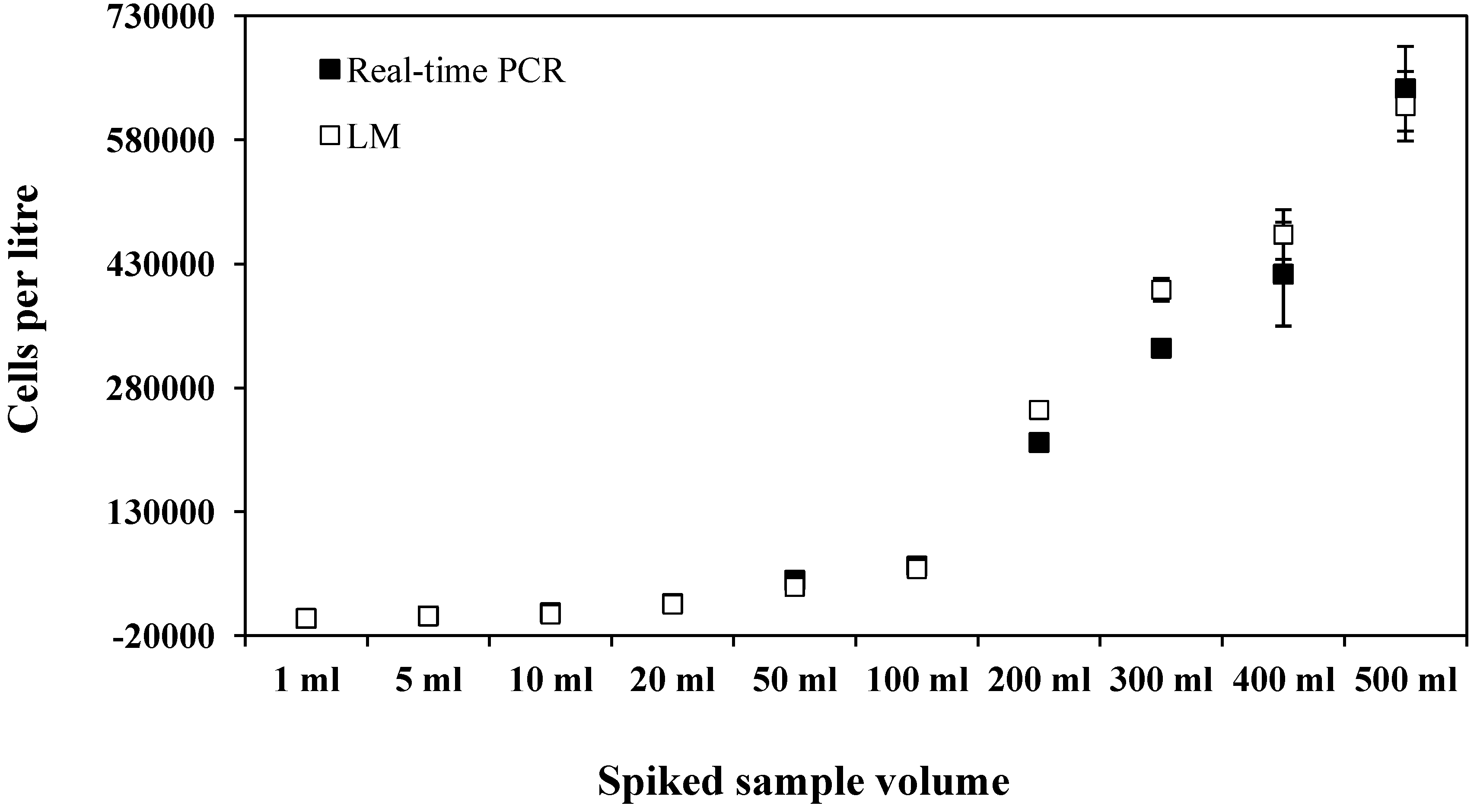
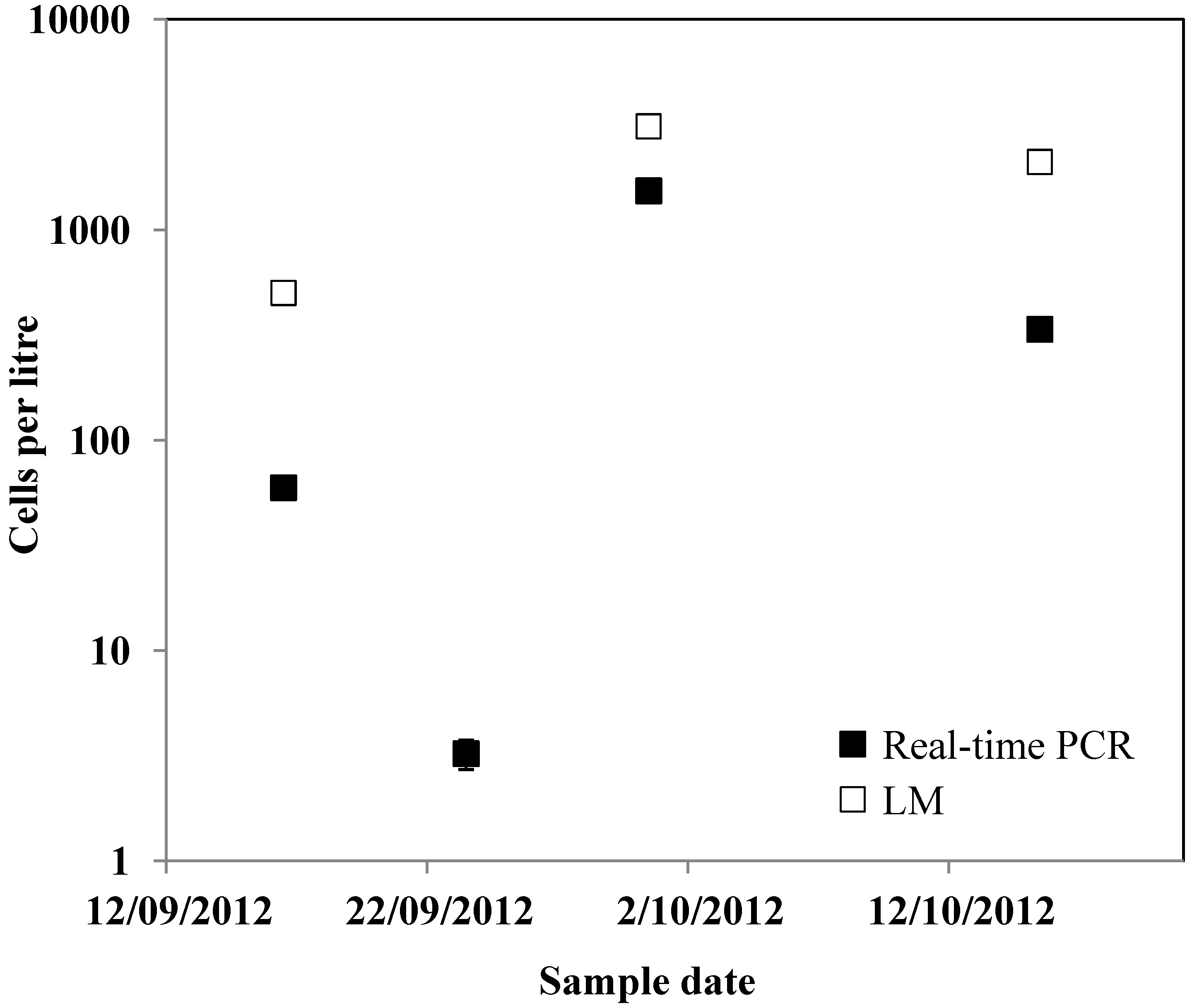
2. Results and Discussion

In this study eight real-time PCR assays were developed for the dinoflagellate species Gymnodinium aureolum, G. catenatum, Karenia brevisulcata, K. mikimotoi, K. papilionacea, K. umbella, Karlodinium veneficum, and Takayama tasmanica, all targeting the D1-D3 region of LSU rRNA gene. Designed assays ranged from 93 to 232 bp in length (Table 1). All of the assays, except for the G. aureolum assay, amplified only the target species as determined via cross-reactivity testing with strains listed in Table 2. The in silico analysis using NCBI blast also showed that primers and probes did not match with other species. Positive results for the G. aureolum assay were obtained for the strains Gymnodinium sp. (CAWD71), G. chlorophorum (CAWD62) and G. cf. microreticulatum (CAWD191). However, the assay did not cross-react with K. mikimotoi. The assay was primarily developed to differentiate the non-toxic G. aureolum from the morphologically similar toxic K. mikimotoi and so is still useful for this purpose.
Table 1. Sequences of primers and probes designed in this study including optimised final concentrations for real-time PCR assays.
Target SpeciesPrimer Name and SequenceProduct Size (bp)Final Concentration
Gymnodinium aureolumGA519-F: GGACATGGTAGCCTGCC153500 nM
GA683-R: GTCAGGAAGGTGCTCAGC500 nM
GA560-P: 6FAM-CAGAACTCACTGTCATATTGCTCCTCC-BHQ-150 nM
Gymnodinium catenatumGC397-F: CTTGGTGAGATTGTCGCAC93500 nM
GC471-R: GCAAGAAACATCACACCGA1000 nM
GC426-P: 6FAM-TGATCACCTTCTATTCCAGCGAAAGC-BHQ-180 nM
Karenia brevisulcataKBS460-F: GATCTGGATGCGATACTGAAT153300 nM
KBS585-R: AGCACTGCTACAAGACATATAA900 nM
KBS544-P: 6FAM-TGACTGAATGTCCCTAGTTGAACTC-BHQ-150 nM
Karenia mikimotoiKM541-F: CGAGTGACTGAATGTCCTCA112500 nM
KM645-R: CCAACAACCTTCATGCAGAG250 nM
KM578-P: 6FAM-CTACCAGACACACAGAGAGCAG-BHQ-150 nM
Karenia papilionaceaKP449-F: TCTGGATGCGATACTGGTTG2321000 nM
KP682-R: TACTTATGTCAAGGATGTGTTC750 nM
KP630-P: 6FAM-CTTGTTAGTTACCTGGCATGAGAC-BHQ-1125 nM
Karenia umbellaKU480-F: ATGTCAACGTCAGTTCACAAT161750 nM
KU623-R: GCACGAGACGAGGCTTA250 nM
KU542-P: 6FAM-TTCGACTAGGCACATTCAGTCAC-BHQ-150 nM
Karlodinium veneficumKV590-F: TGCCTGGTAGAACTCATGTC1001000 nM
KV672-R: ACGAGTAACAGAAGCTACAAG1000 nM
KDV640-P: 6FAM-TGTTCTCATTACCTGCGTCTGGG-BHQ-150 nM
Takayama tasmanicaTT533-F: ACTTCTGGGTGACTGAACGT134100 nM
TT665-R: CCACGTCCTGTCCCATGC1000 nM
TT616-P: 6FAM-CTGGGCTTTGTTCACTGCTCTTAA-BHQ-1125 nM
Table 2. The dinoflagellate strains with corresponding Cawthron Institute Culture Collection of Micro-algae (CICCM) codes used in this study. Accession numbers included are for sequences from the target species used to design the real-time PCR assays.
Species NameCICCM CodeAccession Number
Gymnodinium aureolumCAWD59, 87AY947659
Gymnodinium catenatumCAWD102, 101, 109, 126AY036128
Gymnodinium cf. impudicumCAWD139
Gymnodinium cf. microreticulatumCAWD191
Gymnodinium chlorophorumCAWD62
Gymnodinium impudicumCAWD03
Gymnodinium instriatumCAWD137
Gymnodinium simplexCAWD86
Gymnodinium sp.CAWD172
Karenia bidigitataCAWD80
Karenia brevisCAWD08
Karenia brevisulcataCAWD82AY243032
Karenia mikimotoiCAWD63, 117, 133, 134, 192U92249
Karenia papilionaceaCAWD91U92252
Karenia selliformisCAWD79
Karenia umbellaCAWD131, 65AY947664
Karlodinium veneficumCAWD84AY947665
Takayama helixCAWD128
Takayama tasmanicaCAWD115AY947669
Figure 1. Mean A260/A280 ratios and cycle threshold (Ct) values for replicate DNA extractions of Karenia mikimotoi. Error bars are ± standard error of three replicate DNA extractions.
Marinedrugs 12 01361 g001
The assays all had various optimised primer and probe concentrations (Table 1). The DNA extraction method that gave the best A260/A280 ratios and lowest Ct values was the PowerSoil® DNA isolation kit (Mo Bio, Carlsbad, CA, USA) (Figure 1). The PowerSoil® DNA isolation kit was also selected as these kits are optimised for the removal of environmental PCR inhibitors and environmental samples showed no evidence of PCR inhibition.
The real-time PCR assays had a linear range of detection six to eight orders of magnitude with a limit of detection (LOD) well below one cell for all assays (Table 3). This is similar to the LOD reported for other real-time PCR assays for Gymnodiniaceae species [50,51]. The amplification efficiency of all assays was between 93% and 106% (Table 3).
Table 3. Range of detection, amplification efficiency and R2 values.
Target SpeciesLower Limit of Detection (Cells/Reaction, 1 s.f.)Amplification EfficiencyR2 Value
K. mikimotoi0.007101%1.00
K. umbella0.09105%0.99
K. papilionacea0.295%0.99
K. brevisulcata0.2102%0.99
K. veneficum0.393%0.98
G. catenatum0.006106%1.00
G. aureolum0.006105%0.99
T. tasmanica0.09102%1.00
For the last two decades, species belonging to the family Gymnodiniaceae have caused a number of toxic events along the New Zealand coastline. Additionally, blooms of these species have caused major damage to marine ecosystems and aquaculture internationally during the past 60 years [15]. All assays developed in this study are regularly utilised by the micro-algae laboratory at the Cawthron Institute for confirmation of species identification during routine examination of samples as part of the New Zealand Marine Phytoplankton Monitoring Programme [24]. The identification of Karenia species by LM is particularly difficult and thus cells in field samples are often identified as K. cf. mikimotoi. The conclusive identification of a potentially toxic species is most difficult when cell concentrations are low or the species is rarely encountered in routine monitoring. For example, in 1998 the southern coast of the North Island of New Zealand experienced a severe HAB, which devastated almost all the marine life in Wellington Harbour [52]. A new Karenia species was isolated from the bloom and named Karenia brevisulcata. This species is similar in morphology to other Karenia species and has never been reported since. If this species were to bloom again it could have devastating impacts on marine ecosystems, aquaculture activities and human health. Additionally, the toxins produced by this and other Karenia species are novel bioactive compounds of great interest [15]. Accurate and early identification is vital and the assays designed in this study enable the conclusive identification of species from environmental samples with results available the same day as sample receipt. This is an important consideration for routine monitoring programmes that require a 24-h turn-around for results, but also for the mining of environmental samples for specific bioactive compound producers.
The copy number of the LSU rRNA gene per cell was determined for G. catenatum. The standard curve of serially diluted PCR product had a regression equation of y = −3.17 + 19.60x, R2 = 1.0 and an amplification efficiency of 107%. This is similar to the regression curve and amplification efficiency of the standard curve generated by cell number (Table 3). The mean copy number of the cultured strain was 20,800 ± 1566 copies per cell cell. This is comparable to values calculated for other dinoflagellate species [43,46,53]. The estimated LSU rRNA gene copy number calculated in this study also corresponds to the finding from Godhe et al. [54] that found the number of rDNA copies per cell is significantly correlated to the biovolume of dinoflagellate cells (y = −0.61 + 1.22x, R2 = 0.75; x = log rDNA molecules cell−1, y = log biovolume μm3 cell−1). The average biovolume of G. catenatum cells from the culture used to calculate copy number was 38,438 μm3, which equates to 23,335 copies of rDNA molecules cell−1 from the regression equation in Godhe et al. [54].
The G. catenatum assay was further developed for quantification to demonstrate the potential for the analysis of environmental samples and in response to the detection of naturally occurring cells in Manukau Harbour, New Zealand. The saxitoxin producing species G. catenatum was first detected in New Zealand in 2000 [7], although there is some evidence that this species may have been present in New Zealand prior to this, with a large range expansion in 2000 [8]. Gymnodinium catenatum has increased its geographic range around the entire North Island coastline of New Zealand over the last 12 years, and regular blooms are common in some areas (New Zealand Food Safety Authority, [55]). The ability to differentiate G. catenatum from morphologically similar, non-toxic species (e.g., G. impudicum) during routine monitoring using LM can be difficult, particularly at the onset of blooms when only a few individual cells per litre are present in seawater samples. Additionally, the morphology of G. catenatum cells can also be variable and be present as either single cells or chains [26].
Estimates of G. catenatum cells per litre determined by the real-time PCR assay were generally similar to or slightly fewer than LM estimates, except at low levels below the limit of detection for LM (Figure 2 and Figure 3). Real-time PCR cell estimates ranged between 70% and 108% of LM cell estimates. This is comparable to what has been found for other real-time PCR assays [50] and has been proposed to be due to factors such as DNA recovery, PCR inhibitors and the exponential nature of PCR [46]. To test for PCR inhibition, the DNA extracts were diluted 1:10 and re-amplified. This did not alter the results and the assay successfully detected cell numbers ranging from below 10 cells per reaction to over 5000 cells per reaction in the spiked samples, which equated to 700 to over 600,000 cells per litre. For natural samples the assay detected a range of cell estimates from 0.07 to 16 cells per reaction, which equated to 3 to 1528 cells per litre. The sample size analysed by the two methods was very different (i.e., 10 mL versus 300 mL) and as G. catenatum can occur as both single cells and chains of variable length (more than 60 cells/chain) [7] reliable subsampling can be difficult. LM analysis did not detect G. catenatum from samples collected in Manukau Harbour on the 23 September 2012, but the real-time PCR assay detected approximately three cells per litre (Figure 3). All positive environmental samples from Manukau Harbour were sequenced and sequences were identical. Using blastn searches, the highest homology found was with G. catenatum (e.g., GenBank acc. no. AY036128, coordinates 395-487: query coverage = 100%, E-value = 5 × 10⁻¹⁵, percent identity = 100%). Due to the difficulties in thoroughly testing all assays for specificity, we recommend DNA sequencing to confirm species identification especially during the initial stages of assay development.
Figure 2. Gymnodinium catenatum cell number estimates by real-time PCR and light microscopy (LM) from natural phytoplankton samples spiked with cultured G. catenatum cells. Error bars are ±standard error from triplicate LM analyses and real-time PCR assays.
Marinedrugs 12 01361 g002
Figure 3. Gymnodinium catenatum cell number estimates by real-time PCR and light microscopy (LM) from samples collected at Manukau Bay, New Zealand. Error bars are ±standard error from replicate real-time PCR assays.
Marinedrugs 12 01361 g003
The G. catenatum quantitative real-time PCR assay not only enabled conclusive identification but also detected the presence of cells at the LOD for LM. In this study the analysis of samples by LM and Utermöhl chambers has a lower LOD of 100 cells per litre [31]. This is at the threshold for triggering toxin testing in shellfish. As the real-time PCR assay has a LOD of less than one cell, G. catenatum could be reliably detected in field samples that were negative using LM. This lower LOD allows far greater forewarning of potentially toxic blooms for shellfish harvesters.

3. Experimental Section

3.1. Culture Maintenance

Dinoflagellate cultures (Table 2) were maintained in GP, 50% GP [56], or F/2 [57] medium at 100 µmol photons m−2 s−1 (14:10 h light:dark), 19 °C (±1 °C). The Cawthron Institute Culture Collection of Micro-algae (CICCM) provided the strains used in this study (Table 2). Cultures were harvested during exponential growth phase for assay optimisation or cross-reactivity testing.

3.2. DNA Extraction

Exponentially growing cultures of Karenia mikimotoi (CAWD63, CAWD117, CAWD133, CAWD134, CAWD192) were pooled and twelve 30 mL subsamples were filtered (Durapore membrane filters, 0.45 µm, Millipore, Billerica, MA, USA) and frozen overnight (−20 °C). DNA extraction was assessed using four methods (three replicates of each); (i-genomic CTB DNA extraction mini kits (Intron, Gyeonggi-do, South Korea), PowerSoil® DNA isolation kit (Mo Bio, Carlsbad, CA, USA), DNeasy mini plant kit (Qiagen, Alameda, CA, USA), and a cetyltrimethylammonium bromide (CTAB) method [58]. All DNA extractions were eluted into 50 μL and quantified using a NanoPhotometer (Implen, Munich, Germany) to check for DNA quantity and quality (A260/A280 ratio). Each DNA extraction was assessed using 10 ng of K. mikimotoi genomic DNA with the real-time PCR conditions described above.

3.3. Primers and Probe Design for Real-Time PCR Assays

The target positions for forward and reverse primers and the hydrolysis probes were designed using a multiple LSU rRNA gene (D1-D3 region) alignments (ClustalW) [59] of the target species and sequences from closely related species obtained from GenBank. Separate assays were designed for the detection of eight species including Gymnodinium aureolum, G. catenatum, Karenia brevisulcata, K. mikimotoi, K. papilionacea, K. umbella, Karlodinium veneficum, and Takayama tasmanica. The specificity of the primer sequences was then confirmed using BLAST (Basic Local Alignment Search Tool) at NCBI (National Centre for Biotechnology Information). The hydrolysis probes were synthesized (GeneWorks, Adelaide, Australia) with 6-FAM reporter dye at the 5′-end and Black Hole Quencher 1 at the 3′-end (Table 1).

3.4. Real-Time PCR Assay Optimisation, Specificity and Sensitivity

Real-time PCR assays were optimised on a Rotor-Gene 6000 (Corbett, Sydney, NSW, Australia), using genomic DNA extracted from an exponentially growing culture of the target species. The optimised assays consisted of a 20 μL reaction containing 10 μL of Platinum® Quantitative PCR SuperMix-UDG (Invitrogen, Carlsbad, CA, USA), 0.8 μg non-acetylated bovine serum albumin (BSA; Sigma-Aldrich, Auckland, New Zealand), and 10 ng of DNA template. Optimised primer and probe concentrations for each assay are shown in Table 1. All PCR reactions in this study were set up manually and all included no template control reactions. Assays were run in clear 0.2 mL thin-wall PCR tubes (Axygen, Union City, CA, USA). All assays had an optimised annealing temperature of 60 °C and PCR cycling conditions were: 50 °C for 2 min, 95 °C for 2 min and 45 cycles of 95 °C for 15 s and 60 °C for 45 s.
The specificity of each assay was verified using DNA from closely related species (Table 2). DNA from each species (10 ng) was used in the real-time PCR assays as described above. The sensitivity of each assay was evaluated with genomic DNA extracted using PowerSoil® DNA isolation kits from known cell concentrations of the target species. The amplification efficiency of the assay was determined by using serially diluted genomic DNA samples (analysed in duplicate) ranging from approximately 100,000 to 1 × 10⁻⁴ cells per reaction and the corresponding cycle threshold (Ct) data.

3.5. Determination of Copy Number and Quantification of the Gymnodinium catenatum Assay

The assay specific for G. catenatum was further developed in order to demonstrate the potential for quantification of field samples. DNA was extracted from replicate samples of known numbers of cells of the CAWD126 strain of G. catenatum. Cell concentrations of culture were estimated during exponential growth phase using the Utermöhl technique [60]. Replicates samples consisting of 150,000 cells were were filtered (Durapore membrane filters, 0.45 µm, Millipore, Billerica, MA, USA), frozen overnight (−20 °C) and transferred to the first tube of a PowerSoil® DNA isolation kit. These extractions were serially diluted and used to generate a standard curve of known cell number per reaction versus Ct data.
To estimate the LSU rRNA gene copy number per cell a dilution series of a known concentration of LSU rRNA gene PCR product, ranging from 1 to 0.001 ng was analysed together with extractions of known cell number from above. The number of molecules of PCR product was then determined by the formula: (A × 6.022 × 1023) × (660 × B)−1, with A being the concentration of the PCR product and B the length of the PCR product. The number of molecules in the extractions with known cell number was determined using the PCR product standard curve to obtain the copy number of the LSU rRNA gene per cell. The average biovolume of cells from the culture used was also calculated to determine the relationship between cell size and gene copy number following Godhe et al. [54].

3.6. Spiked Environmental and Natural Sample Testing

The effectiveness of the real-time PCR assay for the identification and discrimination of G. catenatum from phytoplankton samples was examined using samples spiked with cultured cells. A surface seawater sample (10 L) was collected from Nelson Marina (Nelson, South Island, New Zealand). Different volumes (1, 5, 10, 20, 50, 100, 200, 300, 400 and 500 mL) of G. catenatum culture (mix of CAWD101, CAWD102, CAWD109 and CAWD126) were made up to one litre volume with the natural phytoplankton sample to create ten contrived samples. From the one litre samples triplicate 10 mL aliquots were preserved in Lugol’s solution analysed by Light Microscopy (LM) and triplicate 300 mL aliquots were analysed using the real-time PCR assay. For real-time PCR analyses samples were filtered and genomic DNA extracted with PowerSoil® DNA isolation kits (Mo Bio, Carlsbad, CA, USA) as described above. To estimate the abundance of G. catenatum in environmental samples Ct values were used to calculate cell number per reaction based on the standard curve generated with the serial dilution of DNA extracts of known cell number (ranging from 6000 to 0.006 cells per reaction). The real-time PCR assays all included standard curves, positive controls, negative controls and blank extraction controls. DNA samples were also diluted 1:10 to determine evidence of PCR inhibition.
Samples were collected from Manukau Harbour (Auckland, North Island, New Zealand) as part of the New Zealand Marine Phytoplankton Monitoring Programme (by the New Zealand Food Safety Authority, Ministry for Primary Industries, Wellington, New Zealand). Unpreserved and preserved (Lugol’s solution [60]) replicate samples were received within 24 h of collection. Grab samples (100 mL) from three depths (0, 3 and 6 m) were pooled (total 300 mL). G. catenatum cells were identified by LM and cell number estimates estimated using the Utermöhl technique [61] by the micro-algae laboratory at the Cawthron Institute, New Zealand (International Accreditation New Zealand: ISO 17025). One 10 mL subsample, from the pooled 300 mL, was analysed by LM for each environmental sample. For real-time PCR analysis the unpreserved grab samples filtered and genomic DNA extracted with PowerSoil® DNA isolation kits (Mo Bio, Carlsbad, CA, USA) as described above. The abundance of G. catenatum in environmental samples was calculated as above. Environmental samples were also PCR amplified for DNA sequencing (Sanger sequencing) to confirm positive results. PCR amplifications were carried out in 50 μL reaction volumes containing i-Taq 26 PCR master mix (25 μL; Intron, Gyeonggi-do, Korea), both forward and reverse primers (0.4 mM) and template (ca. 50–150 ng of DNA). Thermocycling conditions were the same as for real-time PCR. Amplification products were purified (AxyPrep PCR cleanup kits, Axygen, Union City, CA, USA) and sequenced in both directions using the primers from real-time PCR assay by an external contractor (University of Waikato DNA Sequencing Facility, Hamilton, New Zealand). The resulting sequences were compared to existing sequences in GenBank using the BLAST online software.

4. Conclusions

At present real-time PCR is the most cost-effective, sensitive, and rapid molecular technique for the detection and quantification of dinoflagellates from environmental samples [62]. The assays developed in this study demonstrate great potential for aiding in monitoring programmes for both food safety purposes and rapid screening of samples for species of interest. These assays are all utilised regularly as part of the New Zealand Marine Phytoplankton Monitoring Programme [24], as support for LM analyses by confirming the identification of toxic species in water samples. However, as multiplexing techniques, sequencing technologies, genomic and bioinformatic resources all improve, it is likely that molecular techniques will be increasingly utilised for routine phytoplankton monitoring programmes [34,45,62].

Acknowledgements

We would like to thank K. Ponikla (Cawthron Institute) for technical support, L. MacKenzie for Gymnodinium catenatum measurements, and the Ministry of Primary Industries for kindly supplying the environmental samples. This work was supported by funding from the New Zealand Ministry of Science and Innovation, contract CAW0703.

Author Contributions

K.F.S., L.L.R. and M.d.S. designed the experiments. M.d.S. designed the primers. K.F.S. collected the field samples except for samples collected by the New Zealand Food Safety Authority. K.F.S., M.d.S. and J.A. performed the laboratory work. K.F.S., L.L.R. and M.d.S. wrote the manuscript.

Conflicts of Interest

The authors declare no conflict of interest.

References

  1. Daugbjerg, N.; Hansen, G.; Larsen, J.; Moestrup, O. Phylogeny of some of the major genera of dinoflagellates based on ultrastructure and partial LSU rDNA sequence data, including the erection of three new genera of unarmoured dinoflagellates. Phycologia 2000, 39, 302–317. [Google Scholar] [CrossRef]
  2. de Salas, M.F.; Bolch, C.J.S.; Botes, L.; Nash, G.; Wright, S.W.; Hallegraeff, G.M. Takayama gen. nov (Gymnodiniales, Dinophyceae), a new genus of unarmored dinoflagellates with sigmoid apical grooves, including the description of two new species. J. Phycol. 2003, 39, 1233–1246. [Google Scholar] [CrossRef]
  3. Brand, L.E.; Campbell, L.; Bresnan, E. Karenia: the biology and ecology of a toxic genus. Harmful Algae 2012, 14, 156–178. [Google Scholar] [CrossRef]
  4. Taylor, F.J.R.; Fukuyo, Y.; Larsen, J.; Hallegraeff, G.M. Taxonomy of Harmful Dinoflagellates. In Manual on Harmful Marine Microalgae; Hallegraeff, G.M., Anderson, D.M., Cembella, A.D., Eds.; UNESCO Intergovernmental Oceanographic Commission: Pairs, France, 2003; pp. 389–432. [Google Scholar]
  5. Rhodes, L.L.; Haywood, A.J.; Ballantine, W.J.; MacKenzie, A.L. Algal blooms and climate anomalies in north-east New Zealand, August–December 1992. N. Z. J. Mar. Freshw. Res. 1993, 27, 419–430. [Google Scholar] [CrossRef]
  6. Wear, R.G.; Gardner, J.P.A. Biological effects of the toxic algal bloom of February and March 1998 on the benthos of Wellington Harbour, New Zealand. Mar. Ecol. Prog. Ser. 2001, 218, 63–76. [Google Scholar] [CrossRef]
  7. MacKenzie, L.A.; Beauchamp, T. Gymnodinium catenatum in New Zealand: A New Problem for Public Health and the Shellfish Industry; Cawthron Institute: Nelson, New Zealand, 2002. [Google Scholar]
  8. Irwin, A.; Hallegraeff, G.M.; McMinn, A.; Harrison, J.; Heijnis, H. Cyst and radionuclide evidence demonstrate historic Gymnodinium catenatum dinoflagellate populations in Manukau and Hokianga Harbours, New Zealand. Harmful Algae 2003, 2, 61–74. [Google Scholar] [CrossRef]
  9. Yang, Z.B.; Takayama, H.; Matsuoka, K.; Hodgkiss, I.J. Karenia digitata sp nov. (Gymnodiniales, Dinophyceae), a new harmful algal bloom species from the coastal waters of west Japan and Hong Kong. Phycologia 2000, 39, 463–470. [Google Scholar] [CrossRef]
  10. Yang, Z.B.; Hodgkiss, I.J.; Hansen, G. Karenia longicanalis sp nov. (Dinophyceae): A new bloom-forming species isolated from Hong Kong, May 1998. Bot. Mar. 2001, 44, 67–74. [Google Scholar]
  11. Haywood, A.J.; Steidinger, K.A.; Truby, E.W.; Bergquist, P.R.; Bergquist, P.L.; Adamson, J.; MacKenzie, L. Comparative morphology and molecular phylogenetic analysis of three new species of the genus Karenia (Dinophyceae) from New Zealand. J. Phycol. 2004, 40, 165–179. [Google Scholar] [CrossRef]
  12. Jasperse, J.A. Marine Toxins and New Zealand Shellfish: Proceedings of a Workshop on Research Issues, 10–11 June 1993; Royal Society of New Zealand: Wellington, New Zealand; p. 1993.
  13. Todd, K. A Review of NSP Monitoring in New Zealand in Support of a New Programme; Cawthron Report No. 660; Marine Biotoxin Technical Committee: Nelson, New Zealand, 2003. [Google Scholar]
  14. MacKenzie, L.A.; Haywood, A.J.; Adamson, J.; Truman, P.; Till, D.; Seki, T.; Satake, M.; Yasumoto, T. Gymnodimine Contamination of Shellfish in New Zealand. In Harmful and Toxic Algal Blooms; Yasumoto, T., Oshima, Y., Fukuyo, Y., Eds.; Intergovernmental Oceanographic Commission of UNESCO: Paris, France, 1996; pp. 97–100. [Google Scholar]
  15. Holland, P.T.; Shi, F.; Satake, M.; Hamamoto, Y.; Ito, E.; Beuzenberg, V.; McNabb, P.; Munday, R.; Briggs, L.; Truman, P.; et al. Novel toxins produced by the dinoflagellate Karenia brevisulcata. Harmful Algae 2012, 13, 47–57. [Google Scholar] [CrossRef]
  16. Satake, M.; Shoji, M.; Oshima, Y.; Naoki, H.; Fujita, T.; Yasumoto, T. Gymnocin-A, a cytotoxic polyether from the notorious red tide dinoflagellate, Gymnodinium mikimotoi. Tetrahedron Lett. 2002, 43, 5829–5832. [Google Scholar] [CrossRef]
  17. Satake, M.; Tanaka, Y.; Ishikura, Y.; Oshima, Y.; Naoki, H.; Yasumoto, T. Gymnocin-B with the largest contiguous polyether rings from the red tide dinoflagellate, Karenia (formerly Gymnodinium) mikimotoi. Tetrahedron Lett. 2005, 46, 3537–3540. [Google Scholar] [CrossRef]
  18. de Salas, M.F.; Bolch, C.J.S.; Hallegraeff, G.M. Karlodinium australe sp. nov. (Gymnodiniales, Dinophyceae), a new potentially ichthyotoxic unarmoured dinoflagellate from lagoonal habitats of south-eastern Australia. Phycologia 2005, 44, 640–650. [Google Scholar] [CrossRef]
  19. García Camacho, F.; Gallardo Rodríguez, J.; Sánchez Mirón, A.; Cerón García, M.C.; Belarbi, E.H.; Chisti, Y.; Molina Grima, E. Biotechnological significance of toxic marine dinoflagellates. Biotechnol. Adv. 2007, 25, 176–194. [Google Scholar] [CrossRef]
  20. Waters, A.L.; Hill, R.T.; Place, A.R.; Hamann, M.T. The expanding role of marine microbes in pharmaceutical development. Curr. Opin. Biotechnol. 2010, 21, 780–786. [Google Scholar] [CrossRef]
  21. Beuzenberg, V.; Mountfort, D.; Holland, P.; Shi, F.; MacKenzie, L. Optimization of growth and production of toxins by three dinoflagellates in photobioreactor cultures. J. Appl. Phycol. 2012, 24, 1023–1033. [Google Scholar] [CrossRef]
  22. Selwood, A.I.; van Ginkel, R.; Wilkins, A.L.; Munday, R.; Ramsdell, J.S.; Jensen, D.J.; Cooney, J.M.; Miles, C.O. Semisynthesis of S-Desoxybrevetoxin-B2 and Brevetoxin-B2, and assessment of their acute toxicities. Chem. Res. Toxicol. 2008, 21, 944–950. [Google Scholar] [CrossRef]
  23. Rhodes, L.L.; Scholin, C.; Tyrrell, J.; Adamson, J.; Todd, K. The integration of DNA probes into New Zealand’s routine phytoplankton monitoring programmes. In Harmful Algal Blooms; Hallegraeff, G.M., Blackburn, S.I., Bolch, C.J., Lewis, R.J., Eds.; Intergovernmental Oceanographic Commission of UNESCO: Paris, France, 2001; pp. 429–432. [Google Scholar]
  24. Rhodes, L.L.; Smith, K.F.; Moisan, C. Shifts and stasis in marine HAB monitoring in New Zealand. Environ. Sci. Pollut. Res. 2013, 20, 6872–6877. [Google Scholar] [CrossRef]
  25. NZFSA. Non-Commercial Marine Biotoxin Monitoring Programme: NZFSA VA Operating and Response Manual; New Zealand Food Safety Authority (NZFSA): Wellington, New Zealand, 2010; p. 39. [Google Scholar]
  26. Rhodes, L.L.; Smith, K.F.; de Salas, M. DNA probes, targeting large sub-unit rRNA, for the rapid identification of the paralytic shellfish poison producing dinoflagellate, Gymnodinium catenatum. N. Z. J. Mar. Freshw. Res. 2007, 41, 385–390. [Google Scholar] [CrossRef]
  27. Burkholder, J.M. Implications of harmful microalgae and heterotrophic dinoflagellates in management of sustainable marine fisheries. Ecol. Appl. 1998, 8, S37–S62. [Google Scholar]
  28. Van Dolah, F.M. Marine algal toxins: origins, health effects, and their increased occurrence. Environ. Health Perspect. 2000, 108, 133–141. [Google Scholar] [CrossRef]
  29. Landsberg, J.H. The effects of harmful algal blooms on aquatic organisms. Rev. Fish. Sci. 2002, 10, 113–390. [Google Scholar] [CrossRef]
  30. Hallegraeff, G.M. Ocean climate change, phytoplankton community responses, and harmful algal blooms: a formidable predictive challenge. J. Phycol. 2010, 46, 220–235. [Google Scholar] [CrossRef]
  31. Godhe, A.; Cusack, C.; Pedersen, J.; Anderson, P.; Anderson, D.M.; Breasnan, E.; Cembella, A.; Dahl, E.; Diercks, S.; Elbrachter, M.; et al. Intercalibration of classical and molecular techniques for identification of Alexandrium fundyense (Dinophyceae) and estimation of cell densities. Harmful Algae 2007, 6, 56–72. [Google Scholar] [CrossRef] [Green Version]
  32. John, U.; Medlin, L.K.; Groben, R. Development of specific rRNA probes to distinguish between geographic clades of the Alexandrium tamarense species complex. J. Plankton Res. 2005, 27, 199–204. [Google Scholar] [CrossRef]
  33. Penna, A.; Bertozzini, E.; Battocchi, C.; Galluzzi, L.; Giacobbe, M.G.; Vila, M.; Garces, E.; Luglie, A.; Magnani, M. Monitoring of HAB species in the Mediterranean Sea through molecular methods. J. Plankton Res. 2007, 29, 19–38. [Google Scholar]
  34. Wood, S.A.; Smith, K.F.; Banks, J.C.; Tremblay, L.A.; Rhodes, L.L.; Mountfort, D.; Cary, S.C.; Pochon, X. Molecular genetic tools for environmental monitoring of New Zealand’s aquatic habitats, past, present and the future. N. Z. J. Mar. Freshw. Res. 2013, 47, 90–119. [Google Scholar] [CrossRef]
  35. Miller, P.E.; Scholin, C.A. Identification and enumeration of cultured and wild Pseudo-nitzschia (Bacillariophyceae) using species-specific LSU rRNA-targeted fluorescent probes and filter-based whole cell hybridization. J. Phycol. 1998, 34, 371–382. [Google Scholar] [CrossRef]
  36. Rhodes, L.L.; Scholin, C.; Garthwaite, I. Pseudo-nitzschia in New Zealand and the role of DNA probes and immunoassays in refining marine biotoxin monitoring programmes. Nat. Toxins 1998, 6, 105–111. [Google Scholar] [CrossRef]
  37. Ayers, K.; Rhodes, L.L.; Tyrrell, J.V.; Gladstone, M.; Scholin, C.A. International accreditation of sandwich hybridisation assay format DNA probes for micro-algae. N. Z. J. Mar. Freshw. Res. 2005, 39, 1225–1231. [Google Scholar] [CrossRef]
  38. Haywood, A.J.; Scholin, C.A.; Marin, R., III; Steidinger, K.A.; Heil, C.; Ray, J. Molecular detection of the brevetoxin-producing dinoflagellate Karenia brevis and closely related species using rRNA-targeted probes and a semiautomated sandwich hybridization assay. J. Phycol. 2007, 43, 1271–1286. [Google Scholar] [CrossRef]
  39. Bowers, H.A.; Tengs, T.; Goto, S.; Tomas, C.; Ono, C.; Yoshimatsu, S.; Oldach, D.; Steidinger, K.A.; Landsberg, J.H.; Tomas, C.R.; et al. Development of real-time PCR assays for the detection of Chattonella species in culture and environmental samples. In Harmful Algae 2002; Steidinger, K.A., Landsberg, J.H., Tomas, C.R., Vargo, G.A., Eds.; Florida Institute of Oceanography, and Intergovernmental Oceanographic Commission of UNESCO: Pairs, France, 2004; pp. 231–233. [Google Scholar]
  40. Coyne, K.J.; Handy, S.M.; Demir, E.; Whereat, E.B.; Hutchins, D.A.; Portune, K.J.; Doblin, M.A.; Cary, S.C. Improved quantitative real-time PCR assays for enumeration of harmful algal species in field samples using an exogenous DNA reference standard. Limnol. Oceanogr. 2005, 3, 381–391. [Google Scholar] [CrossRef]
  41. Patil, J.; Gunasekera, R.; Deagle, B.; Bax, N.; Blackburn, S. Development and evaluation of a PCR based assay for detection of the toxic dinoflagellate, Gymnodinium catenatum (Graham) in ballast water and environmental samples. Biol. Invasions 2005, 7, 983–994. [Google Scholar] [CrossRef]
  42. Dyhrman, S.T.; Erdner, D.L.; La Du, J.; Galac, M.; Anderson, D.M. Molecular quantification of toxic Alexandrium fundyense in the Gulf of Maine using real-time PCR. Harmful Algae 2006, 5, 242–250. [Google Scholar] [CrossRef]
  43. Murray, S.A.; Wiese, M.; Stüken, A.; Brett, S.; Kellmann, R.; Hallegraeff, G.; Neilan, B.A. sxtA-based quantitative molecular assay to identify saxitoxin-producing harmful algal blooms in marine waters. Appl. Environ. Microb. 2011, 77, 7050–7057. [Google Scholar] [CrossRef]
  44. Perini, F.; Casabianca, A.; Battocchi, C.; Accoroni, S.; Totti, C.; Penna, A. New approach using the real-time PCR method for estimation of the toxic marine dinoflagellate Ostreopsis cf. ovata in marine environment. PLoS One 2011, 6, e17699. [Google Scholar]
  45. Penna, A.; Galluzzi, L. The quantitative real-time PCR applications in the monitoring of marine harmful algal bloom (HAB) species. Environ. Sci. Pollut. Res. 2013, 20, 6851–6862. [Google Scholar] [CrossRef]
  46. Vandersea, M.W.; Kibler, S.R.; Holland, W.C.; Tester, P.A.; Schultz, T.F.; Faust, M.A.; Holmes, M.J.; Chinain, M.; Litaker, R.W. Development of semi-quantitative PCR assays for the detection and enumeration of Gambierdiscus species (Gonyaulacales, Dinophyceae). J. Phycol. 2012, 48, 902–915. [Google Scholar] [CrossRef]
  47. Hariganeya, N.; Tanimoto, Y.; Yamaguchi, H.; Nishimura, T.; Tawong, W.; Sakanari, H.; Yoshimatsu, T.; Sato, S.; Preston, C.M.; Adachi, M. Quantitative PCR method for enumeration of cells of cryptic species of the toxic marine dinoflagellate Ostreopsis spp. in coastal waters of Japan. PLoS One 2013, 8, e57627. [Google Scholar] [CrossRef]
  48. Gescher, C.; Metfies, K.; Medlin, L.K. The ALEX CHIP—Development of a DNA chip for identification and monitoring of Alexandrium. Harmful Algae 2008, 7, 485–494. [Google Scholar] [CrossRef]
  49. Galluzzi, L.; Cegna, A.; Casabianca, S.; Penna, A.; Saunders, N.; Magnani, M. Development of an oligonucleotide microarray for the detection and monitoring of marine dinoflagellates. J. Microbiol. Methods 2011, 84, 234–242. [Google Scholar] [CrossRef]
  50. Zamor, R.M.; Glenn, K.L.; Hambright, K.D. Incorporating molecular tools into routine HAB monitoring programs: using qPCR to track invasive Prymnesium. Harmful Algae 2012, 15, 1–7. [Google Scholar] [CrossRef]
  51. Yuan, J.; Mi, T.; Zhen, Y.; Yu, Z. Development of a rapid detection and quantification method of Karenia mikimotoi by real-time quantitative PCR. Harmful Algae 2012, 17, 83–91. [Google Scholar] [CrossRef]
  52. Chang, F.H. Gymnodinium brevisulcatum sp nov (Gymnodiniales, Dinophyceae), a new species isolated from the 1998 summer toxic bloom in Wellington Harbour, New Zealand. Phycologia 1999, 38, 377–384. [Google Scholar] [CrossRef]
  53. Galluzzi, L.; Bertozzini, E.; Penna, A.; Perini, F.; Garces, E.; Magnani, M. Analysis of rRNA gene content in the Mediterranean dinoflagellate Alexandrium catenella and Alexandrium taylori: Implications for the quantitative real-time PCR-based monitoring methods. J. Appl. Phycol. 2010, 22, 1–9. [Google Scholar] [CrossRef]
  54. Godhe, A.; Asplund, M.E.; Härnström, K.; Saravanan, V.; Tyagi, A.; Karunasagar, I. Quantification of diatom and dinoflagellate biomasses in coastal marine seawater samples by real-time PCR. Appl. Environ. Microb. 2008, 74, 7174–7182. [Google Scholar] [CrossRef]
  55. New Zealand Food Safety Authority, Ministry for Primary Industries, Wellington, New Zealand. Unpublished data. 2013.
  56. Loeblich, A.R.; Smith, V.E. Chloroplast pigments of the marine dinoflagellate Gyrodinium resplendens. Lipids 1968, 3, 5–13. [Google Scholar] [CrossRef]
  57. Keller, M.D.; Selvin, R.C.; Claus, W.; Guillard, R.R.L. Media for the culture of oceanic ultraphytoplankton. J. Phycol. 1987, 23, 633–638. [Google Scholar]
  58. Doyle, J.J.; Doyle, J.L. A rapid DNA isolation procedure for small quantities of fresh leaf tissue. Phytochem. Bull. 1987, 19, 11–15. [Google Scholar]
  59. Thompson, J.D.; Higgins, D.G.; Gibson, T.J. CLUSTAL W: Improving the sensitivity of progressive multiple sequence alignment through sequence weighting, position-specific gap penalties and weight matrix choice. Nucleic Acids Res. 1994, 22, 4673–4680. [Google Scholar] [CrossRef]
  60. Throndsen, J. Preservation and storage. In Phytoplankton Manual; Sournia, A., Ed.; United Nations Educational, Scientific and Cultural Organization (UNESCO): Paris, France, 1978; pp. 69–74. [Google Scholar]
  61. Utermöhl, H. Zur vervollkommung der quantitativen phytoplankton methodik (Towards a perfection of quantitative phytoplankton methodology). Mitt. Int. Ver. Theor. Angew. Limnol. 1958, 9, 1–38. [Google Scholar]
  62. Ebenezer, V.; Medlin, L.K.; Ki, J.S. Molecular detection, quantification, and diversity evaluation of microalgae. Mar. Biotechnol. 2012, 14, 129–142. [Google Scholar] [CrossRef]

