Article

Variegatusides: New Non-Sulphated Triterpene Glycosides from the Sea Cucumber Stichopus variegates Semper

1
Research Center for Marine Drugs, School of Pharmacy, Second Military Medical University, 325 Guohe Road, Shanghai 200433, China
2
Department of Pharmacy, 202th Hospital of PLA, 5 Guangrong Street, Shenyang 110003, China
3
College of Life Science, Jiangxi Normal University, 99 Ziyang Road, Nanchang 330022, China
4
School of Medicine, Tongji University, 1239 Siping Road, Shanghai 200092, China
5
Department of Pharmacy, 306th Hospital of PLA, 9 Anxiang North Road, Beijing 100101, China
*
Authors to whom correspondence should be addressed.
These authors contributed equally to this work.
Mar. Drugs 2014, 12(4), 2004-2018; https://doi.org/10.3390/md12042004
Submission received: 11 December 2013 / Revised: 29 January 2014 / Accepted: 7 February 2014 / Published: 2 April 2014
(This article belongs to the Collection Bioactive Compounds from Marine Invertebrates)

Abstract

:
Four new triterpene glycosides, variegatusides C–F (14), together with three structurally known triterpene glycosides, variegatusides A and B (5, 6), and holothurin B (7), were isolated from the sea cucumber Stichopus variegates Semper (Holothuriidae), collected from the South China Sea. Their structures were elucidated on the basis of extensive spectral analysis (nuclear magnetic resonance (NMR) and electrospray ionization mass spectrometry (ESIMS)) and chemical evidence. Variegatusides C–F exhibit the same structural feature consisting of the presence of a 23-hydroxyl group at the holostane-type triterpene aglycone side chain. Variegatuside C (1) has a double bond (24, 25) in this same chain, while variegatuside D (2) exhibits a 8(9)-ene bond in the holostane-type triterpene aglycone, which has not been extracted from other sea cucumber species. Compound 4 is a native compound from the sea cucumber S. variegates Semper, which has been reported to be desacetylstichloroside B1. Except for holothurin B, these glycosides have no sulfate group in their sugar chain and show potent antifungal activities in vitro biotests.

1. Introduction

Triterpene glycosides are the predominant secondary metabolites of holothurians and are responsible for their general toxicity. These glycosides have been reported to have a wide spectrum of biological effects, including antifungal, cytotoxic, hemolytic, and immunomodulatory activities [1,2]. In the search for new pharmacologically active substances from marine organisms, attention has been paid to echinoderms, and among them, to sea cucumbers (class Holothuridea). More than 100 of these glycosides have been described, and the majority are usually lanosterol type triterpenes with an 18(20) lactone and a sugar chain of up to six monosaccharide units (princinally d-xylose, d-glucose, d-quinovose, d-3-O-methylglucose and d-3-O-methylxylose) linked to the C-3 of the aglycone [3]. As a continuation of our studies on the structure and biological role of triterpene oligoglycosides from holothurians [4,5,6,7,8,9,10], we have firstly investigated the ethanol (EtOH) extracts of the Stichopodidae-type sea cucumber Stichopus variegates Semper collected from the Hainan province in the South China sea which is used as a tonic in China [11]. Herein, we report the isolation and structure elucidation of the four new, unprecedented, non-sulfated triterpene glycosides, variegatusides C (1), D (2), E (3) and F (4). All isolated compounds revealed different antifungal activities.

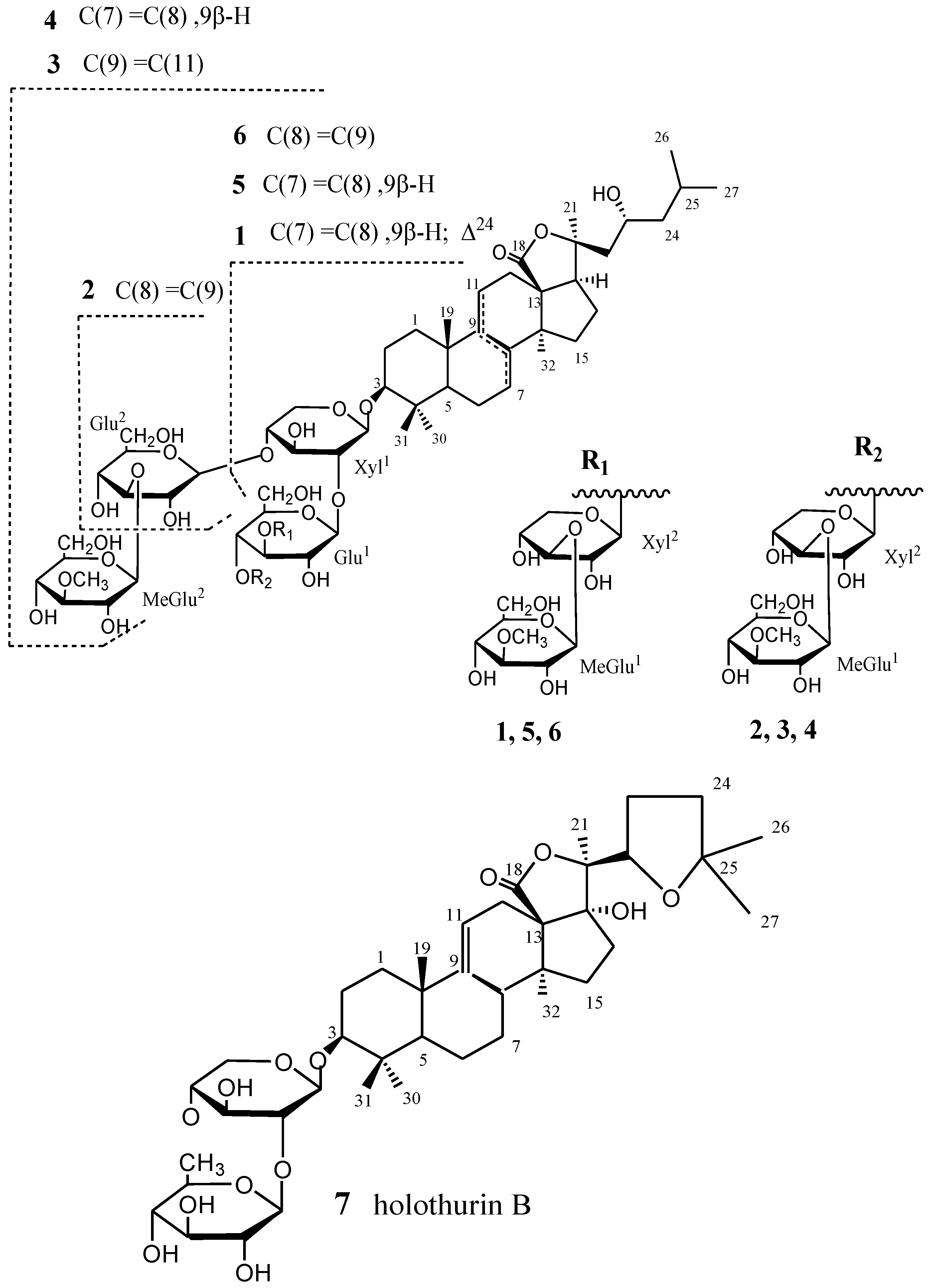
2. Results and Discussion

The 70% ethanolic extract of S. variegates (15 kg, wet weight) was suspended in H2O and partitioned successively with petroleum ether and n-BuOH. The n-BuOH extract was subjected to silica gel and reversed-phase silica (Lichroprep RP-18, 40–63 μm). Final purification of individual compounds was achieved by reversed-phase HPLC on Zobax SB C-18 to give variegatusides C (1), D (2), E (3), F (4) and three known compounds 57 (Figure 1). Structures of the glycosides have been elucidated by extensive analysis (NMR and ESIMS) and chemical method.
Variegatuside C (1), obtained as colorless amorphous powder, was positive to Liebermann-Burchard and Molish test. Its molecular formula was determined as C53H84O22 from the pseudomolecular ion peak at m/z 1071.5419 [M − H]+ in negative ion mode HRESIMS and at m/z 1095 [M + Na]+ in positive ion mode ESIMS and 13C NMR. The IR spectrum showed the presence of hydroxyl (3417 cm−1), carbonyl (1761 cm−1), and olefinic (1652 cm−1) groups.
The 1H NMR, 13C NMR and DEPT spectra of glycoside 1 showed the close aglycone with desacetylstichloroside B1 [12] and variegatuside A [13], differing only by the presence of an olefinic bond at 24(25). The 1H and 13C NMR spectral data of 1 (Table 1) suggested the presence of a triterpene aglycone with two olefinic bonds, one ester, and one hydroxyl group bonded to an oligosaccharide chain composed of four sugar units. Resonances for a 7(8)-double bond at δC 143.6 (C-8), 120.3 (C-7); δH 5.62 (m, H-7) and one 24(25)-double bond at δC 131.2 (C-25), 123.2 (C-24); δH 5.01 (d, J = 7.8 Hz, H-24) were present. The positions of two C=C bonds in 7,8 and 24,25 were corroborated by the HMBC correlations H-32/C-8, H-9/C-8, H-5/C-7, H-9/C-7, and H-27/C-25, H-24/C-25, H-27/C-24, H-23/C-24, H-22/C-24, respectively. The 13C NMR chemical shift inventory of 1 closely parallel that of frondoside A [14] except for signals assigned to C-22 (δC 48.9) and C-23 (δC 66.0) which are shifted downfield by δ 9.77 and 43.24, respectively, in agreement with the presence of an OH group at C-23. The side chain of aglycone is thus comparable to that of stichlorogenol [10], a C-23-hydroxy aglycone from the sea cucumber Stichopus chloronotus for which stereochemistry has been confirmed by X-ray crystallography [12]. Comparable signals in the 13C NMR spectrum (pyridine-d5) of the latter are observed at δC 47.62 and 65.70 [15], suggesting the 23S configuration in variegatuside C (1).
Figure 1. The structures of compounds (17).
Marinedrugs 12 02004 g001
Table 1. 1H (600 MHz) and 13C NMR (150 MHz) Data and Selected HMBC Correlations for the Aglycone Moieties of 1 and 2 (in C5D5N:D2O, 4:1) a.
No.12
δH mult. (J in Hz)δCHMBC (H→C)δH mult. (J in Hz)δCHMBC (H→C)
11.20 m; 1.70 m36.2, CH2 1.17 m; 1.72 m36.2, CH2
21.94 m; 2.21 m27.2, CH2 1.90 m; 2.10 m27.1, CH2
33.31 dd (4.8, 9.6)88.9, CH4, 30, 31, 1 (Xyl1)3.23 dd (4.8, 10.4)88.7, CH4, 30, 31, 1 (Xyl1)
4 39.8, C 39.6, C
51.26 m51.1, CH3, 4, 10, 19, 301.13 m51.1, CH3, 4, 10, 19, 30, 31
61.65 m; 1.86 m22.1, CH2 1.67 m18.4, CH2
75.62 m120.3, CH5, 6, 82.14 m; 2.20 m27.1, CH25, 6, 8
8 143.6, C 131.1, C
93.72 m48.7, CH8, 10, 11, 19 135.6, C8, 10, 11, 19
10 37.2, C 37.1, C
111.32 m; 1.46 m23.3, CH2 2.22 m; 2.34 m21.8, CH2
122.05 m33.2, CH29,11,13,182.12 m; 2.19 m28.2, CH29,11,13,18
13 59.0, C 58.9, C
14 51.1, C 49.0, C
151.80 m33.6, CH2 1.48 m; 1.60 m33.2, CH2
161.62 m; 1.74 m28.2, CH2 1.96 m; 2.10 m25.1, CH2
172.48 m52.7, CH16, 20, 212.41 m51.4, CH16, 20, 21
18 177.6, C 177.4, C
191.11 s22.3, CH31, 5, 9, 101.26 s19.2, CH31, 5, 9, 10
20 83.7, CH 84.0, C
211.67 s27.4, CH317, 20, 221.81s28.5, CH317, 20, 22
221.64 m; 2.34 m48.9, CH2 2.02 m, 2.15 m47.9, CH2
234.20 m66.0, CH24, 254.02 m65.7, CH24, 25
245.01 d (7.8 )123.2,CH23, 25, 26,271 34 m; 1.68 m49.2, CH223, 25, 26, 27
25 131.2, C 2.04 m24.7, CH
260.96 d (6.6)26.4, CH324, 25, 271.00 d (6.8)23.8, CH324, 25, 27
271.30 d (6.0)19.4, CH324, 25, 261.01 d (6.8)22.1, CH324, 25, 26
301.10 s16.7, CH33, 4, 5, 311.07s16.5, CH33, 4, 5, 31
311.26 s28.2, CH33, 4, 5, 301.23 s28.0, CH33, 4, 5, 30
321.04 s28.3, CH38, 13, 14, 151.05 s25.4, CH38, 13, 14, 15
a Assignments aided by DQFCOSY, TOCSY, HMQC, HMBC and NOESY experiments.
The presence of four monosaccharide units in the sugar chain of the glycoside 1 was deduced from its 13C NMR and DEPT spectra, which showed four anomeric carbons at 105.8, 105.9, 105.0, and 105.6 ppm, correlated by HMQC to their corresponding anomeric protons at 4.83 (d, J = 7.2 Hz), 5.28 (d, J = 7.8 Hz), 5.11 (d, J = 7.8 Hz), and 5.25 (d, J = 7.2 Hz) ppm (Table 1). The coupling constants of the anomeric protons were indicative in all cases of a β-configuration for the glycosidic bonds [16]. The monosaccharide units in 1 were identified as xylose, glucose, and 3-O-methylglucose in a 2:1:1 ratio by acidic hydrolysis with aqueous 2 mol/L trifluoroacetic acid and preparation of the corresponding standard aldonitrile peracetates (Sigma), which were analyzed by GC-MS. The NMR spectral data of the carbohydrate part of glycoside 1 were coincident with those of thelenotoside B from Thelenota ananas [17], indicating that these two glycosides contain the same carbohydrate chain.
The DQFCOSY experiment allowed the sequential assignment of most of the resonances of each sugar ring, starting from the easily distinguished signals due to anomeric protons. Complete assignment was achieved by a combination of DQFCOSY and TOCSY results. The HMQC experiment correlated all proton resonances with those of their corresponding carbons. These data (Table 2) indicated that the four sugar residues are in their pyranose forms. The locations of the interglycosidic linkages were deduced from the chemical shifts of Xyl1 C-2 (δC 83.7), Glu C-3 (δC 80.4) and Xyl2 C-3 (δC 87.6), which were downfield relative to shifts expected for the corresponding methyl glycopyranosides [16]. The sequence of the sugar residues in 1 was determined by analysis of HMBC correlations: Xyl1 H-1/C-3 of the aglycone, Glu H-1/Xyl1 C-2, Xyl2 H-1/ Glu C-3 and MeGlu H-1/Xyl2 C-3. This conclusion was also confirmed by the NOESY correlations as shown in Figure 2.
Table 2. 1H- (600 MHz) and 13C-NMR (150 MHz) Data and Key HMBC Correlations for the Sugar Moieties of 1 and 2 (in C5D5N:D2O, 4:1) a.
No.12
δH mult. (J in Hz)δCHMBC (H→C)δH mult. (J in Hz)δCHMBC (H→C)
Xyl1
14. 83 d (7.2)105.8, CH3 (Aglycone)4.72 d (7.6)104.9, CH3 (Aglycone)
24.17 m83.7, CH 4.08 m82.9, CH
34.10 m78.0, CH 4.18 m75.5, CH
43.99 m71.0, CH 3.95 m77.4, CH
53.70 m, 4.34 m66.6, CH2 3.66 m; 4.40 m63.6, CH2
Glu1
15.28 d (7.8)105.9, CH2 (Xyl1)5.20 d (6.8)105.6, CH2 (Xyl1)
23.94 m76.9, CH 4.02 m75.6, CH
34.41 m80.4, CH 4.04 m69.1, CH
43.74 m69.2, CH 4.32 m80.5, CH
54.21 m78.4, CH 3.98 m75.7, CH
64.26 m; 4.44 m62.2, CH24, 5 (Glu1)4.20 m; 4.44 m62.3, CH2C-4, 5 (Glu1)
Xyl2
15.11 d (7.8)105.0, CH3 (Glu1)5.05 d (6.8)105.2, CH4 (Glu1)
23.83 m73.8, CH 3.96 m73.1, CH
34.07 m87.6, CH 4.02 m87.7, CH
44.17 m70.6, CH 4.06 m69.8, CH
53.58 m; 4.16 m66.8, CH2 3.55 m; 4.15m66.5, CH2
Meglu1
15.25 d (7.2)105.6, CH3 (Xyl2)5.18 d (7.6)105.4, CHC-3 (Xyl2)
24.05 m76.8, CH 3.98 m73.6, CH
33.73 m88.1, CH 3.68 m87.8, CH
44.14 m75.2, CH 4.09 m70.7, CH3, 5 (Meglu1)
53.82 m75.7, CH3 (Meglu1)3.82 m76.6, CH
64.42 m; 4.60 m61.2, CH2 4.38 m; 4.54 m61.3, CH2
OMe3.87 s60.9, CH33 (Meglu1)3.85 s60.6, CH33 (Meglu1)
Glu2
1 4.96 d (7.6)102.9, CH4 (Xyl1)
2 3.98 m75.1, CH
3 3.94 m78.2, CH
4 4.10 m70.7, CH5 (Glu2)
5 4.20 m77.7, CH
6 4.22 m; 4.42 m62.3, CH2
MeGlu2
1
2
3
4
5
6
OMe
a Assignments aided by DQFCOSY, TOCSY, HMQC, HMBC and NOESY experiments.
Figure 2. The 1H–1H COSY (bold lines) and selected HMBC (arrows) correlations for 1.
Marinedrugs 12 02004 g002
All these data indicate that variegatuside C (1) is 3β-O-[3-O-methyl-β-d-glucopyranosyl-(1→3)-β-d-xylopyranosyl-(1→3)-β-d-glucopyranosyl-(1→2)-β-d-xylopyranosyl]-23(S)-hydroxylholosta-7,24-diene.
Variegatuside D (2) was isolated as a colorless amorphous powder. It had a molecular formula of C59H96O27 on the basis of pseudomolecular ion peaks at m/z 1259.6031 [M + Na]+ (calcd. 1259.6037) in positive ion mode HRESIMS and at m/z 1235 [M − H] in the negative ion mode ESIMS. The IR spectrum showed the presence of hydroxyl (3355 cm−1), carbonyl (1760 cm−1), and olefinic (1633 cm−1) groups.
The 13C NMR spectral data of the aglycone parts of the glycoside 2 (Table 1) were found to be identical to those of the aglycone of variegatuside B [13], which had been previously identified as 23(S)-hydroxylholosta-8(9)-ene-3β-ol. And in the downfield region of the 13C NMR spectrum of 2, signals at δC 131.1 and 135.6 are present. Such signals were not characteristic for a 7(8)- or 9(11)-double bond. The DEPT and HMQC spectra of variegatuside D (2) indicated that these signals are signals of quaternary carbons belong to a 8(9)-double bond. This structure for the glycoside 2 was confirmed by the NMR spectra and 1H–1H COSY, HMBC, HMQC, TOCSY and NOESY.
The five β-monosaccharide units were identified as xylose, glucose, and 3-O-methyl glucose in a 2:2:1 ratio based on the 1H and 13C NMR spectra, which showed five anomeric carbons (δC 104.9, 105.6, 105.2, 105.4 and 102.9) and five corresponding anomeric protons (δH 4.72, 5.20, 5.05, 5.18 and 4.96) resonances with coupling constants (J values) of 6.8–7.8 Hz (Table 2) and by acidic hydrolysis with aqueous 2 mol/L trifluoroacetic acid followed by GC-MS analysis of the corresponding aldononitrile peracetates. The sequence of the sugar residues [3-O-methylglc (1→3)-xyl (1→4)-glu(1→2) [glu (1→4)]-xyl (1→3)-aglycone] in compound 2 was determined by careful analysis of the HMBC cross-peaks, δ 4.72/88.7 H-1 (Xyl1)/C-3 (aglycone), 5.20/82.9 H-1 (Glu1)/C-2 (Xyl1), 5.05/80.5 H-1 (Xyl2)/C-4 (Glu1), 5.18/87.7 H-1 (Meglu1)/C-3 (Xyl2), and 4.96/77.4 H-1(Glu2)/C-4(Xyl1).
Therefore, the structure of compound 2 was deduced as 3β-O-[3-O-methyl-β-d-glucopyranosyl-(1→3)-β-d-xylopyranosyl-(1→4)-β-d–glucopyranosyl-(1→2)[β-d-glucopyranosyl-(1→4)]-β-d-xylopyranosyl]-23(S)-hydroxylholosta-8(9)-ene.
Variegatuside E (3) obtained as a colorless amorphous powder and minor component, was positive to Liebermann-Burchard and Molish test. The molecular formula of variegatuside E (3) was determined as C66H108O32 by 13C NMR and ESIMS. The positive ion mode ESIMS showed an [M + Na]+ ion peak at m/z 1435 [M + Na]+ and at m/z 1411 [M − H] in the negative-ion mode ESIMS. The IR spectrum showed the presence of hydroxyl (3438 cm−1), carbonyl (1743 cm−1), and olefinic (1649 cm−1).
The NMR data of compound 3 (Table 3) suggested that the aglycone of 3 was quite comparable to those of variegatuside A [13], differing only from the position of an olefinic bond at 9(11). Resonances for a 9(11)-double bond [δC 151.2 (C-9) and 111.0 (C-11); δH 5.60 (1H, brd, H-11)] were present. The position of C=C bond in 9, 11 was corroborated by the HMBC correlations Me-19/C-9, H-5/C-9, and H-2/C-11. This conclusion was confirmed from the TOCSY and 1H–1H COSY spectrum.
The presence of six monosaccharide units in the sugar chain of the glycoside 3 was deduced from its 13C NMR and DEPT spectra, which showed six anomeric carbons at δC 104.9, 105.0, 104.5, 103.1, 102.3 and 105.2 ppm, correlated by HMQC to δH 4.65 (d, J = 7.2 Hz), 5.11 (d, J = 7.8 Hz), 4.95 (d, J = 7.2 Hz), 5.26 (d, J = 7.8 Hz), 4.88 (d, J = 7.2 Hz), and 5.28 (d, J = 7.2 Hz) ppm (Table 4). The oligosaccharide chain of 3 was quite identical to that of desacetystichloroside B1 [12]. The monosaccharide units in 3 were identified as xylose, glucose, and 3-O-methylglucose in a 1:1:1 ratio by acidic hydrolysis with aqueous 2 mol/L trifluoroacetic acid followed by GC-MS analysis of the corresponding aldonitrile peracetates.
Table 3. 1H (600 MHz) and 13C NMR (150 MHz) Data and Selected HMBC Correlations for the Aglycone Moieties of 3 and 4 (in C5D5N:D2O, 4:1) a.
No.34
δH mult. (J in Hz)δCHMBC (H→C)δH mult. (J in Hz)δCHMBC (H→C)
11.21m; 1.67 m36.1, CH2 1.47 m36.3, CH2
21.95 m26.6, CH2 1.95 m27.2, CH2
33.24 dd (6.0,12.0)88.5, CH4, 30, 31, 1 (Xyl1)3.27 dd (4.2, 11.4)89.0, CH4, 30, 31, 1 (Xyl1)
439.6, C 39.6, C
50.90 m52.6, CH3, 4, 10, 19, 30, 311.01 m48.1, CH3, 4, 10, 19, 30
61.20 m; 1.40 m20.8, CH2 1.97 m23.1, CH2
71.26 m27.8, CH2 5.69 m120.0, CH5, 6, 8
83.24 m39.7, CH 147.1, C
9151.2, C8, 10, 11, 193.44 m47.6, CH8, 10, 11, 19
1039.2, C 35.6, C
115.60 brd (4.8)111.0, CH 1.80 m23.4, CH2
122.04 m29.2, CH29, 11, 13, 181.90 m; 2.10 m30.6, CH29, 11, 13, 18
1358.0, C 58.7, C
1447.2, C 51.4, C
151.33 m; 1.67 m35.3, CH2 1.70 m; 1.82 m34.4, CH2
161.90 m24.5, CH2 1.88 m; 2.10 m24.7, CH2
172.50 dd (4.8, 10.8)51.7, CH 2.44 dd (4.8, 9.6)53.8, CH16, 20, 21
18178.9, C 180.7, C
191.38 s21.7, CH31, 5, 9, 101.21 s28.9, CH31, 5, 9, 10
2084.5, C 85.0, C
211.80 s28.3, CH317, 20, 221.83 s28.1, CH317, 20, 22
222.02 m; 2.19 m47.0; CH2 2.02 m; 2.13 m47.5, CH2
234.08 m65.0, CH254.01 m65.5, CH24, 25
241.26 m; 1.63 m48.8, CH223, 25, 26, 271.28 m; 1.64 m49.3, CH223, 25, 26, 27
252.03 m24.3, CH 1.91 m25.3, CH
261.02 s23.5, CH324, 25, 271.00 d (6.0)22.2, CH324, 25, 27
270.98 s21.9, CH324, 25, 260.96 d (6.6)23.8, CH324, 25, 26
301.07 s16.4, CH33, 4, 5, 311.10 s17.5, CH33, 4, 5, 31
311.22 s27.8, CH33, 4, 5, 301.12 s30.0, CH33, 4, 5, 30
320.87 s19.6, CH38, 13, 14, 151.11 s31.0, CH38, 13, 14, 15
a Assignments aided by DQFCOSY, TOCSY, HMQC, HMBC and NOESY experiments.
The interglycosidic linkages in the hexasaccharide chain of 3 and its bonding to aglycone were confirmed by NOESY experiments that showed cross-peaks between H-1 of the first xylose residue and H-3 of the aglycone, between H-1 of glucose and H-2 of the first xylose residue, between H-1 of the second xylose residue and H-4 of the first glucose residue, between H-1 of the first 3-O-methylglucose and H-3 of the second xylose residue, between H-1 of the second glucose and H-4 of the first xylose residue, and between H-1 of the second 3-O-methylglucose and H-3 of the second glucose residue. Therefore, the structure of variegatuside E (3) is 3β-O-[3-O-methyl-β-d-glucopyranosyl-(1→3)-β-d-xylopyranosyl-(1→4)-β-d-glucopyranosyl-(1→2)[3-O-methyl-β-d-glucopyranosyl-(1→3)-β-d-glucopyranosyl-(1→4)]–β-d-xylopyranosyl]-23(S)-hydroxylholosta-9(11)-ene.
Table 4. 1H- (600 MHz) and 13C-NMR (150 MHz) Data and Key HMBC Correlations for the Sugar Moieties of 3 and 4 (in C5D5N:D2O, 4:1) a.
No.34
δH mult. (J in Hz)δCHMBC (H→C)δH mult. (J in Hz)δCHMBC (H→C)
Xyl1
14.65 d (7.2)104.9, CH3 (Aglycone)4.77 d (7.2)105.3, CH3 (Aglycone)
24.06 m82.0, CH 4.11 m83.2, CH
34.12 m75.3, CH 4.18 m75.7, CH
43.95 m77.2, CH 4.00 m76.8, CH
53.60 m; 4.12 m64.7, CH2 3.64 m; 4.40 m64.2, CH2
Glu1
15.11 d (7.8)105.0, CH2 (Xyl1)5.23 d (7.8)105.7, CH2 (Xyl1)
23.96 m73.2, CH 3.98 m73.2, CH
33.99 m68.8, CH 4.06 m69.2, CH
44.20 m80.2, CH 4.38 m80.3, CH
54.18 m75.4, CH 4.22 m75.4, CH
64.11 m; 4.43 m61.9, CH24, 5 (Glu1)4.26 m; 4.42 m62.1, CH24, 5 (Glu1)
Xyl2
14.95 d (7.2)104.5, CH4 (Glu1)5.11 d (7.8)105.0, CH4 (Glu1)
23.95 m73.5, CH 4.02 m73.1, CH
34.12 m87.4, CH1 (Meglu1)4.10 m87.0, CH1 (Meglu1)
44.00 m69.3, CH 4.04 m68.6, CH
53.56 m; 4.10 m66.1, CH2 3.56 m; 4.19 m66.6, CH2
Meglu1
15.26 d (7.8)103.1, CH3 (Xyl2)5.03 d (7.2)105.8, CH3 (Xyl2)
24.01 m76.2, CH3 (Meglu1)3.98 m75.2, CH3 (Meglu1)
33.73 m87.6, CH 3.76 m88.1, CH
43.95 m70.4, CH 4.13 m70.6, CH
53.90 m77.9, CH4 (Meglu1)3.95 m78.4, CH4 (Meglu1)
64.10 m; 4.32 m61.9, CH2 4.26 m; 4.46 m62.2, CH2
OMe3.87 s60.5, CH33 (Meglu1)3.86 s60.8, CH33 (Meglu1)
Glu2
14.88 d (7.2)102.3, CH4 (Xyl1)4.98 d (7.2)102.9, CH4 (Xyl1)
23.93 m75.9, CH 4.00 m73.7, CH
34.08 m87.0, CH1 (Meglu2)4.06 m87.6, CH1 (Meglu2)
44.03 m70.4, CH 4.08 m69.7, CH
53.94 m77.6, CH 3.94 m78.4, CH
64.10 m; 4.40 m61.0, CH2 4.24 m; 4.48 m62.2, CH2
MeGlu2
15.28 d (7.2)105.2, CH3 (Glu2)5.25 d (7.8)105.6, CH3 (Glu2)
24.02 m76.1, CH 3.99 m75.2, CH
33.72 m87.5, CH2, -4 (Meglu2,OMe)3.74 m88.1, CH2, 4 (Meglu2,OMe)
44.05 m71.0, CH 4.15 m70.6, CH
53.80 m78.0, CH 3.96 m77.5, CH
64.10 m; 4.44 m61.9, CH2 4.20 m; 4.41 m61.1, CH2
OMe3.86 s60.5, CH33 (Meglu2)3.85 s60.8, CH33 (Meglu2)
a Assignments aided by DQFCOSY, TOCSY, HMQC, HMBC and NOESY experiments.
Variegatuside F (4), white powder, the molecular formula was established as C66H108O32 from pseudomolecular ion peak at m/z 1411.6458 [M − H]+ in negative ion mode HRESIMS and at m/z 1435 [M + Na]+ in the positive ion mode ESIMS and 13C NMR. The IR spectrum showed the presence of hydroxyl (3418 cm−1), carbonyl (1750 cm−1), and olefinic (1635 cm−1) groups.
The analysis of 13C NMR spectral data of the aglycone moiety of variegatuside F (4) showed the presence of the signal at δC 180.7 (C-18) characteristic for the 18(20)-lactone. The signals of carbons C-22~C-27 were coincident with the corresponding signals in the 13C NMR spectrum of variegatuside A [13]. This indicated its side chains have a hydroxyl group at δC 65.5 (C-23) (Table 3). Glycoside 4 is a native compound from the sea cucumber S. variegates Semper which has been reported as desacetylstichloroside B1 [12]. The 1H and 13C NMR spectral data of 4 (Table 3) suggested the presence of a triterpene aglycone with an olefinic bond, one ester, and one hydroxyl group bonded to an oligosacchaide chain. Resonances for a 7(8)-double bond at δC 147.1 (C-8), 120.0 (C-7); δH 5.69 (m, H-7) were present. The position of the C=C bonds in 7, 8 was corroborated by the HMBC correlations H-32/C-8, H-9/C-8, H-5/C-7, H-9/C-7, respectively. The aglycone structure proposed for variegatuside F (4) containing an 7(8)-double bond and a 23-hydroxyl group is confirmed by 1H–1H COSY, HMQC, TOCSY, NOESY, and HMBC spectral data (Table 3).
Comparison of the NMR spectra of carbohydrate moieties of variegatuside E (3) and variegatuside F (4) (Table 4) shows coincidence of all monosaccharide residue signals. This indicates the identity of the sugar parts of two glycosides. The sequence of the sugar residues in 4 was determined by analysis of HMBC correlations: Xyl1 H-1/C-3 of the aglycone, Glu1 H-1/Xyl1 C-2, Xyl2 H-1/Glu1 C-3, MeGlu1 H-1/Xyl2 C-3, Glu2 H-1/Xyl1 C-4 and MeGlu2 H-1/Glu2 C-3. This conclusion was also confirmed by the NOESY correlations.
Therefore, variegatuside F (4) is 3β-O-[3-O-methyl-β-d-glucopyranosyl-(1→3)-β-d-xylopyranosyl-(1→4)-β-d-glucopyranosyl-(1→2)[3-O-methyl-β-d-glucopyranosyl-(1→3)-β-d-glucopyranosyl-(1→4)]-β-d-xylopyranosyl]-23(S)-hydroxylholosta-7(8)-ene. Compound 4 is a native compound from the sea cucumber S. variegates Semper which has been reported as desacetylstichloroside B1.
Table 5. Antifugal effects of triterpene glycosides (MIC80: μg/mL).
C. albicansC. pseudotropicalisC. neoformansC. parapsilosisM. gypseumC. tropicalis
112.525502512.512.5
23.43.46.83.43.413.6
32512.512.512.512.512.5
42512.52512.512.512.5
5252512.512.512.512.5
6100125502525>125
718.818.89.4>125>12537.6
FCZ a1441>641
ICZ a0.250.1250.50.1250.1250.5
KCZ a10.1250.50.1250.1250.125
a Positive antifungal activity control; MIC80: the minimum concentration to inhibit ≤80% growth for 17 against six fungi strains.
Some triterpene glycosides hitherto isolated from sea cucumber exhibited significant antifungal activity [15]. Glycosides 17 exhibited selective antifungal activities against six strains while 2, 3 had significant growth inhibitory activities against six strains (Table 5). These facts suggest that the Δ25 terminal double bond may increase the activity. The component of the carbohydrate chain seems play an important role whereas the position of trisubstituted double bond in aglycone moiety (Δ7, Δ8 or Δ9(11)) contributes little to the bioactivity. Therefore, more extensive studies are needed before a clear structure-activity relationship can be reached.

3. Experimental Section

3.1. General Experimental Procedures

Melting points were determined on an XT5-XMT apparatus. Optical rotations were measured on a Perkin-Elmer-341 polarimeter (PerkinElmer). IR spectra were recorded on a Bruker Vector-22 infrared spectrometer (Bruker). NMR spectra were obtained from Varian Inova-600 spectrometer with standard pulse sequence. ESI- and HR-ESI-MS were acquired on Micromass Quattro mass spectrometer (Varian). GC-MS were performed on a Finnigan Voyager apparatus (Finnigan Voyager, San Jose, CA, USA) using a DB-5 column (30 m × 0.25 mm i.d., 0.25 μm). HPLC was carried out on an Agilent 1100 liquid chromatograph (Agilent Technologies, Palo Alto, CA, America) equipped with a refractive index detector using a Zorbax 300 SB-C18 column (250 × 9.4 mm i.d.) (Agilent). Column chromatographic separations were performed on silica gel H (200–300 mesh, 10–40 μm, Qingdao Marine Chemical Inc.; Qingdao, China), Lobar Lichroprep RP-C18 (40–63 μm; Merck) and Sephadex LH-20 (Pharmacia). Fractions were monitored by TLC on precoated silica gel HSGF254 plates (CHCl3-EtOAc-MeOH-H2O, 4:4:2.5:0.5) or RP-C18 (MeOH-H2O, 1:1), (Qingdao Marine Chemical Inc., Qingdao, China) and spots were visualized by heating Si gel plates (Qingdao Marine Chemical Inc., Qingdao, China) sprayed with 15% H2SO4 in EtOH.

3.2. Animal Material

Specimens of S. variegates were collected in the South China Sea (Hainan, China) in October 2004, and authenticated by Yu-Lin Liao (Institute of Oceanology, Chinese Academy of Science, Qingdao, China). A voucher specimen (No.HYSC-200410) was deposited in the Research Center for Marine Drugs, School of Pharmacy, Second Military Medical University (Shanghai, China).

3.3. Extraction and Isolation

The sea cucumber (15 kg, wet weight) were crumbled and extracted at room temperature three times with 70% ethanol (20 L, 7 days for each extraction). The combined extracts were concentrated to leave a rufous residue, which was suspended in H2O and then partitioned successively with petroleum ether and n-BuOH. The n-BuOH fraction (17.6 g) was chromatographed on silica gel eluting with CHCl3-MeOH-H2O (8.2:1.8:1 to 6.5:3.5:1, lower phase) gradient to give three fractions (A–C) based on thin-layer chromatography (TLC) analysis. Fractions B and C mainly contained triterpene glycosides. Fr. B (2.1g) was subjected to reversed-phase silica MPLC (Lichroprep RP-C18, 40–63 μm,) eluting with an aq. MeOH (30%–70%) gradient to give 5 sub-fractions, and the Fr. B3 (165 mg) was purified by HPLC (Zorbax 300 SB-C18, 5 μm; 250 × 9.4 mm i.d.; 65% aq. MeOH, 1.5 mL/min) to yield glycoside 1 (9 mg, tR 43.3 min), 5 (19.6 mg; tR 36.2 min) and 6 (51 mg; tR 60.1 min). Fr. C (910 mg) was subjected to size exclusion chromatography on a Sephadex LH-20 column equilibrated with MeOH:H2O (4:6) to give 2 sub-fractions. The Fr. C1 (210 mg) was purified by HPLC (using MeOH-H2O 76:24 as the mobile phase at flow rate of 1.5 mL/min) to yield pure glycosides 4 (9.5 mg; tR 28.4 min), 2 (16.8 mg; tR 33.5 min) and 3 (3.6 mg; tR 36.8 min).

3.3.1. Variegateside C (1)

Colorless amorphous powder; m.p. 235–237 °C; Marinedrugs 12 02004 i001: −16.7 (c 0.5, pyridine); IR (KBr) νmax = 3417, 1761 and 1652 cm−1; 1H and 13C NMR data, see Table 1 and Table 2; ESIMS m/z 1095 [M + Na]+, m/z 1071 [M − H]; HRESIMS m/z 1071.5419 [M − H] (calcd. for C53H83O22, 1071.5431).

3.3.2. Variegateside D (2)

Colorless amorphous powder; m.p. 200–203 °C; Marinedrugs 12 02004 i001: −26.4 (c 0.55, pyridine); IR (KBr) νmax = 3355, 1760 and 1633 cm−1; 1H and 13C NMR data, see Table 1 and Table 2; ESIMS m/z 1259 [M + Na]+, m/z 1235 [M − H]; HRESIMS m/z 1259.6031 [M + Na]+ (calcd. for C59H96O27Na, 1259.6037).

3.3.3. Variegateside E (3)

Colorless amorphous powder; m.p. 250–251 °C; Marinedrugs 12 02004 i001: −32 (c 0.5, pyridine); IR (KBr) νmax = 3438, 1743 and 1649 cm−1; 1H and 13C NMR data, see Table 3 and Table 4; ESIMS m/z 1435 [M + Na]+, m/z 1411 [M − H]; HRESIMS m/z 1411.6709 [M − H] (calcd. for C66H107O32, 1411.6714).

3.3.4. Variegateside F (4)

Colorless amorphous powder; m.p. 210–212 °C; Marinedrugs 12 02004 i001: −34 (c 0.5, pyridine); IR (KBr) νmax = 3418, 1750 and 1635 cm−1; 1H and 13C NMR data, see Table 3 and Table 4; ESIMS m/z 1435 [M + Na]+, m/z 1411 [M − H]; HRESIMS m/z 1411.6458 [M − H] (calcd. for C66H107O32, 1411.6465).

3.4. Acid Hydrolysis of Compounds 14

Each glycoside (1 mg) was heated in an ampule with 2 mol/L trifluoroacetic acid (1 mL) at 120 °C for 2h. The reaction mixture was evaporated to dryness, and the residue was partitioned between CH2Cl2 and H2O. The aqueous phase was concentrated under reduced pressure. Then 1 mL of pyridine and 2 mg of NH2OH·HCl were added to the dried residue, and the mixture was heated at 90 °C for 30 min. After the reaction mixtures were cooled, Ac2O (0.8 mL) was added and the mixtures were heated at 90 °C for 1 h. The reaction mixtures were evaporated under reduced pressure, and the resulting aldononitrile peracetates were analyzed by GC-MS using standard aldononitrile peracetates (Sigma) as reference samples. d-xylose, d-glucose, 3-O-methyl-d-glucose were identified for variegateside C (1) in a 2:1:1 ratio, and variegateside D (2) in a 2:2:1 ratio, and in a 1:1:1 ratio for variegatesides E (3) and F (4).

3.5. Bioassay

The antifungal activities of the compounds 17 were tested against six strains (provided by the Changhai hospital, the Second Military Medical University, China): Candida albicans (ATCC76615), Cryptococcus neoformans (ATCC32609), Microsporum gypseum (ATCC31388), Candida pseudotropicalis (Clinic Strains), Candida parapsilosis (Clinic Strains) and Candida tropicalis (Clinic Strains). The antifungal activity data were evaluated by measuring optical density (OD) at 630 nm using Automatic Microplate Reader [18]. The drug MIC80 was defined as the first well with an approximate 80% reduction in growth compared to the growth of the drug-free well. The data represented the means of three independent experiments in which each compound concentration was tested in three replicate wells. Itraconazole (ICZ), Ketoconazole (KCZ), and Fluconazole (FCZ) were used as positive controls (Table 5).

4. Conclusions

This work represents the first study on the glycosidic contents of this South China sea cucumber S. variegates, a traditional tonic in China, led to the isolation of four holostane-type triterpene glycosides, together with three known triterpene glycosides. Variegatuside C–F (14) are new triterpene glycosides, which contain 23 (S)-hydroxyl in the side chain of the glycoside and non-sulphate in the sugar chain. These glycosides have Δ7(8) and Δ9(11), especially compounds 2 and 6 contain a rare 8(9)-double bond in both aglycone structures. All of these demonstrate an example of the chemical diversity from sea cucumber.
In vitro assays on the compounds indicated their potential as antifungal lead compounds. The structural novelty and biological activity of the secondary metabolites isolated from S. variegates Semper taxonomic group suggest that the sea cucumber has been overlooked in the search for new bioactive compounds, could potentially provide an interesting source of antifungal natural products and may warrant further biomedical investigation.

Acknowledgments

This investigation was supported by a grant from National High-Tech Research and Development Project (863 Project, 2006AA09Z423) and National Natural Science Foundation of China (No. 20502035 and No. 20772155). The authors wish to thank Yulin Liao of the Qingdao Institute of Oceanic Research for the taxonomic identification of the sea cucumber.

Author Contributions

Xiao-Hua Wang and Zheng-Rong Zou completed the main research work, such as extraction, separation and identification of compounds from the sea cucumber. Hua Han took part in some research work and finished writing a research paper, and Zheng-Rong Zou did a little revision of paper. Yang-Hua Yi and Ling Li guided the research work and supported this investigation. Min-Xiang Pan attended collection and preprocessing of the sea cucumber samples.

Conflicts of Interest

The authors declare no conflict of interest.

References

  1. Maier, M.S.; Roccatagliata, A.J.; Kuriss, A.; Cludil, H.; Seldes, A.M.; Pujol, C.A.; Damonte, E.B. Two new cytotoxic and virucidal trisulfated triterpene glycosides from the Antarctic sea cucumber Staurocucumis liouvillei. J. Nat. Prod. 2001, 64, 732–736. [Google Scholar] [CrossRef]
  2. Kalinin, V.A.; Anisimov, M.M.; Prokofieva, N.G.; Avilov, S.A.; Afiyatullov, S.S.; Stonik, V.A. Biological Activities and Biological Role of Triterpene Glycosides from Holothuroids (Echinodermata). In Echinoderm Studies; Jangoux, M., Lawrence, J.M., Eds.; A.A. Balkema: Rotterdam, The Netherlands, 1996; Volume 5, pp. 139–181. [Google Scholar]
  3. Stonik, V.A.; Elyakov, G.B. Secondary Metabolites from Echinoderms as Chemotaxonomic Markers. In Bioorganic Marine Chemistry; Scheuer, P.J., Ed.; Springer: Berlin, Germany, 1988; pp. 43–86. [Google Scholar]
  4. Zou, Z.-R.; Yi, Y.-H.; Wu, H.-M.; Yao, X.-S.; Du, L.-J.; Wu, J.-H.; Liaw, C.C.; Lee, K.H. Intercedensides A–C, three new cytotoxic triterpene glycosides from the sea cucumber Mensamaria intercedens Lampert. J. Nat. Prod. 2003, 66, 1055–1060. [Google Scholar] [CrossRef]
  5. Zhang, S.-L.; Li, L.; Yi, Y.-H.; Zou, Z.-R.; Sun, P. Philinopgenin A, B, and C, three new triterpenoid aglycones from the sea cucumber Pentacta quadrangulasis. Mar. Drugs 2004, 2, 185–191. [Google Scholar] [CrossRef]
  6. Zou, Z.-R.; Yi, Y.-H.; Wu, H.-M.; Wu, J.-H.; Liaw, C.C.; Lee, K.H. Intercedensides D–I, cytotoxic triterpene glycosides from the sea cucumber Mensamaria intercedens Lampert. J. Nat. Prod. 2005, 68, 540–547. [Google Scholar] [CrossRef]
  7. Yi, Y.-H.; Xu, Q.-Z.; Li, L.; Zhang, S.-L.; Wu, H.-M.; Ding, J.; Tong, Y.-G.; Tan, W.-F.; Li, M.-H.; Tian, F.; et al. Philinopsides A and B, two new sulfated triterpene glycosides from the sea cucumber Pentacta quadrangularis. Helv. Chim. Acta 2006, 89, 54–64. [Google Scholar] [CrossRef]
  8. Zhang, S.-Y.; Yi, Y.-H.; Tang, H.-F. Bioactive triterpene glycosides from the sea cucumber Holothuria fuscocinerea. J. Nat. Prod. 2006, 69, 1492–1495. [Google Scholar] [CrossRef]
  9. Han, H.; Yi, Y.-H.; Xu, Q.-Z.; La, M.-P.; Zhang, H.-W. Two new cytotoxic triterpene glycosides from the sea cucumber Holothuria scabra. Planta Med. 2009, 75, 1680–1612. [Google Scholar]
  10. Han, H.; Xu, Q.-Z.; Tang, H.-F.; Yi, Y.-H.; Gong, W. Cytotoxic holostane-type triterpene glycosides from the sea cucumber Pentacta quadrangularis. Planta Med. 2010, 76, 1900–1904. [Google Scholar] [CrossRef]
  11. Liao, Y.-L. Chinese Fauna Echinodermata Holothuroidea; Science Press: Beijing, China, 1997; pp. 154–156. [Google Scholar]
  12. Kitagawa, I.; Kobayashi, M.; Inamoto, T.; Yasuzawa, T.; Kyogoku, Y.M. The structure of six antifungal oligoglycosides, stichloroside-A1, stichloroside-A2, stichloroside-B1, stichloroside-B2, stichloroside-C1, and stichloroside-C2, from the sea cucumber Stichopus chloronotus (Brandt). Chem. Pharm. Bull. 1981, 29, 2387–2391. [Google Scholar] [CrossRef]
  13. Wang, X.-H.; Li, L.; Yi, Y.-H.; Sun, P.; Yan, B.; Pan, M.-X.; Han, H.; Wang, X.-D. Two new triterpene glycosides from sea cucumber Stichopus variegatus Semper. Chin. J. Nat. Med. 2006, 4, 177–180. [Google Scholar]
  14. Nurettin, Y.; John, A.F. A triterpenoid saponin from Cucumaria frondosa. Phytochemistry 1999, 50, 135–138. [Google Scholar] [CrossRef]
  15. Kitagawa, I.; Kobayashi, M.; Hori, M.; Kyogoku, Y. Marine natural products. XVIII. Four lanostane-type triterpene oligoglycosides, bivittosides A, B, C, and D, from the Okinawan sea cucumber Bohadschia bivittata Mitsukuri. Chem. Pharm. Bull. 1989, 37, 61–67. [Google Scholar] [CrossRef]
  16. Shashkov, A.S.; Chizhov, O.S. C13 NMR spectroscopy in chemistry of carbohydrates and related compounds. Bioorg. Khim. 1976, 2, 437–497. [Google Scholar]
  17. Greta, M.; Peter, C.N.; Vladimir, I.K.; Sergey, A.A.; Alexandra, S.S.; Pavel, S.D.; Valentin, A.S.; Valery, S.L. Structure of the major triterpene glycosiden from the sea cucumber Stichopus mollis and evidence to reclassify this species into the new genus Australostichopus. Biochem. Syst. Ecol. 2004, 32, 637–650. [Google Scholar] [CrossRef]
  18. Zhang, J.-D.; Xu, Z.; Cao, Y.-B.; Chen, H.; Yan, L.; An, M.-M.; Gao, P.-H.; Wang, Y.; Jia, X.-M.; Jiang, Y.-Y. Antifungal activities and action mechanisms of compounds from Tribubuls terrestris L. J. Ethnopharmacol. 2006, 103, 76–84. [Google Scholar] [CrossRef]

