Review

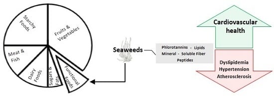
Seaweeds as Preventive Agents for Cardiovascular Diseases: From Nutrients to Functional Foods

by
Susana M. Cardoso
1,*,
Olívia R. Pereira
2,
Ana M. L. Seca
1,3,
Diana C. G. A. Pinto
1 and
Artur M. S. Silva
1
1
Department of Chemistry & QOPNA, University of Aveiro, Aveiro 3810-193, Portugal
2
Department of Diagnostic and Therapeutic Technologies, School of Health Sciences, Polytechnic Institute of Bragança, Bragança 5300-121, Portugal
3
Department of Technological Science and Development, University of Azores, Ponta Delgada 9501-801, Portugal
*
Author to whom correspondence should be addressed.
Mar. Drugs 2015, 13(11), 6838-6865; https://doi.org/10.3390/md13116838
Submission received: 8 August 2015 / Revised: 16 October 2015 / Accepted: 30 October 2015 / Published: 12 November 2015
(This article belongs to the Special Issue Marine Functional Food)

Abstract

:
Being naturally enriched in key nutrients and in various health-promoting compounds, seaweeds represent promising candidates for the design of functional foods. Soluble dietary fibers, peptides, phlorotannins, lipids and minerals are macroalgae’s major compounds that can hold potential in high-value food products derived from macroalgae, including those directed to the cardiovascular-health promotion. This manuscript revises available reported data focusing the role of diet supplementation of macroalgae, or extracts enriched in bioactive compounds from macroalgae origin, in targeting modifiable markers of cardiovascular diseases (CVDs), like dyslipidemia, oxidative stress, vascular inflammation, hypertension, hypercoagulability and activation of the sympathetic and renin-angiotensin systems, among others. At last, the review also describes several products that have been formulated with the use of whole macroalgae or extracts, along with their claimed cardiovascular-associated benefits.

Graphical Abstract

1. Introduction

1.1. Cardiovascular Diseases (CVDs)

CVDs are a group of disorders of the heart and blood vessels, which include coronary heart disease, cerebrovascular disease, peripheral arterial disease, rheumatic heart disease, congenital heart disease and deep vein thrombosis and pulmonary embolism [1]. Overall, CVDs represent the main cause of death worldwide, with an estimated number of 17.5 million in 2012 (i.e., 31% of the global deaths). From those, approximately 6.7 and 7.4 million deaths were due to coronary heart disease and stroke, respectively. These two diseases are projected to remain the leading causes of death in the coming years and, according to the World Health Organization (WHO), about 23.6 million people will die from CVDs in 2030 [2].
Although there are some non-modifiable risk factors for CVDs such as family history, ethnicity and age, most CVDs arise as a consequence of modern-society habits, including an unhealthy diet, physical inactivity, tobacco use and harmful use of alcohol [3]. These behavior risk factors are normally reflected in individuals by a dyslipidemia, raised blood pressure, overweight and obesity. Considered as traditional biomarkers of CVDs diseases, these parameters are normally screened when evaluating the risk of developing CVDs. Still, the recent growing understanding of multiple biological pathways underlying CVDs, including those of inflammation and oxidative stress [4,5], allowed the identification of additional biomarkers that can be useful for a more authentic and reliable estimation of cardiovascular risk, as well as to monitor the efficacy of treatments and to develop new pharmacological tools [6].
For the last two decades, policies and key actions have been developed in order to effectively control the fast growing burden of CVDs worldwide. Programs offering healthy choices affordable and available, stimulating a healthy life style in populations are a main preemptive approach [7]. In this context, dietary interventions such as reduction of salt in the diet, consuming of fruits and vegetables, food enriched in unsaturated fats and potassium, are accepted as strongly associated with a decreased risk for CVDs [8].

1.2. Relevant Macroalgae Components in the Area of Functional Food

Seaweeds, i.e., marine macroalgae are presently pointed out as the plant-based food of the future [9], since besides not competing with food crops for the use of arable land and fresh water resources, they are a good supply of key nutrients including carbohydrates, protein and minerals [10,11], as well as a rich source of health-promoting compounds capable of acting on a wide spectrum of disorders and/or diseases [10,12,13]. This latter fact is becoming particularly evident as macroalgae are presently under the spotlight of many investigations. Note also that macroalgae have been consumed as food in East Asia (mostly in Japan, China and Korea) since ancient times and epidemiological studies associating their regular consumption to several health benefits have been reported [14]. Remarkably, the Japanese have the longest life expectancy in the world and the lowest rates of CVDs, and these scores have been partly associated with their different dietary patterns, which include the regular consumption of macroalgae [15,16].
All these facts are pushing the Western culture to increase the interest in the manufacturing and consumption of high-value products derived from macroalgae, with the main aim of taking advantage of their potential health effects. Indeed, the global functional food market, evaluated for the year 2013, was around $168 billion, and forecast to be more than $305.4 billion by 2020 [17], is believed to be one of such exploiting opportunities where direct applications of seaweeds, crude extracts or purified fractions of seaweeds might hold potential.
Soluble dietary fibers, peptides, phlorotannins, carotenoids and minerals are amongst the most promising macroalgae’s compounds in the area of functional foods/nutraceuticals. Major soluble fibers include alginate from brown macroalgae, carrageenan and agar from red macroalgae, which overall can represent up to half of seaweed’s dried weight (DW). This fact renders macroalgae a leading position with regard to fiber content, ahead of the majority of fruits and vegetables [18,19,20]. This is even more notable because human consumption of fiber-enriched products from macroalgae origin are documented to promote heath benefits, including the prevention of colon cancer, type II diabetes, obesity and CVDs [10,21].
Along with the major soluble fibers mentioned above, fucoidans, i.e., sulfated polysaccharides composed of l-fucose and sulfate ester groups, and ulvans (i.e., sulfated polysaccharides mainly composed of glucuronic acid and iduronic acid units together with rhamnose and xylose sulfates) [13] are also central when it comes to the potential applications in the formulations of functional foods, since over the last few years they have been closely associated to numerous health benefits [22,23]. Note that being natural polysaccharides, their structure is of course very dependent on the species of seaweed. Still, the composition and bioactivities of the final product are also affected by other factors like the method of extraction [13,24].
Phlorotannins are phenolic compounds exclusive to brown algae that consist of dehydro-oligomers or dehydro-polymers of phloroglucinol (Figure 1), occurring in a wide range of molecular arrangements including variations in the nature of the structural linkages between phloroglucinol units (aryl-aryl or diaryl ether bonds) and the number of hydroxyl groups [25,26]. These phenolic compounds are known to accumulate mainly in the cell cytoplasm in specialized secretion vesicles named physode, representing up to 25% of their DW [26]. Although studies focusing on the characterization and bioactivity of individual phlorotannins are still very limited, several of these compounds have already been isolated and characterized (e.g., phloroglucinol, eckol, phlorofucofuroeckol A and dieckol, see Figure 1) and claimed to provide a wide range of biological activities, including potent antioxidant effects [27].
Macroalgae lipids also have potential applications for exploitation. Seaweeds’ lipids represent only 1%–5% of DW, but almost half of that fraction is polyunsaturated, where omega-3 and omega-6 fatty acids like eicosapentanoic acid and arachidonic acid are abundant [19]. In addition to this, the carotenoid fucoxanthin (Figure 1) has great commercial value since it is accepted to be helpful in the treatment of obesity, while also decreasing the risk of other associated diseases [28].
Besides having high proteic content (10%–47% for red and green) [13,14], macroalgae also contains all essential amino acids, which are undoubtedly of importance for health maintenance. In addition to that, macroalgae peptides containing 3–20 amino acid residues are emerging as bioactive constituents, since distinct authors reported their beneficial actions against several disorders like oxidative stress, hypertension, thrombosis and cancer [10,12,29].
Macroalgae also contain an incomparable wealth of minerals (this may vary from 8% to 40% of algae DW) and have therefore been employed as mineral additives to feed and food supplements [30]. Worth noting is, amongst others, their high content in essential minerals namely Na, K, Mg, P, I, Zn and Fe [11,30]. Indeed, most algae contain high levels of Na and K, but their Na/K ratios are usually low [11,30], which is important to compensate the modern Western diets, typically rich in NaCl. Furthermore, many edible macroalgae have higher contents of Mg than terrestrial plants and animals and some of them are particularly enriched in Ca (up to 2 g/100 g DW) and iodine (up to 0.5 g/100 g DW) [30,31].
Figure 1. Chemical structures of some phlorotannins (phloroglucinol, eckol, phlorofucofuroeckol-A and dieckol) and fucoxanthin.
Marinedrugs 13 06838 g001

2. Overview of Relevant Biological Pathways Underlying CVDs

Research studies and accumulated clinical evidence indicate that risk factors of CVDs overlap and intertwine, overall contributing to the onset and growth of the disease. For example, obesity contributes to or directly causes most other modifiable risk factors, including dyslipidemia and hypertension [32] and these in turn are closely related to atheroclerosis and thrombosis. Together with behavior factors, the non-modifiable markers contribute to the onset and exacerbation of CVDs through a cascade of mechanisms including vascular inflammation, oxidative stress, hypercoagulability and activation of the sympathetic and renin-angiotensin systems [4,33]. Attending to the huge complexity of events underlying CVDs, this review will mainly address lipid-metabolic changes and hypertension biomarkers, along with their major complications.
Dyslipidemia is as a major risk factor for CVDs, being in general characterized by an elevated fasting and postprandial concentration of total triglycerides (TG) and of free fatty acids, in combination with the preponderance of low-density lipoproteins cholesterol (LDL-C) along with low levels of high-density lipoproteins cholesterol (HDL-C) and of apolipoprotein B (apo B) plasmatic concentrations [5]. Note that HDL-C exerts cardioprotective effects through the uptake of cholesterol from peripheral tissues and arterial wall to the liver and by preventing LDL oxidation. In opposition, hypertriglyceridemia leads to an incremented formation of small dense LDL, causing the delay of their metabolization and consequent atherogenicity [34]. The latter, in combination with endothelial damage, are key events in the most common pathological processes underlying CVDs [35,36].
Along with dyslipidemia, hypertension is one of the major, yet controllable, highly significant risk factors for the development of all manifestations of CVDs and the main predictor of stroke. It is noteworthy that this condition can also arise from dyslipidemia, through endothelial dysfunction and the loss of physiological vasomotor activity of endothelium, which ultimately result in increased blood pressure and hypertension [35].
By definition, hypertension is established when the systolic blood pressure (i.e., the maximum blood pressure during contraction of the ventricles) is above 140 mmHg and/or the diastolic blood pressure (i.e., the minimum pressure recorded just prior to the next contraction) is above 90 mmHg [37]. Mechanistic causes of hypertension include the reduction of glomerular filtration rate or increased renal tubular reabsorption of salt and water, the excessive activation of renin-angiotensin-aldosterone and sympathetic nervous systems, increased formation of reactive oxygen species (ROS), the vasoconstrictor peptide endothelin and of inflammatory cytokines and decreased synthesis of endothelial nitric oxide. Other important factors for hypertension include an excessive weight gain and dietary factors [38,39].
The intake of minerals and, in particular that of sodium/potassium (Na/K) ratio is believed to be vital in blood pressure control. It is known that the dietary NaCl ingestion increases arterial constriction and peripheral vascular resistance, thus elevating the blood pressure [40]. In opposition, it is well established that K intake leads to the decrement of blood pressure and thus prevents hypertension-associated complications [41]. In this way, WHO recommends a K intake of at least 3.51 g/day and less than 2 g Na/day (equivalent to 5 g salt/day) in adults [41,42]. Besides Na and K, magnesium has also been proven to prevent high blood pressure and metabolic syndrome [43]. Additionally, its deficiency has been suggested to play a role in the onset of type II diabetes and in lipid metabolic changes, thus consequently contributing for atherosclerosis [43,44].
Along with minerals, the renin-angiotensin-aldosterone system has an important function in hypertension, as its activation results in the conversion of angiotensin I to angiotensin II, being the latter a potent vasoconstrictor [45,46]. The clinical use of drugs acting in this system is frequent and includes: (i) inhibitors of angiotensin-converting-enzyme (ACE-I); (ii) angiotensin II receptor blockers (ARBs); and (iii) renin inhibitors. Captopril, cilazapril, enalapril, perindopril, lisinopril and ramipril are authorized ACE-I inhibitor drugs, which competitively block the conversion of angiotensin-I to angiotensin-II [47], while ARBs (e.g., losartan and valsartan) act by competitive antagonism of the angiotensin II receptors [48]. These drugs are used in cases of moderate hypertension and are prescribed alone or in combination with other antihypertensive drugs [46]. Direct renin inhibitors are a novel class of antihypertensive drugs developed to inhibit the renin-angiotensin-aldosterone axis and consequently reduce the angiotensin II concentrations by inhibition of the conversion of angiotensinogen to angiotensin-I [49].
Hypertension and dyslipidemia are also the most common remediable risk factors for atherosclerosis, which is a main cause of the most common pathological process leading to CVDs including myocardial infarction, heart failure and stroke [33,50]. The atherosclerotic plaque is characterized by an increasing lesion grown from a fatty streak to an atheroma, with a fibrous cap. In this process, the passage of lipids into the subendothelial space comes after endothelial damage. It is accepted that endothelium stress can be caused by several factors, including an excess of ROS, or the exposure to toxic agents (e.g., oxidized LDL (oxLDL)), to infectious agents or advanced glycosylated end products (the result of an oxidation reaction with glucose that are found in the blood of diabetics), as well as by hypertension and its proinflammatory effects (smooth muscle lipoxygenase activity and oxide radical formation, among others) [36,50].
The injury of endothelium causes an increase of prothrombotic factors, proinflammatory adhesion molecules (e.g., vascular adhesion molecule-1 (VCAM-1)), proinflammatory cytokines (interleukines 1 and 6 (IL-1 and IL-6, respectively), and tumor necrosis factor alfa (TNF-α)), chemotactic factors (MCP-1), ROS production and increase of leukocyte adhesion. Other important feature is the diminished release of nitric oxide (NO) into the arterial wall, which is known to contribute to endothelial homeostasis through diverse mechanisms, including modulation of vascular tone [51,52]. All these events allow amplifying the inflammatory cascade in atherosclerotic lesions, which is characterized by an immunologic and inflammatory infiltrate composed of distinct cells, including monocyte-derived macrophages, smooth muscle cells and T lymphocytes [53]. The development and progression of the lesion depends on the interaction of these cells with the connective tissue, ending on the formation of stable or instead, vulnerable atherosclerotic plaques. The latter are typically characterized by active proteolytic enzymes, including metalloproteinases (MMPs) [45].
Lesions in vulnerable plaques can progress to the thinning of the fibrous cap rendering it susceptible to rupture and thrombosis. In case of rupture, the inflammatory signaling (e.g., T lymphocytes and macrophages) induce the formation of the procoagulant tissue factor, promoting the thrombus formation which is an important complication of atherosclerosis [45,52]. Briefly, local thrombus produces thrombosis-associated mediators (e.g., serotonin, thromboxane A2, thrombin), which cause local and disseminated vasoconstriction. Besides coagulation, platelet activation results in a well-coordinated series of events which includes the trapped platelets to subendothelium, recruitment and activation of additional platelets and the formation of platelet aggregates, which overall contribute for acute atherothrombosis [54].

3. Evidences of Protective Effects of Seaweeds with Impact on CVDs

3.1. Dyslipidemia

The common side effects of the current synthetic lipid-lowering drugs, i.e., hepatic and/or rhabdomyolysis [55], have increased the tendency to move toward traditional and alternative treatments [56]. In this field, the diet supplementation of seaweeds and/or extracts might have a potential protective role. This is supported by the fact that epidemiological observations show satisfactory results when associating the consumption of seaweeds, medicinal plants and fruits to the prevention of hyperlipidemia in many societies [57]. Having this in mind, the potential effect of distinct macroalgae and/or its isolates in hyperlipidemia control has been largely tested in animal models. Due to the high number of reported studies in this topic, only the most recent ones have been selected for this review.
When supplementing the diets of hypercholesterolemic Wistar rats with 21% of Himanthalia elongata or 23% of Gigartina pistillata, Villanueva and coworkers [58] showed that Himanthalia-treated rats presented a reduction in the plasmatic levels of TG (28%) while increasing those of HDL-C (20%). In turn, the Gigartina-supplemented diet produced a significant decrease of 31% in TG, 18% in total cholesterol (TC) and 16% in LDL-C [58]. Identical effect was observed in studies using diets supplemented with tropical green seaweed (5% of dried Derbesia tenuissima (DT)). DT did not change total body fat mass but it could decrease the plasmatic levels of TG by 38% and TC by 17% [59].
In addition, the diet supplementation of high-cholesterol/high-fat (HF) Sprague-Dawley rats with 10% freeze dried red seaweed Gracilaria changii (G. changii) powder for eight weeks significantly lowered the plasmatic TC, LDL-C and TG contents by 40.34%, 35.95% and 30.91% respectively, as compared to the HF-induced rats group although no effect on HDL-C could be observed [60]. Its hypolipidemic action was comparable to statin. As suggested by the authors, one of the potential mechanisms of action explaining the hypolipidemic effects of G. changii might be due to its high dietary fiber (61.29%) [60]. Supplementation of G. changii to the normal rats showed less significant effect on the plasma TC, HDL-C, LDL-C and TG levels [60]. These differences suggest that this seaweed is probably more effective for hyperlipidemia treatment purposes instead of preventive ones. In addition, brown algae Ecklonia cava supplementation dose dependently suppressed TG, TC, and LDL-C concentrations in the serum in both normal and STZ-diabetic mice (supplementation of 5% of seaweed in diabetic mice causes a decrease in serum levels of 72%, 53%, and 78%, respectively) but failed to affect HDL-C concentrations in normal mice [61].
For the last years, there are also distinct works reporting positive effects on lipid metabolism as a result of diet supplementation of extracts obtained from macroalgae. Ruqqia et al. [62] showed that amongst the ethanol extracts of 13 seaweed species, those of Jolyna laminarioides and Sargassum binderi exhibited comparable hypolipidemic potential to common hypolipidemic drugs such as bezafibrate and fenofibrate (through decrement of TC, TG and LDL-C) in diet-induced hyperlipidemic rats and in triton-induced hyperlipidemic rats [62]. The extract from Melanothamnus afaqhusainii was also moderately active in lowering the levels of TC, TG and LDL-C in triton-induced hyperlipidemic rats [62]. Liver and cardiac enzymes like lactate dehydrogenase, alkaline phosphatase, aspartate alanine aminotransferase were not adversely affected by administration of these three extracts [62].
In addition, the ethanol extract of the brown seaweed Iyengaria stellate (10 mg/200 g body weight to rabbits for 30 days) showed an overall decrease in total plasmatic lipid levels, although an increase in the contents of the liver enzymes alkaline phosphatase, glutamic-pyruvic transaminase (SGPT), glutamic oxaloacetic transaminase and γ-transaminase (with exception for SGPT) were also registered [63]. Note that as SGPT is a more specific indicator of liver injury, the overall results gathered by the authors suggest that the intake of Iyengaria stellate extract as a hypolipidemic agent should be followed by the monitoring of liver enzymes to ensure liver safety.
Furthermore, Dousip et al. [64] investigated the antihyperlipidemic effect of red seaweed Kappaphycus alvarezii and brown seaweed Sargassum polycystum aqueous extracts, as well as their synergistic effects, in high-cholesterol diet fed rats. The results showed that Sargassum polycystum was more effective in decreasing plasma cholesterol (by 37.52%) when compared to Kappaphycus alvarezii or with the mixture of the two extracts, though an increase in plasmatic TG levels by 16.66% was also observed. In turn, the Kappaphycus alvarezii extract most effectively decreased the TG levels by 40.11% and the mixture of the two extracts further increased HDL-C (by 56.71%). All the three tested conditions (individual extracts or mixture) were able to reduce LDL-C levels when compared to the high-cholesterol group [64]. Overall, hypercholesterolemic rats fed with the mixture of the extracts had the lowest atherogenic index among all groups [64].
Besides the evidences of dietary supplementation of seaweeds and/or crude extracts on lipid metabolism, many authors have also been described positive effects for purified fractions and/or isolated compounds. Amongst them, the majority of the works have been focusing two major groups of compounds, namely seaweeds’ sulfated polysaccharides and lipids. Some recent examples are described below.
Borai et al. [65] reported that the oral administration of sulfated polysaccharides (SP) from Ulva fasciata to induced-hypercholesterolemic rats for four consecutive weeks did not exert any side effects and, simultaneously, it caused a significant decrement in serum lipid profile by reducing the plasmatic TC, TG, LDL-C and of very low density lipoproteins-cholesterol (VLDL-C). Notably, the scores for Ulva fasciata ulvans effects were better than those observed for the reference drug fluvastatin [65]. In addition, Hoang and coworkers [66] reported that two types of SP isolated from the green alga Monostroma nitidum showed the ability to reduce cellular lipid concentrations in lipid-loaded hepatocytes, when compared with controls, being this reduction accompanied by a reduced expression of cholesterol synthesis genes and an increment in the expression of genes dictating cholesterol degradation, LDL uptake and peroxisomal β-oxidation.
Another relevant work focusing the effects of ulvans in lipid metabolism has been performed by Hassan et al. [67]. These authors described that the intragastric administration of Ulva lactuca sulfated polysaccharides to dietary-induced hypercholesterolemic rats could cause a more evident effect in the increment of HDL-C level (by 180%) when compared to that induced by its oral administration [67]. This paves the way for discussion of the effect of the administration mode and how to take advantage of this difference in results.
Along with ulvans, fucoidans have been largely evaluated as antihyperlipidemic agents. Recently, a fucoidan extracted from the brown seaweed Sargassum henslowianum and whose structure was established as an α(1→3)-linked l-fucopyranose backbone with sulfate groups mostly present at C-2, C-4 and occasionally at C-3, was tested in an obese mice model. The administration of this fucoidan at a dose of 100 mg/kg P/day to the animals caused a decrement in the plasmatic levels of cholesterol, TG and LDL-C [68]. Unfortunately, definite conclusions on the effective effect of this polysaccharide still requires additional studies on dosage, administration time, in between other parameters.
In addition, Kim et al. [69] have recently demonstrated that the dietary supplementation of a commercial fucoidan (Haewon Biotech, Inc., Seoul, Korea) induced a significant decrease on the plasmatic levels of TG, total cholesterol and of LDL-C. As reported by the authors, the beneficial effects of the fucoidan were probably partially associated to the down-regulation of adipogenic transcription factor [69].
Carrageenans, another type of sulfated polysaccharide typically present in red algae, were recently used in a clinical trial study. Patients with ischemic heart disease (IHD) were reported to exhibit a significant effect on lipid profile by a short-term carrageenan supplement [70]. In fact, the prophylactic administration of the carrageenan food supplement in the complex therapy of IHD patients could significantly decrease the plasmatic TC levels by 16.5% and LDL-C by 33.5% as compared with the baseline measurements (background therapy control and experimental groups) [70].
Amongst the macroalgae lipids, fucoxanthin and fatty acids are widely mentioned when it comes to dyslipidemia regulation. Notably, the beneficial effects of fucoxanthin on cardiovascular diseases were recently reviewed by Gammone et al. [71]. In agreement with distinct reported studies, these authors highlighted that fucoxanthin metabolites (amarouciaxanthin A and fucoxanthinol) are the active in vivo forms of fucoxanthin and that their ability in regulating lipids plasmatic levels are mainly due to their antioxidant activity [71]. In addition, fucoxanthin and its metabolite fucoxanthinol are accepted to exhibit extra-metabolic benefic effects, including the regulation of polyunsaturated fatty acids (PUFA) biosynthesis by the promotion of the upregulation of enzymatic activities related to the bioconversion of omega-3 PUFA and omega-6 PUFA to docosahexaenoic acid (DHA) and arachidonic acid (AA), respectively [72,73,74]. Furthermore, it has been demonstrated that both these compounds could induce a decrease in the content of eicosapentaenoic acid (EPA) on cultured rat hepatoma BRL-3A, thus suggesting a down-regulation of metabolic enzymes such as fatty acid desaturase and elongase [75]. In rodents, fed fucoxanthin promotes the synthesis of DHA in the liver [72], resulting in the improvement of the lipid profile since this acid inhibits the synthesis of thromboxane A2 and enhances the production of prostacyclin, a prostaglandin that produces vasodilation and less sticky platelets [76].
As previously mentioned, fatty acids from seaweeds may have itself beneficial properties relevant to cardiovascular disease since they are good sources of the long-chain omega-3 PUFAs EPA and DHA acids [74,77,78,79,80], which in turn promote beneficial effects in serum EPA, TC and TG levels and in LDL-C/HDL-C ratio [77,81,82]. As demonstrated in KK-Ay mouse, DHA and AA levels are significantly increased by the feeding of lipids from Sargassum horneri and Cystoseira hakodatensis, but not by those obtained from Undaria pinnatifida [83]. As suggested by the authors, this difference can be due to the higher fucoxanthin content in the first two seaweeds. The authors have additionally reported that the plasmatic levels of TC, HDL-C and phospholipid of mouse fed with seaweeds lipids were significantly increased while those of hepatic cholesterol and triacylglycerol were decreased as compared with the control group [83]. The increase of serum cholesterol levels by fucoxanthin, in KK-Ay mice, was studied by Beppu et al. [84]. The results suggest that fucoxanthin exerts its effects on cholesterol metabolism and in the transport system by down-regulation of the LDL receptor and the class B type 1 scavenger receptor, along with inducing sterol regulatory element binding protein expression [84]. Further studies however are required for clarifying whether the responses to cholesterol metabolism are specific for rodents or extended to Human.

3.2. Hypertension

The preventive hypertensive effects of seaweeds have since ever been associated with their richness in dietary fibers and mineral contents and, although reported literature regarding this issue is clearly scarce when compared to that dealing with dyslipidemia, in vitro and in vivo studies support this association. In addition, recent literature data suggests that bioactive components like fucoxanthin, phlorotannins and peptides can also play a role in seaweeds’ antihypertensive effects.
As mentioned before, inhibition of ACE-I is a well-established approach in the treatment of hypertension and, because of that, many authors have screened the potential of seaweeds in inhibiting this enzyme, either using crude extracts, purified fractions and/or isolated components (see selected examples in Table 1).
Cha et al. [85] screened the in vitro ACE-I inhibitory activity of methanol and aqueous extracts from twenty-six red Korean algae, obtained at 20 °C or at 70 °C. The authors have found several potential extracts, with IC50 values for ACE-I in the range of 12.21–124.69 μg·mL−1, being the lowest value found for the aqueous extract of Lomentaria catenata at 20 °C. In addition, Jung et al. [86] found that among the ethanol extracts of ten Korean seaweeds, including four Rhodophyta (Gelidium amansii, Gigartina tenella, Porphyra tenera and Chondria crassicaulis), five Phaeophyta (Ecklonia stolonifera, Ecklonia cava, Hizikia fusiforme, Pelvetia siliquosa and Undaria pinnatifida) and one Chlorophyta (Capsosiphon fulvescens), those of E. cava, E. stolonifera, P. siliquosa, G. tenella and U. pinnatifida were the most promising in targeting ACE-I, all causing inhibition above 50% at 163.9 μg·mL−1. Since several purified fractions and isolated phlorotannins from the extract of E. stolonifera also exhibited marked ACE-I inhibitory activity, the authors concluded that phlorotannins, in particular eckol, phlorofucofuroeckol A and dieckol could be partially responsible for the protective activity of the crude extract [86].
In another study, Wisesingh and coworkers [87] also suggested that phlorotannins could be key constituents of extracts when concerning ACE-I targeting. The authors demonstrated that the ethanol extract of E. cava was the most active against ACE-I activity when compared to other organic extracts obtained with ethyl acetate, chloroform, hexane or diethyl ether. Besides, they reported that the isolated phlorotannins from this seaweed were active against ACE-I.
Antioxidants, including phlorotannins and fucoxanthin, were also recently shown to be components of Saccharina japonica and Sargassum horneri oils, either obtained by environmental friendly supercritical-CO2 with ethanol as a co-solvent or by conventional extraction using ethanol, hexane or a (1:1, v/v) mixture of acetone:methanol. All the extracts exhibited moderate-to-high activity against ACE-I, and authors have suggested that this activity could be attributed in part to fucoxanthin, although no direct correlations between its concentration in the extracts and the inhibitory activity were observed.
Recently, distinct studies applying proteolytic enzymatic digestion to seaweeds has led to detection of a number of renin- or ACE-I-inhibitory bioactive peptide. Examples of potential peptides have been described for red macroalgae (e.g., from Porphyra columbina, Porphyra yezoensis, and Palmaria palmata), as well as for green algae (Caulerpa microphysa) and brown algae (Undaria pinnafita) (Table 1). As seaweed hydrolysates consist of a complex mixture of constituents and the amino acid sequence of bioactive peptides in ACE-active hydrolysates has not been commonly determined, more experimental data should be gathered in order to allow solid conclusions on structural-active relations. However, according to Suetsuna and coworkers [88], the presence of Tyr residues in dipeptides seems to improve their ability of targeting ACE-I.
Table 1. Selected studies reporting inhibitory abilities of seaweeds extracts and isolates on the renin-angiotensin-system.
Seaweed SpeciesExtractionInhibitionRef
ACE-I inhibition of extracts
Twenty-six red algaeMeOH and Aq Ext 20 °C and 70 °CAq Ext 20 °C IC50 (µg/mL): Lomentaria catenata = 13.78; Lithophyllum okamurae = 12.21; MeOH Ext 20 °C IC50 (µg/mL): Ahnfeltiopsis flabelliformis = 13.84; Laurencia okamurae = 106.15; MeOH Ext70 °C: Bonnemaisonia hamifera, Grateloupia filicina, Sinkoraena lancifolia, Grateloupia lanceolata, Gracilaria vermiculophylla and L. okamurae ranging from 25.82 to 124.69[85]
Ten Korean seaweeds EtOH ExtEcklonia stolonifera, E. cava, Pelvetia siliquosa, Undaria Pinnatifida and Gigartina tenella: above 50% inhibition of ACE at 163.93 µg/mL[86]
Ecklonia cavaEtOH, EtAc, CHCl3, Hex, DEBest inhibition for EtOH Ext, IC50 = 0.96 mg/mL[87]
ACE-I inhibition associated with antioxidants
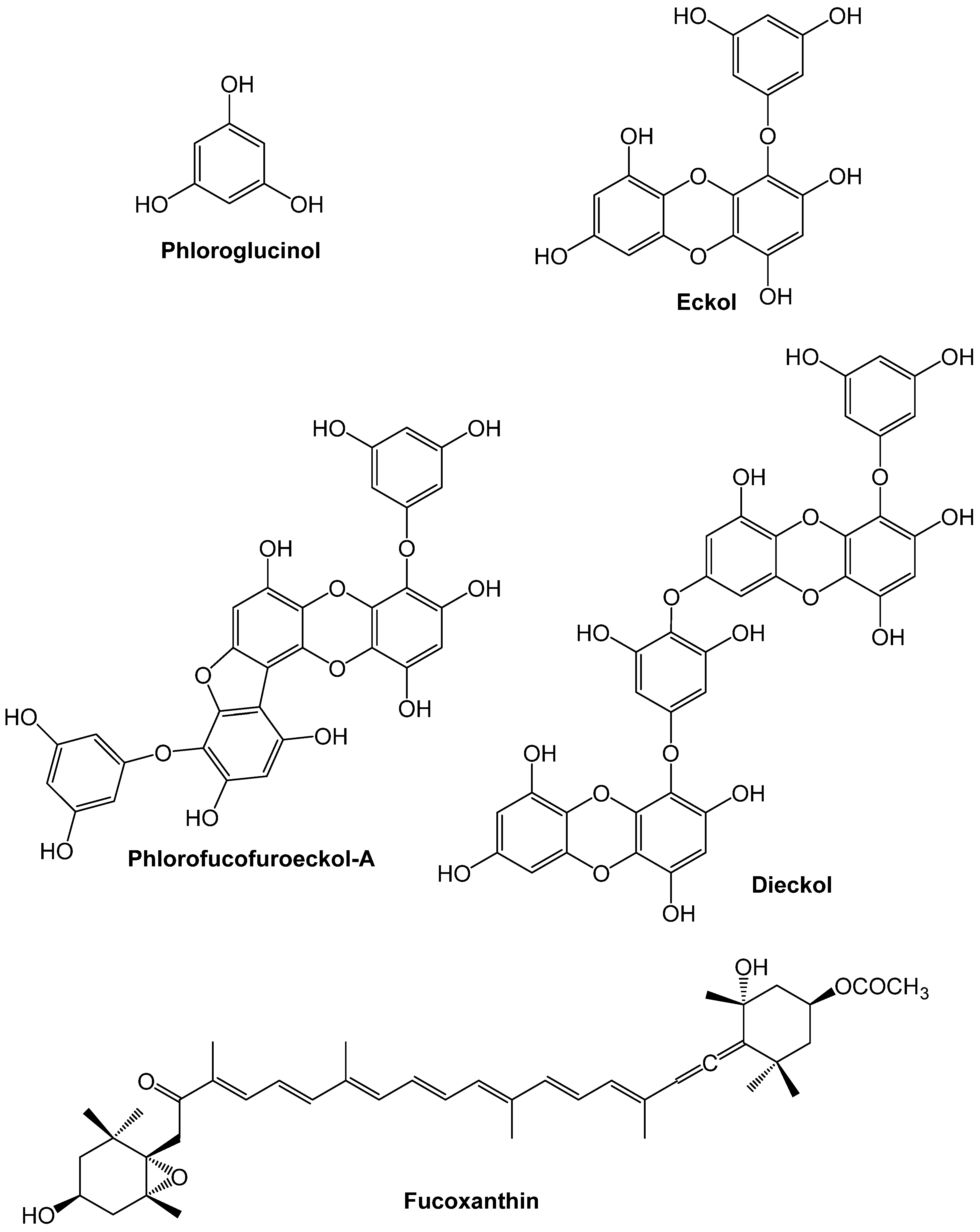
Ecklonia stoloniferaPurified PhlorotanninsBest inhibition recorded for eckol, dieckol and phlorofucofuroeckol. IC50 (μM): eckol = 70.82; phlorofucofuroeckol A = 12.74; dieckol = 34.25[86]
Ecklonia cavaPurified PhlorotanninsIC50 (mM): phloroglucinol = 2.57 ± 0.09; eckol = 2.27 ± 0.08; triphlorethol-A = 2.01 ± 0.36; dieckol = 1.47 ± 0.04; eckstolonol = 2.95 ± 0.28[87]
Saccharina japonica (SJ) Sargassum horneri (SH)Supercritic CO2 vs. Acet: MeOHIC50 (µg/mL): SJ CO2 Ext = 0.89 ± 0.07; SJ Acet:MeOH Ext = 1.05 ± 0.14; SH CO2 Ext = 0.97 ± 0.11; SH Acet:MeOH Ext = 1.28 ± 0.50;[89]
ACE-I or Renin inhibition associated with peptides
Porphyra columbinaEnzymatic in thermostatic reactor (A/AF)ACE-I IC50 (g/L): A = 1.2 ± 0.1; AF = 1.7 ± 0.0[90]
Porphyra yezoensispH and enzymatic ACE-I IC50 (g/L): 1.6[91]
Palmaria palmataPapain Ile-Arg-Leu-Ile-Ile-Val-Leu-Met-Pro-Ile-Leu-Met-Ala Renin inhibitory bioassay: ↓ renin activities by 58.97% (±1.26) at 1 mg/mL.[92]
Solieria chordalis (SC) Palmaria palmata (PP)Chymotrypsin (ChTr) or trysin (Tr)<10 kDa fractions of SC: hydrolyzed with ChTr (IC50 ACE 3.50 mg/mL) or Tr (IC50 ACE 20.34 mg/mL); <10 kDa fractions of PP: hydrolyzed with ChTr (ACE IC50 460.05 mg/mL)[93]
Caulerpa microphysaPepsin, alcalase, flavourzymeACE-I IC50 (mg/L): pepsin = 0.20; flavourzyme = 29.74; alcalase = 31.71[94]
Undaria pinnafidaPepsin ACE-I IC50 (µM): Ala-Ile-Tyr-Lys = 213; Tyr-Lys-Tyr-Tyr = 64.2; Lys-Phe-Tyr-Gly = 90.5; Tyr-Asn-Lys-Leu = 21[95]
Undaria pinnatifidaAq hot Ext dyalisis, chromatographyACE-I IC50 (µM): Tyr-His = 5.1; Lys-Trp = 10.8; Lys-Tyr = 7.7; Lys-Phe = 28.3; Phe-Tyr = 3.7; Val-Trp = 10.8; Val-Phe = 43.7; Ile-Tyr = 2.7; Ile-Trp = 12.4; Val-Tyr = 11.3[88]
ACE—angiotensin I converting enzyme; Acet—acetone; Aq—aqueous; Ext—extract; DE—diethyl ether; EtAc—ethyl acetate; EtOH—ethanol; Hex—hexane; MeOH—methanol; ↓ decrement.
Because non-invasive measure of blood pressure in lab animals is a difficult task, this parameter has also not been widely screened when evaluating the beneficial effects of the dietary supplementation of seaweeds or isolates in CVDs-related animal models. Still, some authors described the diet-supplementation of seaweeds and/or seaweed isolates in blood pressure (see resume of selected studies in Table 2).
Table 2. Selection of studies that include the assessment of blood pressure or antioxidant, anti-inflammatory or endothelium restoring as a consequence of diet supplementation of seaweeds or isolates, as evaluated in vivo models.
Seaweed Species (Extract)Model DoseEffectsRef.
Ulva ohnoi (UO) Derbesia tenuissima (DT)High-carbohydrate, HF diet-fed rats 5% for 8 weeksUO: ↓ total final body fat mass by 24% and sBP by 29 mmHg; ↑ Glc utilization and insulin sensitivity; DT: ↓ TG by 38% and TC by 17%[59]
Ulva linza (UL) Lessonia trabeculata (LT)High-sucrose, HF diet-fed rats|400 mg·kg−1 for 8 weeksUL, LT: ↓ levels of intra-abdominal fat, arterial BP, insulin resistance, TC, TG, SOD; ↓ liver expression levels SOD and GPx and ↑ CAT in control groups and ↓ in algae-fed rats; LT: ↓ GPx activity[96]
Gracilaria changiiHF, HC diet-induced rats|5% and 10% for 8 weeks5%: ↓ TC (−39.19%), LDL-C (−36.36%), and TG (−25.45%); 10%: ↓ TC, LDL-C and TG content by 40.34%, 35.95% and 30.91%, respectively; lowest AI; 5% and 10%: in plasma = ↓ LipPerox; ↓ AST and ALT levels; in erythrocyte = ↑ SOD, CAT and GSH-Px[60]
Not detailedHealthy children from 3 to 6 years diet including seaweed intake using 3-day dietary recordsCross-section study in healthy preschoolers: Girls with higher seaweed intake had significantly lower systolic BP (102.4, 99.2 and 96.9 mmHg for girls with the lowest, middle and highest tertiles of seaweed intake, respectively); seaweed intake was negatively related to dBP in boys and to sBP in girls.[97]
Undaria pinnatifida (UP)Men/Women with MS|Gr1:1 month (m) 4 g/day UP; Gr2: 1 m 4 g plus 1 m g/day UP (pills)Randomized double-blinded placebo-controlled trial. Gr2: ↓ systolic BP 10.5 mmHg after a month of 6 g/day seaweed (primarily in subjects with high-normal baseline BP); ↓ waist circumference for women participants (↓ 2.1 cm after 4 g/day and further 1.8 cm after 1 m 6 g/day seaweed). No changes in lipid profile.[98]
Undaria pinnatifida (UP)19 patients MS|3.3 g in capsules sBP: ↓ 13 mmHg below the baseline after 4 weeks and 8 mmHg after 8 weeks. dBP: ↓ 9 mmHg after 4 weeks and 8 mmHg after 8 weeks; hypercholesterolemia ↓ 8% by week 4[99]
Extracts
Sargassum subrepandum (MeOH Ext)Rats with atherogenic diet|100 mg/kg b.wt↓ TC, TG, LDL-C and ↑ HDL-C; ↓ MDA, NO, leptin, TNF-alpha levels; ↑ adiponectin level;[100]
Ulva fasciata (Ulvans/Aq Ext at 4 °C or 100 °C plus EtOH pp)HC rats|175 mg/kg for 4 weeksBoth Ext: No side effects; ↓ TC, TG, TG, LDL-C and VLDL-C; ↓ liver NO; ↓ ICAM-1 and VCAM-1; ↑ IL-10; ↓ atherogenic plaques in the aorta more than fluvastatin; [65]
Ulva lactuca (Ulvans/Aq Ext at 100 °C plus EtOH pp)HC rats|175 mg/kg for 4 weeks↓TL, TG, TC, LDL-C and VLDL-C; ↑ HDL-C; ↓ AI, creatine kinase and LDH; ↓ liver ALT, AST and ALP activities and serum urea, creatinine and urea/creatinine ratio; ↑ hepatic CAT, GSH-Px; ↑ GSH, Total thiol levels[67]
Not detailed (Low-MW Commercial alginates)DOCA salt-induced hypertensive rats|250 or 500 mg/kg for 30 days↓ sBP; dose-dependent normalization of changes induced by DOCA salt, with the exception of further increasing sodium excretion[101]
Gloiopeltis complanata (Funoran/Aq hot Ext plus various purification steps)HC, high-sal fed rats|1000 mg/kg for 20 days↓ sBP; ↓ TC, TG, LDL-C, AI; ↑ urine excretion of sodium, chloride; ↑ urine Na/K ratio[102]
Not detailed (Seaweed fiber (SF))Hypertensive Patients|Pills with 0.33 g; 25 min before meals for 4 weeks↓ mean and sBP; ↑ plasma renin activity; ↓ urinary secretion of Na, K and Na/K ratio[103]
Palmaria palmata (protein hydrolysate and tridecapeptide IRLIIVLMPILMA)Spontaneously Hypertensive rats|50 mg/kg b.wtAfter 24 h ingestion: ↓ 34 mm Hg in sBP; IRLIIVLMPLIMA: ↓ 33 mm Hg[104]
AI—atherogenic index; Aq—aqueous; ALP—alkaline phosphatase; ALT—alanine aminotransferase; AST—aspartate aminotransferase; BP—blood pressure; CAT—catalase; dBP—diastolic blood pressure; DOCA—deoxycorticosterone acetate; EtOH—etanol; Ext—extract; Glc—glucose; Gr—group; GSH—hepatic reduced glutathione; GSH-Px—glutathione peroxidase; HC—high cholesterol; HDL-C—high density lipoprotein cholesterol; HF—high-fat; LDL-C—low density lipoprotein cholesterol; iCAM—intercelular adhesion molecule-1; LipPerox—lipid peroxidation; MDA—malonaldialdehyde; MeOH—methanol; MS—metabolic syndrome; NO—nitric oxide; pp—precipitation; sBP—sistolic blood pressure SOD—superoxide dismutase; TC—plasma total cholesterol; TG—plasma total triglycerides; VCAM—vascular cell adhesion molecule-1; ↓ decrement; ↑ increase.
When evaluating the response of dietary supplementation of Ulva ohnoi and Derbesia tenuissima in a rat model of human metabolic syndrome, Kumar et al. [59] reported that Ulva ohnoi could induce a marked decrease in systolic blood pressure (by 29 mmHg), along with a decrease in the final body fat mass (by 24%) and the improvement of glucose utilization and insulin sensitivity. On the basis of chemical composition of the two algae, the authors have suggested that the better scores of Ulva ohnoi with respect to those induced by Derbesia tenuissima could be due to its richness in soluble fibers and magnesium. A similar trend on blood pressure were also registered by Ramirez-Huigera et al. [96], when testing the effects of diet-suplementation of Ulva linza and Lessonia trabeculata in rats fed with a hypercaloric diet.
Despite scarce and sometimes inconsistent, the effect of diet supplementation of macroalgae in human blood pressure, either in healthy or in hypertensive scenarios has also been evaluated. The cross-section studies reported by Wada et al. [97] in healthy Japanese preschoolers allowed to observe that girls with higher seaweed intake had lower systolic blood pressure while diastolic bold pressure in boys also decreased. Additionally, systolic and diastolic blood pressure in hypertensive elderly Japanese patients have been shown to be significantly decreased after daily doses of 5 g of dried Undaria pinnatifida powder for eight weeks [99]. The same trend was registered in a randomized double-blinded placebo-controlled trial with metabolic syndrome patients (also including hypertension) taking daily pill dosages of 6 g of U. pinnafita [98].
The benefit of seaweeds’ dietary fiber supplementation in hypertensive rats has also been previously demonstrated by distinct authors. Some authors suggested that their positive effects could be due to retention of dietary Na [102] or to an enhanced intestinal K absorption [101] or to these two effects in simultaneous [103]. It should however be remarked that as the cationic exchange ability of fibers is dependent on factors like the type of fiber and the mineral content of the diet, further information regarding this topic is still required.
Antihypertensive abilities of macroalgaes’ peptides have also been investigated in vivo models. Fitzgerald et al. [104] showed that the diet supplementation of a tridecapeptide derived from a papain digest of Palmaria palmata (IRLIIVLMPILMA, previously shown to be active against renin [92]) could cause a decrease in systolic blood pressure of about 33 mmHg in spontaneously-hypertensive rats. Further simulation of gastric digestion allowed the authors to conclude that the active forms of IRLIIVLMPILMA are probably dipeptides originated along the passage through the gastrointestinal tract. In addition, Suetsuna and coworkers have characterized several di- and tetrapeptides from digests of Undaria pinnafita [88,95]. Amongst them, the dipetides Tyr-His, Lys-Tyr, Phe-Tyr and Ile-Tyr, as well as the tetrapeptides Ala-Ile-Tyr-Lys, Tyr-Lys-Tyr-Tyr, Lys-Phe-Tyr-Gly and Tyr-Asn-Lys-Leu, were shown to efficiently decrease the blood pressure of spontaneous hypertensive rats through diet supplementation [88,95].

3.3. Biological Pathways Underlying Atherosclerotic-Related Events

As previously referred, the formation and progression of atherosclerotic plaques involves a multitude of factors, including oxidative and inflammatory events. In this context, antioxidants and anti-inflammatory sources are of interest in the quest for antiatherogenic activity. Still, it is noteworthy that only a scarce number of research studies have previously monitored these biological pathways when evaluating the potential diet supplementation of macroalgae in atherosclerosis-related models, being those studies major tested with extracts or isolated compounds (included in Table 2). Besides, structure-bioactive studies are almost non-existent. Because of that, this topic demands urgent research.
Phlorotannins are one the seaweeds’ bioactive compounds that could hold a potential in this particular field. In fact, Costa-Mugica and coworkers [105] demonstrated an effective protection of LDL oxidation by phlorotannin-enriched fractions isolated from Halimeda incrassata, as measured in two heparin-precipitated LDL models. Moreover, the phlorotannins phloroglucinol, eckol, and dieckol (Figure 1) have been shown to protect against proinflammatory responses in human umbilical vein endothelial cells (HUVEC) and in an animal model of acute inflammation, in response to the endotoxin high-mobility group protein B1 (HMGB1) [106]. Phlorotannins inhibited lipopolysaccharide (LPS)-induced HMGB1 release, HMGB1-mediated barrier disruption, the expressions of CAMs and the adhesion/transendothelial migration of leukocytes to human endothelial cells. The three phlorotannins could also suppress acetic acid-induced hyperpermeability and carboxymethylcellulose-induced leukocytes migration in the animal model [106]. Notably, this study also highlighted the importance of phlorotannins’ hydroxyl groups regarding the vascular barrier protective effects, since the magnitude of protection on HMGB1-mediated hyperpermeability and monocytes adhesion noticed in HUVEC cells followed the pattern dieckol > eckol > phloroglucinol and were even lowered when the hydroxyl groups of dieckol were replaced by methyl groups. Based on the overall results, the authors hypothesized that the presence of hydrogen bond donors and hydrophilic moieties in phlorotannins may play a central role in its affinity for membrane receptors on human endothelial cells [106].
Besides phlorotannins, fucoxanthin has also been associated to the positive effects of a methanol extract of Sargassum subrepandum [100]. In their study, the authors showed that in addition to the improvement of lipid profile, the oral administration of the extract in a dose of 100 mg/kg.wt for four months caused a marked decrease in serum malonaldialdehyde (MDA, i.e., a oxidative stress biomarker) and in the proinflammatory markers leptin and TNF-α levels, with a simultaneous increase in the level of adiponectin, i.e., an adipocyte-specific protein that is thought to act as anti-inflammatory, anti-atherogenic and insulin enhancer [107].
Sulfate polysaccharides, including ulvans, carragenans and fucoidans, are the most exploited compounds when regarding the protective effects on atheroclerotic-plaque formation and progression-related problems. e.g., Hassan and coworkers [67] proved that the oral supplementation of hypercholesterolemic rats with an ulvan purified extract from Ulva lactuca could ameliorate the activity of the antioxidant enzymes catalase (CAT), glutathione peroxidase (GSH-Px) and superoxide dismutase (SOD) by 110%, 77% and 23%, respectively, when compared with the hypercholesterolemic control rats. Apart from that, the authors also described beneficial effects on non-enzymatic antioxidants parameters (hepatic reduced glutathione, total thiol and lipid peroxidation), suggesting that the defensive effect of ulvan was in part mediated by protection of liver against free radicals.
The atheropreventive activity of ulvans from Ulva fasciata was also recently reported by Borai and coworkers [65]. After the oral administration of crude cold and hot extracts to hypercholesterolemic rats extracts at a dose 175 mg/kg body weight for four weeks, the authors registered an improvement of serum lipid profile and of the endothelial dysfunction. In particular, in comparison to hypercholesterolemic rats, the ulvans-supplemented animals showed a decrease of the NO liver levels of 38.95% and 69.46% (for cold and hot extracts, respectively), while this effect was of 58.19% for the reference drug (fluvastatin). Both extracts also caused a significant decrease in the levels of the soluble adhesion molecules ICAM-1 and VCAM-1, along with an increase in the amounts of the anti-inflammatory marker IL-10 (about 30%, 18% and 34% compared to control, for cold, hot ulvan-enriched extracts and fluvastatin, respectively). In addition, morphometric measurements of atherosclerotic lesion in untreated/treated rats showed that the oral supplementation of U. fasciata extracts induced a reduction in the medial cross-section area of the aorta (19.50% and 26.46%, respectively). This result was also consistent with histological data, which demonstrated that in contrast with the untreated rats, those subjected to algae-enriched diet exhibited no oblivious lesions in the aorta, showing thin intimae and no visible swelling, endothelial cells basically intact and without desquamation, no migration of smooth muscle cells to the under-intimae, no proliferation of smooth muscle cells and smooth muscle cells arranged in a regular pattern.
Amongst SP, fucoidans have very promising biomedical/medical applications with regard to atherosclerosis and associated progressive complications. The anti-inflammatory potential of these polysaccharides in hypercholesterolemic rats has been reported by Preetha et al. [108], using a hot water-extracted fucoidan from Sargassum wightii and a comercial fucoidan from Fucus vesiculosos, which overall resulted in almost equally effects. The subcutaneous treatment with the algae fucoidans at a dose of 5 mg/kg bwt/day during seven days reduced the increased serum levels of the inflammatory biomarkers TNF-α and C-reactive protein, the NO concentrations in plasma and cardiac tissues, as well as the levels of cardiac mRNA iNOS and of COX-2 (two potent proinflammatory enzymes). In addition, the authors also demonstrated the anticoagulant effect of these polysaccharides, as estimated by the levels of plasmatic fibrinogen, a central protein in the formation of blood clots.
At this point, one should remark that fucoidans and in general SP have been proved to exert potent antithrombotic effects. Indeed, these polysaccharides share common structural features with heparin, i.e., a highly sulfated glycosaminoglycan that is widely used in clinical practice as an injectable anticoagulant. The anticoagulant activities of SP, along with their inhibition of platelet aggregation and thrombolytic activities has been the focus of many investigations and have been recently revised by distinct authors [22,109,110], which also highlight the high molecular weight and the high sulfation level (particularly with substitution at C-2 or C-2 and C-3 positions of fucose) as positive structural factors affecting these properties [22,111]. Still, reported data focusing these benefits do not take into account bioacessibility and bioavailability issues, as SP in vivo models have been administered subcutaneously (as for the above mentioned work of Preetha et al. [108]) or, in the majority of cases, as an intravenous injection. In this context, although there are a growing body of evidences that fucoidans and other SP can be partly absorbed [112] or suffer favorable changes by intestinal microbiota [113,114], these topics are not elucidated, hampering the understanding if SP exert effective antithrombotic activities when orally ingested.

4. Functional Food with Macroalgae for Promoting Cardiovascular Health

Although there is no consensual definition for the term “functional food” worldwide, this is vastly accepted for foods and food components that have been demonstrated to provide specific health benefits beyond the basic nutrition [19,115]. The design of functional foods is hence undoubtedly associated to the concept of preventing diseases and/or improving optimal health of consumers, besides the basic nutrient needs.
Given the evidence of the beneficial health effects of seaweeds and/or isolates of macroalgae origin, there is a strong case for their inclusion in regular foods (food and beverages), in order to take advantages of their benefits [115,116,117]. It is expected that the combined efforts of industry and research in this field will result in the coming years in a high number of new functional food products reaching to the market, including those intended to promote cardiovascular health.
The development of functional foods with seaweeds for cardiovascular-health promotion have been particularly tested in meat-based products [116,117,118] (Table 3) and patents have also been registered [119]. In these products, it is important to improve the fatty acid composition and the content of functional ingredients, while reducing contents of cholesterol, fat and salt [117,120]. Distinct authors have reported that nutritional values of meat products can be significantly improved by the incorporation of whole seaweeds, without hampering quality and sensory properties [118,121,122,123,124,125,126]. Besides, a remarkable work has been done by Schultz-Moreira et al., since along with describing the enhanced nutritional value of restructured meat when fortified with seaweeds, they also evaluated distinct parameters (e.g., lipid profile, antioxidant enzymes and arylesterase) with impact on cardiovascular system [127,128,129,130,131,132], as demonstrated in hypertensive rats. In addition, Lim et al. [133] also showed that chicken and pork patties fortified with Laminaria japonica could improve postprandial plasma glucose and lipids profiles in borderline-hyperlipidemic adults.
Table 3. Selected studies reporting CVDs-related parameters in foods/beverages using macroalgae or isolates as ingredients.
ProductSeaweed SpeciesRelevant ResultsRef
Meat-based products
Gel/emulsion meat systemsHimanthalia elongate (HE), Undaria pinnatifida (UP), Porphyra umbilicalis (PU) at 2.5% or 5% ↑ water- and fat-binding properties except in the case of PU at 2.5%.[121]
Gel/emulsion meat systemsHimanthalia elongate (HE), Undaria pinnatifida (UP), Porphyra umbilicalis (PU) at 5.6%All: ↑ n-3 PUFA and ↓ n-6/n-3 PUFA ratio; ↓ Na and ↑ K, Ca, Mg, Mn, antioxidants ↓ TI by for PU and UP[122]
Low-fat frankfurters Himanthalia elongata (HE) at 5.5% (algae plus 50% substitution of animal fat by olive oil)Effect of HE add: little effect on lipid and amino acid profiles but ↑ dietary fiber content and Ca and ↓ Na/K ratios[123]
Restructured meatsHimanthalia elongata at 5%Effects in hypercholesterolemic rats: ↓ TC; ↑ expression CYP7A1 and Cu, Zn-SOD; ↓ expression CAT, Mn-SOD and GPx;[127]
Restructured meatsUndaria pinnatifida (UP), Porphyra umbilicalis (PU) at 5%Effects in hypercholesterolemic rats: PU = ↓ TC; ↑ expression Mn-SOD and CAT and AE activity; UP meat mainly had benefits as antioxidant [131]
Restructured meatsUndaria pinnatifida (UP), Porphyra umbilicalis (PU) at 5%UP moderately ameliorated the lipid profile in hypercholesterolemic rats: ↓ TC and VLDL-C [132]
Restructured meatsHimanthalia elongata at 5%Effects on hypercholesterolemic rats: ↑ AE activity; ↓ VLDL-C, ILDL-C + LDL-C[130]
Pork/chicken patties Laminaria japonica (LJ) (replacement of 2.25 g pork/chichen by 2.25 g LJ)↓ increased in postprandial glucose blood levels; ↓ TC and LDL-C[133]
Others
BreadTridecapeptide IRLIIVLMPILMA from Palmaria palmata at 4%Activity against renin IRLIIVLMPILMA maintained after baking process[134]
BreadAscophyllum nodosum at 4% Single blind cross over trial: ↓ in energy intake at a test meal 4 h later; no significant changes in Glc and cholesterol[135]
TeaEcklonia cava (EC), Undaria pinnatifida (UP), Hizikia fusiforme (HF), Ulva pertusa (UP)ACE inhibition IC50 (mg DW/mL): EC = 5.33 ± 0.24, UP = 26.4 ± 1.05, HF = 7.79 ± 0.46; UP = ND[136]
AE—arylesterase; ACE—angiotensin I converting enzyme; AI—atherogenic index; CAT—catalase; Glc—glucose; IDL-C—intermediate density lipoprotein cholesterol; LDL-C—low density lipoprotein cholesterol; PUFA—polynsaturated fatty acids; SOD—superoxide dismutase; TC—plasma total cholesterol; TG—plasma total triglycerides; VLDL-C—very low density lipoprotein cholesterol; ↓ decrement; ↑increase.
Bakery and pasta products are other excellent choices for introducing bioactive ingredients, including algae, since they are greatly consumed worldwide. When testing the incorporation of a renin-inhibitory Palmaria palmata protein at 4% in bread, Fitzgerald et al. [134] reported that the bioactivity was maintained after the baking process and hence concluded that this peptide could be used for production of bread with enriched renin-inhibitory capacity, while not affecting the texture or sensory properties of the bread to a large degree.
Hall et al. [135], in a single blind cross trial also reported that addition of Ascophyllum nodosum into bread could significantly reduce energy intake at a subsequent test meal and in the total energy intake in the 24 h period following consumption of the A. nodosum enriched bread, though differences in blood glucose and cholesterol were not significant. The authors however suggested that a long-term interventional study should be done in order to establish the real potential of A. nodosum enriched bread energy intake as well as in the metabolism of glucose and lipids.
For the last years, development of beverages with seaweeds and/or extracts has also been the focus of distinct investigations and of several patent registrations. Amongst those, Fu and coworkers have patented a beverage containing water-insoluble algal dietary fibers (0.01% to 20%) and citric acid, sugar, fruit juice, plant thickeners and water, which can prevent from distinct diseases, including cardiovascular disorders [137]. Likewise, Kim patented a Hizikia fusiforme fortified drink with antihypertensive effects [138], while Korea Bio Polymer Co. Ltd. patented a functional beverage able to ameliorate cardiovascular disease [139]. This latter contained Polymann™ (Korea Bio Polymer Co. Ltd., Seoul, South Korea), a purified form of polymannuronic acid from the kelp Undaria.
Besides patents, scientific studies reporting potential effects of functional beverages including distinct macroalge has been reported by Nagai and coworkers [136,140]. Overall, these studies highlighted that drinks formulated with incorporation of macroalgae, in particular with Ecklonia cava, could be of benefit not only because of their minerals and phenolics richness, but also because of their ability to target ACE-I.
In between other products, functional salts containing seaweeds components have been studied [141]. Related to this, it is worth noting that there is a commercially available seaweed-derived salt substitute for hypertensive patients (Saloni K, from NMR Pharma, India).

5. Conclusions

Seaweeds are a great source of compounds with diverse applications, including in the field of cardiovascular-health. This fact renders macroalgae and crude/purified extracts, a potential for application as ingredients in the formulation of new functional foods in that health area. Indeed, there is evidence that diet supplementation with whole macroalgae or products of macroalgae origin can ameliorate several mechanisms underlying the onset and propagation of CVDs. Still, let us remark that the challenge of using these ingredients in novel foods should not be restricted to the improvement of their nutritional formulations, but instead, efforts should be done in order to test the claimed health benefits of the new products.

Acknowledgments

The authors gratefully acknowledge the financial support provided by the Foundation for Science and Technology (FCT) and QREN, FEDER, and COMPETE, for funding the QOPNA research unit (project PEst-C/QUI/UI0062/2013; FCOMP-01-0124-FEDER-037296).

Conflicts of Interest

The authors declare no conflict of interest.

References

  1. World Health Organization (WHO). Cardiovascular Diseases (CVDs). Fact Sheet N°317. Updated January 2015. Available online: http://www.who.int/mediacentre/factsheets/fs317/en/ (accessed on 15 July 2015).
  2. World Health Organization (WHO). Cardiovascular Diseases. Available online: http://www.who.int/cardiovascular_diseases/about_cvd/en/ (accessed on 15 July 2015).
  3. World Hearth Federation. Cardiovascular Disease Risk Factors. Available online: http://www.world-heart-federation.org/cardiovascular-health/cardiovascular-disease-risk-factors/ (accessed on 15 July 2015).
  4. Siti, H.N.; Yusof, K.; Kamsiah, J. The role of oxidative stress, antioxidants and vascular inflammation in cardiovascular disease (a review). Vascul. Pharmacol. 2015, 71, 40–56. [Google Scholar] [CrossRef] [PubMed]
  5. Klop, B.; Elte, J.W.F.; Castro Cabezas, M. Dyslipidemia in obesity: Mechanisms and potential targets. Nutrients 2013, 5, 1218–1240. [Google Scholar] [CrossRef] [PubMed]
  6. Stoner, L.; Lucero, A.A.; Palmer, B.R.; Jones, L.M.; Young, J.M.; Faulkner, J. Inflammatory biomarkers for predicting cardiovascular disease. Clin. Biochem. 2013, 46, 1353–1371. [Google Scholar] [CrossRef] [PubMed]
  7. De Pascual-Teresa, S.; Moreno, D.A.; García-Viguera, C. Flavanols and anthocyanins in cardiovascular health: A review of current evidence. Int. J. Mol. Sci. 2010, 11, 1679–1703. [Google Scholar] [CrossRef] [PubMed] [Green Version]
  8. Word Health Organization (WHO). Diet, Nutrition and the Prevention of Cardiovascular Diseases; WHO Tecnhical Report Series 916; Word Health Organization: Geneva, Switzerland, 2003. [Google Scholar]
  9. Winterman, D. Future Foods: What Will We Be Eating in 20 Years’ Time? Available online: http://www.bbc.com/news/magazine-18813075 (accessed on 13 July 2015).
  10. Lordan, S.; Ross, R.P.; Stanton, C. Marine bioactives as functional food ingredients: Potential to reduce the incidence of chronic diseases. Mar. Drugs 2011, 9, 1056–1100. [Google Scholar] [CrossRef] [PubMed]
  11. Bocanegra, A.; Bastida, S.; Benedí, J.; Ródenas, S.; Sánchez-Muniz, F.J. Characteristics and nutritional and cardiovascular-health properties of seaweeds. J. Med. Food 2009, 12, 236–258. [Google Scholar] [CrossRef] [PubMed]
  12. Fitzgerald, C.; Gallagher, E.; Tasdemir, D.; Hayes, M. Heart health peptides from macroalgae and their potential use in functional foods. J. Agric. Food Chem. 2011, 59, 6829–6836. [Google Scholar] [CrossRef] [PubMed]
  13. Cardoso, S.M.; Carvalho, L.G.; Silva, P.J.; Rodrigues, M.S.; Pereira, O.R.; Pereira, L. Bioproducts from seaweeds: A review with special focus on the Iberian Peninsula. Curr. Org. Chem. 2014, 18, 896–917. [Google Scholar] [CrossRef]
  14. Brownlee, I.; Fairclough, A.; Hall, A.; Paxman, J. The potential health benefits of seaweed and seaweed extract. In Seaweed: Ecology, Nutrient Composition and Medicinal Uses. Marine Biology: Earth Sciences in the 21st Century; Pomin, V.H., Ed.; Nova Science Publishers: Hauppauge, NY, USA, 2012; pp. 119–136. [Google Scholar]
  15. Shimazu, T.; Kuriyama, S.; Hozawa, A.; Ohmori, K.; Sato, Y.; Nakaya, N.; Nishino, Y.; Tsubono, Y.; Tsuji, I. Dietary patterns and cardiovascular disease mortality in Japan: A prospective cohort study. Int. J. Epidemiol. 2007, 36, 600–609. [Google Scholar] [CrossRef] [PubMed]
  16. Yamori, Y.; Miura, A.; Taira, K. Implications from and for food cultures for cardiovascular diseases: Japanese food, particularly Okinawan diets. Asia Pac. J. Clin. Nutr. 2001, 10, 144–145. [Google Scholar] [CrossRef] [PubMed]
  17. Research and Markets. Functional Food Market: GCC Industry Analysis and Opportunity Assessment 2014–2020; Research and Markets: Dublin, UK, 2014. [Google Scholar]
  18. Mišurcová, L.; Škrovánková, S.; Samek, D.; Ambrožová, J.; Machů, L. Health benefits of algal polysaccharides in human nutrition. Adv. Food Nutr. Res. 2012, 66, 75–145. [Google Scholar] [PubMed]
  19. Holdt, S.L.; Kraan, S. Bioactive compounds in seaweed: Functional food applications and legislation. J. Appl. Phycol. 2011, 23, 543–597. [Google Scholar] [CrossRef]
  20. Dawczynski, C.; Schubert, R.; Jahreis, G. Amino acids, fatty acids, and dietary fibre in edible seaweed products. Food Chem. 2007, 103, 891–899. [Google Scholar] [CrossRef]
  21. Jiménez-Escrig, A.; Sánchez-Muniz, F. Dietary fibre from edible seaweeds: Chemical structure, physicochemical properties and effects on cholesterol metabolism. Nutr. Res. 2000, 20, 585–598. [Google Scholar] [CrossRef]
  22. De Jesus Raposo, M.; de Morais, A.; de Morais, R. Marine Polysaccharides from Algae with Potential Biomedical Applications. Mar. Drugs 2015, 13, 2967–3028. [Google Scholar] [CrossRef] [PubMed]
  23. Patel, S. Therapeutic importance of sulfated polysaccharides from seaweeds: Updating the recent findings. 3 Biotech 2012, 2, 171–185. [Google Scholar] [CrossRef]
  24. Ale, M.T.; Mikkelsen, J.D.; Meyer, A.S. Important determinants for fucoidan bioactivity: A critical review of structure-function relations and extraction methods for fucose-containing sulfated polysaccharides from brown seaweeds. Mar. Drugs 2011, 9, 2106–2130. [Google Scholar] [CrossRef] [PubMed]
  25. Isaza Martínez, J.H.; Torres Castañeda, H.G. Preparation and chromatographic analysis of phlorotannins. J. Chromatogr. Sci. 2013, 51, 825–838. [Google Scholar] [CrossRef] [PubMed]
  26. Freile-Pelerguín, Y.; Robledo, D. Bioactive Compounds from Algae. In Bioactive Compounds from Marine Foods: Plant and Animal Sources; Blanca, H.-L., Miguel, H., Eds.; John Wiley & Sons: West Sussex, UK, 2013; pp. 113–130. [Google Scholar]
  27. Li, Y.-X.; Wijesekara, I.; Li, Y.; Kim, S.-K. Phlorotannins as bioactive agents from brown algae. Process Biochem. 2011, 46, 2219–2224. [Google Scholar] [CrossRef]
  28. D’Orazio, N.; Gemello, E.; Gammone, M.A.; de Girolamo, M.; Ficoneri, C.; Riccioni, G. Fucoxantin: A treasure from the sea. Mar. Drugs 2012, 10, 604–616. [Google Scholar] [CrossRef] [PubMed]
  29. Kim, S.K.; Wijesekara, I. Development and biological activities of marine-derived bioactive peptides: A review. J. Funct. Foods 2010, 2, 1–9. [Google Scholar] [CrossRef]
  30. Rupérez, P. Mineral content of edible marine seaweeds. Food Chem. 2002, 79, 23–26. [Google Scholar] [CrossRef]
  31. Yeh, T.S.; Hung, N.H.; Lin, T.C. Analysis of iodine content in seaweed by GC-ECD and estimation of iodine intake. J. Food Drug Anal. 2014, 22, 189–196. [Google Scholar] [CrossRef]
  32. Zalesin, K.C.; Franklin, B.A.; Miller, W.M.; Peterson, E.D.; McCullough, P.A. Impact of obesity on cardiovascular disease. Med. Clin. North Am. 2011, 95, 919–937. [Google Scholar] [CrossRef] [PubMed]
  33. Sowers, J.R.; Epstein, M.; Frohlich, E.D. Diabetes, Hypertension, and Cardiovascular Disease An Update. Hypertension 2001, 37, 1053–1059. [Google Scholar] [CrossRef] [PubMed]
  34. Jellinger, P.S. American Association of Clinical Endocrinologists’ Guidelines for Management of Dyslipidemia and Prevention of Atherosclerosis. Endocr. Pract. 2012, 18, 1–78. [Google Scholar] [PubMed]
  35. Halperin, R.O.; Sesso, H.D.; Ma, J.; Buring, J.E.; Stampfer, M.J.; Gaziano, J.M. Dyslipidemia and the risk of incident hypertension in men. Hypertension 2006, 47, 45–50. [Google Scholar] [CrossRef] [PubMed]
  36. Hadi, H.A.R.; Carr, C.S.; al Suwaidi, J. Endothelial dysfunction: Cardiovascular risk factors, therapy, and outcome. Vasc. Health Risk Manag. 2005, 1, 183–198. [Google Scholar] [PubMed]
  37. Lichstein, P.R. The Medical Interview. In Clinical Methods: The History, Physical, and Laboratory Examinations; Walker, H.K., Hall, W.D., Hurst, J.W., Eds.; Butterworths: Boston, CA, USA, 1990. [Google Scholar]
  38. Hall, J.E.; Granger, J.P.; do Carmo, J.M.; da Silva, A.A.; Dubinion, J.; George, E.; Hamza, S.; Speed, J.; Hall, M.E. Hypertension: Physiology and pathophysiology. Compr. Physiol. 2012, 2, 2393–2442. [Google Scholar] [PubMed]
  39. Oparil, S.; Zaman, M.A.; Calhoun, D.A. Review Pathogenesis of Hypertension. Ann. Intern. Med. 2003, 139, 761–776. [Google Scholar] [CrossRef] [PubMed]
  40. Blaustein, M.P.; Leenen, F.H.H.; Chen, L.; Golovina, V.A.; Hamlyn, J.M.; Pallone, T.L.; van Huysse, J.W.; Zhang, J.; Wier, W.G. How NaCl raises blood pressure: A new paradigm for the pathogenesis of salt-dependent hypertension. Am. J. Physiol. Heart Circ. Physiol. 2012, 302, H1031–H1049. [Google Scholar] [CrossRef] [PubMed]
  41. World Health Organization (WHO). Guideline: Potassium Intake for Adults and Children; World Health Organization: Geneva, Switzerland, 2012. [Google Scholar]
  42. World Health Organization (WHO). Guideline: Sodium Intake for Adults and Children; World Health Organization: Geneva, Switzerland, 2012. [Google Scholar]
  43. Kass, L.; Weekes, J.; Carpenter, L. Effect of magnesium supplementation on blood pressure: A meta-analysis. Eur. J. Clin. Nutr. 2012, 66, 411–418. [Google Scholar] [CrossRef] [PubMed]
  44. Geiger, H.; Wanner, C. Magnesium in disease. CKJ Clin. Kidney J. 2012, 5, i25–i38. [Google Scholar] [CrossRef] [PubMed]
  45. Libby, P.; Ridker, P.M.; Maseri, A. Inflammation and atherosclerosis. Circulation 2002, 105, 1135–1143. [Google Scholar] [CrossRef] [PubMed]
  46. McAreavey, D.; RoMcAreavbertson, J.I. Angiotensin converting enzyme inhibitors and moderate hypertension. Drugs 1990, 40, 326–345. [Google Scholar] [CrossRef] [PubMed]
  47. Uk, J.M.; Uk, H.D.; Poland, M.T.; Kjekshus, J.; France, P.L.; Denmark, C.T.; Committee, E.S.C.; Cpg, G.; Priori, S.G.; Angeles, M.; et al. Expert consensus document on angiotensin converting enzyme inhibitors in cardiovascular disease: The Task Force on ACE-inhibitors of the European Society of Cardiology. Eur. Heart J. 2004, 25, 1454–1470. [Google Scholar] [CrossRef] [PubMed]
  48. Barreras, A.; Gurk-Turner, C. Angiotensin II receptor blockers. Proc. (Bayl. Univ. Med. Cent.) 2003, 16, 123–126. [Google Scholar] [PubMed]
  49. Sanoski, C.A. Aliskiren: An oral direct renin inhibitor for the treatment of hypertension. Pharmacotherapy 2009, 29, 193–212. [Google Scholar] [CrossRef] [PubMed]
  50. Spagnoli, L.G.; Bonanno, E.; Sangiorgi, G.; Mauriello, A. Role of inflammation in atherosclerosis. J. Nucl. Med. 2007, 48, 1800–1815. [Google Scholar] [CrossRef] [PubMed]
  51. Tousoulis, D.; Kampoli, A.-M.; Tentolouris, C.; Papageorgiou, N.; Stefanadis, C. The role of nitric oxide on endothelial function. Curr. Vasc. Pharmacol. 2012, 10, 4–18. [Google Scholar] [CrossRef] [PubMed]
  52. Patrick, L.; Uzick, M. Cardiovascular Disease: C-Reactive Protein and the Inflammatory Disease Paradigm: HMG-CoA Reductase Inhibitors, alpha-Tocopherol, Red Yeast Rice, and Olive Oil Polyphenols. A Review of the Literature. Altern. Med. Rev. 2001, 6, 248–271. [Google Scholar] [PubMed]
  53. Cardoso, S.M.; Catarino, M.D.; Semião, M.S.; Pereira, O.R. Virgin Olive Oil As a Source of Anti-Inflammatory Agents. In Virgin Olive Oil: Production, Composition, Uses and Benefits for Man; de Leonardis, A., Ed.; Nova Science Publishers, Inc.: Hauppauge, NY, USA, 2014; pp. 187–209. [Google Scholar]
  54. Badimon, L.; Padró, T.; Vilahur, G. Atherosclerosis, platelets and thrombosis in acute ischaemic heart disease. Eur. Hear. J. Acute Cardiovasc. Care 2012, 1, 60–74. [Google Scholar]
  55. Pahan, K. Lipid-lowering drugs. Cell Mol. Life Sci. 2006, 63, 1165–1178. [Google Scholar] [CrossRef] [PubMed]
  56. Chu, S.-M.; Shih, W.-T.; Yang, Y.-H.; Chen, P.-C.; Chu, Y.-H. Use of traditional Chinese medicine in patients with hyperlipidemia: A population-based study in Taiwan. J. Ethnopharmacol. 2015, 168, 129–135. [Google Scholar] [CrossRef] [PubMed]
  57. Bahmani, M.; Mirhoseini, M.; Shirzad, H.; Sedighi, M.; Shahinfard, N.; Rafieian-Kopaei, M. A Review on Promising Natural Agents Effective on Hyperlipidemia. J. Evid. Based Complement. Altern. Med. 2015, 20, 228–238. [Google Scholar] [CrossRef] [PubMed]
  58. Villanueva, M.J.; Morcillo, M.; Tenorio, M.D.; Mateos-Aparicio, I.; Andrés, V.; Redondo-Cuenca, A. Health-promoting effects in the gut and influence on lipid metabolism of Himanthalia elongata and Gigartina pistillata in hypercholesterolaemic Wistar rats. Eur. Food Res. Technol. 2014, 238, 409–416. [Google Scholar] [CrossRef]
  59. Kumar, S.; Magnusson, M.; Ward, L.; Paul, N.; Brown, L. Seaweed Supplements Normalise Metabolic, Cardiovascular and Liver Responses in High-Carbohydrate, High-Fat Fed Rats. Mar. Drugs 2015, 13, 788–805. [Google Scholar] [CrossRef] [PubMed]
  60. Chan, P.T.; Matanjun, P.; Yasir, S.M.; Tan, T.S. Antioxidant and hypolipidaemic properties of red seaweed, Gracilaria changii. J. Appl. Phycol. 2014, 26, 1–11. [Google Scholar] [CrossRef]
  61. Kim, M.J.; Kim, H.K. Insulinotrophic and hypolipidemic effects of Ecklonia cava in streptozotocin-induced diabetic mice. Asian Pac. J. Trop. Med. 2012, 5, 374–379. [Google Scholar] [CrossRef]
  62. Ruqqia, K.; Sultana, V.; Ara, J.; Ehteshamul-Haque, S.; Athar, M. Hypolipidaemic potential of seaweeds in normal, triton-induced and high-fat diet-induced hyperlipidaemic rats. J. Appl. Phycol. 2015, 27, 571–579. [Google Scholar] [CrossRef]
  63. Riaz, B.; Najam, R.; Anser, H.; Ali, M.S. Evaluation of Iyengariastellata for its hypolipidemic and hepatoprotective activity. Pak. J. Pharm. Sci. 2014, 27, 1775–1779. [Google Scholar] [PubMed]
  64. Dousip, A.; Matanjun, P.; Sulaiman, M.R.; Tan, T.S.; Ooi, Y.B.H.; Lim, T.P. Effect of seaweed mixture intake on plasma lipid and antioxidant profile of hyperholesterolaemic rats. J. Appl. Phycol. 2014, 26, 999–1008. [Google Scholar] [CrossRef]
  65. Borai, I.H.; Ezz, M.K.; Rizk, M.Z.; Matloub, A.A.; Aly, H.F.; El, A.; Farrag, R.; Fouad, G.I. Hypolipidemic and Anti-atherogenic Effect of Sulphated Polysaccharides from the Green Alga Ulva fasciata. Int. J. Pharm. Sci. Rev. Res. 2015, 31, 1–12. [Google Scholar]
  66. Hoang, M.H.; Kim, J.-Y.; Lee, J.H.; You, S.G.; Lee, S.-J. Antioxidative, hypolipidemic, and anti-inflammatory activities of sulfated polysaccharides from Monostroma nitidum. Food Sci. Biotechnol. 2015, 24, 199–205. [Google Scholar] [CrossRef]
  67. Hassan, S.; El-Twab, S.A.; Hetta, M.; Mahmoud, B. Improvement of lipid profile and antioxidant of hypercholesterolemic albino rats by polysaccharides extracted from the green alga Ulva lactuca Linnaeus. Saudi J. Biol. Sci. 2011, 18, 333–340. [Google Scholar] [CrossRef] [PubMed]
  68. Cuong, H.D.; Thuy, T.T.T.; Huong, T.T.; Ly, B.M.; Van, T.T.T. Structure and hypolipidaemic activity of fucoidan extracted from brown seaweed Sargassum henslowianum. Nat. Prod. Res. 2015, 29, 411–415. [Google Scholar] [CrossRef] [PubMed]
  69. Kim, M.J.; Jeon, J.; Lee, J.S. Fucoidan prevents high-fat diet-induced obesity in animals by suppression of fat accumulation. Phyther. Res. 2014, 28, 137–143. [Google Scholar] [CrossRef] [PubMed]
  70. Sokolova, E.V.; Bogdanovich, L.N.; Ivanova, T.B.; Byankina, A.O.; Kryzhanovskiy, S.P.; Yermak, I.M. Effect of carrageenan food supplement on patients with cardiovascular disease results in normalization of lipid profile and moderate modulation of immunity system markers. PharmaNutrition 2014, 2, 33–37. [Google Scholar] [CrossRef]
  71. Gammone, M.A.; Riccioni, G.; D’Orazio, N. Carotenoids: Potential allies of cardiovascular health? Food Nutr. Res. 2015, 59, 26762. [Google Scholar] [CrossRef] [PubMed]
  72. Tsukui, T.; Konno, K.; Hosokawa, M.; Maeda, H.; Sashima, T.; Miyashita, K. Fucoxanthin and fucoxanthinol enhance the amount of docosahexaenoic acid in the liver of KKAy obese/diabetic mice. J. Agric. Food Chem. 2007, 55, 5025–5029. [Google Scholar] [CrossRef] [PubMed]
  73. Tsukui, T.; Baba, N.; Hosokawa, M.; Sashima, T.; Miyashita, K. Enhancement of hepatic docosahexaenoic acid and arachidonic acid contents in C57BL/6J mice by dietary fucoxanthin. Fish. Sci. 2009, 75, 261–263. [Google Scholar] [CrossRef]
  74. Miyashita, K.; Narayan, B.; Tsukui, T.; Kamogawa, H.; Abe, M.; Hosokawa, M. Brown seaweed lipids as potential source of omega-3 PUFA in biological systems. In Handbook of Marine Macroalgae: Biotechnology and Applied Phycology; John Wiley & Sons: West Sussex, UK, 2011; pp. 329–339. [Google Scholar]
  75. Aki, T.; Yamamoto, M.; Takahashi, T.; Tomita, K.; Toyoura, R.; Iwashita, K.; Kawamoto, S.; Hosokawa, M.; Miyashita, K.; Ono, K. Regulation of polyunsaturated fatty acid biosynthesis by seaweed fucoxanthin and its metabolite in cultured hepatocytes. Lipids 2014, 49, 133–141. [Google Scholar] [CrossRef] [PubMed]
  76. Riccioni, G.; D’Orazio, N.; Franceschelli, S.; Speranza, L. Marine carotenoids and cardiovascular risk markers. Mar. Drugs 2011, 9, 1166–1175. [Google Scholar] [CrossRef] [PubMed]
  77. Guil-Guerrero, J.L. Stearidonic acid (18:4n-3): Metabolism, nutritional importance, medical uses and natural sources. Eur. J. Lipid Sci. Technol. 2007, 109, 1226–1236. [Google Scholar] [CrossRef]
  78. Pereira, H.; Barreira, L.; Figueiredo, F.; Custódio, L.; Vizetto-Duarte, C.; Polo, C.; Rešek, E.; Aschwin, E.; Varela, J. Polyunsaturated fatty acids of marine macroalgae: Potential for nutritional and pharmaceutical applications. Mar. Drugs 2012, 10, 1920–1935. [Google Scholar] [CrossRef] [PubMed]
  79. Rodrigues, D.; Freitas, A.C.; Pereira, L.; Rocha-Santos, T.A.P.; Vasconcelos, M.W.; Roriz, M.; Rodríguez-Alcalá, L.M.; Gomes, A.M.P.; Duarte, A.C. Chemical composition of red, brown and green macroalgae from Buarcos bay in Central West Coast of Portugal. Food Chem. 2015, 183, 197–207. [Google Scholar] [CrossRef] [PubMed]
  80. Santos, S.A.O.; Vilela, C.; Freire, C.S.R.; Abreu, M.H.; Rocha, S.M.; Silvestre, A.J.D. Chlorophyta and Rhodophyta macroalgae: A source of health promoting phytochemicals. Food Chem. 2015, 183, 122–128. [Google Scholar] [CrossRef] [PubMed]
  81. Russo, G.L. Dietary n-6 and n-3 polyunsaturated fatty acids: From biochemistry to clinical implications in cardiovascular prevention. Biochem. Pharmacol. 2009, 77, 937–946. [Google Scholar] [CrossRef] [PubMed]
  82. Brown, E.M.; Allsopp, P.J.; Magee, P.J.; Gill, C.I.; Nitecki, S.; Strain, C.R.; Mcsorley, E.M. Seaweed and human health. Nutr. Rev. 2014, 72, 205–216. [Google Scholar] [CrossRef] [PubMed]
  83. Airanthi, M.K.W.-A.; Sasaki, N.; Iwasaki, S.; Baba, N.; Abe, M.; Hosokawa, M.; Miyashita, K. Effect of brown seaweed lipids on fatty acid composition and lipid hydroperoxide levels of mouse liver. J. Agric. Food Chem. 2011, 59, 4156–4163. [Google Scholar] [CrossRef] [PubMed]
  84. Beppu, F.; Hosokawa, M.; Niwano, Y.; Miyashita, K. Effects of dietary fucoxanthin on cholesterol metabolism in diabetic/obese KK-Ay mice. Lipids Health Dis. 2012, 11, 112. [Google Scholar] [CrossRef] [PubMed]
  85. Cha, S.-H.; Lee, K.-W.; Jeon, Y.-J. Screening of Extracts from Red Algae in Jeju for Potentials Marine Angiotensin-I Converting Enzyme (ACE) Inhibitory Activity. Algae 2006, 21, 343–348. [Google Scholar] [CrossRef]
  86. Jung, H.A.; Hyun, S.K.; Kim, H.R.; Choi, J.S. Angiotensin-converting enzyme I inhibitory activity of phlorotannins from Ecklonia stolonifera. Fish. Sci. 2006, 72, 1292–1299. [Google Scholar] [CrossRef]
  87. Wijesinghe, W.A.; Ko, S.C.; Jeon, Y.J. Effect of phlorotannins isolated from Ecklonia cava on angiotensin I-converting enzyme (ACE) inhibitory activity. Nutr. Res. Pract. 2011, 5, 93–100. [Google Scholar] [CrossRef] [PubMed]
  88. Suetsuna, K.; Maekawa, K.; Chen, J.R. Antihypertensive effects of Undaria pinnatifida (wakame) peptide on blood pressure in spontaneously hypertensive rats. J. Nutr. Biochem. 2004, 15, 267–272. [Google Scholar] [CrossRef] [PubMed]
  89. Sivagnanam, S.; Yin, S.; Choi, J.; Park, Y.; Woo, H.; Chun, B. Biological Properties of Fucoxanthin in Oil Recovered from Two Brown Seaweeds Using Supercritical CO2 Extraction. Mar. Drugs 2015, 13, 3422–3442. [Google Scholar] [CrossRef] [PubMed]
  90. Cian, R.E.; Caballero, M.S.; Sabbag, N.; González, R.J.; Drago, S.R. Bio-accessibility of bioactive compounds (ACE inhibitors and antioxidants) from extruded maize products added with a red seaweed Porphyra columbina. LWT—Food Sci. Technol. 2014, 55, 51–58. [Google Scholar] [CrossRef]
  91. Qu, W.; Ma, H.; Pan, Z.; Luo, L.; Wang, Z.; He, R. Preparation and antihypertensive activity of peptides from Porphyra yezoensis. Food Chem. 2010, 123, 14–20. [Google Scholar] [CrossRef]
  92. Fitzgerald, C.; Mora-Soler, L.; Gallagher, E.; O’Connor, P.; Prieto, J.; Soler-Vila, A.; Hayes, M. Isolation and characterization of bioactive pro-peptides with in vitro renin inhibitory activities from the macroalga Palmaria palmata. J. Agric. Food Chem. 2012, 60, 7421–7427. [Google Scholar] [CrossRef] [PubMed]
  93. Bondu, S.; Bonnet, C.; Gaubert, J.; Deslandes, É.; Turgeon, S.L.; Beaulieu, L. Bioassay-guided fractionation approach for determination of protein precursors of proteolytic bioactive metabolites from macroalgae. J. Appl. Phycol. 2014, 27, 2059–2074. [Google Scholar] [CrossRef]
  94. Lin, H.C.; Chou, S.T.; Chuang, M.Y.; Liao, T.Y.; Tsai, W.S.; Chiu, T.H. The effects of Caulerpa microphysa enzyme-digested extracts on ACE-inhibitory activity and in vitro anti-tumour properties. Food Chem. 2012, 134, 2235–2241. [Google Scholar] [CrossRef] [PubMed]
  95. Suetsuna, K.; Nakano, T. Identification of an antihypertensive peptide from peptic digest of wakame (Undaria pinnatifida). J. Nutr. Biochem. 2000, 11, 450–454. [Google Scholar] [CrossRef]
  96. Ramirez-Higuera, A.; Quevedo-Corona, L.; Paniagua-Castro, N.; Chamorro-Ceballos, G.; Milliar-Garcia, A.; Jaramillo-Flores, M.E. Antioxidant enzymes gene expression and antihypertensive effects of seaweeds Ulva linza and Lessonia trabeculata in rats fed a high-fat and high-sucrose diet. J. Appl. Phycol. 2014, 26, 597–605. [Google Scholar] [CrossRef]
  97. Wada, K.; Nakamura, K.; Tamai, Y.; Tsuji, M.; Sahashi, Y.; Watanabe, K.; Ohtsuchi, S.; Yamamoto, K.; Ando, K.; Nagata, C. Seaweed intake and blood pressure levels in healthy pre-school Japanese children. Nutr. J. 2011, 10, 83. [Google Scholar] [CrossRef] [PubMed]
  98. Teas, J.; Baldeón, M.E.; Chiriboga, D.E.; Davis, J.R.; Sarriés, A.J.; Braverman, L.E. Could dietary seaweed reverse the metabolic syndrome? Asia Pac. J. Clin. Nutr. 2009, 18, 145–154. [Google Scholar] [PubMed]
  99. Hata, Y.; Nakaijima, K.; Uchida, J.; Hidaka, H.; Nakano, T. Clinical effects of brown seaweed, Undaria pinnatifida (wakame), on blood pressure in hypertensive subjects. J. Clin. Biochem. Nutr. 2001, 30, 43–53. [Google Scholar] [CrossRef]
  100. Ahmed, H.H.; Abdalla, M.S.; Eskander, E.F.; Al-Khadragy, M.F.; Massoud, M.N. Hypolipidemic influence of Sargassum subrepandum: Mechanism of action. Eur. Rev. Med. Pharmacol. Sci. 2012, 16 (Suppl. 3), 112–120. [Google Scholar] [PubMed]
  101. Chen, Y.Y.; Ji, W.; Du, J.R.; Yu, D.K.; He, Y.; Yu, C.X.; Li, D.S.; Zhao, C.Y.; Qiao, K.Y. Preventive effects of low molecular mass potassium alginate extracted from brown algae on DOCA salt-induced hypertension in rats. Biomed. Pharmacother. 2010, 64, 291–295. [Google Scholar] [CrossRef] [PubMed]
  102. Ren, D.; Noda, H.; Amano, H.; Nishino, T.; Nishizawa, K. Study on Antihypertensive and Antihyperlipidemic Effects of Marine Algae. Fish. Sci. 1994, 60, 83–88. [Google Scholar]
  103. Krotkiewski, M.; Aurel, M.; Holm, G.; Grimby, G.; Szczepanik, J. Effects of a sodium-potassium ion-exchanging seaweed preparation in mild hypertension. Am. J. Hypertens. 1991, 4, 483–488. [Google Scholar] [CrossRef] [PubMed]
  104. Fitzgerald, C.; Aluko, R.E.; Hossain, M.; Rai, D.K.; Hayes, M. Potential of a Renin Inhibitory Peptide from the Red Seaweed Palmaria palmata as a Functional Food Ingredient Following Confirmation and Characterization of a Hypotensive Effect in Spontaneously Hypertensive Rats. J. Agric. Food Chem. 2014, 62, 8352–8356. [Google Scholar] [CrossRef] [PubMed]
  105. Costa-Mugica, A.; Batista-Gonzalez, A.E.; Mondejar, D.; Soto-López, Y.; Brito-Navarro, V.; Vázquez, A.M.; Brömme, D.; Zaldívar-Muñoz, C.; Vidal-Novoa, A.; e Silva, A.M.D.O.; et al. Inhibition of LDL-oxidation and antioxidant properties related to polyphenol content of hydrophilic fractions from seaweed Halimeda Incrassata (Ellis) Lamouroux. Brazilian J. Pharm. Sci. 2012, 48, 31–37. [Google Scholar] [Green Version]
  106. Kim, T.H.; Ku, S.K.; Lee, T.; Bae, J.S. Vascular barrier protective effects of phlorotannins on HMGB1-mediated proinflammatory responses in vitro and in vivo. Food Chem. Toxicol. 2012, 50, 2188–2195. [Google Scholar] [CrossRef] [PubMed]
  107. Lihn, A.S.; Pedersen, S.B.; Richelsen, B. Adiponectin: Action, regulation and association to insulin sensitivity. Obes. Rev. 2005, 6, 13–21. [Google Scholar] [CrossRef] [PubMed]
  108. Preetha, S.P.; Devaraj, H. Role of sulphated polysaccharides from Sargassum Wightii in control of diet-induced hyperlipidemia and associated inflammatiory complications in rats. Eur. J. Inflamm. 2010, 8, 23–30. [Google Scholar]
  109. De Jesus Raposo, M.F.; de Morais, R.M.S.C.; de Morais, A.M.M.B. Bioactivity and applications of sulphated polysaccharides from marine microalgae. Mar. Drugs 2013, 11, 233–252. [Google Scholar] [CrossRef] [PubMed]
  110. Wijesekara, I.; Pangestuti, R.; Kim, S.K. Biological activities and potential health benefits of sulfated polysaccharides derived from marine algae. Carbohydr. Polym. 2011, 84, 14–21. [Google Scholar] [CrossRef]
  111. Jiao, G.; Yu, G.; Zhang, J.; Ewart, H.S. Chemical structures and bioactivities of sulfated polysaccharides from marine algae. Mar. Drugs 2011, 9, 196–233. [Google Scholar] [CrossRef] [PubMed]
  112. Li, B.; Lu, F.; Wei, X.; Zhao, R. Fucoidan: Structure and bioactivity. Molecules 2008, 13, 1671–1695. [Google Scholar] [CrossRef] [PubMed]
  113. Lynch, M.B.; Sweeney, T.; Callan, J.J.; O’Sullivan, J.T.; O’Doherty, J.V. The effect of dietary Laminaria-derived laminarin and fucoidan on nutrient digestibility, nitrogen utilisation, intestinal microflora and volatile fatty acid concentration in pigs. J. Sci. Food Agric. 2010, 90, 430–437. [Google Scholar] [CrossRef] [PubMed]
  114. O’Sullivan, L.; Murphy, B.; McLoughlin, P.; Duggan, P.; Lawlor, P.G.; Hughes, H.; Gardiner, G.E. Prebiotics from marine macroalgae for human and animal health applications. Mar. Drugs 2010, 8, 2038–2064. [Google Scholar] [CrossRef] [PubMed]
  115. Plaza, M.; Cifuentes, A.; Ibanez, E. In the search of new functional food ingredients from algae. Trends Food Sci. Technol. 2008, 19, 31–39. [Google Scholar] [CrossRef]
  116. Mendis, E.; Kim, S.K. Present and future prospects of seaweeds in developing functional foods. In Advances in Food and Nutrition Research; Kim, S.-K., Ed.; Elsevier Inc.: Amsterdam, The Netherlands, 2011; Volme 64, pp. 1–15. [Google Scholar]
  117. Cofrades, S.; Serdaroğlu, M.; Jiménez-Colmenero, F. Design of healthier foods and beverages containing whole algae. In Functional Ingredients from Algae for Foods and Nutraceuticals; Dominguez, H., Ed.; Elsevier Inc.: Amsterdam, The Netherlands, 2013; pp. 609–633. [Google Scholar]
  118. Cofrades, S.; López-López, I.; Jiménez-Colmenero, F. Applications of seaweed in meat-based functional foods. In Handbook of Marine Macroalgae: Biotechnology and Applied Phycology; Kim, S.-K., Ed.; John Wiley & Sons, Ltd.: West Sussex, UK, 2012; pp. 491–499. [Google Scholar]
  119. Lee, D.S.; Shin, M.K. Functional Meat Sauce Containing Polymann Having Effect of Reducing Neutral Lipids and Cholesterol Without Any Adverse Effect as Main Component. KR-20030045232-A, 2005. [Google Scholar]
  120. Zhang, W.; Xiao, S.; Samaraweera, H.; Lee, E.J.; Ahn, D.U. Improving functional value of meat products. Meat Sci. 2010, 86, 15–31. [Google Scholar] [CrossRef] [PubMed]
  121. Cofrades, S.; López-López, I.; Solas, M.T.; Bravo, L.; Jiménez-Colmenero, F. Influence of different types and proportions of added edible seaweeds on characteristics of low-salt gel/emulsion meat systems. Meat Sci. 2008, 79, 767–776. [Google Scholar] [CrossRef] [PubMed]
  122. López-López, I.; Bastida, S.; Ruiz-Capillas, C.; Bravo, L.; Larrea, M.T.; Sánchez-Muniz, F.; Cofrades, S.; Jiménez-Colmenero, F. Composition and antioxidant capacity of low-salt meat emulsion model systems containing edible seaweeds. Meat Sci. 2009, 83, 492–498. [Google Scholar] [CrossRef] [PubMed]
  123. López-López, I.; Cofrades, S.; Ruiz-Capillas, C.; Jiménez-Colmenero, F. Design and nutritional properties of potential functional frankfurters based on lipid formulation, added seaweed and low salt content. Meat Sci. 2009, 83, 255–262. [Google Scholar] [CrossRef] [PubMed]
  124. Jeon, M.R.; Choi, S.H. Quality Characteristics of the Hamburger Patties with Sea Tangle (Laminaria japonica) Powder and/or Cooked Rice. Korean J. Food Sci. Anim. Resour. 2012, 32, 77–83. [Google Scholar] [CrossRef]
  125. Choi, Y.S.; Choi, J.H.; Han, D.J.; Kim, H.Y.; Kim, H.W.; Lee, M.A.; Chung, H.J.; Kim, C.J. Effects of Laminaria japonica on the physico-chemical and sensory characteristics of reduced-fat pork patties. Meat Sci. 2012, 91, 1–7. [Google Scholar] [CrossRef] [PubMed]
  126. Kim, H.W.; Choi, J.H.; Choi, Y.S.; Han, D.J.; Kim, H.Y.; Lee, M.A.; Kim, S.Y.; Kim, C.J. Effects of sea tangle (Lamina japonica) Powder on quality characteristics of breakfast sausages. Korean J. Food Sci. Anim. Resour. 2010, 30, 55–61. [Google Scholar] [CrossRef]
  127. Schultz Moreira, A.R.; Benedí, J.; González-Torres, L.; Olivero-David, R.; Bastida, S.; Sánchez-Reus, M.I.; González-Muñoz, M.J.; Sánchez-Muniz, F.J. Effects of diet enriched with restructured meats, containing Himanthalia elongata, on hypercholesterolaemic induction, CYP7A1 expression and antioxidant enzyme activity and expression in growing rats. Food Chem. 2011, 129, 1623–1630. [Google Scholar] [CrossRef]
  128. Schultz Moreira, A.R.; Benedi, J.; Bastida, S.; Sánchez-Reus, I.; Sánchez-Muniz, F.J. Nori- and sea spaghetti- but not wakame-restructured pork decrease the hypercholesterolemic and liver proapototic short-term effects of high-dietary cholesterol consumption. Nutr. Hosp. 2013, 28, 1422–1429. [Google Scholar] [PubMed]
  129. Schultz Moreira, A.R.; Olivero-David, R.; Vázquez-Velasco, M.; González-Torres, L.; Benedí, J.; Bastida, S.; Sánchez-Muniz, F.J. Protective Effects of Sea Spaghetti-Enriched Restructured Pork Against Dietary Cholesterol: Effects on Arylesterase and Lipoprotein Profile and Composition of Growing Rats. J. Med. Food 2014, 17, 921–928. [Google Scholar] [CrossRef] [PubMed]
  130. Moreira, A.R.S.; Garcimartín, A.; Bastida, S.; Jiménez-escrig, A.; Rupérez, P.; Green, B.D.; Rafferty, E.; Sánchez-muniz, F.J.; Benedí, J. Effects of Undaria pinnatifida, Himanthalia elongata and Porphyra umbilicalis extracts on in vitro α-glucosidase activity and glucose diffusion. Nutr. Hosp. 2014, 29, 1434–1446. [Google Scholar] [PubMed]
  131. Moreira, A.S.; González-Torres, L.; Olivero-David, R.; Bastida, S.; Benedi, J.; Sánchez-Muniz, F.J. Wakame and Nori in Restructured Meats Included in Cholesterol-enriched Diets Affect the Antioxidant Enzyme Gene Expressions and Activities in Wistar Rats. Plant Foods Hum. Nutr. 2010, 65, 290–298. [Google Scholar] [CrossRef] [PubMed]
  132. Olivero-David, R.; Schultz-Moreira, A.; Vázquez-Velasco, M.; González-Torres, L.; Bastida, S.; Benedí, J.; Isabel Sanchez-Reus, M.; José González-Muñoz, M.; Sánchez-Muniz, F.J. Effects of Nori- and Wakame-enriched meats with or without supplementary cholesterol on arylesterase activity, lipaemia and lipoproteinaemia in growing Wistar rats. Br. J. Nutr. 2011, 106, 1476–1486. [Google Scholar] [CrossRef] [PubMed]
  133. Lim, H.-S.; Kim, H.-H. Effects of the sea tangle-added patty on postprandial blood glucose and lipid profiles in borderline-hyperlipidemic adults. FASEB J. 2013, 27, 1079.22. [Google Scholar]
  134. Fitzgerald, C.; Gallagher, E.; Doran, L.; Auty, M.; Prieto, J.; Hayes, M. Increasing the health benefits of bread: Assessment of the physical and sensory qualities of bread formulated using a renin inhibitory Palmaria palmata protein hydrolysate. LWT—Food Sci. Technol. 2014, 56, 398–405. [Google Scholar] [CrossRef]
  135. Hall, A.C.; Fairclough, A.C.; Mahadevan, K.; Paxman, J.R. Ascophyllum nodosum enriched bread reduces subsequent energy intake with no effect on post-prandial glucose and cholesterol in healthy, overweight males. A pilot study. Appetite 2012, 58, 379–386. [Google Scholar] [CrossRef] [PubMed]
  136. Nagai, T.; Suzuki, N.; Nagashima, T. Angiotensin I-converting enzyme inhibitory activities of beverages made from sea algae and commercially available tea extracts. J. Food Agric. Environ. 2006, 4, 17–19. [Google Scholar]
  137. Fu, X.; Gao, Y.; Li, L.; Wang, J.; Xue, C.; Xu, J.; Yang, Q. Beverage Containing Water Insoluble Dietary Fiber Useful for Preventing and/or Treating e.g., Cardiovascular Disease, Diabetes and Gallstone, Comprises Algae Dietary Fiber, Citric Acid, Sugar, Fruit Juice, Plant Hardener and Water. CN 101427835 A, 13 May 2009. [Google Scholar]
  138. Kim, K.S. Beverage Composition Using Sea Weed Fusiforme and Onion for the Prevention of Hypertension. WO 2008032958 A1, 20 March 2008. [Google Scholar]
  139. Lee, D.S.; Shin, M.K. Functional Beverage Useful for Cardiovascular Disease and Liver Function Containing Polymann. KR 2005003746 A, 2005. [Google Scholar]
  140. Nagai, T.; Yukimoto, T. Preparation and functional properties of beverages made from sea algae. Food Chem. 2003, 81, 327–332. [Google Scholar] [CrossRef]
  141. Kim, Y.M.; Byun, J.Y.; Namgung, B.; Jo, J.H.; Do, J.R.; In, J.P. Studies on Functional Salt Fortified with Seaweed Components. Food and Agriculture Organization of the United Nations, 2007. Available online: http://agris.fao.org/agris-search/search.do?recordID=KR2008001412 (accessed on 6 November 2015).

