Article

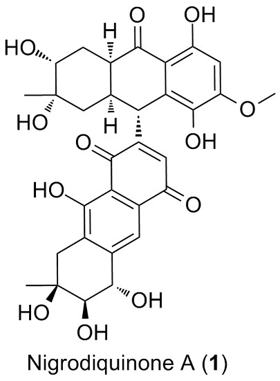
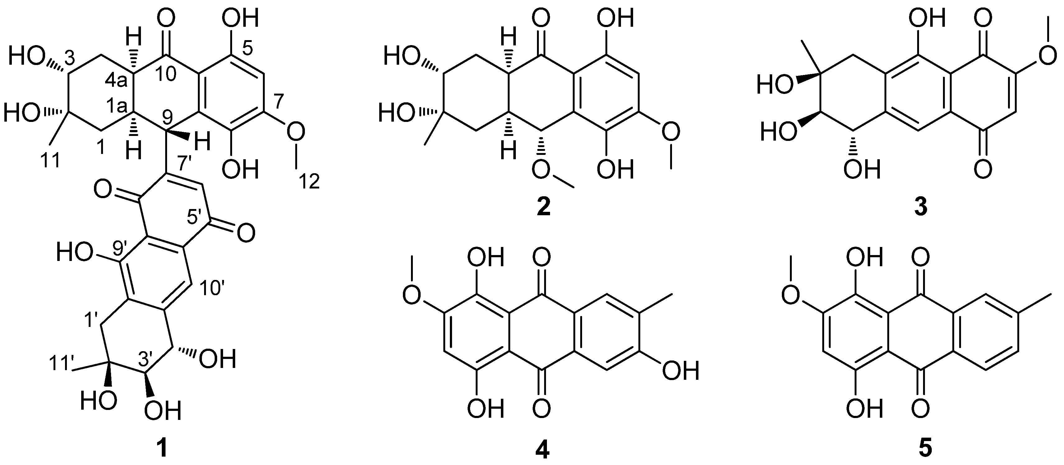
Nigrodiquinone A, a Hydroanthraquinone Dimer Containing a Rare C-9–C-7′ Linkage from a Zoanthid-Derived Nigrospora sp. Fungus

1
State Key Laboratory for the Chemistry and Molecular Engineering of Medicinal Resources, College of Chemistry and Pharmaceutical Sciences, Guangxi Normal University, Guilin 541004, China
2
Key Laboratory of Marine Drugs, Chinese Ministry of Education, School of Medicine and Pharmacy, Ocean University of China, Qingdao 266003, China
3
College of Pharmaceutical Sciences, Hebei University, Baoding 071002, China
4
Laboratory for Marine Drugs and Bioproducts, Qingdao National Laboratory for Marine Science and Technology, Qingdao 266200, China
*
Authors to whom correspondence should be addressed.
These authors contributed equally to this work.
Mar. Drugs 2016, 14(3), 51; https://doi.org/10.3390/md14030051
Submission received: 27 January 2016 / Revised: 29 February 2016 / Accepted: 4 March 2016 / Published: 9 March 2016

Abstract

:
One new hydroanthraquinone dimer with a rare C-9–C-7′ linkage, nigrodiquinone A (1), and four known anthraquinone monomers 25, were isolated from a fungus Nigrospora sp. obtained from the zoanthid Palythoa haddoni collected in the South China Sea. The structure of 1 was established through extensive NMR spectroscopy, and the absolute configuration was elucidated by comparing computed electronic circular dichroism (ECD) and optical rotations (OR) with experimental results. All the compounds were evaluated for antiviral activity, and 1 was also evaluated for antibacterial activity. Compound 4 displayed mild antiviral activity against coxsackie virus (Cox-B3) with the IC50 value of 93.7 μM, and 5 showed an IC50 value of 74.0 μM against respiratory syncytial virus (RSV).

Graphical Abstract

1. Introduction

Anthraquinone dimers are widespread and structurally-diverse family which have been isolated from marine-derived organisms including fungi, actinomycetes, tunicates and echinoderms [1,2,3,4,5,6]. They feature intriguing variety of linkages between the component anthraquinones. The structural features of these compounds may display different biological profiles. Bisanthraquinones with C-10–C-1′ and C-5a–C-2′ linkages obtained from Streptomyces sp., displayed potent methicillin resistant Staphylococcus aureus (MRSA) inhibiting activity [7]. Albopunctatone possessing a C-10–C-2′ connection isolated from the ascidian Didemnum albopunctatum showed equipotent activity towards chloroquines-ensitive and chloroquine-resistant strains of Plasmodium falciparum [8]. Another two dimmers, tetrahydroaltersolanol C and alterporriol Q featuring C-4–C-4′ and C-8–C-2′ linkages, isolated from a fungus Alternaria sp. exhibited obvious activity against the porcine reproductive and respiratory syndrome (PRRS) virus [9].
In our investigation of bioactive anthraquinone derivatives from marine-derived fungi, a series of anthraquinone monomers and anthraquinone dimers have been previously isolated from Alternaria sp. (ZJ-2008003) [9] and Nigrospora sp. (ZJ-2010006) [10]. Further chemical investigation of the fermentation broth of the Nigrospora sp. resulted in the isolation of a new hydroanthraquinone dimer, nigrodiquinone A (1), with a rare C-9–C-7′ linkage, together with four known hydroanthraquinone monomers, 4a-epi-9α-methoxydihydrodeoxybostrycin (2), 10-deoxybostrycin (3), 3,5,8-trihydroxy-7-methoxy-2-methylanthracene-9,10-dione (4), and austrocortirubin (5) (Figure 1). Herein, we report the isolation and structure elucidation of nigrodiquinone A (1), and antibacterial and antiviral activities of these five compounds.

2. Results and Discussion

Nigrodiquinone A (1) was isolated as a yellow amorphous powder. Its molecular formula C31H32O12 (sixteen degrees of unsaturation) was determined on the basis of HRESIMS data and combined with 1H and 13C NMR spectroscopic data. The deshielded area of 1H NMR spectrum (Table 1) revealed the presences of two hydrogen-bonded hydroxyl groups at δH 12.90 and 12.54 and three aromatic protons at δH 7.76, 6.51, and 6.13. Besides, one methoxy group (δH 3.93) and three oxymethine protons (δH 4.74, 3.54, and 3.31) were also observed by the 1H NMR spectrum analysis. Three carbonyl (δC 203.5, 189.9, and 183.7), six oxygen quaternary (δC 159.6, 159.1, 155.7, 136.0, 70.9, and 70.6), and three oxymethine (δC 77.4, 71.5, and 71.0) carbons were presented in 1 on the basis of careful analysis on the 13C NMR (Table 1) and DEPT spectra.
The 1H and 13C NMR data were suggested that 1 was a dimeric compound of 4a-epi-9α-methoxydihydrodeoxybostrycin (2) and 10-deoxybostrycin (3) [10]. The chemical shifts of C-9 and C-7′ were shifted to upfield (δC 36.8 and 129.7 in 1 vs. 72.8 in 2 and 160.9 in 3) and the absence of two methoxy groups (δH 3.38 in 2 and δH 3.86 in 3) indicated that this two monomers could be linked between C-9 and C-7′. Furthermore, the HMBC correlations from H-9 to C-6′, C-8′, and H-6′ to C-9 further confirmed that two units were joined via a C-9–C-7′ linkage. To the best of our knowledge, this is the first time to report the anthraquione dimer with a C-9–C-7′ linkage.
In the selective 1D NOE experiments, the irradiation of H-9 resulted in no obvious enhancement of H-1a suggested that H-9 and H-1a might be trans oriented. The irradiation of H-3′ resulted in enhancement of H-11′ and no obvious enhancement of H-4′, and the key NOESY data (Figure 2) indicated that the relative configurations of all asymmetric carbons in 1 should be identical to those of 2 and 3 [10]. Thus, the relative configuration of 1 was determined as (1aR*,2S*,3R*,4aR*,9R*,2′S*,3′R*, and 4′S*).
To compare computed electronic circular dichroism (ECD) and optical rotations (OR) with experimental results is a valid method to assign absolute configurations of natural products [11,12,13,14,15,16]. Thus, the absolute configuration of (1) was investigated by quantum chemical TDDFT calculations of their ECD and optical rotations (OR) spectra. ECD computations for four possible absolute configurations ((1aR,2S,3R,4aR,9R,2′S,3′R,4′S)-1, (1aS,2R,3S,4aS,9S,2′R,3′S,4′R)-1, (1aR,2S,3R,4aR,9R,2′R,3′S,4′R)-1, and (1aS,2R,3S,4aS,9S,2′S,3′R,4′S)-1) of 1 were carried out at the B3LYP/6-311G+(2d, p) level in the gas phase. Only the predicted ECD for (1aR,2S,3R,4aR,9R,2′S,3′R,4′S)-1 look similar to the experimental result of 1 (Figure 3, and supporting information). The computed ORs in the gas phase were −39.1 for (1aR,2S,3R,4aR,9R,2′S,3′R,4′S)-1, +38.7 for (1aS,2R,3S,4aS,9S,2′R,3′S,4′R)-1, respectively, and the experimental optical rotation value was −36.0. Based on both of ECD and OR, the absolute configuration of 1 was assigned as 1aR,2S,3R,4aR,9R,2′S,3′R,4′S.
With regard to the connecting positions of monomers in the dimmers, the C-1–C-1′ [6], C-1–C-5′ [6], C-2–C-2′ [17], C-4–C-4′ [8], C-4–C-6′ [8], C-5–C-5′ [18], C-5–C-7′ [6], C-7–C-5′ [19], and C-8–C-8′ [8] connections have been reported to present in anterporriols family. Besides, the C-9–C-9′ [20], C-10–C–2′ [8], and C-10–C-1′/C-4a–C-2′ [7] linkages also presented in the anthraquinone dimmers. Nigrodiquinone A (1) is the first report to possess the rare C-9–C-7′ linkage.
Compounds 15 were evaluated for antiviral activity against Cox-B3 and RSV, with ribavirin as a positive control (Table 2). Compound 4 displayed mild antiviral activity against Cox-B3 with the IC50 value of 93.7 μM, and 5 showed an IC50 value of 74.0 μM against RSV. Compound 1 was evaluated for antibacterial activity against nine bacterial strains, Gram-positive B. subtilis, B. cereus, M. luteus, M. tetragenus, S. albus, S. aureus, Gram-negative E. coli, V. anguillarum, and V. parahemolyticus, however 1 was inactive at the concentration of 50 μM.

3. Materials and Methods

3.1. General Experimental Procedures

Optical rotations were measured on a JASCO P-1020 digital polarimeter. CD spectra were recorded on a MOS-450 spectrometer. IR spectra were recorded on a Bruker EQUINOX 55 spectrometer using KBr pellets. 1H and 13C spectra were recorded on a JEOL Eclips-600 spectrometer at 600 MHz for 1H and 150 MHz for 13C in DMSO-d6. Chemical shifts δ are reported in ppm, using TMS as internal standard and coupling constants (J) are in Hz. HRESIMS were measured on a Thermo MAT95XP High Resolution mass spectrometer. Silica gel (Qing Dao Hai Yang Chemical Group Co., Qingdao, China; 200–300 mesh), octadecylsilyl silica gel (Unicorn; 45–60 μm) and Sephadex LH-20 (Amersham Biosciences, Uppsala, Sweden) were used for column chromatography (CC). Precoated silica gel plates (G60, F-254, Yan Tai Zi Fu Chemical Group Co., Qingdao, China) were used for thin layer chromatography (TLC). Semi-preparative HPLC was performed on a Waters 1525 system using a semi-preparative C18 (Kromasil 7 μm, 10 × 250 mm) column coupled with a Waters 2996 photodiode array detector, at a flow rate of 2.0 mL/min.

3.2. Fungal Material

The fungal strain Nigrospora sp. (ZJ-2010006, Genbank NO. HM565952) [10] was isolated from a piece of fresh tissue from the inner part of the zoanthid Palythoa haddoni (GX-WZ-20100026) [21], collected from the Weizhou coral reefs in the South China Sea in April 2010. The strain was deposited at the Key Laboratory of Marine Drugs, the Ministry of Education of China, School of Medicine and Pharmacy, Ocean University of China, Qingdao, PR China. The fungal strain was cultivated in potato glucose liquid medium (composition of medium: 200 g/L cooked and sliced potatoes, 20 g/L glucose in artificial seawaters, in 1L Erlenmeyer flasks each containing 400 mL of culture broth) at 27 °C without shaking for 4 weeks.

3.3. Extraction and Isolation

The culture (80 L) was filtered to separate the culture broth from the mycelia. The culture broth was extracted with an equal volume of EtOAc and the fungal mycelia were extracted with CHCl3-MeOH (1:1, v/v) for three times, respectively. The organic extracts were evaporated under vacuum, and then the crude extract was suspended in H2O and partitioned with EtOAc. All the EtOAc layers were combined and evaporated to dryness under reduced pressure to give an EtOAc extract (98.4 g), which was subjected to silica gel column chromatography using gradient mixtures of petroleum ether (PE)/EtOAc (EA) (100% PE, 60:40, 40:60, 20:80, and 100% EA) to yield five fractions (Fr. 1–Fr. 5). Fr. 2 was subjected to silica gel column chromatography (CHCl3/MeOH) to obtain Fr. 2-1 (20:1, v/v) and Fr. 2-2 (10:1, v/v). Fr. 2-1 was repeated subjected to silica gel column chromatography to get 5 (7.2 mg). Fr. 2-2 was subjected to Sephadex LH-20 chromatography with mixtures of CHCl3-MeOH (1:1, v/v), purified by ODS column chromatography and preparative by HPLC on a C18 column (Kromasil, 7 μm, 10 × 250 mm, 2 mL/min) to obtain 2 (MeOH-H2O (75:25, v/v), 5.3 mg), 3 (MeOH-H2O (55:45, v/v), 12.5 mg) and 4 (MeOH-H2O (55:45, v/v), 50.1 mg). Fr. 3 was further purified by Sephadex LH-20 chromatography with mixtures of CHCl3-MeOH (1:1, v/v), and then semi-preparative HPLC, eluting with MeOH-H2O (50:50, v/v) to yield 1 (1.5 mg).
Nigrodiquinone A (1): yellow amorphous powder; [α] D 24 −36.0 (c 0.025, acetone); CD (0.84 mmol, MeOH) λmax (Δε) 212 (−3.97), 239 (0.16), 254 (−1.00), 283 (1.79), 354 (−0.78) nm. IR (KBr) νmax 3407, 2928, 1631, 1294 cm–1; mp 226 °C; 1H NMR and 13C NMR see Table 1; ESIMS m/z 597.1 [M + H]+; HRESIMS m/z 597.1975 (calcd for C31H33O12, 597.1972 [M + H]+).
The structures of 25 were assigned by spectroscopic methods and comparison of the 1H- and 13C NMR data with those reported in literature [10].

3.4. Biological Assays

Antiviral activity was evaluated by the cytopathic effect (CPE) inhibition assay according to established procedures, with ribavirin as a positive control [22]. Two viruses, coxsackie virus (Cox-B3) and respiratory syncytial virus (RSV) were used. Antibacterial activities were evaluated by the method as described previously [10]. Nine bacterial strains, Gram-positive Bacillus cereus (ACCC 11077), Bacillus subtilis (ATCC 6633), Micrococcus luteus (ATCC 49732), Micrococcus tetragenus (ATCC 13623), Staphylococcus albus (ATCC 8799), Staphylococcus aureus (ATCC 27154), Gram-negative Escherichia coli (ATCC 25922), Vibrio anguillarum (ATCC 19019), and Vibrio parahemolyticus (ATCC 17802) were used, and ciprofloxacin was used as a positive control.

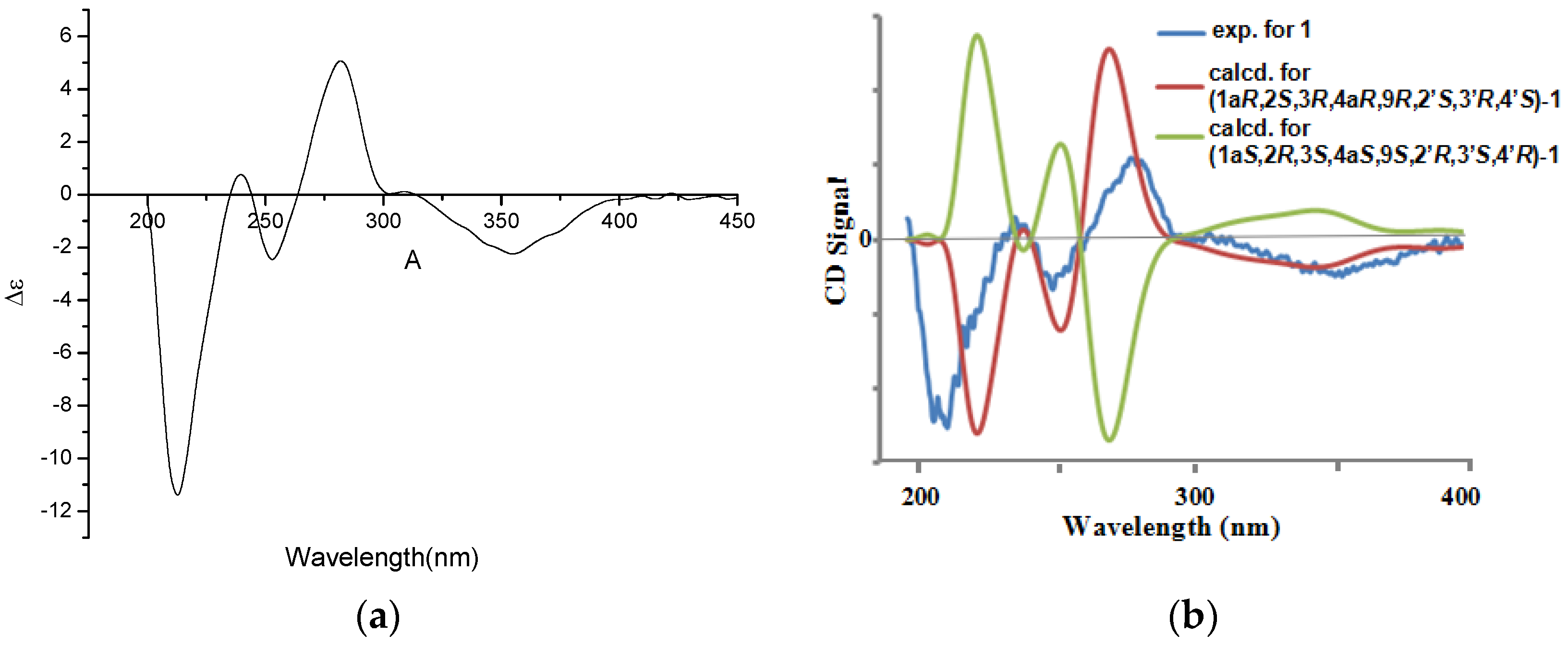
3.5. Computational Section

Conformational searches were performed using MMFF94S force field for (1aR,2S,3R,4aR,9R,2′S,3′R,4′S)-1, (1aS,2R,3S,4aS,9S,2′R,3′S,4′R)-1, (1aR,2S,3R,4aR,9R,2′R,3′S,4′R)-1, and (1aS,2R,3S,4aS,9S,2′S,3′R,4′S)-1. All geometries (15 lowest energy conformers for (1aR,2S,3R,4aR,9R,2′S,3′R,4′S)-1, 15 for (1aS,2R,3S,4aS,9S,2′R,3′S,4′R)-1, 15 lowest energy conformers for (1aR,2S,3R,4aR,9R,2′R,3′S,4′R)-1, and 15 for (1aS,2R,3S,4aS,9S,2′S,3′R,4′S)-1, respectively) with relative energy from 0–10 kcal/mol used in optimizations at the B3LYP/6-311+G(d) level using Gaussian 09 package [23]. The B3LYP/6-311+G(d)-optimized conformers (3 lowest energy conformers for (1aR,2S,3R,4aR,9R,2′S,3′R,4′S)-1, 4 for (1aS,2R,3S,4aS,9S,2′R,3′S,4′R)-1, 3 lowest energy conformers for (1aR,2S,3R,4aR,9R,2′R,3′S,4′R)-1 and 4 for (1aS,2R,3S,4aS,9S,2′S,3′R,4′S)-1; see Supporting Information for details) with relative energy from 0 to 4.6 kcal/mol were then re-optimized at the B3LYP/6-311+G(d) level. ECD and OR computations for all conformers were carried out at the B3LYP/6-311G+(2d, p) level in the gas phase. Boltzmann statistics were performed for ECD simulations with standard deviation of σ 0.16 eV.

4. Conclusions

As part of our ongoing investigation on discovering for biological secondary metabolites from zoanthid-derived fungi in the South China Sea, a new hydroanthraquinone dimer with a rare C-9–C-7′ linkage, nigrodiquinone A (1), was isolated from the culture of Nigrospora sp. The absolute configuration of nigrodiquinone A was determined by quantum chemical TDDFT calculations of theirs ECD and OR spectra. Compound 4 displayed mild antiviral activity against Cox-B3 with the IC50 value of 93.7 μM, and 5 showed an IC50 value of 74.0 μM against RSV.

Acknowledgments

This work was supported by the Program of National Natural Science Foundation of China (No. 41322037), the Program of Natural Science Foundation of Shandong Province of China (No. JQ201510), the Marine Special Public Welfare Scientific Research, State Oceanic Administration of China (No. 201405038), the Guangxi Natural Science Foundation of China (2014GXNSFAA118055), the Project of Key Laboratory for the Chemistry and Molecular Engineering of Medicinal Resources (Guangxi Normal University), Ministry of Education of China (CMEMR2013-A06), and the Taishan Scholars Program, China.

Author Contributions

Wei-Feng Xu, Xue-Mei Hou and Kai-Lin Yang contribute to extraction, isolation, identification, and manuscript preparation; Fei Cao contribute to quantum chemistry calculation; Chang-Yun Wang contribute to NMR analysis and structure elucidation; Kai-Lin Yang contribute to bioactivities test; Rui-Yun Yang and Chang-Lun Shao were the project leaders organizing and guiding the experiments and manuscript writing.

Conflicts of Interest

The authors declare no conflict of interest.

References

  1. Fsulkner, D.J. Marine natural products. Nat. Prod. Rep. 1987, 4, 539–576. [Google Scholar] [CrossRef]
  2. Blunt, J.W.; Copp, B.R.; Hu, W.P.; Munro, M.H.G.; Northcote, P.T.; Prinsep, M.R. Marine natural products. Nat. Prod. Rep. 2008, 25, 35–94. [Google Scholar] [CrossRef] [PubMed]
  3. Blunt, J.W.; Copp, B.R.; Munro, M.H.G.; Northcote, P.T.; Prinsep, M.R. Marine natural products. Nat. Prod. Rep. 2011, 28, 196–268. [Google Scholar] [CrossRef] [PubMed]
  4. Blunt, J.W.; Copp, B.R.; Keyzers, R.A.; Munro, M.H.G.; Prinsep, M.R. Marine natural products. Nat. Prod. Rep. 2012, 29, 144–222. [Google Scholar] [CrossRef] [PubMed]
  5. Blunt, J.W.; Copp, B.R.; Keyzers, R.A.; Munro, M.H.G.; Prinsep, M.R. Marine natural products. Nat. Prod. Rep. 2014, 31, 160–258. [Google Scholar] [CrossRef] [PubMed]
  6. Zhou, X.M.; Zheng, C.J.; Chen, G.Y.; Song, X.P.; Han, C.R.; Li, G.N.; Fu, Y.H.; Chen, W.H.; Niu, Z.G. Bioactive anthraquinone derivatives from the mangrove-derived fungus Stemphylium sp. 33231. J. Nat. Prod. 2014, 77, 2021–2028. [Google Scholar] [CrossRef] [PubMed]
  7. Socha, A.M.; Garcia, D.; Sheffer, R.; Rowley, D.C. Antibiotic bisanthraquinones produced by a Streptomycete isolated from a cyanobacterium associated with Ecteinascidia turbinate. J. Nat. Prod. 2006, 69, 1070–1073. [Google Scholar] [CrossRef] [PubMed]
  8. Carroll, A.R.; Nash, B.D.; Duffy, S.; Avery, V.M. Albopunctatone, an antiplasmodial anthrone-anthraquinone from the Australian ascidian Didemnum albopunctatum. J. Nat. Prod. 2012, 75, 1206–1209. [Google Scholar] [CrossRef] [PubMed]
  9. Zheng, C.J.; Shao, C.L.; Guo, Z.Y.; Chen, J.F.; Deng, D.S.; Yang, K.L.; Chen, Y.Y.; Fu, X.M.; She, Z.G.; Lin, Y.C.; et al. Bioactive hydroanthraquinones and anthraquinone dimmers from a soft coral-derived Alternaria sp. fungus. J. Nat. Prod. 2012, 75, 189–197. [Google Scholar] [CrossRef] [PubMed]
  10. Yang, K.L.; Wei, M.Y.; Shao, C.L.; Fu, X.M.; Guo, Z.Y.; Xu, R.F.; Zheng, C.J.; She, Z.G.; Lin, Y.C.; Wang, C.Y. Antibacterial anthraquinone derivatives from a sea anemone-derived fungus Nigrospora sp. J. Nat. Prod. 2012, 75, 935–941. [Google Scholar] [CrossRef] [PubMed]
  11. Zhu, H.J. Organic Stereochemsitry—Experimental and Theoretical Methods; Wiley-VCH: Weinheim, Germany, 2015. [Google Scholar]
  12. Zhu, H.J. Current Organic Stereochemistry; Science Presses of China: Beijing, China, 2009. [Google Scholar]
  13. Zhu, H.J.; Li, W.X.; Hu, D.B.; Wen, M.L. Discussion of absolute configuration for bioactive Griseusins by comparing computed optical rotations and electronic circular dichroism with the experimental results. Tetrahedron 2014, 70, 8236–8243. [Google Scholar] [CrossRef]
  14. Yu, H.; Li, W.X.; Wang, J.C.; Yang, Q.; Wang, H.J.; Zhang, C.C.; Ding, S.S.; Li, Y.; Zhu, H.J. Pestalotiopsin C, stereochemistry of a new caryophyllene from a fungus of Trichoderma sp. and its tautomerization characteristics in solution. Tetrahedron 2015, 71, 3491–3494. [Google Scholar] [CrossRef]
  15. He, P.; Wang, X.F.; Guo, X.J.; Zhou, C.Q.; Shen, S.G.; Hu, D.B.; Yang, X.L.; Luo, D.Q.; Dukor, R.; Zhu, H.J. Vibrational circular dichroism study for natural bioactive schizandrin and reassignment of its absolute configuration. Tetrahedron Lett. 2014, 55, 2965–2968. [Google Scholar] [CrossRef]
  16. He, J.B.; Ji, Y.N.; Hu, D.B.; Zhang, S.; Yan, H.; Liu, X.C.; Luo, H.R.; Zhu, H.J. Structure and absolute configuration of penicilliumine, a new alkaloid from Penicillium commune 366606. Tetrahedron Lett. 2014, 55, 2684–2686. [Google Scholar] [CrossRef]
  17. Huang, C.H.; Pan, J.H.; Chen, B.; Yu, M.; Huang, H.B.; Zhu, X.; Lu, Y.J.; She, Z.G.; Lin, Y.C. Three bianthraquinone derivatives from the mangrove endophytic fungus Alternaria sp. ZJ9-6B from the South China Sea. Mar. Drugs 2011, 9, 832–843. [Google Scholar] [CrossRef] [PubMed]
  18. Suemitsu, R.; Horiuchi, K.; Kubota, M.; Okamatse, T. Production of alterporriols, altersolanols and macrosporin by Alternaria porri and A. Solani. Phytochemistry 1990, 29, 1509–1511. [Google Scholar] [CrossRef]
  19. Debbab, A.; Aly, A.H.; Edrada-Ebel, R.; Wray, V.; Müller, W.E.G.; Totzke, F.; Zirrgiebel, U.; Schächtele, C.; Kubbutat, M.H.G.; Lin, W.H.; et al. Bioactive metabolites from the endophytic fungus Stemphylium globuliferum isolated from Mentha pulegium. J. Nat. Prod. 2009, 72, 626–631. [Google Scholar] [CrossRef] [PubMed]
  20. Rideout, J.A.; Sutherland, M.D. Pigments of marine animals. XV. Bianthrones and related polyketides from Lamprometra palmate gyges and other species of crinoids. Aust. J. Chem. 1985, 38, 793–808. [Google Scholar] [CrossRef]
  21. Qin, X.Y.; Yang, K.L.; Li, J.; Wang, C.Y.; Shao, C. L. Phylogenetic diversity and antibacterial activity of culturable fungi derived from the zoanthid Palythoa haddoni in the South China Sea. Mar. Biotechnol. 2015, 17, 99–109. [Google Scholar] [CrossRef] [PubMed]
  22. Grassauer, A.; Weinmuellner, R.; Meier, C.; Pretsch, A.; Prieschl-Grassauer, E.; Unger, H. Iota-carrageenan is a potent inhibitor of rhinovirus infection. Virol. J. 2008, 5. [Google Scholar] [CrossRef] [PubMed]
  23. Frisch, M.J.; Trucks, G.W.; Schlegel, H.B.; Scuseria, G.E.; Robb, M.A.; Cheeseman, J.R.; Scalmani, G.; Barone, V.; Mennucci, B.; Petersson, G.A.; et al. Gaussian 09; Gaussian Inc.: Wallingford, CT, USA, 2009. [Google Scholar]
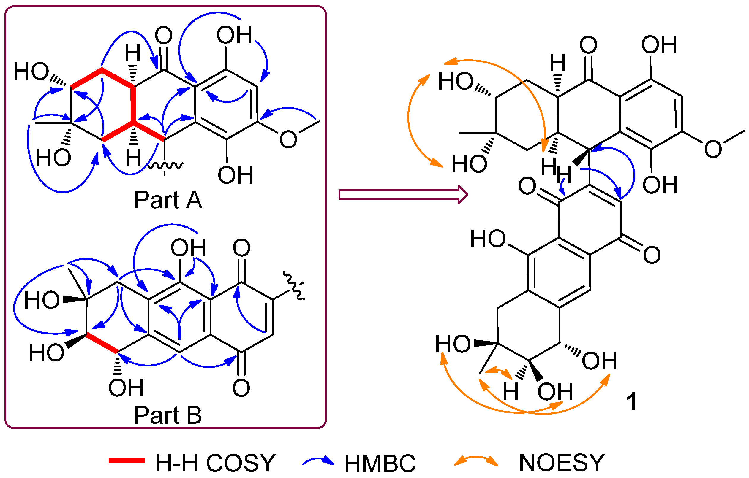
Figure 1. Chemical structures of 15.
Marinedrugs 14 00051 g001
Figure 2. Key correlations for compound 1.
Marinedrugs 14 00051 g002
Figure 3. (a) Experimental CD spectra for 1 and (b) calculated electronic circular dichroism (ECD) spectra of 1.
Marinedrugs 14 00051 g003
Table 1. NMR spectroscopic data of 1.a
PositionδH (J in Hz)δC, Mult.H–H COSYHMBC
11.48, t, (13.5)
1.88, ddd, (13.5, 3.6, 2.3)
41.5, CH2H-1aC-3
1a2.86, m34.5, CHH-1, 4a, 9
2 70.6, C
33.31, m71.0, CHH-4
41.65, ddd, (12.6, 12.0, 4.8)
2.52, ddd, (12.7, 4.5, 2.7)
36.8, CH2H-3, 4aC-2, 10
4a3.07, m41.5, CHH-1a, 4
5 159.1, C
66.51, s98.7, CH C-10a
7 155.7, C
8 136.0, C
94.68, br s36.8, CH C-1, 1a, 9a, 10a, 6′, 8′
9a 124.9, C
10 203.5, C
10a 109.6, C
111.17, s26.8, CH3 C-1, 2, 3
123.93, s55.8, CH3 C-7
1′2.68, d (19.0),
3.07, d (19.0)
36.8, CH2 C-3′, 4a′, 9′
1a′ 130.2, C
2′ 70.9, C
3′3.54, d (8.7)77.4, CHH-4′
4′4.74, br d (8.7)71.5, CHH-3′
4a′ 148.1, C
5′ 183.7, C
6′6.13, d (1.1)135.3, CH C-8′, 9
7′ 129.7, C
8′ 189.9, C
9′ 159.6, C
9a′ 113.0, C
10′7.76, br s117.3, CH C-1a′, 4′, 5′, 9a′
10a′ 100.0, C
11′1.43, s26.3, CH3 C-1′, 2′, 3′
2-OH3.17, br s
3-OH3.71, d (4.9)
5-OH12.90, s C-6, 10a
8-OH7.61, s
2′-OH3.67, br s
3′-OH4.27, br s
4′-OH4.87, br s
9′-OH12.54, s C-1a′, 9′, 9a′
a Measured at 600 MHz for 1H NMR and 150 MHz for 13C NMR in acetone-d6.
Table 2. Antiviral activities of compounds 15. a
VirusIC50 (μM)
12345Ribavirin b
RSV----74.078.0
Cox-B3---93.7-39.0
a Data are expressed in IC50 values (μM). b Ribavirin was used as a positive control. “-” means no antiviral activities.

