
Influence of Chitosan Swelling Behaviour on Controlled Release of Tenofovir from Mucoadhesive Vaginal Systems for Prevention of Sexual Transmission of HIV

 , , , ,
Abstract
:1. Introduction
2. Results and Discussion
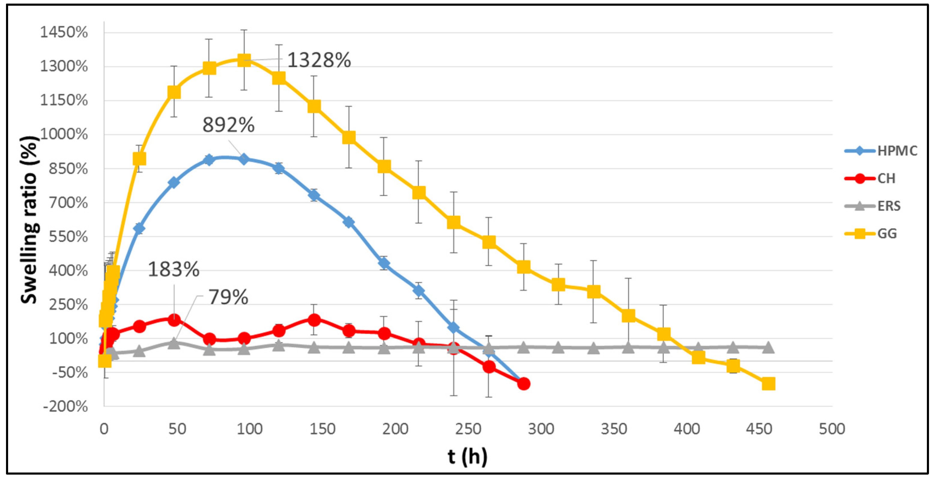
2.1. Swelling Tests
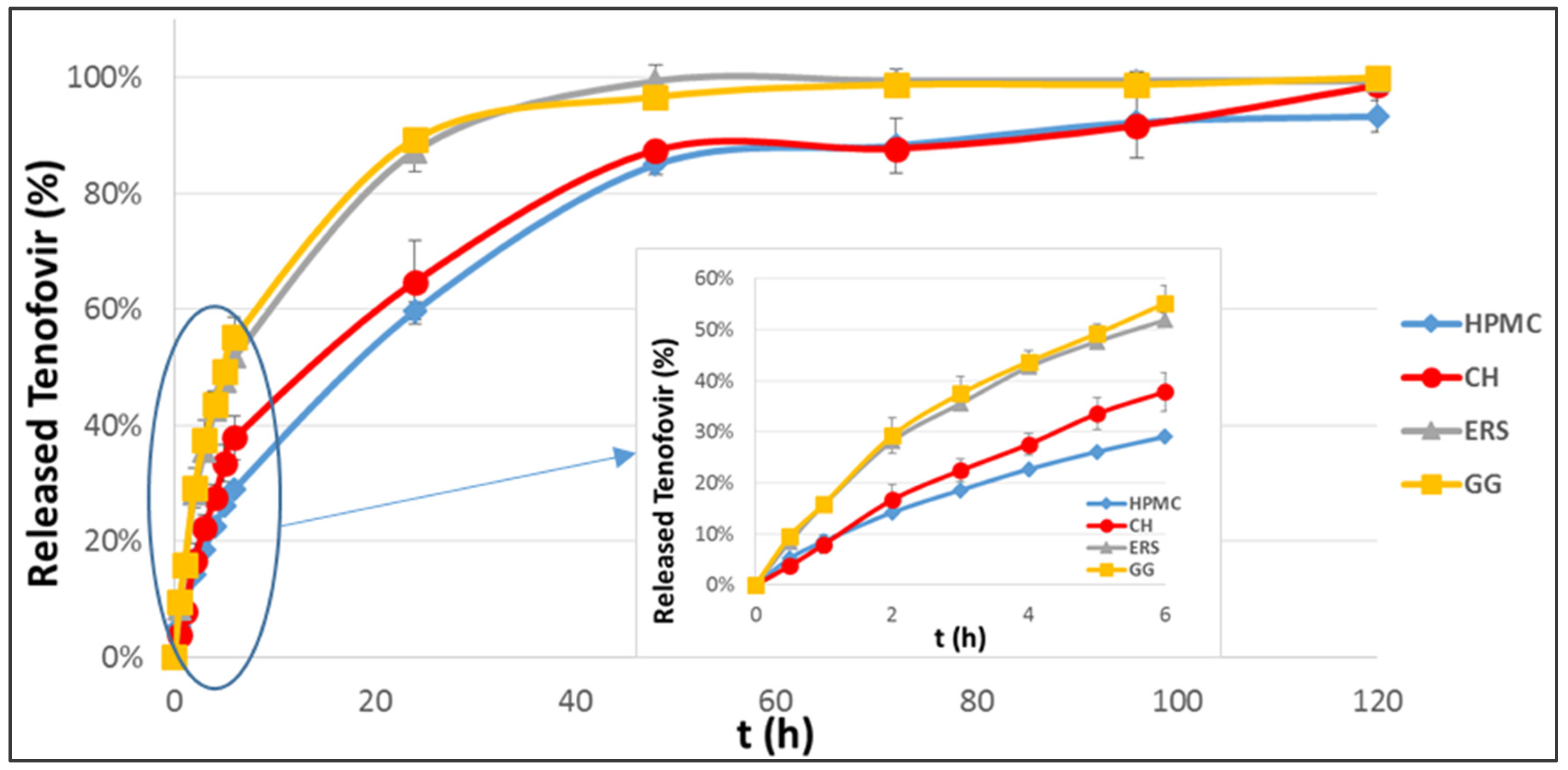
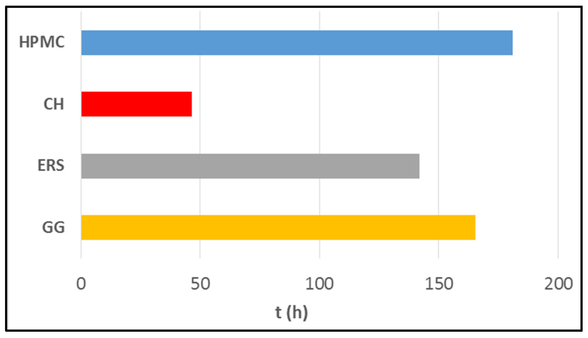
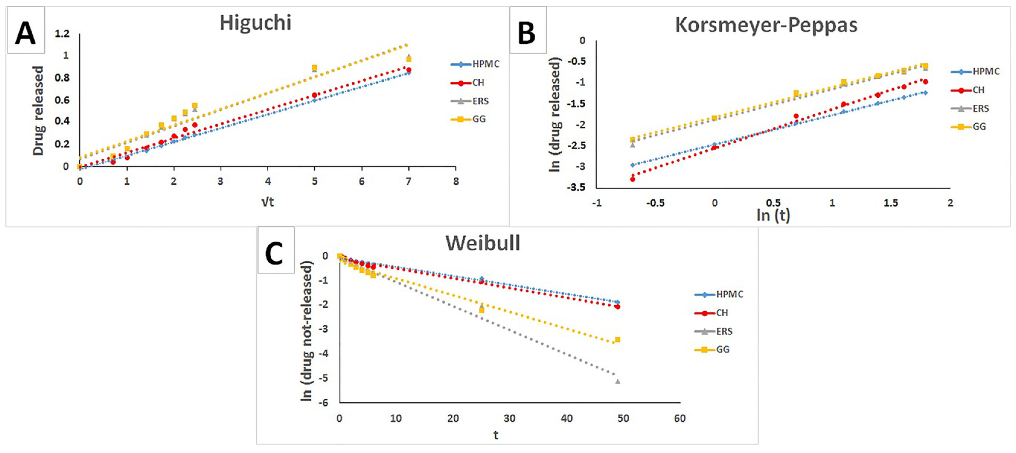
2.2. Release Study
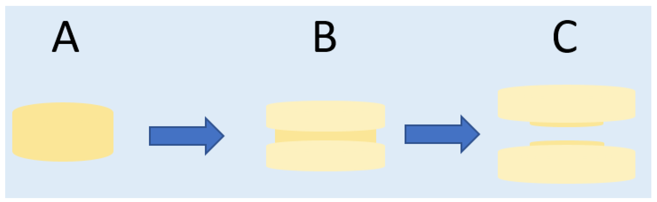
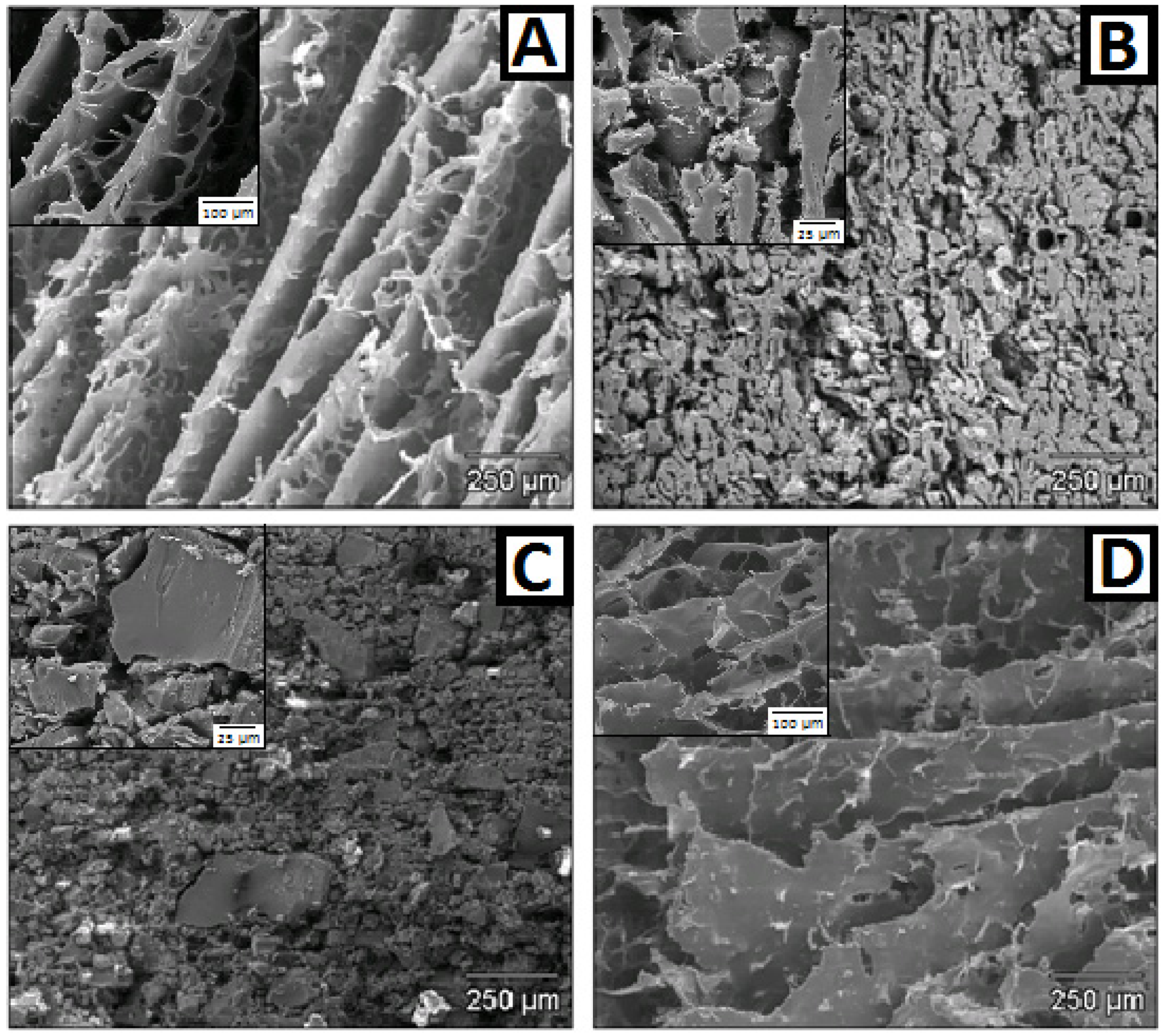
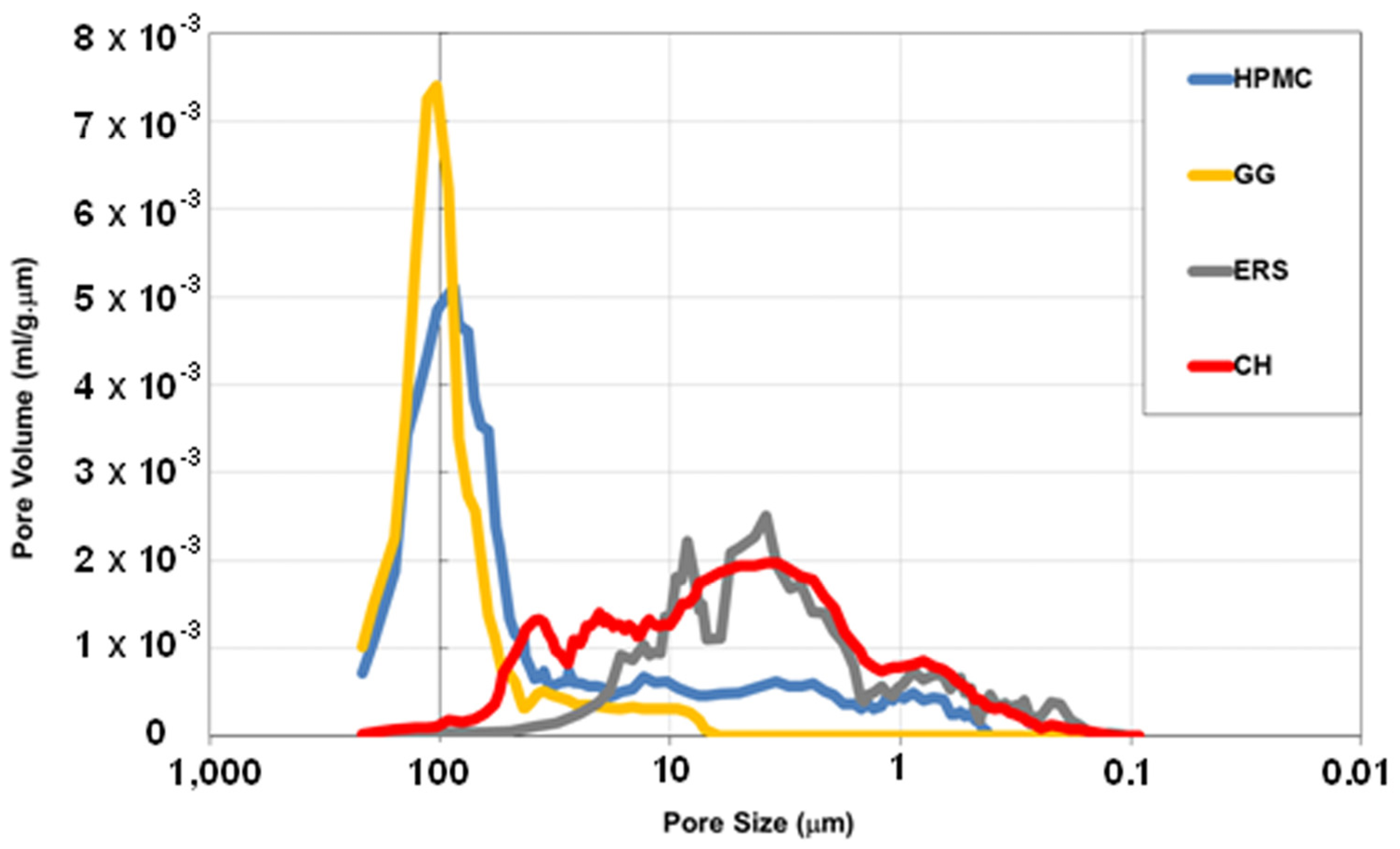
2.3. Microstructure of Witnesses. FE-SEM, and Hg Porosimetry
2.4. Evaluation of Mucoadhesion
2.5. Cell Toxicity
3. Experimental Section
3.1. Materials and Preparation of the Compact
3.2. Methods
3.2.1. Swelling Tests
3.2.2. Release Study
3.2.3. Swelling Witnesses
3.2.4. Assessment of Mucoadhesion
3.2.5. Cytotoxicity Assessment
4. Conclusions
Acknowledgments
Author Contributions
Conflicts of Interest
References
- World Health Organization. HIV/AIDS. Available online: http://www.who.int/topics/hiv_aids/en/ (accessed on 9 September 2015).
- World Health Organization. Global HIV Prevalence Has Levelled off. Available online: http://www.who.int/mediacentre/news/releases/2007/pr61/en/ (accessed on 9 September 2015).
- UNAIDS. AIDS by the Numbers. Available online: http://www.unaids.org/sites/default/files/media_asset/20131120_AIDSbynumbers_A5brochure_en_2.pdf (accessed on 9 September 2015).
- UNAIDS. AIDS by the Numbers 2015. Available online: http://www.unaids.org/sites/default/files/media_asset/AIDS_by_the_numbers_2015_en.pdf (accessed on 9 September 2015).
- UNAIDS. The Gap Report. Available online: http://www.unaids.org/sites/default/files/media_asset/UNAIDS_Gap_report_en.pdf (accessed on 9 September 2015).
- McConville, C.; Friend, D.R.; Clark, M.R.; Malcolm, K. Preformulation and development of a once-daily sustained-release tenofovir vaginal tablet containing a single excipient. J. Pharm. Sci. 2013, 102, 1859–1868. [Google Scholar] [CrossRef] [PubMed]
- Viread®, European Medicines Agency (EMA). Available online: http://www.ema.europa.eu/docs/en_GB/document_library/EPAR_-_Product_Information/human/000419/WC500051737.pdf (accessed on 9 September 2015).
- Mayer, K.H.; Maslankowski, L.A.; Gai, F.; El-Sadr, W.M.; Justman, J.; Kwiecien, A.; Mâsse, B.; Eshleman, S.H.; Hendrix, C.; Morrow, K.; et al. Safety and tolerability of tenofovir vaginal gel in abstinent and sexually active HIV-infected and uninfected women. AIDS 2006, 20, 543–551. [Google Scholar] [CrossRef] [PubMed]
- Johnson, T.J.; Gupta, K.M.; Fabian, J.; Albright, T.H.; Kiser, P.F. Segmented polyurethane intravaginal rings for the sustained combined delivery of antiretroviral agents dapivirine and tenofovir. Eur. J. Pharm. Sci. 2010, 39, 203–212. [Google Scholar] [CrossRef] [PubMed]
- Abdool Karim, Q.; Abdool Karim, S.S.; Frohlich, J.A.; Grobler, A.C.; Baxter, C.; Mansoor, L.E.; Kharsany, A.B.; Sibeko, S.; Mlisana, K.P.; Omar, Z.; et al. Effectiveness and safety of tenofovir gel, an antiretroviral microbicide, for the prevention of HIV infection in women. Science 2010, 329, 1168–1174. [Google Scholar] [CrossRef] [PubMed]
- Celum, C.; Baeten, J.M. Tenofovir-based pre-exposure prophylaxis for HIV prevention: Evolving evidence. Curr. Opin. Infect. Dis. 2012, 25, 51–57. [Google Scholar] [CrossRef] [PubMed]
- Akil, A.; Agashe, H.; Dezzutti, C.S.; Moncla, B.J.; Hillier, S.L.; Devlin, B.; Shi, Y.; Uranker, K.; Rohan, L.C. Formulation and characterization of polymeric films containing combinations of antiretrovirals (ARVs) for HIV prevention. Pharm. Res. 2015, 32, 458–468. [Google Scholar] [CrossRef] [PubMed]
- Akil, A.; Devlin, B.; Cost, M.; Rohan, L.C. Increased Dapivirine tissue accumulation through vaginal film codelivery of dapivirine and Tenofovir. Mol. Pharm. 2014, 11, 1533–1541. [Google Scholar] [CrossRef] [PubMed]
- Baum, M.M.; Butkyavichene, I.; Churchman, S.A.; Lopez, G.; Miller, C.S.; Smith, T.J.; Moss, J.A. An intravaginal ring for the sustained delivery of tenofovir disoproxil fumarate. Int. J. Pharm. 2015, 495, 579–587. [Google Scholar] [CrossRef] [PubMed]
- Smith, J.M.; Srinivasan, P.; Teller, R.S.; Lo, Y.; Dinh, C.T.; Kiser, P.F.; Herold, B.C. Tenofovir disoproxil fumarate intravaginal ring protects high-dose depot medroxyprogesterone acetate-treated macaques from multiple SHIV exposures. J. Acquir. Immune Defic. Syndr. 2015, 68, 1–5. [Google Scholar] [CrossRef] [PubMed]
- Moss, J.A.; Malone, A.M.; Smith, T.J.; Kennedy, S.; Nguyen, C.; Vincent, K.L.; Motamedi, M.; Baum, M.M. Pharmacokinetics of a Multipurpose Pod-Intravaginal Ring Simultaneously Delivering Five Drugs in an Ovine Model. Antimicrob. Agents Chemother. 2013, 57, 3994–3997. [Google Scholar] [CrossRef] [PubMed]
- Smith, J.M.; Rastogi, R.; Teller, R.S.; Srinivasan, P.; Mesquita, P.M.; Nagaraja, U.; McNicholl, J.M.; Hendry, R.M.; Dinh, C.T.; Martin, A.; et al. Intravaginal ring eluting tenofovir disoproxil fumarate completely protects macaques from multiple vaginal simian-HIV challenges. Proc. Natl. Acad. Sci. USA 2013, 110, 16145–16150. [Google Scholar] [CrossRef] [PubMed]
- Valenta, C. The use of mucoadhesive polymers in vaginal delivery. Adv. Drug Deliv. Rev. 2005, 57, 1692–1712. [Google Scholar] [CrossRef] [PubMed]
- Rodriguez, I.C.; Cerezo, A.; Salem, I.I. Bioadhesive delivery systems. ARS Pharm. 2000, 41, 115–128. [Google Scholar]
- Sánchez-Sánchez, M.P.; Martín-Illana, A.; Ruiz-Caro, R.; Bermejo, P.; Abad, M.J.; Carro, R.; Bedoya, L.M.; Tamayo, A.; Rubio, J.; Fernández-Ferreiro, A.; et al. Chitosan and Kappa-Carrageenan Vaginal Acyclovir Formulations for Prevention of Genital Herpes. In Vitro and Ex Vivo Evaluation. Mar. Drugs 2015, 13, 5976–5992. [Google Scholar] [CrossRef] [PubMed]
- Frank, L.A.; Sandri, G.; D’Autilia, F.; Contri, R.V.; Bonferoni, M.C.; Caramella, C.; Frank, A.G.; Pohlmann, A.R.; Guterres, S.S. Chitosan gel containing polymeric nanocapsules: A new formulation for vaginal drug delivery. Int. J. Nanomed. 2014, 9, 3151–3161. [Google Scholar]
- Senyiğit, Z.A.; Karavana, S.Y.; Eraç, B.; Gürsel, O.; Limoncu, M.H.; Baloğlu, E. Evaluation of chitosan based vaginal bioadhesive gel formulations for antifungal drugs. Acta Pharm. 2014, 64, 139–156. [Google Scholar] [CrossRef] [PubMed]
- Szymańska, E.; Winnicka, K.; Amelian, A.; Cwalina, U. Vaginal chitosan tablets with clotrimazole-design and evaluation of mucoadhesive properties using porcine vaginal mucosa, mucin and gelatine. Chem. Pharm. Bull. 2014, 62, 160–167. [Google Scholar] [CrossRef] [PubMed]
- Sánchez, R.; Damas, R.; Domínguez, P.; Cerezo, P.; Salcedo, I.; Aguzzi, C.; Viseras, C. Uso de la HidroxiPropilMetilCelulosa (HPMC) en la Liberación Modificada de Fármacos. Available online: http://innovacion.gob.sv/inventa/attachments/article/8566/HPMC%20en%20f%C3%A1rmacos.pdf (accessed on 9 September 2015).
- Ghosal, K.; Hazra, B.T.; Bhowmik, B.B.; Thomas, S. Formulation Development, Physicochemical Characterization and In Vitro-in vivo drug release of vaginal Films. Curr. HIV Res. 2016, 14, 295–306. [Google Scholar] [CrossRef] [PubMed]
- Grammen, C.; Van den Mooter, G.; Appeltans, B.; Michiels, J.; Crucitti, T.; Ariën, K.K.; Augustyns, K.; Augustijns, P.; Brouwers, J. Development and characterization of a solid dispersion film for the vaginal application of the anti-HIV microbicide UAMC01398. Int. J. Pharm. 2014, 475, 238–244. [Google Scholar] [CrossRef] [PubMed]
- Tuğcu-Demiröz, F.; Acartürk, F.; Özkul, A. Preparation and characterization of bioadhesive controlled-release gels of cidofovir for vaginal delivery. J. Biomater. Sci. Polym. Ed. 2015, 26, 1237–1255. [Google Scholar] [CrossRef] [PubMed]
- Wang, L.; Tang, X. A novel ketoconazole bioadhesive effervescent tablet for vaginal delivery: Design, in vitro and ‘in vivo’ evaluation. Int. J. Pharm. 2008, 350, 181–187. [Google Scholar] [CrossRef] [PubMed]
- Karasulu, H.Y.; Hilmioğlu, S.; Metin, D.Y.; Güneri, T. Efficacy of a new ketoconazole bioadhesive vaginal tablet on Candida albicans. Farmaco 2004, 59, 163–167. [Google Scholar] [CrossRef] [PubMed]
- Hiorth, M.; Nilsen, S.; Tho, I. Bioadhesive mini-tablets for vaginal drug delivery. Pharmaceutics 2014, 6, 494–511. [Google Scholar] [CrossRef] [PubMed] [Green Version]
- Baloğlu, E.; Ozyazici, M.; Yaprak Hizarcioğlu, S.; Senyiğit, T.; Ozyurt, D.; Pekçetin, C. Bioadhesive controlled release systems of ornidazole for vaginal delivery. Pharm. Dev. Technol. 2006, 11, 477–484. [Google Scholar] [CrossRef] [PubMed]
- Ahmad, F.J.; Alam, M.A.; Khan, Z.I.; Khar, R.K.; Ali, M. Development and in vitro evaluation of an acid buffering bioadhesive vaginal gel for mixed vaginal infections. Acta Pharm. 2008, 58, 407–419. [Google Scholar] [CrossRef] [PubMed]
- Eudragit® RS PO, Evonik Industries. Available online: http://eudragit.evonik.com/product/eudragit/en/products-services/eudragit-products/sustained-release-formulations/rs-po/pages/default.aspx (accessed on 9 September 2015).
- Gupta, N.V.; Natasha, S.; Getyala, A.; Bhat, R.S. Bioadhesive vaginal tablets containing spray dried microspheres loaded with clotrimazole for treatment of vaginal candidiasis. Acta Pharm. 2013, 63, 359–372. [Google Scholar] [CrossRef] [PubMed]
- Parodi, B.; Russo, E.; Caviglioli, G.; Baldassari, S.; Gaglianone, N.; Schito, A.M.; Cafaggi, S. A chitosan lactate/poloxamer 407-based matrix containing Eudragit RS microparticles for vaginal delivery of econazole: Design and in vitro evaluation. Drug Dev. Ind. Pharm. 2013, 39, 1911–1920. [Google Scholar] [CrossRef] [PubMed]
- Maderuelo, C.; Zarzuelo, A.; Lanao, J.M. Critical factors in the release of drugs from sustained release hydrophilic matrices. J. Control. Release 2011, 154, 2–19. [Google Scholar] [CrossRef] [PubMed]
- Chandrika, K.P.; Singh, A.; Rathore, A.; Kumar, A. Novel cross linked guar gum-g-poly(acrylate) porous superabsorbent hydrogels: Characterization and swelling behaviour in different environments. Carbohydr. Polym. 2016, 149, 175–185. [Google Scholar] [CrossRef] [PubMed]
- Chen, Y.C.; Ho, H.O.; Chiu, C.C.; Sheu, M.T. Development and characterization of a gastroretentive dosage form composed of chitosan and hydroxyethyl cellulose for alendronate. Drug Des. Dev. Ther. 2013, 8, 67–78. [Google Scholar]
- Palmeira-de-Oliveira, R.; Duarte, P.; Palmeira-de-Oliveira, A.; das Neves, J.; Amaral, M.H.; Breitenfeld, L.; Martinez-de-Oliveira, J. Women’s experiences, preferences and perceptions regarding vaginal products: Results from a cross-sectional web-based survey in Portugal. Eur. J. Contracept. Reprod. Health Care 2015, 20, 259–271. [Google Scholar] [CrossRef] [PubMed]
- Siepmann, J.; Siepmann, F. Mathematical modeling of drug dissolution. Int. J. Pharm. 2013, 453, 12–24. [Google Scholar] [CrossRef] [PubMed]
- Zakaria, A.S.; Afifi, S.A.; Elkhodairy, K.A. Newly Developed Topical Cefotaxime Sodium Hydrogels: Antibacterial Activity and In Vivo Evaluation. Biomed. Res. Int. 2016, 2016, 6525163. [Google Scholar] [CrossRef] [PubMed]
- Musumeci, G.; Bon, I.; Lembo, D.; Cagno, V.; Re, M.C.; Signoretto, C.; Diani, E.; Lopalco, L.; Pastori, C.; Martin, L.; et al. M48U1 and Tenofovir combination synergistically inhibits HIV infection in activated PBMCs and human cervicovaginal histocultures. Sci. Rep. 2017, 7, 41018. [Google Scholar] [CrossRef] [PubMed]
- Dash, S.; Murthy, P.N.; Nath, L.; Chowdhury, P. Kinetic modeling on drug release from controlled drug delivery systems. Acta Pol. Pharm. 2010, 67, 217–223. [Google Scholar] [PubMed]
- Mamani, P.L.; Ruiz-Caro, R.; Veiga, M.D. Matrix tablets: The effect of hydroxypropyl methylcellulose/anhydrous dibasic calcium phosphate ratio on the release rate of a water-soluble drug through the gastrointestinal tract I. In vitro tests. AAPS PharmSciTech 2012, 13, 1073–1083. [Google Scholar] [CrossRef] [PubMed]
- Costa, P.; Sousa Lobo, J.M. Modeling and comparison of dissolution profiles. Eur. J. Pharm. Sci. 2001, 13, 123–133. [Google Scholar] [CrossRef]
- Bruschi, M.L. Strategies to Modify the Drug Release from Pharmaceutical Systems; Elsevier: Amsterdam, The Netherlands, 2015. [Google Scholar]
- Hazzah, H.A.; Farid, R.M.; Nasra, M.M.; El-Massik, M.A.; Abdallah, O.Y. Lyophilized sponges loaded with curcumin solid lipid nanoparticles for buccal delivery: Development and characterization. Int. J. Pharm. 2015, 492, 248–257. [Google Scholar] [CrossRef] [PubMed]
- Kulinowski, P.; Dorożyński, P.; Młynarczyk, A.; Węglarz, W.P. Magnetic resonance imaging and image analysis for assessment of HPMC matrix tablets structural evolution in USP Apparatus 4. Pharm. Res. 2011, 28, 1065–1073. [Google Scholar] [CrossRef] [PubMed]
- Agarwal, S.; Murthy, R.S. Effect of Different Polymer Concentration on Drug Release Rate and Physicochemical Properties of Mucoadhesive Gastroretentive Tablets. Indian J. Pharm. Sci. 2015, 77, 705–714. [Google Scholar] [CrossRef] [PubMed]
- Odeniyi, M.A.; Khan, N.H.; Peh, K.K. Release and mucoadhesion properties of diclofenac matrix tablets from natural and synthetic polymer blends. Acta Pol. Pharm. 2015, 72, 559–567. [Google Scholar] [PubMed]
- Tasdighi, E.; Jafari Azar, Z.; Mortazavi, S.A. Development and In Vitro Evaluation of a Contraceptive Vagino-Adhesive Propranolol Hydrochloride Gel. Iran. J. Pharm. Res. 2012, 11, 13–26. [Google Scholar] [PubMed]
- Morales, J.O.; Su, R.; McConville, J.T. The influence of recrystallized caffeine on water-swellable polymethacrylate mucoadhesive buccal films. AAPS PharmSciTech 2013, 14, 475–484. [Google Scholar] [CrossRef] [PubMed]
- Ruiz-Caro, R.; Veiga-Ochoa, M.D. Characterization and dissolution study of chitosan freeze-dried systems for drug controlled release. Molecules 2009, 14, 4370–4386. [Google Scholar] [CrossRef] [PubMed]
- Owen, D.H.; Katz, D.F. A vaginal fluid simulant. Contraception 1999, 59, 91–95. [Google Scholar] [CrossRef]
- Harada, S.; Koyanagi, Y.; Yamamoto, N. Infection of HTLV-III/LAV in HTLV-I-carrying cells MT-2 and MT-4 and application in a plaque assay. Science 1985, 229, 563–566. [Google Scholar] [CrossRef] [PubMed]
- Krug, H.F. Quality Handbook: Standard Procedures for Nanoparticle Testing; Nanommune Deliverable XYZ (EMPA): Zurich, Switzerland, 2011; pp. 130–137. [Google Scholar]


| Batch | Korsmeyer-Peppas | Higuchi | Weibull | ||||
|---|---|---|---|---|---|---|---|
| KKP | n | R2 | KH | R2 | KW | R2 | |
| HPMC | 0.088 | 0.63 | 0.9899 | 0.124 | 0.9980 | 0.036 | 0.9931 |
| CH | 0.077 | 0.92 | 0.9926 | 0.130 | 0.9815 | 0.040 | 0.9839 |
| ERS | 0.152 | 0.73 | 0.9887 | 0.148 | 0.9453 | 0.098 | 0.9859 |
| GG | 0.161 | 0.71 | 0.9929 | 0.145 | 0.9231 | 0.068 | 0.9756 |
| Witness | Vp (cm3·g−1) | Sp (m2·g−1) | Dp (μm) | ρB (cm3·g−1) | ρA (cm3·g−1) | P (%) |
|---|---|---|---|---|---|---|
| HPMC | 5.97 | 0.36 | 91.89 | 0.14 | 0.90 | 84 |
| CH | 1.74 | 0.43 | 28.74 | 0.38 | 1.19 | 67 |
| ERS | 0.35 | 3.59 | 9.16 | 0.77 | 1.06 | 27 |
| GG | 5.89 | 0.25 | 106.08 | 0.14 | 0.97 | 85 |
| Evaluated Substance | Cell Line | CC50 |
|---|---|---|
| TFV | MT-2 | >1000 µg/mL |
| HEC-1A | >1000 µg/mL | |
| GG | MT-2 | >1000 µg/mL |
| HEC-1A | >1000 µg/mL | |
| CH | MT-2 | >1000 µg/mL |
| HEC-1A | >1000 µg/mL | |
| ERS | MT-2 | >1000 µg/mL |
| HEC-1A | >1000 µg/mL | |
| HPMC | MT-2 | >1000 µg/mL |
| HEC-1A | >1000 µg/mL |
© 2017 by the authors. Licensee MDPI, Basel, Switzerland. This article is an open access article distributed under the terms and conditions of the Creative Commons Attribution (CC BY) license ( http://creativecommons.org/licenses/by/4.0/).
Share and Cite
Notario-Pérez, F.; Martín-Illana, A.; Cazorla-Luna, R.; Ruiz-Caro, R.; Bedoya, L.-M.; Tamayo, A.; Rubio, J.; Veiga, M.-D. Influence of Chitosan Swelling Behaviour on Controlled Release of Tenofovir from Mucoadhesive Vaginal Systems for Prevention of Sexual Transmission of HIV. Mar. Drugs 2017, 15, 50. https://doi.org/10.3390/md15020050
Notario-Pérez F, Martín-Illana A, Cazorla-Luna R, Ruiz-Caro R, Bedoya L-M, Tamayo A, Rubio J, Veiga M-D. Influence of Chitosan Swelling Behaviour on Controlled Release of Tenofovir from Mucoadhesive Vaginal Systems for Prevention of Sexual Transmission of HIV. Marine Drugs. 2017; 15(2):50. https://doi.org/10.3390/md15020050
Chicago/Turabian StyleNotario-Pérez, Fernando, Araceli Martín-Illana, Raúl Cazorla-Luna, Roberto Ruiz-Caro, Luis-Miguel Bedoya, Aitana Tamayo, Juan Rubio, and María-Dolores Veiga. 2017. "Influence of Chitosan Swelling Behaviour on Controlled Release of Tenofovir from Mucoadhesive Vaginal Systems for Prevention of Sexual Transmission of HIV" Marine Drugs 15, no. 2: 50. https://doi.org/10.3390/md15020050