1. Introduction
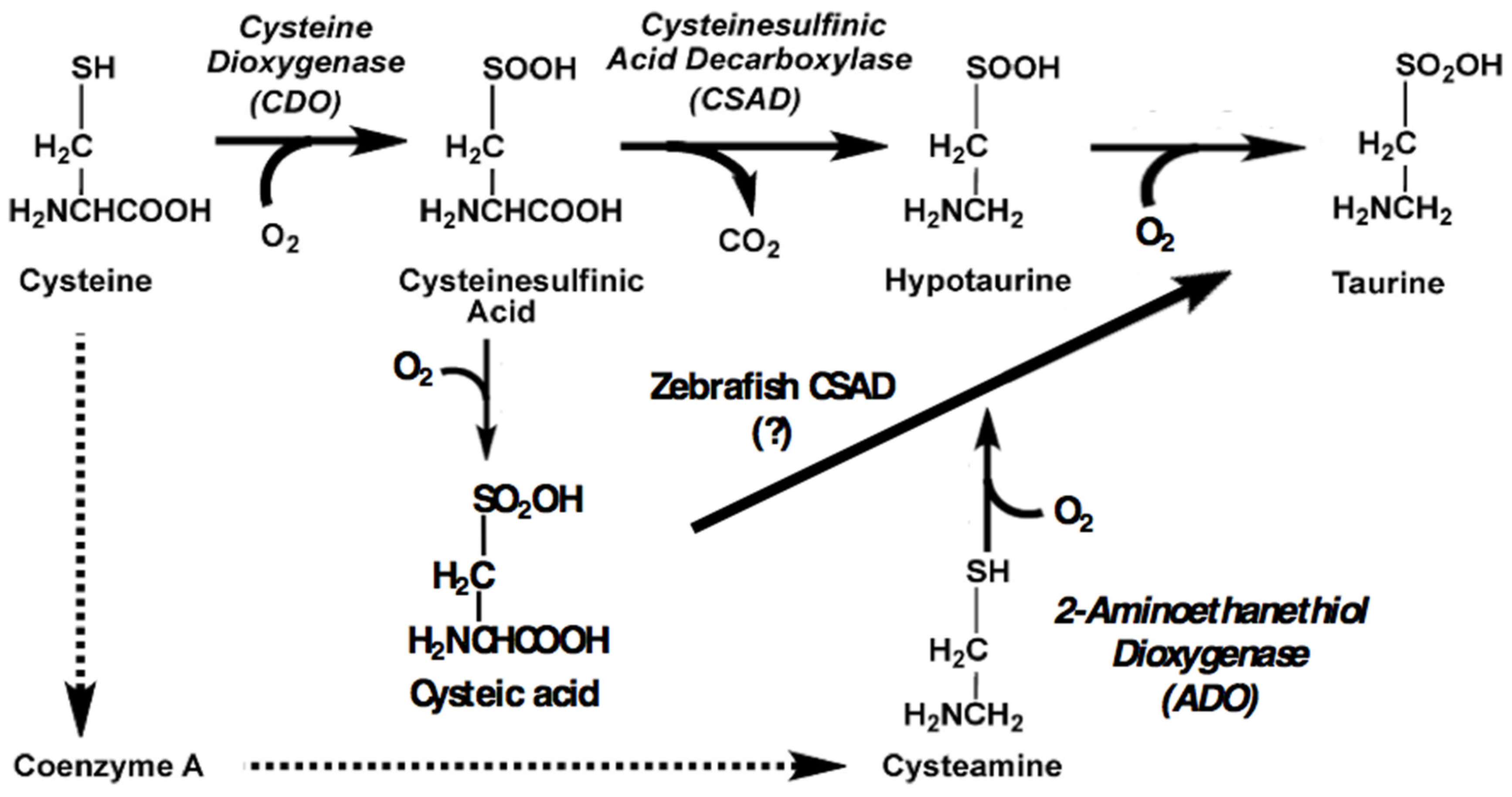
To assess a possible amelioratory role for taurine in dietary induced inflammation, our laboratory seeks to procure a strain of zebrafish deficient in endogenous taurine synthesis. This will enable clear observation of the effects of graded dietary supplementation. Taurine (2-aminoethanesulfonic acid) is a critical amino acid for animals and must be synthesized de novo or obtained through the diet. For taurine synthesizers, the cysteine sulfinic acid decarboxylase (CSAD) enzyme (XP_009295318.1) catalyzes the terminal reaction in the primary biosynthetic pathway (
Figure 1). Essential roles for taurine include osmoregulation, bile salt conjugation, and protection from oxidative stress [
1]. Species lacking sufficient endogenous synthesis require dietary supplementation. This is particularly apparent in carnivores who tend to have low levels of CSAD or none at all. Strictly carnivorous cats have low levels of CSAD but require high levels of taurine as well as methionine, which along with cysteine is a precursor of taurine synthesis [
2,
3]. In fish, deficiencies in taurine production have become increasingly apparent as aquaculture feeds exchange greater proportions of fish meal for plant protein sources containing no taurine. The supplementation requirements of several commercially relevant species have been described, including by our laboratory for the strict marine carnivore, cobia (
Rachycentron canadum). Taurine synthesizers also benefit in terms of growth from supplementation in the feed, with 1.5% being the standard amount in commercial formulations. [
4,
5,
6,
7]. The U.S. FDA (United States Food and Drug Administration) recently approved taurine supplementation of feed for farmed fish intended for human consumption [
8].
Knocking out the
csad gene in mice leads to an 83% reduction in plasma levels of taurine, with resulting mortality within 24 h of birth by the third generation of inbred homozygotes [
9]. Residual production of taurine may be via the cysteamine pathway (
Figure 1). Cats, as insufficient synthesizers of taurine, exhibit retinal degeneration and hepatic lipidosis when fed a diet without taurine supplementation [
2,
3]. Taurine-deficient fish display similar liver maladies, largely expected since fish conjugate bile salts to taurine [
5,
6]. CSAD mRNA is found in the earliest stages of zebrafish embryonic development, including in the yolk at the gastrula stage, and in both the yolk and notochord in the somite and pharyngula stages. In the pharygula stage, 24–48 hpf (hours post fertilization), CSAD expression is evident in several additional tissues including the liver, brain, and optic cup [
10,
11]. Knockdown of CSAD mRNA or TauT (taurine transporter) mRNA during early embryonic development results in cardiomyopathy and mortality in zebrafish [
11,
12]. Taurine is also an immunomodulator, mitigating inflammation by scavenging free radicals and inhibiting the production of several potent inflammatory agents including tumor necrosis factor-α (TNF-α), interleukins, prostaglandins, superoxide anion, and nitric oxide [
13,
14].
The sa9430 mutant zebrafish strain (Sanger, Zebrafish Mutation Project, [
15]) was acquired as a potential knockout for
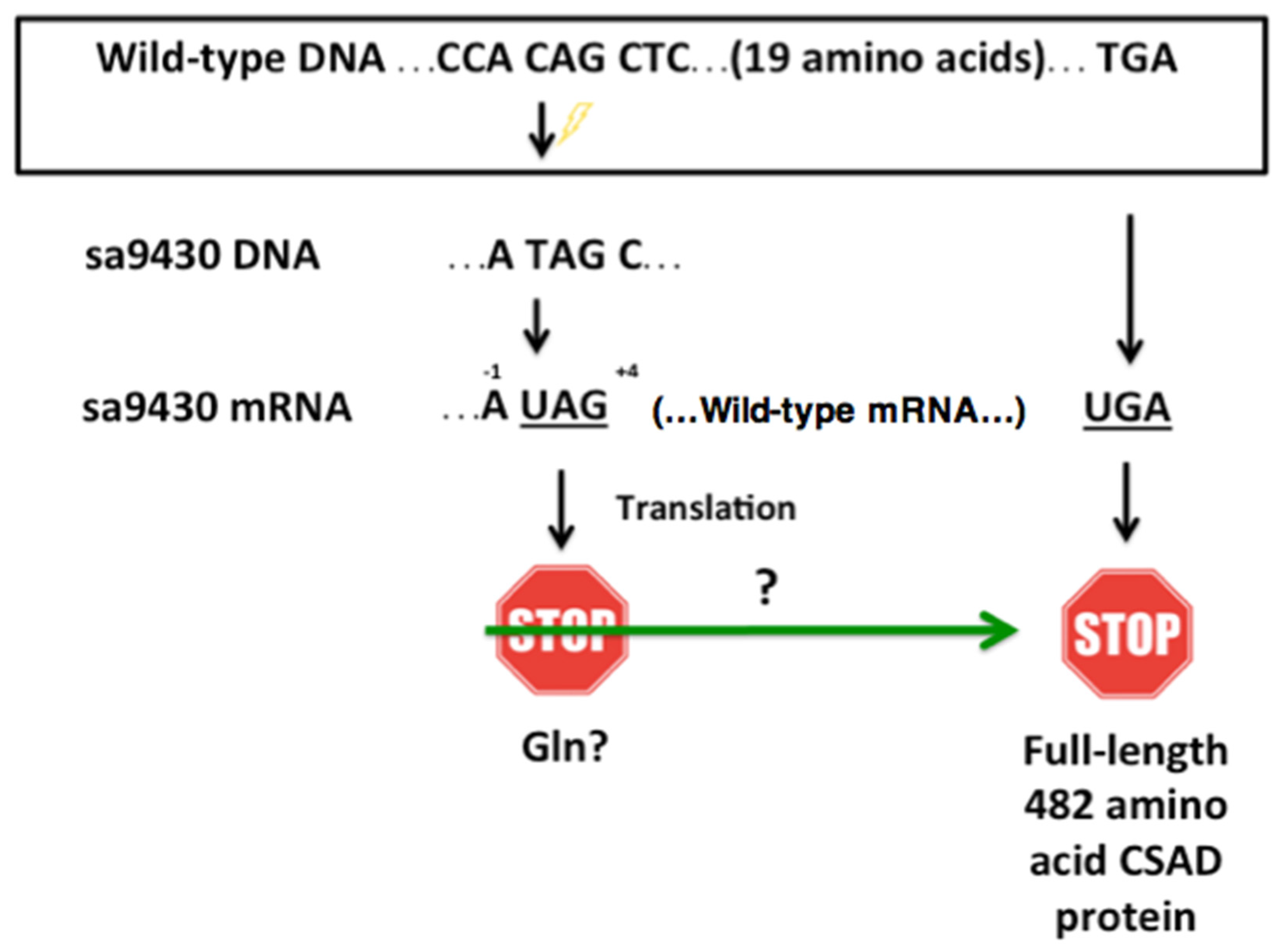
csad suitable for our dietary studies. This strain has a C→T point mutation in the single copy of the
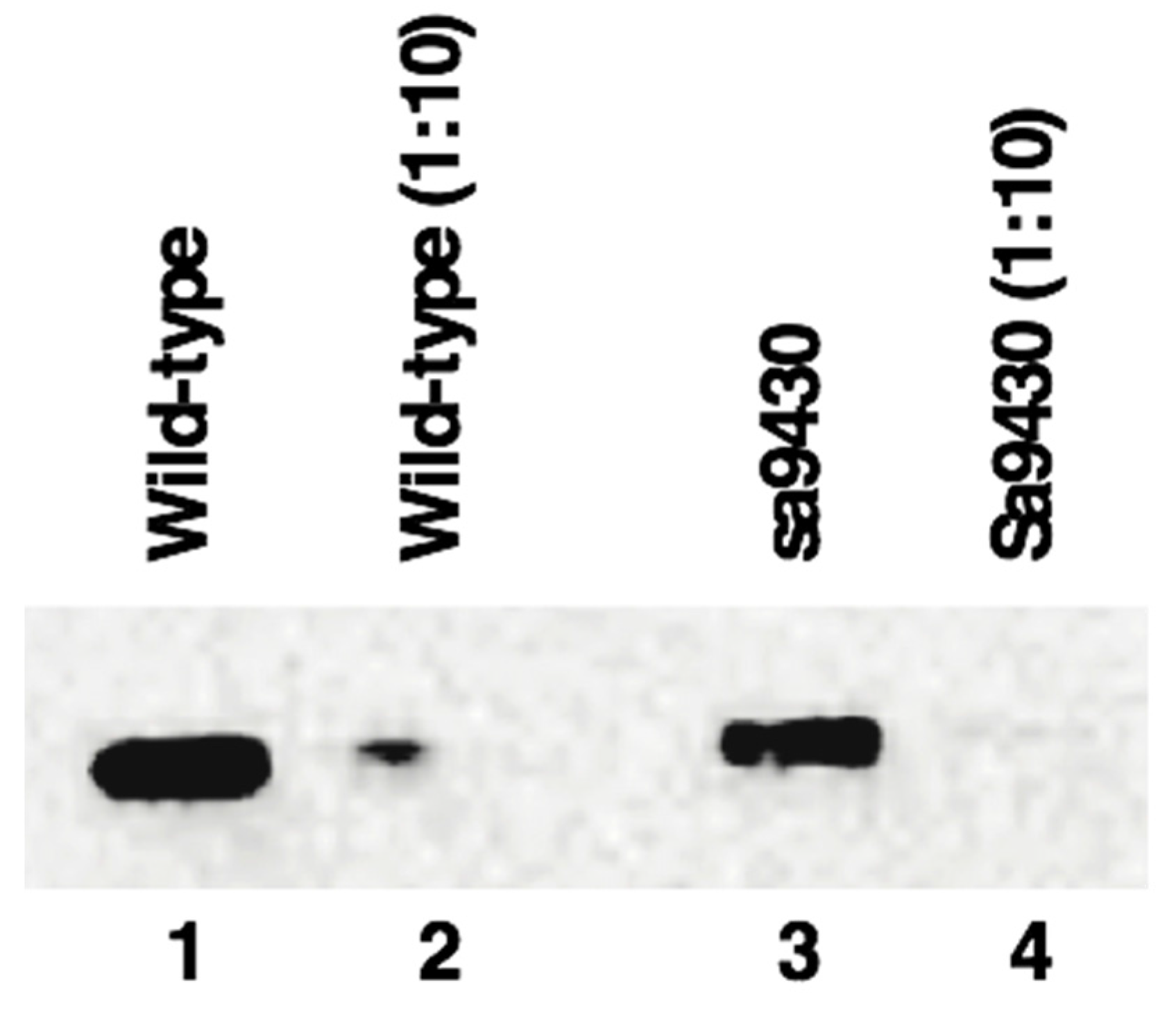
csad gene on chromosome 23. The mutation alters a codon that would normally be translated to glutamine to a UAG stop codon 20 amino acids prior to the normal UGA stop codon. No phenotype has been described for this mutant, but commercial feeds typically contain adequate dietary taurine and the lack of de novo synthesis may be masked. Zebrafish deficient in taurine synthesis that do not receive dietary supplementation are expected to exhibit pathologies similar to those seen in the knockdown studies, including mortality. Since our fish were reared and maintained on a commercial taurine-supplemented diet, as well as taurine-containing live artemia, gross changes in phenotype were not necessarily expected. However, we did not anticipate the results presented here from immunoblotting, enzyme activity assays, and mass spectrometry which show that the sa9430 mutant is producing wild-type CSAD protein.
The CSAD of some animals can utilize cysteic acid as a substrate for taurine synthesis, including cats and rats [
16,
17]. In this pathway, there is the direct conversion of cysteic acid to taurine without a hypotaurine intermediate. Data presented here reveal the ability of zebrafish CSAD to utilize cysteic acid as an alternative to cysteine sulfinic acid as a substrate for taurine synthesis.
3. Discussion
In mammals, the UAG codon has been described as notoriously “leaky”, and the nucleotides preceding and following the stop codon influence the frequency of readthrough. Readthrough frequency is increased in mammals when an adenine is in the −1 position, the nucleotide preceding the UAG stop codon. A pyrimidine in the +4 position, which corresponds to the nucleotide directly following the UAG, is associated with a greater chance of readthrough as compared to a purine [
21]. The sa9430 zebrafish strain has both an adenine in the −1 position and a pyrimidine (cytosine) in the +4 position.
Stop codon efficiency and frequency are correlated. In many organisms, UAG is not only the least efficient in terminating translation, it is also the least frequently occurring codon [
27]. In lower eukaryotes such as
Candida and
Drosophila, UAGA is the most efficient and frequent sequence, while UAGC is the least efficient and frequent sequence [
22]. UAGC is the sequence found in the sa9430 zebrafish strain (
Figure 3). A study on stop codon frequency in blunt snout bream (
Megalobrama amblycephala) reported that UAG has a relative synonymous codon usage (RSCU) value of 0.59 as compared to 1.11 for UAA and 1.29 for UGA [
28]. RCSU value is a measure of codon bias, and the score of <1 for UAG signifies its less frequent use as compared to the other codons [
29]. It may also be less efficient at terminating translation.
Translational readthrough is mediated by two predominant mechanisms. One is through the action of suppressor tRNAs, which incorporate an amino acid at the site of the stop codon. The other is through the pairing of a near-cognate tRNA with the stop codon. In
S. cerevisiae, suppressor tRNA(Gln) have been identified that insert glutamine at UAG stop codons. Tyrosine or lysine can also be inserted [
25]. A study of virally induced termination suppression also demonstrated UAG translation to glutamine [
26]. These same three amino acids have been shown to be inserted at a UAG stop codon as a result of near-cognate tRNA pairing. This is accomplished by mispairing at position 1 as well as at the classic wobble position 3 of the nonsense codon [
23,
24]. In this case, it is probable that mispairing of a tRNA complementary to CAG is occurring at position 1, resulting in the incorporation of a glutamine at UAG.
Taurine has been shown to be incorporated into modified uridines in mitochondrial tRNAs of sea squirts, cows, and humans. Lack of this taurine incorporation at a wobble position uridine results in weak codon-anticodon pairing and is associated with defective translation and subsequent disease [
30,
31]. It is challenging to speculate as to precisely which mechanisms are orchestrating the readthrough. The lower levels of taurine in the sa9430 embryos as compared to the wild-type may have to do with the pool of tRNAs present early in development.
It is notable that activity assays using the standard substrate, cysteine sulfinic acid, did not yield conclusive results (data not shown). Substrate levels reduced over the course of the assay without a resulting increase in taurine levels. Another enzyme present in the liver may compete for this substrate.
It was important to analyze F2 homozygotes in immunoblotting and activity assays. Maternal influence on offspring phenotype can be particularly confounding in the first generation. This was apparent in the studies of CSAD knockout in mice, with deleterious effects increasing over generations [
9]. Analyzing second generation homozygotes strengthens the proposal that mutants are making wild-type protein and there is no change in phenotype.
This work establishes why no aberrant phenotype has been previously described for the sa9430 strain. Despite the altered
csad genotype, there is expression of a wild-type CSAD protein. Since this strain is unsuitable for studying the effects of dietary taurine supplementation in a taurine-deficient fish, our laboratory is in the process of using CRISPR/Cas9 technology to generate a
csad knockout. Assessing the capacity for taurine to alleviate inflammation has implications beyond zebrafish. Findings will be useful in the development of plant-based feeds for commercially relevant fish, with taurine potentially mitigating the inflammatory potential of plant ingredients which are otherwise desirable in terms of supply and cost. This research may also lead to improvement in treatments for IBD (inflammatory bowel disease). In a zebrafish model of IBD, administration of taurine mollified chemically-induced degeneration of the intestinal mucosa and other symptoms of inflammatory bowel disease [
13,
32].
4. Materials and Methods
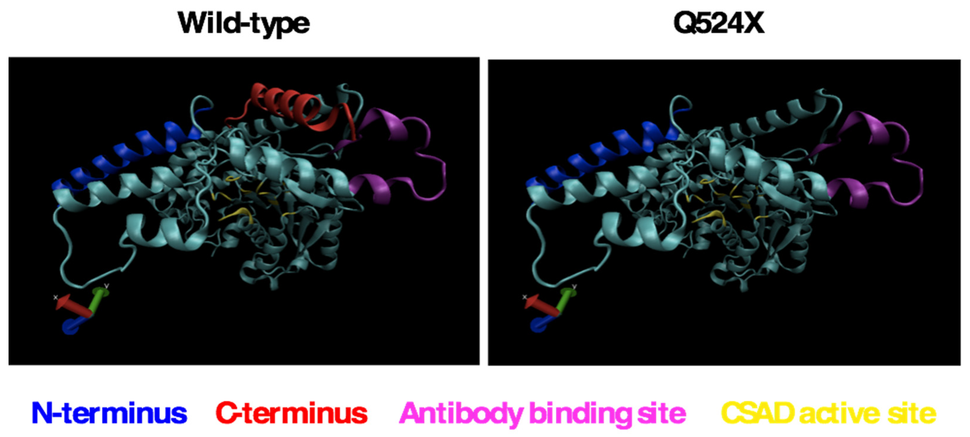
4.1. Computer Modeling of CSAD
Wild-type (ENSDARG00000026348) and sa9430 (ZBD-ALT-130411-5055) CSAD proteins were modeled using the Phyre2 web portal [
20]. Sequences were aligned to models of the human CSAD protein. 3D LigandSite was used to predict the ligand binding site amino acids for both wild-type and sa9430 [
33].
4.2. Zebrafish Strains and Maintenance
Wild-type and sa9430 zebrafish were maintained at the zebrafish facility of the Aquaculture Research Center at the Institute of Marine and Environmental Technology (Institutional Animal Care and Use Committee at University of Maryland, Baltimore #0315011, approved April 2015). Fish were maintained on a 14 h light, 10 h dark cycle at 28.5 °C. Larvae were fed a combination of paramecia, artemia, and GEMMA Micro 75 (Skretting). At the appropriate size, fish transitioned to GEMMA Micro 150, then 300, with occasional additions of artemia, particularly in the week preceding mating. Heterozygous embryos of csad sa9430 Tb (Sanger, Zebrafish Mutation Project) were raised and naturally bred to obtain homozygotes with wild-type csad and homozygotes with sa9430 mutant csad.
4.3. Genotyping
For confirmation of genotype, caudal fin clips were obtained and immersed in genomic DNA extraction buffer (50 mM KCl, 10 mM Tris-HCl (pH = 8.0), 150 mM MgCl
2, 0.3% Tween-20, and 0.3% NP40 in sterile MilliQ water), boiled at 95–100 °C for 15 min, cooled on ice, and digested with proteinase K at 55 °C for 1–3 h. After digestion, lysates were boiled at 95–100 °C for 15 min to inactivate proteinase K and centrifuged for 3 min at 12,000×
g [
34]. The resulting genomic DNA was amplified by PCR using zebrafish
csad-specific primers:
Sequencing of the 364-base-pair PCR product was performed in a 10 µL reaction volume consisting of 40–150 ng PCR product, 3 pmol of primer, 0.5 µL Big Dye v3.1 sequencing mix and 1.5 µL 5X sequencing buffer (Applied Biosystems, ThermoFisher Scientific, Waltham, MA, USA). Cycling parameters were 95 °C for 5 min, followed by 50 cycles at 95 °C for 15 s, 50 °C for 15 s, 60 °C for 4 min. The sequencing product was purified by adding 60 µL 100% isopropanol and 30 µL H2O, mixing thoroughly, incubating at room temperature for 30 min, and centrifuging at 2000× g for 30 min. The supernatant was decanted and 100 µL 70% isopropanol was added to wash the DNA, followed by another centrifugation at 2000× g for 14 min and decanting of the supernatant. Labeled products were air dried for 20–30 min before addition of 10 µL HI-DI formamide from Applied Biosystems. The mixture was heated at 95 °C for 2 min and then immediately put on ice. The denatured product was sequenced using an Applied Biosystems 3130XL Genetic Analyzer (ThermoFisher Scientific, Waltham, MA, USA) and compared with the published sequences for the wild-type (ENSDARG00000026348) and sa9430 (ZBD-ALT-130411-5055) strains using the Sequencher program (Version 5.0.1, Gene Codes, Ann Arbor, MI, USA).
4.4. Zebrafish Feeding Trials with and Without Supplemental Taurine
This work was performed by Aaron Watson, PhD. Descriptions of the feeding trial, sample preparation, and RT-PCR can be found in his thesis [
19], and LC-MS methods to obtain taurine values were performed as described in a publication on taurine leaching [
35].
4.5. Liver Protein Isolation
Zebrafish were euthanized by rapid cooling followed by decapitation. Livers were isolated from wild-type and sa9430 zebrafish and frozen at −80 °C. Liver tissue was homogenized in buffer containing 60 mM Potassium phosphate (pH 7.4), 5 mM DTT, 50 mM sucrose, and 0.5 µM pyridoxal-5’–phosphate (PLP) at a volume of approximately 1:15 weight (mg): lysis buffer volume (μL) [
36]. Homogenization was achieved by vortexing and pipetting up and down with a P200 micropipettor. Samples were centrifuged for 5 min at 1500×
g and supernatant containing the protein fraction collected. For the activity assay, the supernatant was dialyzed in a Slide-a-Lyzer Mini Dialysis Unit (ThermoFisher Scientific, Waltham, MA, USA) for 2 h at 4 °C in the homogenization buffer according to manufacturer’s instructions. Total protein concentration was determined using the Qubit Assay (ThermoFisher Scientific, Waltham, MA, USA).
4.6. Immunoblotting
Protein extracts were prepared as described in
Section 4.5, combined with standard SDS-PAGE sample buffer, heated for 3 minutes at 95 degrees C, and centrifuged for one minute at 10,000×
g. For the immunoblot shown in
Figure 4, lanes 1–4 of a 4%–12% Bis-Tris protein gel (NuPAGE Novex, (ThermoFisher Scientific, Waltham, MA, USA)) were loaded with 3.7 μg, 0.37 μg, 2.6 μg, and 0.26 μg protein, respectively, and run according to the manufacturer’s protocol for 32 min using MOPS running buffer. Proteins were transferred to a PVDF membrane in the Trans-Blot Turbo Transfer System (Bio-Rad, Hercules, CA, USA). Immunoblotting was performed in the iBind Western System (ThermoFisher Scientific, Waltham, MA, USA) using rabbit anti-zebrafish CSAD antibody at a dilution of 1:1000 (#6862, provided by Plant Sensory Systems, LLC, Halethorpe, MD, USA), as the primary antibody, and goat anti-rabbit IgG H&L HRP conjugate at a dilution of 1:2000 (Bio-Rad, Hercules, CA, USA) as the secondary antibody. A chemiluminescent signal was generated with addition of Clarity Western ECL substrate and imaged in a ChemiDoc Touch Imaging System (Bio-Rad, Hercules, CA, USA).
4.7. CSAD Activity Assay
For the activity assay, reaction mixtures were prepared containing 85 μL buffer (same as the homogenization buffer in
Section 4.5) and 5 μL total protein from liver (4.4 μg wild-type or 7.1 μg sa9430,
Section 4.5). The CSAD enzyme and its PLP cofactor in the buffer were allowed to incubate at 23 °C for 15 min before proceeding with the assay at the same temperature. The time points for the assay were 0, 30, and 90 min. Beginning with the 90-min time point, 10 μL 50 mM cysteic acid (substrate for CSAD) was added to the reaction mixtures. Immediately following the addition of the substrate at the 0-time point, all reactions were stopped with addition of 100 μL (volume equal to total reaction volume) ice cold ethanol with 5% acetic acid. Reaction mixtures were centrifuged at 350×
g for 10 min 100 μL supernatant was transferred to a clean microcentrifuge tube and dried by evaporation at 70 °C.
4.8. Amino Acid Analysis by HPLC
Dry samples were suspended in 0.1N HCl and filtered through 0.45 micron filters (EMD Millipore, Billerica, MA, USA). 5 μl of the filtered extracts was derivatized according to the AccQTag Ultra Derivitization Kit protocol (Waters Corporation, Milford, MA, USA). Amino acids were analyzed using an Agilent 1260 Infinity High Performance Liquid Chromatography System equipped with ChemStation (Agilent Technologies, Santa Clara, CA, USA) by injecting 5 μL of the derivatization mix onto an AccQTag Amino Acid Analysis C18 (Waters, Milford, MA, USA) 4.0 μm 3.9 × 150 mm column heated at 37 °C. Amino acids were eluted at 1.0 mL·min−1 flow with a mix of 10-fold diluted AccQTag Ultra Eluent (C; Waters Corporation, Milford, MA, USA), ultra-pure water (A) and acetonitrile (B) according to the following gradient: initial, 98.0% C/2.0% B; 2.0 min, 97.5% C/2.5% B; 25.0 min, 95.0% C/5.0% B; 30.5 min, 94.9% C/5.1% B; 33.0 min, 91.0% C/9.0% B; 38 min, 40.0% A/60.0% B; 43 min, 98.0% C/2.0% B. Derivatized amino acids were detected at 260 nm using a photo diode array detector. Amount of amino acids was expressed in g per g of dry weight of sample (% DW) making reference to AABA signal, external calibration curve of standard hydrolysate amino acids and dry weight of samples.
4.9. Preparation of Embryos for Amino Acid Analysis by HPLC
Fifty 1-hpf F2 embryos were collected from each of two matings of wild-type fish as well as 50 from two matings of homozygous sa9430 fish. Lyophilized embryos were extracted in 70% ethanol containing 0.154 mM
d-norleucine for evaluation of extraction efficiency. Samples were sonicated for 60 min at 25 °C in a bath sonicator (Branson 1200, Emerson, Danbury, CT, USA) followed by centrifugation at 350×
g for 10 min (IEC, ThermoFisher Scientific, Waltham, MA, USA). The ethanol fraction was retained and dried. These samples were then analyzed by HPLC as described in
Section 4.8.
4.10. Preparation of Juveniles for Amino Acid Analysis by HPLC
Six 5-week-old F1 juveniles (2 wild-type, 2 heterozygotes, and 2 sa9430 mutants) were homogenized by bead beating for 30 s at 4 m/s in 300 μL cold PBS with Pierce protease inhibitors (Thermofisher Scientific, Waltham, MA, USA). Samples were centrifuged for 5 min at 1500×
g at 4 °C. Supernatant was filtered through an Amicon Ultra 3K Device (EMD Millipore, Billerica, MA, USA), dried, and then analyzed by HPLC as described in
Section 4.8.
4.11. Preparation of Paramecia and Artemia for Taurine Analysis
Samples of paramecia and artemia used to feed zebrafish in our facility were centrifuged at 350×
g for 10 minutes (IEC, ThermoFisher Scientific, Waltham, MA, USA), and the pellet was retained and lyophilized. Samples were resuspended in 70% methanol and sonicated for 60 min at 25 °C in a bath sonicator (Branson 1200). Following centrifugation at 350×
g for 10 min (IEC), the methanol fraction was retained and dried. These samples were then prepared for taurine analysis by HPLC (
Section 4.8) or LC-MS [
35], respectively. Taurine levels in artemia were analyzed by Aaron Watson, Ph.D
. 4.12. Preparation of GEMMA (Skretting) Feeds for HPLC Analysis
GEMMA Micro 75 and 150, and
GEMMA Wean 0.2, 0.3, and 0.5 mm pellets were homogenized in a bead beater with 70% methanol. The homogenate was centrifuged, and retained supernatant was dried in a SpeedVac. This was performed by Michelle Price, Ph.D., at Plant Sensory Systems, LLC (Halethorpe, MD, USA). Samples were prepared for HPLC analysis as described in
Section 4.8.
4.13. Mass Spectrometry
Liver extracts were prepared as described in
Section 4.5. 100 μL total liver protein extracts contained 74.7 μg and 52.2 μg from wild-type and sa9430 fish, respectively. These extracts were each incubated with 5 μg rabbit anti-zebrafish CSAD (provided by Plant Sensory Systems, LLC, Halethorpe, MD, USA) with shaking at 1400 rpm for 1 h at 4 °C. 5 μg goat anti-rabbit—biotin (ThermoFisher Scientific, Waltham, MA, USA) was added to each tube, followed by shaking at 1400 rpm for 1 h at 4 °C. 30 μL streptavidin beads from the SMART Digest Immunoaffinity Kit (ThermoFisher Scientific, Waltham, MA, USA) were added followed by shaking at 1400 rpm at 4 °C for one hour followed by an additional hour at 23 °C. The wash protocol outlined in the kit was followed. Digestion occurred at 70 °C for one hour. The supernatant was retained and lyophilized.
Peptides were analyzed by LC-MS/MS using a Dionex UltiMate 3000 Rapid Separation nanoLC and a linear ion trap—Orbitrap hybrid mass spectrometer (ThermoFisher Scientific, Waltham, MA, USA). Approximately 1 μg of peptide samples was loaded onto the trap column, which was 150 μm × 3 cm in-house packed with 3 um C18 beads. The analytical column was a 75 um × 10.5 cm PicoChip column packed with 1.9 um C18 beads (New Objectives). The flow rate was kept at 300nL/min. Solvent A was 0.1% FA in water and Solvent B was 0.1% FA in ACN. The peptide was separated on a 90-min analytical gradient from 5% ACN/0.1% FA to 40% ACN/0.1% FA. The mass spectrometer was operated in data-dependent mode. The source voltage was 2.10 kV and the capillary temperature was 275 °C. MS1 scans were acquired from 400–2000 m/z at 60,000 resolving power and automatic gain control (AGC) set to 1 × 10⁶. The top ten most abundant precursor ions in each MS1 scan were selected for fragmentation. Precursors were selected with an isolation width of 1 Da and fragmented by collision-induced dissociation (CID) at 35% normalized collision energy in the ion trap. Previously selected ions were dynamically excluded from re-selection for 60 s. The MS2 AGC was set to 3 × 10⁵. Apex triggering was enabled for the peptide analysis on the Q Exactive HF (ThermoFisher Scientific, Waltham, MA, USA). All setup was same as above except that the top15 most abundant precursor ions in each MS1 scan were selected for fragmentation. Precursors were selected with an isolation width of 2 Da and fragmented by Higher-energy collisional dissociation (HCD) at 30% normalized collision energy in the HCD cell.
Individual raw data files were converted to the vendor neutral mzML format with msconvert [
37] and processed with the Trans-Proteomic Pipeline Version 4.8. [
38]. Sequence determination was performed using Comet software [
39] to search the Swissprot
D. rerio database, with an abbreviated FASTA containing CSAD sequence 482 amino acids in length (NP_001007349) or the predicted X1 isoform 544 amino acids in length (XP_009295318.1). Methionine oxidation and carbamidomethylation of cysteine were allowed as variable and fixed modifications, respectively. The enzyme specificity parameter was set to trypsin allowing for 1 missed cleavage site per peptide. MS1 precursor ion mass tolerance and MS2 product ion mass tolerance parameters were set to 10 ppm and 0.4 Da, respectively. Peptide spectra matched to theoretical spectra calculated from the database were validated using Peptide Prophet and peptides were assembled into protein groups with Protein Prophet. Sequences were analyzed using MacVector 12.7.5.


