Briarenones A‒C, New Briarellin Diterpenoids from the Gorgonian Briareum violaceum
by
 ,
,
Yang Cheng
1, 
Atallah F. Ahmed
2,3 ,
,
Raha S. Orfali
2,
Chang-Feng Dai
4 and
Jyh-Horng Sheu
1,5,6,7,* 1
Department of Marine Biotechnology and Resources, National Sun Yat-sen University, Kaohsiung 804, Taiwan
2
Department of Pharmacognosy, College of Pharmacy, King Saud University, Riyadh 11451, Saudi Arabia
3
Department of Pharmacognosy, Faculty of Pharmacy, Mansoura University, Mansoura 35516, Egypt
4
Institute of Oceanography, National Taiwan University, Taipei 112, Taiwan
5
Graduate Institute of Natural Products, Kaohsiung Medical University, Kaohsiung 807, Taiwan
6
Frontier Center for Ocean Science and Technology, National Sun Yat-sen University, Kaohsiung 804, Taiwan
7
Department of Medical Research, China Medical University Hospital, China Medical University, Taichung 404, Taiwan
*
Author to whom correspondence should be addressed.
Mar. Drugs 2019, 17(2), 120; https://doi.org/10.3390/md17020120
Submission received: 9 January 2019
/
Revised: 1 February 2019
/
Accepted: 12 February 2019
/
Published: 17 February 2019
(This article belongs to the Special Issue Marine Natural Products Discovery: In Memory of Late Prof. Tatsuo Higa)
Abstract
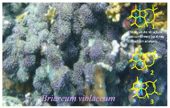
:Three new eunicellin-derived diterpenoids of briarellin type, briarenones A‒C (1‒3), were isolated from a Formosan gorgonian Briareum violaceum. The chemical structures of the compounds were elucidated on the basis of extensive spectroscopic analyses, including two-dimensional (2D) NMR. The absolute configuration of 1 was further confirmed by a single crystal X-ray diffraction analysis. The in vitro cytotoxic and anti-inflammatory potentialities of the isolated metabolites were tested against the growth of a limited panel of cancer cell lines and against the production of superoxide anions and elastase release in N-formyl-methionyl-leucyl-phenyl-alanine and cytochalasin B (fMLF/CB)-stimulated human neutrophils, respectively.
1. Introduction
Gorgonians belonging to genus Briareum (phylum Cnidaria, family Briareidae) are considered to be a rich source of highly oxygenated diterpenoids, particularly those derived from briarein (briarane) [1,2,3,4,5,6,7,8,9,10,11], eunicellin (cladiellin) [1,12,13,14,15,16], asbestinin [11,15,17,18,19], and, to a lesser extent, cembrane [20]. These metabolites were shown to exhibit various bioactivities such as anti-inflammatory [2,4,5,13,21], analgesic [22], cytotoxic [7,8,9,10,14,15,23], antimalarial [12,15], antiviral [10,15], and antimicrobial [15] activities. Briarellins constitute a class of tetracyclic oxygenated diterpenoids in which an additional seven-membered ether ring (oxepane) is formed between C-3 and C-16 of the eunicellin skeleton. These metabolites were originally discovered from Caribbean gorgonians of genus Briareum, e.g., B. asbestinum [13,14,16] and B. polyanthes [12,15], and also showed interesting bioactivities. Yet, briarellins remain to be isolated from the Pacific Briareum genus, although briarellins with the α,β-conjugated enone in the six-membered ring analogs were previously reported from one species of a taxonomically linked Pachyclavularia genus [24,25,26]. This prompted us to extensively investigate the chemical constituents of a gorgonian Briareum violaceum growing wildly in the Taiwanese waters and to screen their cytotoxic and anti-inflammatory activities. The current study led to the discovery of a group of three new briarellin diterpenoids, briarenones A‒C (1‒3), characterized by having cis fused cyclohexane, cyclodecane, and oxepane rings. The chemical structures of these molecules were resolved by various spectroscopic analyses, including two-dimensional (2D) NMR correlation analysis, while their absolute configurations were assigned by single-crystal X-ray diffraction analysis for 1.
2. Results and Discussion
The lyophilized specimen of the gorgonian was extracted with ethyl acetate (EtOAc). The chromatographic fractionation of the solvent-free extract on silica gel and reversed-phase C18 columns and the final separation using reversed-phase high-performance liquid chromatography (RP-HPLC) afforded diterpenoids 1‒3 (Figure 1; Figures S1–S26, Supplementary Materials).
Briarenone A (1) was obtained as a needle crystal, +224.4 (CHCl3). Its molecular formula C22H32O6 was determined by the sodium adduct peak [M + Na]+ at m/z 415.2089 in the high-resolution electrospray ionization mass spectrometry (HRESIMS), inferring seven degrees of unsaturation. The infrared (IR) absorption bands at νmax 3477, 1728, and 1670 cm−1 indicated the presence of hydroxyl, ester carbonyl, and α,β-unsaturated carbonyl functionalities, respectively. As the NMR spectra of 1 showed numerous broad and very weak signals due to the likely presence of a mixture of slowly interconverting conformers on the NMR time scale, remeasuring the spectra at −10 °C allowed us to have better resolution for the signals. The 1H NMR spectrum displayed the signals of four methyls, including one olefinic (δH 1.99 ppm, s), two methyls attached to oxygen-bearing carbons (δH 1.27 ppm, s and 1.28 ppm, s), and one secondary methyl (δH 1.09 ppm, d, J = 7.2 Hz), signifying the terpenoid nature of 1. Furthermore, the NMR data (Table 1) indicated the existence of an acetyl (δC 171.3 ppm, δC/δH 21.5/2.08 ppm) and an α,β-unsaturated ketone (δC 198.0, 157.0, and δC/δH 128.0/5.95 ppm). Taking into account the unsaturation degrees mentioned above, the 22 carbon signals in the 13C NMR spectrum of 1 (Table 1) are, thus, ascribable to the presence of a tetracyclic diterpenoid acetate. Three ring-juncture methines (δC/δH 51.1/2.71, 48.3/2.5, and 36.8/3.13 ppm), two tetrahydrofuran (THF)-oxymethines (δC/δH 83.4/3.72 and 77.9/4.79 ppm), and one oxymethylene (δC/δH 63.7/3.62 and 3.34 ppm) suggested a briarellin-related [13,16,24,25] or an asbestinin-related [15,17,18] structure for 1. However, since the protons of two methyl groups (δH 1.99 ppm, s, H3-20 and 1.09 ppm, d, J = 7.2 Hz, H3-17), but not one methyl group, showed 3JCH correlations in the heteronuclear multiple bond correlation (HMBC) spectrum with two of the angular methine carbons (δC 51.1 and 48.3 ppm for C-10 and C-14, respectively), the briarellin-type structure was designated for 1 (Figure 2). Three partial structures were assigned by the analysis of proton correlation spectroscopy (1H-1H COSY), including that assigned by the allylic correlation of the H3-20 (δH 1.99 ppm) with the olefinic proton H-12 (δH 5.95 ppm, s) (Figure 2). The connectivity of these partial structures, the positions of the hydroxyl, acetoxyl, and ketone carbonyl, and the ring-fusion carbons of THF and oxepane rings were deduced from the complete correlation analyses of the HMBC spectrum as illustrated in Figure 2. Furthermore, a comparison of the 13C NMR data of 1 with those of pachyclavulariaenone F (4), isolated from Pachyclavularia violacea [25], verified the replacement of the C-4 hydroxymethine group (δC 69.7 ppm, CH) in 4 by a methylene group (δC 33.6 ppm, CH2) in 1. This was associated with a significant upfield chemical shift at C-5 (∆δC ‒8.5 ppm) in 1 relative to that in 4. The planar structure of 1 was accordingly established (Figure 2). The relative configuration of 1 at C-1, C-2, C-3, C-6, C-7, C-9, C-10, C-14, and C-15, was assigned by the analysis of nuclear rotating-frame Overhauser effect spectroscopy (ROESY) correlations (Figure 2), which were found to be consistent with that of 4 as illustrated in Figure 2. Consequently, two opposite sets of nuclear Overhauser effect (NOE) interactions were observed for 1. One set displayed NOEs for H-1/H-10, H-1/H-14, H-1/H3-17, H-14/H3-17, H-1/H-8β (δH 2.03 ppm, d, J = 15.0, 12.0 Hz), and H-8β/H3-19, and another set exhibited NOEs for H-2/H3-18 and H3-18/H-6, while H-9 did not show any NOE correlation with H-10. Thus, H-1, H-10, H-14, H3-17, and H3-19 should be positioned in the β-face of the molecule, whereas H-2, H-9, H3-18, and H-6 should be positioned in its α-face. In order to confirm the molecular structure of 1, including the absolute configuration, a single-crystal X-ray structure analysis was further performed (Figure 3). The compound was slowly crystallized from MeOH as a monohydrate C22H32O6·H2O, with H-bond holding the H2O molecule in a hydrophilic pocket. From a dataset collected using copper radiation, the X-ray crystallographic analysis (Tables S1–S3, Supplementary Materials) of 1 determined the absolute configurations of briarenone A (1) as 1S,2R,3R,6S,7S,9R,10R,14S,15S, which forms a hydrogen bond from 7-OH with a molecule of water.
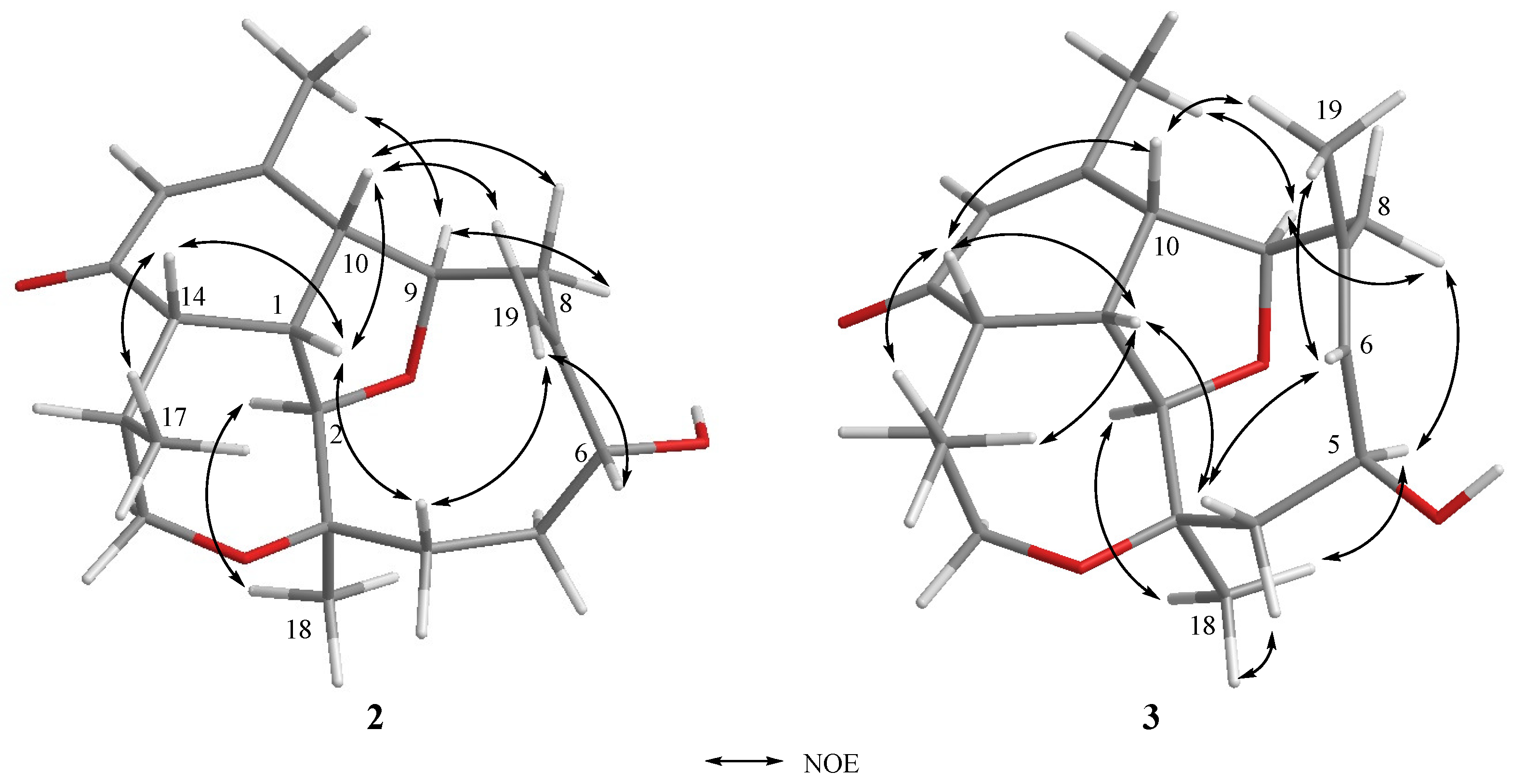
Briarenone B (2) was obtained as a white powder. It possessed a molecular formula of C20H28O4, as established from the sodiated ion peak in the HREIMS (m/z 355.1880 [M + Na]+) of 2, accounting for seven degrees of unsaturations. The IR absorptions at νmax 3420 and 1670 cm−1 indicated the presence of hydroxyl and conjugated carbonyl functionalities, respectively. The NMR data of 2, measured in CDCl3 at −10 °C, were comparable to those of the framework of 1 except in the replacement of an sp3 oxycarbon (δC 74.6 ppm, C, C-7) and a methyl group (δC/δH 23.7/1.28 ppm, CH3, CH3-19) in 1 by an exomethylene group (δC/δH 148.3 ppm, C and 117.7/5.35 and 5.18 ppm, CH2, respectively) in 2. Analysis of NMR data of 2 (Table 1) and correlations found in the 1H-1H COSY and HMBC spectra (Figure 4) enabled the establishment of the gross structure of 2. Furthermore, the observed NOE correlations for 2 (Figure 5) assigned similar β-positioning for H-1, H-10, H-14, and H3-17, and α-orientation for H-2, H-9, and H3-18 as found in 1. Moreover, one of the exomethylene protons (δH 5.18 ppm, s) exhibited NOE interaction with H-10 (δH 2.97 ppm, br d, J = 7.0 Hz), while the other one (δH 5.35, s) displayed NOE with the hydroxymethine proton at C-6 (δH 4.07 ppm, dd, J = 11.0, 4.5 Hz). A molecular modeling investigation revealed that H-6 did not exhibit NOE interaction in the nuclear Overhauser effect spectroscopy (NOESY) with the methylene protons at C-8 (distances >3.7 Å). This designated the α-orientation of 6-OH and, hence, the 6R configuration as shown in Figure 6. Moreover, it was found that eunicellins with similar substitutions in the ten-membered ring, but with an 6β-OH (Figure 6), displayed distinctive chemical shifts at C-6 (δC < 74.0 ppm), C-7 (δC ≥ 150.0 ppm), and H-6 (δH > 4.25 ppm) [27,28,29,30] than those assigned for 2 (δ 78.3, 148.3, and 4.07 ppm, respectively). This observation also suggests the α-orientation of 6-OH in 2. On the basis of the above findings, the absolute configuration of 1, and the biogenetic consideration, the configuration 1S, 2R,3R,6R,9R,10R,14S,15S was, thus, established for 2.
The HREIMS (m/z 355.1880 [M + Na]+) and NMR data (Table 1) indicated a molecular formula C20H28O4 for briarenone C (3), inferring that 3 is an isomer of 2. Therefore, it was found that 3 possessed an α,β-unsaturated carbonyl (IR: νmax 1668 cm−1 δC 198.1 ppm, C, 156.4 ppm, C, and δC/δH 128.6/5.91 ppm, CH) and a hydroxy methine group (IR: νmax 3299 cm−1; δC/δH 65.0/4.84 ppm, CH). However, compound 3 differs in the presence of a trisubstituted double bond (δC/δH 135.4/5.52 ppm, CH and δC 128.8 ppm, C), instead of a 1,1-disubstituted double bond in 2, with the appearance of an olefinic methyl (δC/δH 29.2/1.93 ppm). The hydroxyl group was found to be linked to the allylic carbon (C-5) due to the HMBC correlation observed from the olefinic methyl (δH 1.93 ppm, H3-19) to the sp2 methine carbon (δC 135.4 ppm, C-6), which in turn exhibited 1H-1H COSY correlation with H-5 (δH 4.84 ppm, dd, J = 8.5, 8.5 Hz). Moreover, comparison of NMR data of 3 with those of pachyclavulariaenone D pointed out that 3 is the 5-hydroxy isomer of pachyclavulariaenone D [25]. The gross structure of 3 was further elucidated from the full 2D NMR spectroscopic correlation analyses of 3 (Figure 4). The NOE interaction of H3-19 with the olefinic methine protons H-6 (δH 5.52 ppm, d, J = 8.5 Hz) (Figure 5), in addition to the downfield shift of C-19 (δC 29.2 ppm), indicated the Z geometry of the 6,7-double bond [31]. Moreover, the NOE correlations for H-2/H3-18, H3-18/H-5, H-5/H-8α (δH 2.74 ppm, d, J = 14.5 Hz), and H-8α/H-9 disclosed the 5S configuration. These NOEs were further validated by a molecular modeling study (Figure 6). Finally, full NOE correlation analysis (Figure 5) coupled with the previous identification of the absolute configuration of 1, which was co-isolated along with 3, designated the 1S,2R,3R,5S,9R,10R,14S,15S configuration for 3.
Although briarellin-type diterpenoids (e.g., briarellins A–S) were discovered initially from Caribbean gorgonians of genus Briareum [12,13,14,15,16], since 1995, numerous related analogs were successfully isolated and identified from Pachyclavularia violacea inhabiting Taiwanese [24,25] and Indonesian [26] waters. Unlike the previously identified briarellins, the structures of briarenones A–C (1–3), isolated in this study from B. violaceum, were found to be similar to those of pachyclavulariaenones [24,25] in possessing the three ring-juncture protons (H-1, H-10, and H-14) cis to each other and forming the cis fusion of the cyclohexane, cyclodecane, and oxepane rings. Also, the determination of the absolute configurations of 1–3 by NOE correlation analysis coupled with X-ray crystallographic analysis of 1 using copper radiation, could imply the absolute configurations for similar briarellins isolated from the genera of Briareum and Pachyclavularia, such as pachyclavulariaenones A–G [24,25], to be similar to that of 1. Compounds of this kind possessing a six-membered carbocyclic ring cis-fused to both of the ten-membered carbocyclic and seven-membered ether rings, making three ring-junction protons (H-1, H-10, and H-14) cis to each other, including those reported from Pachyclavularia violacea [24,25,26], can be considered as a useful chemotaxonomic marker in classification of gorgonians and in approaching a resolution of the argument for considering the gorgonian species of Pachyclavularia to be the same as those of genus Briareum [26,32].
The isolated compounds were evaluated against the growth of DLD-1 (human colon adenocarcinoma), HT-29 (human colon carcinoma), and HuCC-T1 (human colon cholangiocellular carcinoma) cell lines. The in vitro anti-inflammatory activities of compounds 1‒3 were also measured against the release of elastase and the production of superoxide anions in N-formyl-methionyl-leucyl-phenyl-alanine and cytochalasin B (fMLF/CB)-activated neutrophils. However, the compounds did not show either cytotoxic or anti-inflammatory activities in the tested in vitro models.
3. Materials and Methods
3.1. General Experimental Procedures
Optical rotations and IR spectra were measured on a JASCO P-1020 polarimeter and a JASCO FT/IR-4100 (JASCO Corporation, Tokyo, Japan) spectrophotometer, respectively. HRESIMS spectra were measured on a Bruker APEX II mass spectrometer (Bruker, Bremen, Germany). 1H and 13C NMR spectra were obtained on a Varian Unity INOVA 600 FT-NMR (or Varian Unity INOVA500 FT-NMR) instruments (Varian Inc., Palo Alto, CA, USA) at 600 MHz (or 500 MHz) for 1H, and 150 MHz (or 125 MHz) for 13C in CDCl3. Thin-layer chromatography (TLC) analyses were performed on precoated silica (Si) gel plates (Kieselgel 60 F-254, 0.2 mm), and Si gel (230–400 mesh) (Merck, Darmstadt, Germany) and C18-reversed phase Si gel (RP-18; 40–63 µM) (Parc-Technologique BLVD, Quebec, Canada) were used for column chromatography. Further purification and the isolation of compounds were performed by reversed-phase high-performance liquid chromatography (RP-HPLC) on a Hitachi L-2455 HPLC apparatus with a Supelco C18 column (250 × 21.2 mm, 5 μm).
3.2. Animal Material
The soft coral B. violaceum was collected from Jihui Fish Port (Taitung, Taiwan) by scuba divers at a depth of 10–15 m on March 2013 and stored in a freezer at −20 °C until extraction. Moreover, the soft coral was taxonomically identified by Prof. Chang-Feng Dai, National Taiwan University, Taipei. A voucher specimen was deposited in the Department of Marine Biotechnology and Resources, National Sun Yat-sen University, Kaohsiung.
3.3. Extraction and Isolation
The freeze-dried soft coral (0.5 kg) was extracted with EtOAc (3 × 3 L) and filtered. The filtrate was evaporated in vacuo to yield the EtOAc extract (3.90 g). The extract was fractionated by Si gel column chromatography using EtOAc in n-hexane (6.25% to 100%) as a gradient elution system to afford 21 fractions (A01–A21). Fraction A04 was further fractionated on an RP-18 Si gel column, using MeOH-H2O (2.5:1) as a mobile phase, into eight subfractions (A0401–A0408). Compound 1 (7.4 mg) was obtained from A405 after two-step purification on RP-HPLC using MeOH-H2O (3:2) and acetyl nitrite (CH3CN)-H2O (1:2). Subfractions A402 and A403 were combined together on the basis of their similar TLC chromatogram and were further separated on RP-HPLC using CH3CN-H2O (1:2) to yield compounds 2 (4.5 mg) and 3 (2.2 mg).
3.3.1. Briarenone A (1)
Colorless needles; +224.4 (c 7.4, CHCl3); IR (neat) νmax 3477, 2964, 2929, 1728, 1670, and 756 cm−1; 1H and 13C NMR data (600/150 MHz; CDCl3), Table 1; ESIMS m/z 415 [M + Na]+; HRESIMS m/z 415.2089 [M + Na]+ (calculated for C22H32O6Na, m/z 415.2091).
3.3.2. Briarenone B (2)
Colorless powder; −10.7 (c 4.5, CHCl3); IR (neat) νmax 3420, 2912, 2851, 1670, and 772 cm−1; 1H and 13C NMR data (500/125 MHz; CDCl3), Table 1; ESIMS m/z 355 [M + Na]+; HRESIMS m/z 355.1880 [M + Na]+ (calculated for C20H28O4Na, m/z 355.1880).
3.3.3. Briarenone C (3)
Colorless powder; −14.8 (c 2.2, CHCl3); IR (neat) νmax 3299, 2921, 2851, and 1668 cm−1; 1H and 13C NMR data (500/125 MHz; CDCl3), Table 1; ESIMS m/z 355 [M + Na]+; HRESIMS m/z 355.1880 [M + Na]+ (calculated for C20H28O4Na, m/z 355.1880).
3.3.4. Single-Crystal X-Ray Crystallography of 1
A suitable colorless prism of compound 1 was obtained from a solution in MeOH by slow evaporation for a month at 4 °C. The crystal (0.20 × 0.18 × 0.17 mm3) was analyzed at 100(2) K, space group P212121 (# 19). Cell: a = 8.3456(3) Å, b = 10.6813(3) Å, c = 23.4466(8) Å, V = 2090.07(12) Å3, Z = 4, Dcalcd = 1.305 mg/m3, and μ(CuKα) = 0.790 mm−1. Intensity data of single-crystal X-ray diffraction were measured on a Bruker APEX DUO diffractometer. Of the 13326 reflections collected, only 3658 independent reflections [R(int) = 0.0331] with I > 2σ(I) were used for the analysis. The structure was solved by direct method and refined by a full-matrix least squares on F2 method. The refinement converged to a final R1 = 0.0401, wR2 = 0.1064, with goodness-of-fit = 1.091. For coordinates corresponding to the absolute stereochemistry represented, absolute structure parameter 0.04(6) was obtained [33].
3.4. Bioassays
3.4.1. Cytotoxicity Assay
Cancer cell (DLD-1, HT-29, and HuCC-T1) lines were purchased from the American Type Culture Collection (ATCC). Compounds 1‒3 were evaluated for cytotoxic activity using an Alamar blue assay. Alamar Blue (resazurin) is an important non-toxic redox indicator that is utilized to assess metabolic function and cell health. A complete description of this test was previously described [34,35]. The absorbance was measured at 570 nm using an ELISA plate reader (Thermo Fisher Scientific Instruments Co., Ltd., Vantaa, Finland).
3.4.2. In Vitro Anti-Inflammatory Assays
Human neutrophils were obtained from blood by dextran sedimentation, Ficoll-Hypaque centrifugation, and hypotonic lysis and then incubated as previously described [36]. Neutrophils (6 × 10⁵ cells∙mL−1) incubated at 37 °C in Hank’s balanced salt solution (HBSS) with MeO-Suc-Ala-Ala-Pro-Val-p-nitroanilide (100 μM) and Ca2+ (1 mM) were treated with dimethyl sulfoxide (DMSO) or the tested compound for 5 min. The activation of neutrophils was challenged for 10 min with fMLF (100 nM)/CB (0.6 and 0.5 μg∙mL−1 for superoxide anion generation and elastase release, respectively). The anti-inflammatory activities of compounds 1‒3 were measured with examining the inhibition of fLMF/CB-induced human neutrophils producing superoxide anion and elastase, using UV spectrometer detection at wavelengths of 550 nm and 405 nm, respectively [36,37].
4. Conclusions
Three new briarellin type diterpenoids, briarenones A‒C (1‒3), were identified from Briareum violaceum inhabiting Taiwanese waters. The compounds have three cis ring-juncture protons (H-1, H-10, and H-14) due to the cis fusion of the cyclohexane, cyclodecane, and oxepane rings. The molecular structures of this type may be considered as a useful chemotaxonomic marker in the identification of some species of genus Briareum. The absolute configurations of the compounds were assigned on the basis NOE correlation analysis coupled with a single-crystal X-ray diffraction analysis for briarenone A. The isolated metabolites showed no in vitro cytotoxicity against DLD-1, HT-29, and HuCC-T1 cells and did not inhibit the superoxide anion generation or elastase release in fMLF/CB-stimulated neutrophils. Compounds 1‒3 did not exhibit cytotoxic and anti-inflammatory activities in this study; however, more biological activity screening should be carried out to discover their pharmaceutical potential. Moreover, the absolute configuration of 1 analyzed by X-ray diffraction would be useful for elucidation of the structurally similar metabolites isolated from the genera Briareum and Pachyclavularia.
Supplementary Materials
HRESIMS, 1H, 13C, HMQC, COSY, HMBC, and NOESY spectra of new compounds 1–3; and crystal data, atomic coordinate bond lengths [Å] and angles [°] for 1 are available online at https://www.mdpi.com/1660-3397/17/2/120/s1. Figure S1: HRESIMS spectrum of 1; Figure S2: 1H NMR spectrum of 1 in CDCl3; Figure S3: 1H NMR spectrum of 1 in CDCl3 (1.08–2.82 and 3.10–5.92 ppm); Figure S4: 13C NMR spectrum of 1 in CDCl3 and DEPT spectra; Figure S5: HSQC spectrum of 1 in CDCl3; Figure S6: 1H-1H COSY spectrum of 1 in CDCl3; Figure S7: HMBC spectrum of 1 in CDCl3; Figure S8: ROESY spectrum of 1 in CDCl3; Figure S9: HRESIMS spectrum of 2; Figure S10: 1H NMR spectrum of 2 in CDCl3; Figure S11: 1H NMR spectrum of 2 in CDCl3 (0.96–3.38 and 3.24–6.08ppm); Figure S12: 13C NMR spectrum of 2 in CDCl3; Figure S13: DEPT spectra of 2 in CDCl3; Figure S14: HSQC spectrum of 2 in CDCl3; Figure S15: 1H-1H COSY spectrum of 2 in CDCl3; Figure S16: HMBC spectrum of 2 in CDCl3; Figure S17: NOESY spectrum of 2 in CDCl3; Figure S18: HRESIMS spectrum of 3; Figure S19: 1H NMR spectrum of 3 in CDCl3; Figure S20: 1H NMR spectrum of 3 in CDCl3 (0.82–3.04 and 3.38–6.03 ppm); Figure S21: 13C NMR spectrum of 1 in CDCl3; Figure S22: DEPT spectra of 3 in CDCl3; Figure S23: HSQC spectrum of 1 in CDCl3; Figure S24: 1H-1HCOSY spectrum of 3 in CDCl3; Figure S25: HMBC spectrum of 3 in CDCl3; Figure S26: NOESY spectrum of 3 in CDCl3; Table S1: Crystal data and structure refinement for 1; Table S2: Atomic coordinates (× 104) and equivalent isotropic displacement parameters (Å2 × 10³) for 1; Table S3: Bond lengths and bond angles for 1.
Author Contributions
J.-H.S. designed the experiment. Y.C. isolated the compounds and performed spectroscopic data measurement and structure interpretation. A.F.A. performed the spectroscopic data analysis, final structure determination, and preparation of the manuscript. R.S.O. contributed to data analysis and editing of the manuscript. C.-F.D. contributed to species identification of the soft coral.
Funding
The research was funded by the Ministry of Science and Technology of Taiwan (MOST104-2113-M-110-006 and 104-2811-M-110-026) and the International Scientific Partnership Program (ISPP) at King Saud University, Saudi Arabia (ISPP-116).
Acknowledgments
Financial supported was mainly provided by the Ministry of Science and Technology (MOST104-2113-M-110-006 and 104-2811-M-110-026) to J.-H.S. The authors extend their appreciation to the International Scientific Partnership Program ISPP at King Saud University for funding this research work through ISPP-116.
Conflicts of Interest
The authors declare no conflicts of interest.
References
- Blunt, J.W.; Carroll, A.R.; Copp, B.R.; Davis, R.A.; Keyzers, R.A.; Prinsep, M.R. Marine natural products. Nat. Prod. Rep. 2018, 35, 8–53. [Google Scholar] [CrossRef] [PubMed] [Green Version]
- Xu, J.H.; Lai, K.H.; Su, Y.D.; Chang, Y.C.; Peng, B.R.; Backlund, A.; Wen, Z.H.; Sung, P.J. Briaviolides K-N, new briarane-type diterpenoids from cultured octocoral Briareum violaceum. Mar. Drugs 2018, 16, 75. [Google Scholar] [CrossRef] [PubMed]
- Su, Y.D.; Su, J.H.; Hwang, T.L.; Wen, Z.H.; Sheu, J.H.; Wu, Y.C.; Sung, P.J. Briarane diterpenoids isolated from octocorals between 2014 and 2016. Mar. Drugs 2017, 15, 44. [Google Scholar] [CrossRef] [PubMed]
- Chen, N.F.; Su, Y.D.; Hwang, T.L.; Liao, Z.J.; Tsui, K.H.; Wen, Z.H.; Wu, Y.C.; Sung, P.J. Briarenols C-E, new polyoxygenated briaranes from the octocoral Briareum excavatum. Molecules 2017, 22, 475. [Google Scholar] [CrossRef] [PubMed]
- Su, Y.D.; Wu, T.Y.; Wen, Z.H.; Su, C.C.; Chen, Y.H.; Chang, Y.C.; Wu, Y.C.; Sheu, J.H.; Sung, P.J. Briarenolides U-Y, new anti-Inflammatory briarane diterpenoids from an octocoral Briareum sp. (Briareidae). Mar. Drugs 2015, 13, 7138–7149. [Google Scholar] [CrossRef] [PubMed]
- Sung, P.J.; Lin, M.R.; Hwang, T.L.; Fan, T.Y.; Su, W.C.; Ho, C.C.; Fang, L.S.; Wang, W.H. Briaexcavatins M-P, four new briarane-related diterpenoids from cultured octocoral Briareum excavatum (Briareidae). Chem. Pharm. Bull. 2008, 56, 930–935. [Google Scholar] [CrossRef] [PubMed]
- Sung, P.J.; Su, J.H.; Duh, C.Y.; Chiang, M.Y.; Sheu, J.H. Briaexcavatolides K-N, new briarane diterpenes from the gorgonian Briareum excavatum. J. Nat. Prod. 2001, 64, 318–323. [Google Scholar] [CrossRef]
- Wu, S.L.; Sung, P.J.; Chiang, M.Y.; Wu, J.Y.; Sheu, J.H. New polyoxygenated briarane diterpenoids, briaexcavatolides O-R, from the gorgonian Briareum excavatum. J. Nat. Prod. 2001, 64, 1415–1420. [Google Scholar] [CrossRef]
- Sheu, J.H.; Sung, P.J.; Su, J.H.; Wang, G.H.; Duh, C.Y.; Shen, Y.C.; Chiang, M.Y.; Chen, I.T. Excavatolides U-Z, new briarane diterpenes from the gorgonian Briareum excavatum. J. Nat. Prod. 1999, 62, 1415–1420. [Google Scholar] [CrossRef]
- Coval, S.J.; Cross, S.; Bernardinelli, G.; Jefford, C.W.; Brianthein, V. A New Cytotoxic and Antiviral Diterpene Isolated from Briareum asbestinum. J. Nat. Prod. 1988, 51, 981–984. [Google Scholar] [CrossRef]
- Dookran, R.; Maharaj, D.; Mootoo, B.S.; Ramsewak, R.; McLean, S.; Reynolds, W.F.; Tinto, W.F. Briarane and asbestinane diterpenes from Briareum asbestinum. Tetrahedron 1994, 50, 1983–1992. [Google Scholar] [CrossRef]
- Ospina, C.A.; Rodriguez, A.D.; Ortega-Barria, E.; Capson, T.L. Briarellins J-P and polyanthellin A: New eunicellin-based diterpenes from the gorgonian coral Briareum polyanthes and their antimalarial activity. J. Nat. Prod. 2003, 66, 357–363. [Google Scholar] [CrossRef] [PubMed]
- Gomez-Reyes, J.F.; Salazar, A.; Guzman, H.M.; Gonzalez, Y.; Fernandez, P.L.; Ariza-Castolo, A.; Gutierrez, M. Seco-Briarellinone and briarellin S, two new eunicellin-based diterpenoids from the Panamanian octocoral Briareum asbestinum. Mar. Drugs 2012, 10, 2608–2617. [Google Scholar] [CrossRef] [PubMed]
- Rodríguez, A.D.; Cóbar, O.M. The briarellins, new eunicellin-based diterpenoids from a Caribbean gorgonian, Briareum asbestinum. Tetrahedron 1995, 51, 6869–6880. [Google Scholar] [CrossRef]
- Ospina, C.A.; Rodríguez, A.D. Bioactive Compounds from the Gorgonian Briareum polyanthes. Correction of the structures of four Asbestinane-Type Diterpenes. J. Nat. Prod. 2006, 69, 1721–1727. [Google Scholar] [CrossRef] [PubMed]
- Rodriguez, A.D.; Cobar, O.M. Studies on the minor constituents of the Caribbean gorgonian octocoral Briareum asbestinum Pallas. Isolation and structure determination of the eunicellin-based diterpenoids briarellins E-I. Chem. Pharm. Bull. 1995, 43, 1853–1858. [Google Scholar] [CrossRef] [PubMed]
- Rodriguez, A.D.; Cobar, O.M.; Martinez, N. Isolation and structures of sixteen new asbestinin diterpenes from the Caribbean gorgonian Briareum asbestinum. J. Nat. Prod. 1994, 57, 1638–1655. [Google Scholar] [CrossRef]
- Rodríguez, A.D.; Cóbar, O.M. Structures and bioactivities of new asbestinin diterpenoids from the Caribbean gorgonian octocoral Briareum asbestinum. Tetrahedron 1993, 49, 319–328. [Google Scholar] [CrossRef]
- Stierle, D.B.; Carte, B.; Faulkner, D.J.; Tagle, B.; Clardy, J. The asbestinins, a novel class of diterpenes from the gorgonian Briareum asbestinum. J. Am. Chem. Soc. 1980, 102, 5088–5092. [Google Scholar] [CrossRef]
- Chang, Y.C.; Huang, I.C.; Chiang, M.Y.N.; Hwang, T.L.; Kung, T.H.; Lin, C.S.; Sheu, J.-H.; Sung, P.J. Briaviodiol A, a new cembranoid from a soft coral Briareum violacea. Chem. Pharm. Bull. 2010, 58, 1666–1668. [Google Scholar] [CrossRef]
- Wei, W.C.; Lin, S.Y.; Chen, Y.J.; Wen, C.C.; Huang, C.Y.; Palanisamy, A.; Yang, N.S.; Sheu, J.H. Topical application of marine briarane-type diterpenes effectively inhibits 12-O-tetradecanoyl-phorbol-13-acetate-induced inflammation and dermatitis in murine skin. J. Biomed. Sci. 2011, 18, 94–106. [Google Scholar] [CrossRef] [PubMed]
- Lin, Y.Y.; Lin, S.C.; Feng, C.W.; Chen, P.C.; Su, Y.D.; Li, C.M.; Yang, S.N.; Jean, Y.H.; Sung, P.J.; Duh, C.Y.; et al. Anti-Inflammatory and analgesic effects of the marine-derived compound excavatolide B isolated from the culture-type Formosan Gorgonian Briareum excavatum. Mar. Drugs 2015, 13, 2559–2579. [Google Scholar] [CrossRef] [PubMed]
- Sheu, J.H.; Sung, P.J.; Huang, L.H.; Lee, S.F.; Wu, T.; Chang, B.Y.; Duh, C.Y.; Fang, L.S.; Soong, K.; Lee, T.J. New cytotoxic briaran diterpenes from the Formosan gorgonian Briareum sp. J. Nat. Prod. 1996, 59, 935–938. [Google Scholar] [CrossRef] [PubMed]
- Wang, G.H.; Sheu, JH.; Chiang, M.Y.; Lee, T.J. Pachyclavulariaenones A–C, three novel diterpenoids from the soft coral Pachyclavularia violacea. Tetrahedron Lett. 2001, 42, 2333–2336. [Google Scholar] [CrossRef]
- Wang, G.H.; Sheu, J.H.; Duh, C.Y.; Chiang, M.Y. Pachyclavulariaenones D-G, new diterpenoids from the soft coral Pachyclavularia violacea. J. Nat. Prod. 2002, 65, 1475–1478. [Google Scholar] [CrossRef] [PubMed]
- Anta, C.; Gonzalez, N.; Rodriguez, J.; Jimenez, C. A new secosterol from the Indonesian octocoral Pachyclavularia violacea. J. Nat. Prod. 2002, 65, 1357–1359. [Google Scholar] [CrossRef] [PubMed]
- Yamada, K.; Ogata, N.; Ryu, K.; Miyamoto, T.; Komori, T.; Higuchi, R. Bioactive terpenoids from Octocorallia. 3. A new eunicellin-based diterpenoid from the soft coral Cladiella sphaeroides. J. Nat. Prod. 1997, 60, 393–396. [Google Scholar] [CrossRef]
- Rao, C.B.; Rao, D.S.; Satyanarayana, C.; Rao, D.V.; Kassuhlke, K.E.; Faulkner, D.J. New cladiellane diterpenes from the soft coral Cladiella australis of the Andaman and Nicobar Islands. J. Nat. Prod. 1994, 57, 574–580. [Google Scholar] [CrossRef]
- Hassan, H.M.; Khanfar, M.A.; Elnagar, A.Y.; Mohammed, R.; Shaala, L.A.; Youssef, D.T.; Hifnawy, M.S.; El Sayed, K.A. Pachycladins A-E, prostate cancer invasion and migration inhibitory eunicellin-based diterpenoids from the Red Sea soft coral Cladiella pachyclados. J. Nat. Prod. 2010, 73, 848–853. [Google Scholar] [CrossRef]
- Chen, B.W.; Wu, Y.C.; Chiang, M.Y.; Su, J.H.; Wang, W.H.; Fan, T.Y.; Sheu, J.H. Eunicellin-based diterpenoids from the cultured soft coral Klyxum simplex. Tetrahedron 2009, 65, 7016–7022. [Google Scholar] [CrossRef]
- Kalinowski, H.O.; Berger, S.; Braun, S. Carbon 13 NMR Spectroscopy; John Wiley & Sons: Chichester, UK, 1988. [Google Scholar]
- Samimi-Namin, K.; van Ofwegen, L.P. Overview of the genus Briareum (Cnidaria, Octocorallia, Briareidae) in the Indo-Pacific, with the description of a new species. Zookeys 2016, 557, 1–44. [Google Scholar] [CrossRef] [PubMed]
- Crystallographic data for compound 1 has been deposited with the Cambridge Crystallographic Data Centre (deposition number CCDC 1885052). Copies of the data can be obtained, free of charge, on application to the Director, CCDC, 12 Union Road, Cambridge CB21EZ, UK (fax: +44-1223-336033 or email: [email protected]. Deposited on 12 December 2018).
- O’Brien, J.; Wilson, I.; Orton, T.; Pognan, F. Investigation of the Alamar Blue (resazurin) fluorescent dye for the assessment of mammalian cell cytotoxicity. Eur. J. Biochem. 2000, 267, 5421–5426. [Google Scholar] [CrossRef] [PubMed] [Green Version]
- Nakayama, G.R.; Caton, M.C.; Nova, M.P.; Parandoosh, Z. Assessment of the Alamar blue assay for cellular growth and viability in vitro. J. Immunol. Methods 1997, 204, 205–208. [Google Scholar] [CrossRef]
- Hwang, T.L.; Li, G.L.; Lan, Y.H.; Chia, Y.C.; Hsieh, P.W.; Wu, Y.H.; Wu, Y.C. Potent inhibition of superoxide anion production in activated human neutrophils by isopedicin, a bioactive component of the Chinese medicinal herb Fissistigma oldhamii. Free Radic. Biol. Med. 2009, 46, 520–528. [Google Scholar] [CrossRef] [PubMed]
- Yang, S.C.; Chung, P.J.; Ho, C.M.; Kuo, C.Y.; Hung, M.F.; Huang, Y.T.; Chang, W.Y.; Chang, Y.W.; Chan, K.H.; Hwang, T.L. Propofol inhibits superoxide production, elastase release, and chemotaxis in formyl peptide-activated human neutrophils by blocking formyl peptide receptor 1. J. Immunol. 2013, 190, 6511–6519. [Google Scholar] [CrossRef] [PubMed]
Figure 1.
Structures of new briarellin diterpenoids (1‒3) isolated from Briareum violaceum and pachyclavulariaenone F (4).
Figure 1.
Structures of new briarellin diterpenoids (1‒3) isolated from Briareum violaceum and pachyclavulariaenone F (4).

Figure 2.
Key 1H-1H COSY, HMBC, and NOE correlations for 1.

Figure 3.
Oak Ridge Thermal Ellipsoid Plot (ORTEP) diagram of the molecular structure of 1 as determined by X-ray analysis.
Figure 3.
Oak Ridge Thermal Ellipsoid Plot (ORTEP) diagram of the molecular structure of 1 as determined by X-ray analysis.

Figure 4.
Key 1H-1H COSY and HMBC correlations for 2 and 3.

Figure 5.
Key NOE correlations for 2 and 3.

Figure 6.
A partial structure of eunicellin-derived diterpenoids: cladiellisin [27], 3-acetyl cladiellisin [28], pachycladin B [29], and klysimplexins C and E [30].


Table 1.
The 1H and 13C NMR chemical shifts for 1‒3.
| 1 | 2 | 3 | ||||
|---|---|---|---|---|---|---|
| # | δH, m (J in Hz) a | δC b, type | δH, m (J in Hz) c | δC d, type | δH, m (J in Hz) c | δCd, type |
| 1 | 3.13 ddd (9.6, 5.4, 4.8) | 36.8, CH | 3.12 ddd (10.0, 7.0, 4.5) | 37.7, CH | 2.89 m | 40.4, CH |
| 2 | 3.72 d (9.6) | 83.4, CH | 3.76 d (10.0) | 86.4, CH | 3.62 d (10.5) | 85.8, CH |
| 3 | 76.5, C | 75.4, C | 75.5, C | |||
| 4α | 1.94 br d (10.8) | 33.6, CH2 | 1.58 m | 31.6, CH2 | 1.72 d (15.0) | 46.4, CH2 |
| 4β | 1.49 m | 1.58 m | 2.36 dd (14.5, 8.5) | |||
| 5α | 1.94 br d (10.8) | 27.2, CH2 | 2.04 m | 28.7, CH2 | 4.84 dd (8.5, 8.5) | 65.0, CH |
| 5β | 1.58 ddd (11.4, 10.8, 3.6) | 1.79 m | ||||
| 6 | 5.77 dd (11.4, 3.6) | 78.0, CH | 4.07 dd (11.0, 4.5) | 78.3, CH | 5.52 d (8.5) | 135.4, CH |
| 7 | 74.6, C | 148.3, C | 128.8, C | |||
| 8α | 1.81 dd (15.0, 3.6) | 45.5, CH2 | 2.77 dd (14.0,4.5) | 37.0, CH2 | 2.74 d (14.5) | 38.6, CH2 |
| 8β | 2.03 dd (15.0, 12.0) | 2.14 dd (14.0,5.0) | 2.01 dd (14.5, 3.5) | |||
| 9 | 4.79 dd (12.0, 3.6) | 77.9, CH | 4.45 dd (5.0, 4.0) | 82.2, CH | 4.29 dd (6.5, 3.0) | 81.2, CH |
| 10 | 2.71 br d (6.0) | 51.1, CH | 2.97 br d (7.0) | 50.3, CH | 2.82 br d (8.5) | 48.3, CH |
| 11 | 157.0, C | 156.6, C | 156.4, C | |||
| 12 | 5.95 s | 128.0, CH | 5.93 s | 127.4, CH | 5.91 s | 128.6, CH |
| 13 | 198.1, C | 198.1, C | 198.1, C | |||
| 14 | 2.50 br d (4.8) | 48.3, CH | 2.42 br d (4.5) | 48.5, CH | 2.31 dd (4.5, 5.5) | 48.4, CH |
| 15 | 2.67 m | 30.0, CH | 2.59 m | 30.4, CH | 2.58 m | 32.3, CH |
| 16α | 3.62 d (13.8) | 63.8, CH2 | 3.35 dd (13.5,3.5) | 63.9, CH2 | 3.52 m | 65.2, CH2 |
| 16β | 3.34 br d (13.8) | 3.54 d (13.0) | 3.52 m | |||
| 17 | 1.09 d (7.2) | 17.2, CH3 | 1.04 d (7.5) | 17.2, CH3 | 1.02 d (7.5) | 18.5, CH3 |
| 18 | 1.27 s | 22.2, CH3 | 1.25 s | 23.8, CH3 | 1.41 s | 27.5, CH3 |
| 19 | 1.28 s | 23.7, CH3 | 5.35 s; 5.18 s | 117.7, CH2 | 1.93 s | 29.2, CH3 |
| 20 | 1.99 s | 21.9, CH3 | 1.98 s | 21.8, CH3 | 1.91 s | 21.9, CH3 |
| Ac | 2.08 s | 21.5, CH3 | ||||
| 171.3, C | ||||||
a Spectra recorded at 600 MHz in CDCl3 at −10 °C; b spectra recorded at 150 MHz in CDCl3 at −10 °C; c spectra recorded at 500 MHz in CDCl3; d spectra recorded at 125 MHz in CDCl3.
© 2019 by the authors. Licensee MDPI, Basel, Switzerland. This article is an open access article distributed under the terms and conditions of the Creative Commons Attribution (CC BY) license (http://creativecommons.org/licenses/by/4.0/).
Share and Cite
MDPI and ACS Style
Cheng, Y.; Ahmed, A.F.; Orfali, R.S.; Dai, C.-F.; Sheu, J.-H. Briarenones A‒C, New Briarellin Diterpenoids from the Gorgonian Briareum violaceum. Mar. Drugs 2019, 17, 120. https://doi.org/10.3390/md17020120
AMA Style
Cheng Y, Ahmed AF, Orfali RS, Dai C-F, Sheu J-H. Briarenones A‒C, New Briarellin Diterpenoids from the Gorgonian Briareum violaceum. Marine Drugs. 2019; 17(2):120. https://doi.org/10.3390/md17020120
Chicago/Turabian StyleCheng, Yang, Atallah F. Ahmed, Raha S. Orfali, Chang-Feng Dai, and Jyh-Horng Sheu. 2019. "Briarenones A‒C, New Briarellin Diterpenoids from the Gorgonian Briareum violaceum" Marine Drugs 17, no. 2: 120. https://doi.org/10.3390/md17020120
Note that from the first issue of 2016, this journal uses article numbers instead of page numbers. See further details here.