Article

An Algal Metabolite-Based PPAR-γ Agonist Displayed Anti-Inflammatory Effect via Inhibition of the NF-κB Pathway

1
College of Pharmacy, Pusan National University, Busan 46241, Korea
2
College of Pharmacy, Kyunghee University, Seoul 02447, Korea
3
Center for Proteome Biophysics, Department of Chemistry, Pusan National University, Busan 46241, Korea
*
Author to whom correspondence should be addressed.
Mar. Drugs 2019, 17(6), 321; https://doi.org/10.3390/md17060321
Submission received: 10 May 2019 / Revised: 23 May 2019 / Accepted: 25 May 2019 / Published: 30 May 2019
(This article belongs to the Collection Marine Drugs in the Management of Metabolic Diseases)

Abstract

:
In our previous study, a synthetic compound, (+)-(R,E)-6a1, that incorporated the key structures of anti-inflammatory algal metabolites and the endogenous peroxisome proliferator-activated receptor γ (PPAR-γ) ligand 15-deoxy-∆12,14-prostaglandin J2 (15d-PGJ2), exerted significant PPAR-γ transcriptional activity. Because PPAR-γ expressed in macrophages has been postulated as a negative regulator of inflammation, this study was designed to investigate the anti-inflammatory effect of the PPAR-γ agonist, (+)-(R,E)-6a1. Compound (+)-(R,E)-6a1 displayed in vitro anti-inflammatory activity in lipopolysaccharides (LPS)-stimulated murine RAW264.7 macrophages. Compound (+)-(R,E)-6a1 suppressed the expression of proinflammatory factors, such as nitric oxide (NO), inducible NO synthase (iNOS), cyclooxygenase-2 (COX-2), interleukin-6 (IL-6), and tumor necrosis factor-α (TNF-α), possibly by the inhibition of the nuclear factor-κB (NF-κB) pathway. In macrophages, (+)-(R,E)-6a1 suppressed LPS-induced phosphorylation of NF-κB, inhibitor of NF-κB α (IκBα), and IκB kinase (IKK). These results indicated that PPAR-γ agonist, (+)-(R,E)-6a1, exerts anti-inflammatory activity via inhibition of the NF-κB pathway.

1. Introduction

Peroxisome proliferator-activated receptor γ (PPAR-γ), which can be activated by natural or synthesized ligands, such as 15-deoxy-∆12,14-prostaglandin J2 (15d-PGJ2) or rosiglitazone, respectively, is a member of the nuclear receptor superfamily [1]. Generally, inactive PPAR-γ may be localized to the cytoplasm rather than the nucleus. Activated PPAR-γ is translocated to the cell nucleus and forms a heterodimer with a second member of the nuclear receptor family, retinoic X receptor (RXR). Then, the PPAR/RXR heterodimer binds to peroxisome proliferator response element (PPRE) on target DNA to regulate the transcription of genes relevant to lipid and glucose metabolism [2]. Recent research also revealed that PPAR-γ plays a key role in the repression of inflammatory genes, especially in macrophages. PPAR-γ expressed in macrophages has been postulated as a negative regulator of inflammation [3,4]. After activation by infection, tissue damage, or exposure to endotoxin (i.e., lipopolysaccharides (LPS)), macrophages secrete a large amount of proinflammatory mediators, including inducible nitric oxide synthase (iNOS), nitric oxide (NO), cyclooxygenase-2 (COX-2), tumor necrosis factor-α (TNF-α), and interleukin-6 (IL-6). Some of these proinflammatory mediators are involved in systemic diseases, such as obesity, diabetes, and cancer [5,6,7]. Currently, two molecular mechanisms have been defined for anti-inflammatory actions of PPAR-γ agonist. (1) PPAR-γ agonist binds to NF-κB (nuclear factor-κB) in the nucleus to inhibit its binding to DNA gene promoter regions, resulting in the suppression of inflammatory gene transcription. Binding of active NF-κB to DNA leads to the expression of proinflammatory mediators, such as TNF-α, IL-1β, IL-6, iNOS, and COX-2 [8,9,10,11,12,13,14]. (2) PPAR-γ agonist, such as 15d-PGJ2, interferes with activation of NF-κB in the cytoplasm. NF-κB resides in the cytoplasm under inactive state while associated with its repressor, IκB (inhibitor of NF-κB). During inflammatory stimulation, IκB may undergo phosphorylation by IκB kinase (IKK) and, thus, be degraded, leading to the release of NF-κB and allowing it to enter the nucleus. The endogenous PPAR-γ ligand, 15d-PGJ2, was shown to covalently bind to IKK, inhibiting its function and subsequently inhibiting the phosphorylation of IκB and then activation of NF-κB [8,9,10,11,12,13].
In a previous study, we isolated oxy fatty acids and prostaglandins from a red alga with substantial anti-inflammatory activity [15]. Thereafter, by incorporating the key structural motifs of these natural products, we synthesized a new class of endocyclic enone jasmonate derivatives as anti-inflammatory leads [16,17]. In a further study, we synthesized an exocyclic enone jasmonate derivative, (+)-(R,E)-6a1, as a potent PPAR-γ agonist that share the key exocyclic enone moiety with the endogenous PPAR-γ ligand, 15d-PGJ2 [18] (See Supplementary Materials). Typical PPAR-γ ligands, such as rosiglitazone and 15d-PGJ2, are composed of three distinct partial structures, a polar head, linker, and a hydrophobic tail (Figure 1). The polar head and hydrophobic tail play important roles in H-bonding and hydrophobic interaction, respectively, with the PPAR-γ LBD (ligand binding domain). The exocyclic α,β-unsaturated ketone (enone) moiety of 15d-PGJ2 was reported to be essential for covalent bonding with Cys285 in the PPAR-γ LBD. Similar to 15d-PGJ2, the enone functionality of (+)-(R,E)-6a1 may contribute to covalent bonding to the PPAR-γ LBD, and this additional covalent bonding may contribute to the activation of PPAR-γ [18,19,20]. In a continuing study, we investigated the in vitro anti-inflammatory effects of the PPAR-γ agonist, (+)-(R,E)-6a1, in RAW264.7 murine macrophages. Based on in vitro results, the possible anti-inflammatory mechanism of (+)-(R,E)-6a1 was also discussed.

2. Results and Discussion

2.1. Compound (+)-(R,E)-6a1 Promoted PPAR-γ Translocation to Cell Nuclei

Generally, after ligand binding, the activated PPAR-γ will translocate to the nucleus and bind to NF-κB to repress the gene expression of proinflammatory mediators. In our previous study, the PPAR-γ agonistic activity of (+)-(R,E)-6a1 was evaluated by luciferase assay by using the PPRE-luciferase reporter plasmid [18]. Herein, we used Western blot to assess the protein level of the translocated PPAR-γ at the nucleus due to activation by (+)-(R,E)-6a1 in RAW264.7 cells. The result showed that the endonuclear PPAR-γ protein level was significantly increased by (+)-(R,E)-6a1 treatment in a concentration-dependent manner, and the activity was comparable to the standard PPAR-γ agonist rosiglitazone (Figure 2). Since PPAR-γ expressed in macrophages can downregulate inflammatory responses [21], we investigated the expression of proinflammatory mediators.

2.2. Cytotoxicity of (+)-(R,E)-6a1 to RAW264.7, Ac2F, and KB Cells

Prior to the in vitro anti-inflammatory assay, compound (+)-(R,E)-6a1 was first evaluated for its cytotoxicity to murine macrophages (RAW264.7), rat liver cells (Ac2F), and human oral epidermoid cancer cells (KB) to gauge the suitable concentration for the cell-based anti-inflammatory assay. As shown in Figure 3, (+)-(R,E)-6a1 showed no significant toxicity to cells at the concentrations of 10 and 50 μM for 24 h; specifically, (+)-(R,E)-6a1 was nontoxic to RAW264.7 cells at the concentration of 50 μM for up to 24 h. However, the viability and cell morphology of RAW264.7 cells appeared to be affected by (+)-(R,E)-6a1 after 48 h at 50 μM (Figure 3D). Therefore, RAW264.7 cells were employed for anti-inflammatory assay with (+)-(R,E)-6a1 concentrations lower than 30 μM.

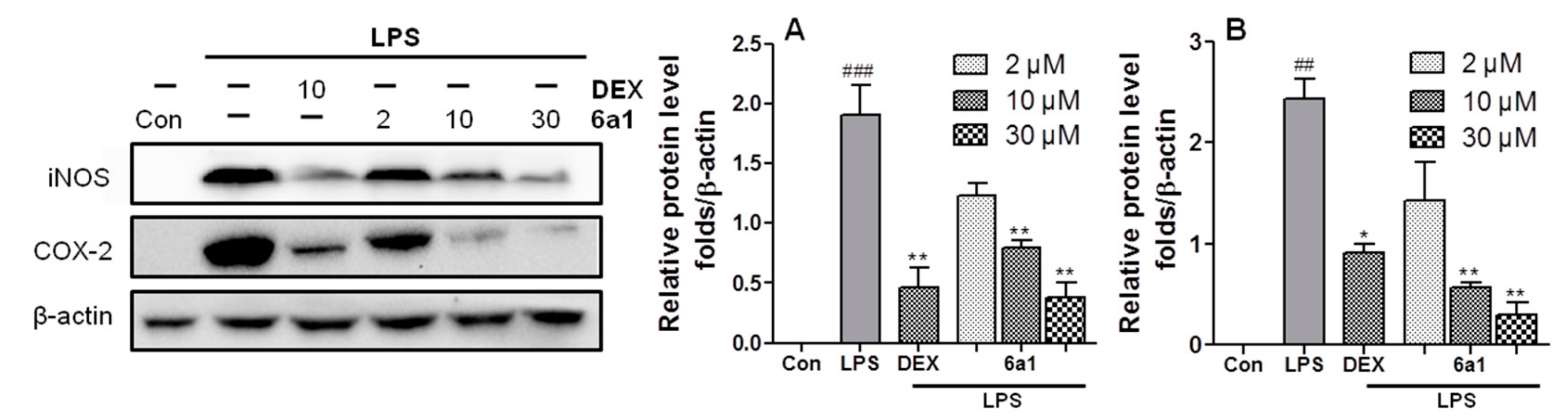
2.3. (+)-(R,E)-6a1 Inhibited LPS-Induced Expression of Proinflammatory Factors in RAW264.7 Cells

In order to verify the in vitro anti-inflammatory effect of (+)-(R,E)-6a1, the protein levels of the proinflammatory factors, iNOS and COX-2, were examined in RAW264.7 cells by Western blot. As expected, LPS stimulation markedly increased iNOS and COX-2 protein levels, but this increase could be diminished in a dose-dependent manner by pretreatment with (+)-(R,E)-6a1 (Figure 4). Notably, at the concentration of 30 μM, (+)-(R,E)-6a1 significantly decreased the protein levels of iNOS and COX-2 with a potency comparable to that of 10 μM dexamethasone. Dexamethasone was employed as a standard anti-inflammatory drug for comparison.
Meanwhile, as an inflammatory mediator, high levels of NO are produced in response to inflammatory stimuli and mediation of inflammatory effects. iNOS is a family of enzymes that catalyze NO production from L-arginine. Thereby, it was hypothesized that the suppression of iNOS by (+)-(R,E)-6a1 (Figure 4) would lead to decreased NO production in macrophages, and thus, we examined NO levels in RAW264.7 cell supernatant using the Griess reagent. Compound (+)-(R,E)-6a1 significantly decreased the NO production in a concentration-dependent manner (Figure 5A). In addition, the amount of TNF-α and IL-6 that was produced was analyzed by enzyme-linked immunosorbent assay (ELISA). TNF-α and IL-6 are inflammatory cytokines that provide a host of defensive effects during the inflammatory response and maintain normal cellular conditions [22]. Proinflammatory mediator levels were markedly increased when murine macrophages RAW264.7 were exposed to LPS. However, these increases in TNF-α and IL-6 were inhibited by (+)-(R,E)-6a1 in a dose-dependent manner (Figure 5B,C), suggesting that (+)-(R,E)-6a1 participates in a signaling pathway activated by LPS in macrophages. This result is consistent with that of other studies, which have shown that treatment of macrophages with various concentrations of PPAR-γ agonists reduces the production of proinflammatory cytokines [23,24].

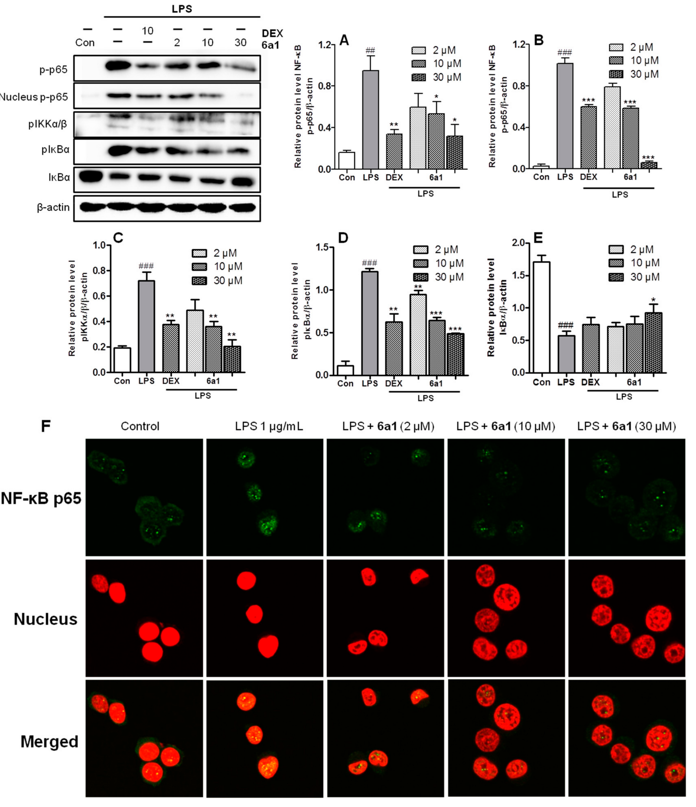
2.4. (+)-(R,E)-6a1 Inhibited LPS-Induced NF-Κb Signal Pathway in RAW264.7 Cells

NF-κB is recognized as a crucial component of many immune responses and inflammation. For example, macrophages rely on NF-κB for the secretion of proinflammatory cytokines [25]. The conventional anti-inflammatory mechanism of PPAR-γ ligands is known via the inhibition of NF-κB [8,9,10,11,12,13,14]. PPAR-γ ligands, such as rosiglitazone, activate PPAR-γ, and activated PPAR-γ is translocated into the nucleus to bind with NF-κB. The NF-κB–PPAR-γ complex cannot bind to the promotor region of DNA; thereby, gene expression of proinflammatory mediators is suppressed (Figure 6, path A). Meanwhile, an endogenous PPAR-γ ligand, 15-deoxy-∆12,14-prostaglandin J2 (15d-PGJ2), was reported to exert anti-inflammatory activity via additional mechanism by inhibiting the activation and nuclear translocation of NF-κB (Figure 6, path B). Recently, Rossi et al. indicated that IKK is the critical target of 15d-PGJ2 in the NF-κB activation pathway. The study showed that the exocyclic enone moiety in 15d-PGJ2 can form Michael adducts that covalently modify Cys179 of IKK, thus leading to the inhibition of IKK phosphorylation, and subsequent interference with the downstream NF-κB activation events [9,11].
Since (+)-(R,E)-6a1 share the same exocyclic enone moiety with 15d-PGJ2, we investigated the effect of (+)-(R,E)-6a1 on the alternative pathway of 15d-PGJ2 involving the inhibition of IKK and subsequent inhibition of NF-κB. As shown in Figure 7, the phosphorylation levels of NF-κB significantly increased after LPS treatment for 30 min, but pretreatment with (+)-(R,E)-6a1 obviously decreased the NF-κB p65 phosphorylation in a dose-dependent manner (Figure 7A). As a result, the phosphorylated protein level of NF-κB in the nucleus was also significantly decreased by (+)-(R,E)-6a1 treatment (Figure 7B). IKK and IκB activate NF-κB, because phosphorylated IκBα by IKK releases the active NF-κB for translocation into the nucleus [26]. As expected, LPS stimulation markedly increased phosphorylated protein levels of IKK and IκBα. However, the phosphorylation of IKK and IκBα was decreased (Figure 7C,D), and IκBα degradation was prevented in a dose-dependent manner (Figure 7E) by (+)-(R,E)-6a1 treatment. At the same time, the immunofluorescence assay showed that the LPS-stimulated translocation of NF-κB into the nucleus was moderately prevented by (+)-(R,E)-6a1, especially at the concentration of 30 μM (Figure 7F). Our findings suggested that (+)-(R,E)-6a1 may exert anti-inflammatory activity not only by the conventional inhibition of NF-κB from DNA binding like typical PPAR-γ ligands (Figure 6, path A), but also by inhibition of the activation and endonuclear translocation of NF-κB (Figure 6, path B) in the same manner as the 15d-PGJ2, which shares the same exocyclic enone moiety.

3. Materials and Methods

3.1. Materials

Compound (+)-(R,E)-6a1 was synthesized by our group [18]. Dimethylsulfoxide (DMSO), dexamethasone (DEX), lipopolysaccharide (LPS), Griess reagent were purchased from Sigma-Aldrich (St. Louis, MO, USA). NE-PERTM Nuclear and Cytoplasmic Extraction Reagents kit was purchased from Thermo Scientific (Rockford, IL, USA).

3.2. Cell Culture and Cell Viability

RAW264.7 murine macrophages were purchased from the Korean Cell Line Bank (KCLB®, Seoul, Korea); rat liver Ac2F cells and human oral epidermoid cancer cells (KB) were obtained from the American Type Culture Collection (ATCC, Rockville, MD, USA). Cells were cultured at 37 °C in a 5% CO2 humidified incubator and maintained in high-glucose Dulbecco’s Modified Eagle Medium (DMEM, Nissui, Tokyo, Japan) containing 100 mg/mL streptomycin, 2.5 mg/L amphotericin B, and 10% heat-inactivated fetal bovine serum (FBS). Suspensions of tested cell lines (cal. 1.0 × 10⁴ cells/well) were seeded in 96-well culture plates, cultured for 12 h, and then treated with various diluted concentrations of (+)-(R,E)-6a1 for 24 h, 48 h, and 72 h, respectively. Control cultures were treated with culture medium alone. The tested compounds were evaluated at twice-fold dilutions, and the highest concentration was 50 μM. Cell viability was evaluated using water soluble tetrazolium (WST) reagent (EZ-CyTox, Daeil Lab Service Co., Ltd., Seoul, Korea), which was added to each well (10 μL) and incubated at 37 °C for 1 h. Absorbances were read using an iMark Microplate Absorbance Reader (Bio-Rad Laboratories, Hercules, CA, USA) at a wavelength of 450 nm. Cells in the exponential phase were used for all experiments.

3.3. Production Levels of NO and Cytokines Released into the Medium

RAW264.7 macrophages (cal. 1 × 10⁴ cells/well) were seeded in a 96-well culture plate and cultured for 12 h. Cells were pretreated with various concentrations of drug for 1 h and then co-incubated with 25 ng/mL of LPS for 24 h. NO concentrations in medium were determined using a Griess assay. Griess reagent (80 μL) was added to media supernatants (80 μL) and then incubated at 37 °C for 15 min in the dark. Absorbance was measured at 520 nm using an iMark Microplate Absorbance Reader (Bio-Rad Laboratories, Hercules, CA, USA). NO concentrations were calculated using 0–100 μM sodium nitrite standards. TNF-α and IL-6 expression levels in culture medium were quantified using a sandwich-type ELISA kit (Biolegend, San Diego, CA, USA). Absorbance was measured at 450 nm.

3.4. Immunofluorescence Staining of NF-Κb P65 in RAW264.7 Cells

Cells were grown on confocal dish and treated with compound treatment for 24 h. After treatment, cells were fixed in 10% formalin solution for 15 min, washed with phosphate buffer saline (PBS) thrice, treated with 0.5% (v/v) Triton X-100/PBS for 15 min, washed with PBS thrice, and then blocked at room temperature for 30 min in 10% FBS/PBS. Cells were incubated with rabbit anti-NFκB-p65 antibody (Cell signaling technology, Danvers, MA, USA) at 4 °C overnight, washed thrice with PBS, incubated for 30 min at room temperature with secondary antibody anti-rabbit Alexa 488 (Cell signaling technology, USA) as a molecular probe, washed thrice with PBS, and then incubated with PI/Rnase (10 µg/mL) at room temperature for 20 min. The location of NFκB-p65 was viewed with a confocal microscopy FluoView FV10i (Olympus, Australia) using an excitation wavelength of 488 nm and an emission wavelength of 537 nm.

3.5. Western Blot Assay

RAW264.7 cells were harvested and suspended in lysis buffer containing protease and phosphatase inhibitor cocktails. The concentration of proteins was determined using a bicinchoninic acid (BCA) protein assay (Thermo Scientific, Rockford, IL, USA). Equal amounts of proteins were resolved by 10% sodium dodecyl sulfate (SDS)-polyacrylamide gel electrophoresis and electrophoretically transferred to polyvinylidene difluoride (PVDF) membranes, which were then blocked in Tris-buffered saline containing 0.1% Tween 20 (TBS-T) and 5% skimmed milk for 1 h at room temperature. Then, the membranes were incubated with specific primary antibodies (Cell Signaling Technology, Danvers, MA, USA) overnight at 4 °C. Anti-rabbit IgG-HRP was used as the secondary antibody. Signals were developed using the ChemiDoc™Touch Imaging System (Bio-Rad Laboratories, Hercules, CA, USA).

3.6. Statistical Analysis

The significance of intergroup differences was determined by ANOVA. Results are expressed as the mean ± SDs of indicated numbers of independent experiments. Values of p < 0.05 were considered statistically significant.

4. Conclusions

To summarize, the in vitro anti-inflammatory activity of PPAR-γ ligand, (+)-(R,E)-6a1, was evaluated, and the anti-inflammatory mechanism of (+)-(R,E)-6a1 was discussed. Compound (+)-(R,E)-6a1 decreased the protein levels of iNOS and COX-2 with a potency comparable to that of dexamethasone. The productions of NO, IL-6, and TNF-α were decreased by (+)-(R,E)-6a1 by a similar degree as dexamethasone. Compound (+)-(R,E)-6a1 suppressed LPS-induced phosphorylation of NF-κB, IKK, and IκBα in macrophages. Collected data indicated that (+)-(R,E)-6a1 may exert anti-inflammatory effect in a similar way as an endogenous PPAR-γ ligand, 15d-PGJ2, via two distinct NF-κB inhibition pathways. The results of the in vitro anti-inflammatory potency of (+)-(R,E)-6a1, which is quite comparable to that of dexamethasone, suggest that (+)-(R,E)-6a1 may serve as a potential anti-inflammatory lead for further study.

Supplementary Materials

The following are available online at https://www.mdpi.com/1660-3397/17/6/321/s1, Figure S1: The 1H-NMR spectrum of compound (+)-(R,E)-6a1; Figure S2: The 13C-NMR spectrum of compound (+)-(R,E)-6a1; Figure S3: The HRMS data and optical rotation result of compound (+)-(R,E)-6a1; Figure S4: Design of PPAR-γ agonist using algal metabolites, and 15d-PGJ2; Figure S5: In vitro PPAR-γ activation by compounds 3l, 6al, 6a2, and by rosiglitazone at 5 μM or 10 μM in rat liver Ac2F cell line.

Author Contributions

Conceptualization, Z.J., and M.S.; formal analysis, Z.J., M.S., D.L. and E.L.K.; writing—original draft preparation, Z.J.; writing—review and editing, J.H., D.-S.I., S.K., and J.H.J.; funding acquisition J.H.J.

Funding

This research was supported by the National Research Foundation of Korea (NRF) (201901920001) and a project entitled “Omics based fishery disease control, technology development, and industrialization” funded by the Korean Ministry of Oceans and Fisheries. M. Su was supported by the PNU postdoc program, and Z. Ju was partially supported by China Scholarship Council (No. 201608260116).

Conflicts of Interest

The authors declare no competing financial interest.

References

  1. Kliewer, S.A.; Xu, H.E.; Lambert, M.H.; Willson, T.M. Peroxisome proliferator-activated receptors: From genes to physiology. Recent Prog. Horm. Res. 2001, 56, 239–263. [Google Scholar] [CrossRef]
  2. Spiegelman, B.M. PPAR-gamma: adipogenic regulator and thiazolidinedione receptor. Diabetes 1998, 47, 507–514. [Google Scholar] [CrossRef] [PubMed]
  3. Ricote, M.; Li, A.C.; Willson, T.M.; Kelly, C.J.; Glass, C.K. The peroxisome proliferator-activated receptor-γ is a negative regulator of macrophage activation. Nature 1998, 391, 79. [Google Scholar] [CrossRef]
  4. Murphy, G.J.; Holder, J.C. PPAR-γ agonists: therapeutic role in diabetes, inflammation and cancer. Trends Pharmacol. Sci. 2000, 21, 469–474. [Google Scholar] [CrossRef]
  5. Ono, M. Molecular links between tumor angiogenesis and inflammation: inflammatory stimuli of macrophages and cancer cells as targets for therapeutic strategy. Cancer Sci. 2008, 99, 1501–1506. [Google Scholar] [CrossRef]
  6. Moller, D.E.; Berger, J.P. Role of PPARs in the regulation of obesity-related insulin sensitivity and inflammation. Int. J. Obes. 2003, 27, S17–S21. [Google Scholar] [CrossRef] [Green Version]
  7. Pascual, G.; Fong, A.L.; Ogawa, S.; Gamliel, A.; Li, A.C.; Perissi, V.; Rose, D.W.; Willson, T.M.; Rosenfeld, M.G.; Glass, C.K. A SUMOylation-dependent pathway mediates transrepression of inflammatory response genes by PPAR-γ. Nature 2005, 437, 759. [Google Scholar] [CrossRef] [PubMed]
  8. Scher, J.U.; Pillinger, M.H. 15d-PGJ2: the anti-inflammatory prostaglandin? Clin. Immunol. 2005, 114, 100–109. [Google Scholar] [CrossRef] [PubMed]
  9. Straus, D.S.; Pascual, G.; Li, M.; Welch, J.S.; Ricote, M.; Hsiang, C.H.; Sengchanthalangsy, L.L.; Ghosh, G.; Glass, C.K. 15-Deoxy-Δ12, 14-prostaglandin J2 inhibits multiple steps in the NF-κB signaling pathway. Proc. Nati. Acad. Sci. 2000, 97, 4844–4849. [Google Scholar] [CrossRef]
  10. Cernuda-Morollón, E.; Pineda-Molina, E.; Pérez-Sala, D. 15-deoxy-∆12,14-prostaglandin J2 Inhibition of NF-κB-DNA Binding through Covalent Modification of the p50 Subunit. J. Biol. Chem. 2001, 276, 35530–35536. [Google Scholar] [CrossRef] [PubMed]
  11. Rossi, A.; Kapahi, P.; Natoli, G.; Takahashi, T.; Chen, Y.; Karin, M.; Santoro, M.G. Anti-inflammatory cyclopentenone prostaglandins are direct inhibitors of IκB kinase. Nature 2000, 403, 103. [Google Scholar] [CrossRef] [PubMed]
  12. Rizzo, G.; Fiorucci, S. PPARs and other nuclear receptors in inflammation. Curr. Opin. Pharmacol. 2006, 6, 421–427. [Google Scholar] [CrossRef]
  13. Na, H.K.; Surh, Y.J. Transcriptional regulation via cysteine thiol modification: a novel molecular strategy for chemoprevention and cytoprotection. Mol. Carcinogenesis. 2006, 45, 368–380. [Google Scholar] [CrossRef] [PubMed]
  14. Pande, V.; Sousa, S.F.; Ramos, M.J. Direct covalent modification as a strategy to inhibit nuclear factor-kappa B. Curr. Med. Chem. 2009, 16, 4261–4273. [Google Scholar] [CrossRef]
  15. Dang, H.T.; Lee, H.J.; Yoo, E.S.; Shinde, P.B.; Lee, Y.M.; Hong, J.K.; Kim, D.K.; Jung, J.H. Anti-inflammatory constituents of the red alga Gracilaria verrucosa and their synthetic analogues. J. Nat. Prod. 2008, 71, 232–240. [Google Scholar] [CrossRef] [PubMed]
  16. Dang, H.T.; Lee, Y.M.; Kang, G.J.; Yoo, E.S.; Hong, J.K.; Lee, S.M.; Lee, S.K.; Pyee, Y.; Chung, H.J.; Moon, H.R.; et al. In vitro stability and in vivo anti-inflammatory efficacy of synthetic jasmonates. Bioorg. Med. Chem. 2012, 20, 4109–4116. [Google Scholar] [CrossRef] [PubMed]
  17. Dang, H.T.; Lee, H.J.; Yoo, E.S.; Hong, J.K.; Bao, B.Q.; Choi, J.S.; Jung, J.H. New jasmonate analogues as potential anti-inflammatory agents. Bioorg. Med. Chem. 2008, 16, 10228–10235. [Google Scholar] [CrossRef]
  18. Ju, Z.R.; Su, M.Z.; Hong, J.K.; Ullah, S.; Kim, E.L.; Zhao, C.H.; Moon, H.R.; Kim, S.M. Design of PPAR-γ agonist based on algal metabolites and the endogenous ligand 15-deoxy-∆12,14-prostaglandin J2. Eur. J. Med. Chem. 2018, 157, 1192–1201. [Google Scholar] [CrossRef]
  19. Shiraki, T.; Kamiya, N.; Shiki, S.; Kodama, T.S.; Kakizuka, A.; Jingami, H. α, β-unsaturated ketone is a core moiety of natural ligands for covalent binding to peroxisome proliferator-activated receptor γ. J. Mol. Biol. 2005, 280, 14145–14153. [Google Scholar] [CrossRef]
  20. Soares, A.F.; Nosjean, O.; Cozzone, D.; D’Orazio, D.; Becchi, M.; Guichardant, M.; Ferry, G.; Boutin, J.A.; Lagarde, M.; Géloën, A. Covalent binding of 15-deoxy-∆12,14-prostaglandin J2 to PPARγ. Biochem. Biophys. Res. Commun. 2005, 337, 521–525. [Google Scholar] [CrossRef]
  21. Szanto, A.; Nagy, L. The many faces of PPARγ: anti-inflammatory by any means? Immunobiology 2008, 213, 789–803. [Google Scholar] [CrossRef] [PubMed]
  22. Medzhitov, R. Origin and physiological roles of inflammation. Nature 2008, 454, 428. [Google Scholar] [CrossRef] [PubMed]
  23. Cuzzocrea, S.; Pisano, B.; Dugo, L.; Lanaro, A.; Maffia, P.; Patel, N.S.A.; Paola, R.D.; Latenti, A.; Genovese, T.; Chatterjee, P.K.; et al. Rosiglitazone, a ligand of the peroxisome proliferator-activated receptor-γ, reduces acute inflammation. Eur. J. Pharmacol. 2004, 483, 79–93. [Google Scholar] [CrossRef]
  24. Su, M.Z.; Cao, J.F.; Huang, J.; Liu, S.; Im, D.S.; Yoo, J.W.; Jung, J.H. The in vitro and in vivo anti-inflammatory effects of a phthalimide PPAR-γ agonist. Mar. Drugs. 2017, 15, 7. [Google Scholar] [CrossRef]
  25. Tak, P.P.; Firestein, G.S. NF-κB: a key role in inflammatory diseases. J. Clin. Invest. 2001, 107, 7–11. [Google Scholar] [CrossRef] [PubMed]
  26. Karin, M.; Ben-Neriah, Y. Phosphorylation meets ubiquitination: the control of NF-κB activity. Annu. Rev. Immunol. 2000, 18, 621–663. [Google Scholar] [CrossRef] [PubMed]
Figure 1. Structures and pharmacophoric regions of (A) rosiglitazone, (B) 15-deoxy-∆12,14 prostaglandin J2 (15d-PGJ2), and (C) (+)-(R,E)-6a1.
Marinedrugs 17 00321 g001
Figure 2. Endonuclear peroxisome proliferator-activated receptor γ (PPAR-γ) protein levels in RAW264.7 macrophages treated with (+)-(R,E)-6a1 for 24 h were determined by Western blotting. Nuclear levels of transcription factor II B (TF II B) were used for reference purposes. Rosiglitazone (Rosi) was employed as a positive control. The results shown are representative of three independent experiments. ## p < 0.01 compared with the control group.
Marinedrugs 17 00321 g002
Figure 3. Cytotoxicity of (+)-(R,E)-6a1 on rat liver Ac2F cells, human oral epidermoid cancer cells (KB), and RAW 264.7 murine macrophages at (A) 24 h, (B) 48 h, and (C) 72 h. (D) The morphological changes of RAW264.7 cells following treatment with (+)-(R,E)-6a1 for 48 h. Magnification 100×. The results are shown as mean ± SD (n = 3) of three independent experiments. * p < 0.05, ** p < 0.01, *** p < 0.001 compared with the control group.
Marinedrugs 17 00321 g003
Figure 4. Effects of (+)-(R,E)-6a1 on liposaccharides (LPS)-induced (A) nitric oxide synthase (iNOS) and (B) cyclooxygenase-2 (COX-2) protein expression in RAW264.7 cells. The cells were treated with different concentrations of (+)-(R,E)-6a1, and cultured in the presence or absence of LPS (1 μg/mL) for 24 h. Dexamethasone (DEX) was employed as a positive control (10 μM). The results are shown as mean ± SD (n = 3) of three independent experiments. ## p < 0.01, ### p < 0.001 compared with the control group; * p < 0.05, ** p < 0.01 compared with the LPS-stimulated group.
Marinedrugs 17 00321 g004
Figure 5. Inhibitory activity of (+)-(R,E)-6a1 on lipopolysaccharides (LPS)-induced nitric oxide (NO) and cytokine production. (A) The production of NO in the medium of RAW264.7 cells after treatment with (+)-(R,E)-6a1 for 1 h followed by treatment with LPS (25 ng/mL, 24 h). The concentration of NO in the medium was determined by Griess method. (B) The production of tumor necrosis factor α (TNF-α) in the RAW264.7 cells after treatment with (+)-(R,E)-6a1 for 1 h, and then with LPS (25 ng/mL, 3 h). (C) The production of interleukin-6 (IL-6) in the medium of RAW264.7 cells after treatment with (+)-(R,E)-6a1 for 1 h, and then with LPS (25 ng/mL, 24 h). Dexamethasone (DEX) was employed as a positive control (30 μM). The results are shown as mean ± SD (n = 3) of three independent experiments. ## p < 0.01, ### p < 0.001 compared with the control group; * p < 0.05, ** p < 0.01 compared with the LPS-stimulated group.
Marinedrugs 17 00321 g005
Figure 6. The speculative anti-inflammatory mechanism of (+)-(R,E)-6a1 on the nuclear factor-κB (NF-κB) signal pathway in RAW 264.7 cells. Path A: Conventional anti-inflammatory mechanism of peroxisome proliferator-activated receptor γ (PPAR-γ) ligands via binding with NF-κB in the nucleus, and thereby blocking it from binding to DNA promotor regions. Path B: Alternative anti-inflammatory mechanism of 15d-PGJ2 via the inhibition of NF-κB activation and endonuclear translocation.
Marinedrugs 17 00321 g006
Figure 7. Effect of compound (+)-(R,E)-6a1 on the nuclear factor-κB (NF-κB) p65 activation in RAW 264.7 macrophages. (A) Phosphorylation of NF-κB p65, (B) phosphorylated endonuclear NF-κB p65, (C) phosphorylation of IκB kinase (IKK), (D) phosphorylation of IκBα, and (E) IκBα protein were determined by Western blot. Cells were pretreated with compound (+)-(R,E)-6a1 for 1 h, and then stimulated with lipopolysaccharide (LPS, 25 ng/mL) for 30 min. β-actin was used as an internal control. (F) Immunofluorescence assay, NF-κB p65 is viewed as green fluorescence, and cell nuclei are viewed as red fluorescence by PI staining using confocal microscopy. The results shown are representative of three independent experiments. ## p < 0.01, ### p < 0.001 compared with the control group; * p < 0.05, ** p < 0.01, *** p < 0.001 compared with the LPS-stimulated group.
Marinedrugs 17 00321 g007

