Secondary Metabolites with α-Glucosidase Inhibitory Activity from the Mangrove Fungus Mycosphaerella sp. SYSU-DZG01


1
School of Chemistry, Sun Yat-Sen University, Guangzhou 510275, China
2
State Key Laboratory of Applied Microbiology Southern China, Guangdong Institute of Microbiology, Guangdong Academy of Sciences, Guangzhou 510070, China
3
Key Laboratory of Tropical Medicinal Plant Chemistry of Ministry of Education, Hainan Normal University, Haikou 571158, China
4
South China Sea Bio-Resource Exploitation and Utilization Collaborative Innovation Center, Guangzhou 510006, China
*
Authors to whom correspondence should be addressed.
Mar. Drugs 2019, 17(8), 483; https://doi.org/10.3390/md17080483
Submission received: 29 July 2019
/
Revised: 13 August 2019
/
Accepted: 18 August 2019
/
Published: 20 August 2019
(This article belongs to the Special Issue Enzyme Inhibitor from Marine Organisms)
Abstract
:Four new metabolites, asperchalasine I (1), dibefurin B (2) and two epicoccine derivatives (3 and 4), together with seven known compounds (5–11) were isolated from a mangrove fungus Mycosphaerella sp. SYSU-DZG01. The structures of compounds 1–4 were established from extensive spectroscopic data and HRESIMS analysis. The absolute configuration of 1 was deduced by comparison of ECD data with that of a known structure. The stereostructures of 2–4 were further confirmed by single-crystal X-ray diffraction. Compounds 1, 8 and 9 exhibited significant α-glucosidase inhibitory activity with IC50 values of 17.1, 26.7 and 15.7 μM, respectively. Compounds 1, 4, 6 and 8 showed antioxidant activity by scavenging DPPH· with EC50 values ranging from 16.3 to 85.8 μM.
1. Introduction
Diabetes mellitus (DM), a chronic metabolic disorder disease, is caused by the lack of insulin secretion (type І diabetes mellitus) or insufficient insulin sensitivity (type ІІ diabetes mellitus) [1,2], and the typical characteristic of the latter is post-prandial hyperglycemia. α-Glucosidase is a kind of membrane-bounded enzyme which is mainly found in intestinal epithelium cells and leads to the increase of blood glucose levels by hydrolyzing the glycosidic bonds of a polysaccharide [3,4,5]. As a result of that, α-Glucosidase inhibitors (AGIs), such as acarbose, miglitol and voglibose have become a widespread medical treatment in type ІІ diabetes mellitus according to their glycemic control ability [6,7]. Nevertheless, existing AGIs often cause many side effects including abdominal pain, flatulence, diarrhea and other gastrointestinal disorders [8,9]. Hence, many natural medicine chemists were attracted to develop α-glucosidase inhibitors with lower toxicity and side effects for potential use. Some new α-glucosidase inhibitors have been researched like flavipesolides A–C [10], asperteretal E [11] and so on [12,13], and the discovery of better α-glucosidase inhibitors is still an urgent need.
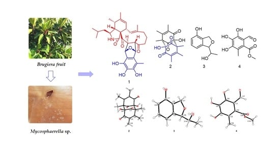
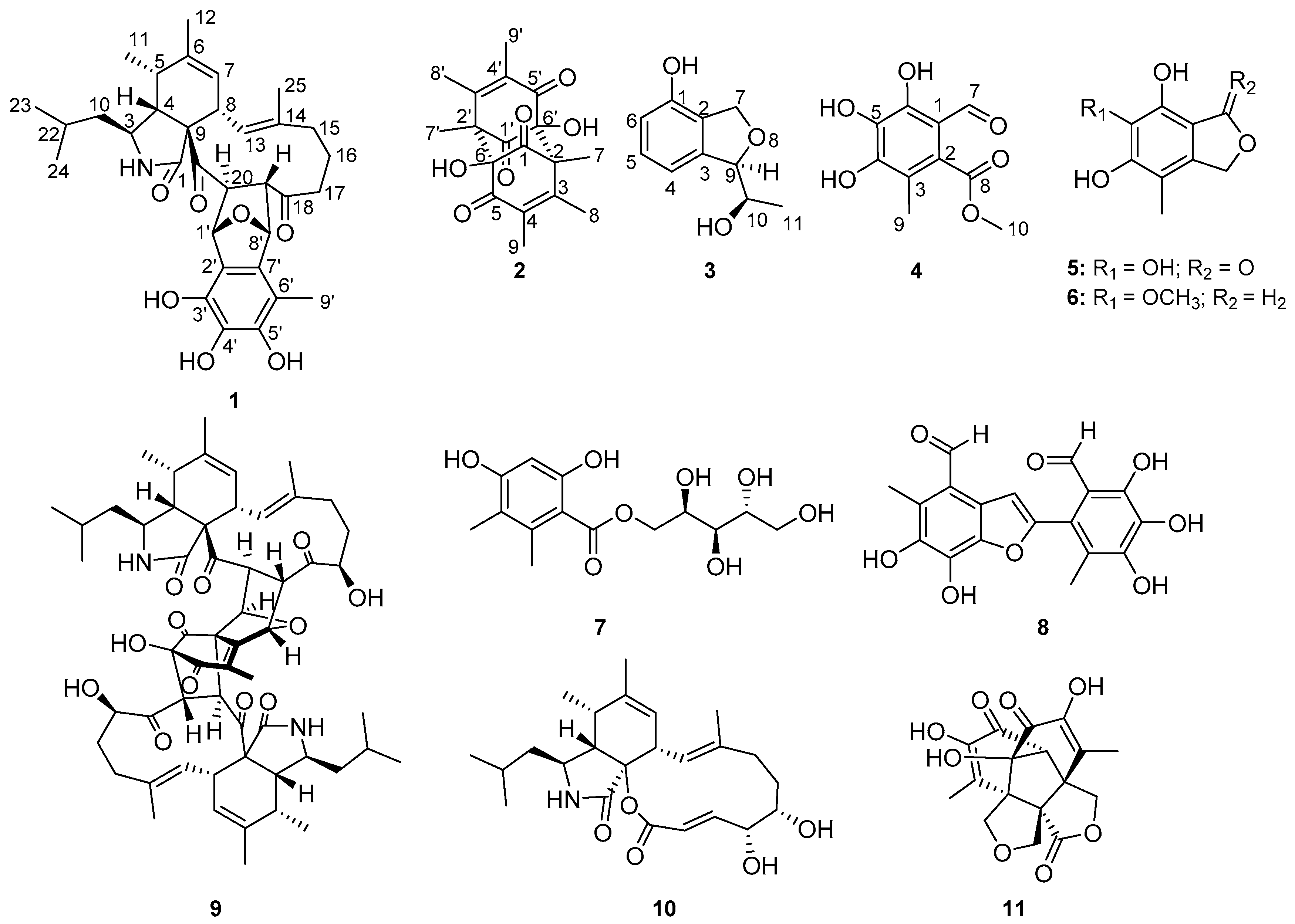
Marine fungi are proved to be rich sources of structurally unique and bioactive secondary metabolites. Mycosphaerella sp., which contributes the largest genus of Ascomycota, is a common plant pathogen widely distributed in terrestrial plant and marine environment [14,15,16]. As part of our ongoing investigation on new secondary metabolites from marine fungi in the South China Sea [17,18,19,20,21], a mangrove fungus, Mycosphaerella sp. SYSU-DZG01, collected from the fruit of the mangrove plant Bruguiera, Hainan Dongzhai Harbor Mangrove Reserve attracted our attention because the EtOAc extract of the solid fermentation medium exhibited significant α-glucosidase inhibitory activity. Chemical investigation of the bioactive extract (Figure 1) lead to the discovery of four new metabolites, asperchalasine I (1), dibefurin B (2) and two epicoccine derivatives, (R)-9-((R)-10-hydroxyethyl)-7,9-dihydroisobenzofuran-1-ol (3), 2-methoxycarbonyl-4,5,6-trihydroxy-3-methyl-benzaldehyde (4), together with seven known compounds, epicoccone B (5) [22], 1,3-dihydro-5-methoxy-7-methylisobenzofuran (6) [23], paeciloside A (7) [24], epicoccolide B (8) [25], asperchalasine A (9) [26], aspochalasin I (10) [27] and epicolactone (11) [28]. Their structures were established by extensive spectroscopic data and single-crystal X-ray diffraction analysis. Asperchalasine I possesses a distinct T-shaped skeleton containing one epicoccine moiety and one cytochalasan moiety. In bioactivity assays, compounds 1, 8 and 9 exhibited α-glucosidase inhibitory activity and 1, 4, 6 and 8 showed antioxidant activity by scavenging DPPH·. Herein, the isolation, structure elucidation, α-glucosidase inhibitory and antioxidant activities of these compounds are reported.
2. Results
2.1. Structure Elucidation
Asperchalasine I (1), the molecular formula, C33H41O7N, was determined on the basis of the HRESIMS ion at m/z 562.2798 ([M − H]− calcd. for C33H40O7N: 562.2799). As shown in Table 1, the 1H NMR data indicated characteristic signals of two double-bond protons (δH 5.93 and 5.36), nine methine protons (δH 5.52, 5.06, 4.46, 3.76, 3.21, 3.05, 2.88, 2.54 and 1.62), four methylene protons (δH 2.09, 1.90, δH 2.01, 1.58, δH 1.35 and δH 1.21) and six methyl groups (δH 2.05, 1.78, 1.26, 1.17, 0.96 and 0.94). Subsequently, the 13C NMR data showed the presence of 33 carbon signals, according to the DEPT and HSQC data, which were identified as three carbonyls (δC 211.6, 203.6 and 176.3), an aromatic ring (δC 141.3, 133.7, 132.6, 132.5, 123.7 and 111.9), two trisubstituted double bonds (δC 141.8, 137.4, 125.8 and 125.1), nine methines, four methylenes, six methyls and a quaternary carbon (δC 67.3). The 1H-1H COSY (Figure 2) correlations of H3-23/H-22/H2-10/H-3/H-4/H-5/H3-11, H-7/H-8/H-13, H2-15/H2-16/H2-17 and H-19/H-20, together with the HMBC correlations from H-4 to C-1, C-9 and C-21, from H3-12 to C-5, C-6 and C-7, from H-13 to C-25, from H3-25 to C-15, from H2-17 and H-19 to C-18, and from H-20 to C-21, suggested the presence of an cytochalasan moiety. Meanwhile, the epicoccine moiety was inferred by HMBC correlations from H-1′ to C-3′, from H-8′ to C-2′ and from H3-9′ to C-5′, C-6′ and C-7′. The key linking relation of the cytochalasan moiety and epicoccine moiety through C-19/C-8′ and C-20/C-1′ C-C bonds was suggested from the 1H-1H COSY cross-peak of H-19/H-8′ and the HMBC correlation from H-1′ to C-20 and C-21. Moreover, the structure of 1 was further confirmed by a detailed comparison of the NMR data of 1 and asperchalasine B [26], which suggested that they shared the same skeleton. The upfield shifted of H2-17 (δH 1.21) in 1 (δH 4.10 in asperchalasine B) and the disappearance of a methoxy group at C-4′ (δH 3.70, δC 61.0), suggesting the replacements of methine at C-17 and a methoxy group at C-4′ in asperchalasine B with a methylene and hydroxy group in 1, respectively.
Detailed analysis of NOESY data determined the relative configuration of chiral carbons in compound 1. The NOESY correlations of H-4/H2-10, H-3/H3-11, H-5/H-8/H3-25, H-13/H-20 and H-19/H3-25 suggested that these protons were cofacial. Neither 1H-1H coupling nor a 1H-1H COSY correlation was observed between the protons of H-20 (δH 4.46, d, J = 5.8 Hz) and H-1′ (δH 5.06, s), which suggested that the dihedral angle of those protons was approximately 90° (Figure 3) [26]. The electron circular dichroism (ECD) spectrum (Figure S8) showed two positive Cotton effects (CE) at 228 nm and 306 nm, which were also consistent with those of asperchalasine B. Therefore, the absolute configuration of 1 was suggested to be 3S, 4R, 5S, 8S, 9S, 19S, 20S, 1′S, 8′R.
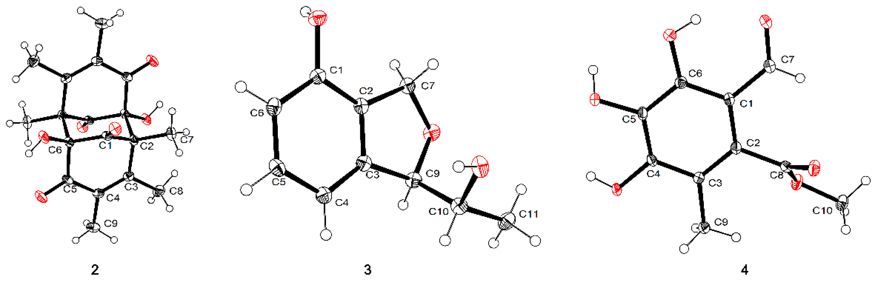
Dibefurin B (2), a colorless monocrystal, was assigned a molecular formula of C18H20O6 by the HRESIMS ion at m/z 331.1187 ([M − H]− calcd. for C18H19O6: 331.1187). For the monomer, the 1H, 13C NMR (Table 2) and HSQC data of 2 revealed diagnostic signals for nine carbons, including two carbonyls (δC 200.6 and 192.6), two disubstituted olefin carbons (δC 157.6 and 131.4), an oxygen-bearing carbon [δH 6.67 (OH-6), δC 91.7 (C-6)], three methyls (δC 19.1, 12.2 and 12.1) and a quaternary carbon (δC 60.4). The HMBC correlations from H3-7 to C-1, C-2, C-3 and C-6′, from H3-8 to C-2, C-3 and C-4, from H3-9 to C-3, C-4 and C-5, revealed that Me-7, Me-8 and Me-9 were connected at C-2, C-3 and C-4, respectively. Similarly, the hydroxyl was attached to C-6, as evidenced by the HMBC correlations from OH-6 to C-1, C-2′, C-5 and C-6. In view of the HRESIMS data and X-ray (Figure 4), it could confirm that compound 2 was a symmetric dimer and the absolute configuration of 2 was assigned as 2S, 6R, 2′R, 6′S.
Compound 3 was purified as a colorless crystal whose molecular formula was deduced as C10H12O3 based on HRESIMS data (179.0716 [M − H]−, calcd. 179.0714). Analysis of the 1H NMR spectrum of 3 (Table 3) displayed three aromatic proton resonances (δH 7.13, 6.82 and 6.69), an oxygenated methylene (δHa 5.12, δHb 5.02), two oxygenated methines (δH 5.06 and 3.97), and a methyl (δH 1.20). The 13C spectrum revealed 10 signals, indicating an aromatic ring, a methylene, two methines and a methyl. In the 1H-1H COSY spectrum, the ortho-trisubstitution on the aromatic ring was confirmed by the cross-peaks of H-4/H-5/H-6. Moreover, the 1H-1H COSY spectrum showed correlations from H-10 to H-9 and H-11, and the chemical shift of C-10 (δC 70.6) showed the hydroxyl was attached to C-10. Subsequently, the HMBC correlations between H-6 and C-1/C-2 determined the linkage of 1-OH to C-1, and the correlations between H-7 and C-1/C-2/C-3, H-9 and C-2/C-3/C-10 established the presence of a phthalan ring. The same relative configuration of C-9 and C-10 was clearly deduced under the guidance of single-crystal X-ray (Figure 4). Hence, the absolute configuration of 3 was determined as 9R, 10R.
Compound 4 was deduced to have a molecular formula of C10H10O6 from its HRESIMS spectrum with a deprotonated molecular ion at m/z 225.0407. The 1H NMR (Table 3) in MeOH-d4 showed three singlets at δH 9.71, 3.90 and 2.08, according to the 13C NMR and HSQC data, which were attributed to an aldehyde group (δC 194.7), a methoxy group (δC 52.9) and a methyl (δC 12.4), respectively. In addition, resonances of a carbonyl and an aromatic ring were observed in the 13C NMR data. In the HMBC spectrum, the correlations of H-9 to C-2, C-3 and C-4 supported the connection of Me-9 to C-3, the correlations of H-7 to C-1 and C-6 indicated the linkage of aldehyde group and C-1. Meanwhile, the carbonyl (δC 169.9, C-8) had the HMBC correlation from H-10 (δH 3.90), further indicated the presence of methyl ester. With the assistance of single-crystal X-ray (Figure 4), the structure of compound 4 was clearly confirmed.
2.2. Biological Evaluation
Compounds 1–11 were tested for their inhibitory effects against α-glucosidase, and antioxidant activity. As seen in Table 4, the results indicated that compounds 1, 8 and 9 showed significant inhibitory effects against α-glucosidase with IC50 values of 17.1, 26.7 and 15.7 μM, respectively, which were better than the positive controls acarbose (610.2 μM) and 1-deoxynojirimycin (71.5 μM). Beyond that, all of the compounds were tested for their antioxidant activity based on DPPH· (2, 2-diphenyl-1-picrylhydrazyl radical) scavenging. The results showed the antioxidant activity of 8 was 89% at the concentration of 100 μM and compound 8 possessed more potent capacity than positive control ascorbic acid in scavenging DPPH· with an EC50 value of 16.3 μM. Compounds 1, 4 and 6 also exhibited weak DPPH· scavenging activity with respective EC50 values of 77.8, 85.8 and 59.1 μM.
3. Experimental Section
3.1. General Experimental Procedures
UV data were measured on a UV-Vis-NIR spectrophotometer (Perkin Elmer, Waltham, UK). IR spectrum data were recorded using a Bruker Vector spectrophotometer 22. Melting points were tested on a Fisher-Johns hot-stage apparatus which were uncorrected. Optical rotations were recorded using an MCP300 (Anton Paar, Shanghai, China). HRESIMS data were conducted on an Ion Mobility-Q-TOF High-Resolution LC-MS (Synapt G2-Si, Waters). The ECD experiment data were measured with J-810 spectropolarimeter (JASCO, Tokyo, Japan). The NMR spectra were recorded on Bruker Avance spectrometer (Bruker, Beijing, China) (Compounds 1 and 3: 500 MHz for 1H and 125 MHz for 13C, respectively; compounds 2 and 4: 400 MHz for 1H and 100 MHz for 13C). Column chromatography (CC) was carried out on silica gel (200–300 mesh, Marine Chemical Factory, Qingdao, China) and sephadex LH-20 (Amersham Pharmacia, Piscataway, NJ, USA).
3.2. Fungal Materials
The fungus used in this research was isolated from the fruit of the marine mangrove plant Bruguiera collected in 2014 in Hainan Dongzhai Harbor Mangrove Reserve by using the standard protocol. The strain was identified as Mycosphaerella sp. (compared to no. KX067865.1) upon the analysis of ITS sequence data of the rDNA gene. The ITS sequence data obtained from the fungal strain has been submitted to GenBank with accession no. MN194208. A voucher strain was deposited in our laboratory.
3.3. Fermentation, Extraction and Isolation
The fungus Mycosphaerella sp. SYSU-DZG01 was grown on solid cultured medium in 100 × 1000 mL Erlenmeyer flasks at room temperature for 30 days under static conditions, each containing 80 g rice and 120 mL 0.3% saline. After incubation, the former was extracted with methanol twice and concentrated to yield 10.9 g of crude extract under reduced pressure. The crude extract was subjected to LC-HRESIMS analysis (Figure S29). Then, the residue was eluted by using gradient elution with petroleum ether/EtOAc from 9:1 to 0:10 (v/v) on silica gel CC to get ten fractions (Fr.1–Fr.10). Fr.2 (630 mg) was further eluted by silica gel CC using CH2Cl2/MeOH (40:1) to obtain Fr.2.1–Fr.2.3. Fr.2.3 (301 mg) was purified by Sephadex LH-20 CC and eluted with MeOH to obtain compound 2 (3.5 mg), 6 (11.1 mg) and 10 (3.6 mg). Fr.4 (217 mg) was applied to silica gel CC by CH2Cl2/MeOH (20:1) to obtain Fr.4.1–Fr.4.7. Fr.4.1 (8.1 mg) was further purified by Sephadex LH-20 CC using MeOH to obtain compound 1 (2.3 mg), 3 (2.2 mg), 8 (20.8 mg) and 9 (2.7 mg). Fr.5 (817 mg) was eluted (by CH2Cl2/MeOH, 25:3) to obtain Fr.5.1–Fr.5.5. Fr.5.1 (13.3 mg), Fr.5.2 (27.7 mg) and Fr.5.4 (10.9 mg) was purified by Sephadex LH-20 CC using CH2Cl2/MeOH (1:1) to yield compound 4 (4.3 mg), 5 (2.0 mg), 7 (3.7 mg) and 11 (2.7 mg).
Asperchalasine I (1): White powder; [α = +61.4 (c 0.1, MeOH); UV (MeOH) λmax (log ε): 206 (4.53) nm; IR (KBr) νmax (cm−1): 3369, 1691, 1440, 1384, 1201, 1120, 1053; HRESIMS m/z 562.2798 [M − H]− (calcd. for C33H40O7N: 562.2799); 1H and 13C NMR data: see Table 1.
Dibefurin B(2): Colorless crystal; m.p. 67.8–69.2 °C; [α = +0.3 (c 0.1, MeOH); UV (MeOH) λmax (log ε): 237 (3.98) nm; IR (KBr) νmax (cm−1): 3448, 1747, 1664, 1645, 1238, 1037; HRESIMS m/z 331.1187 [M − H]− (calcd. for C18H19O6, 331.1187); 1H and 13C NMR data: see Table 2.
Compound 3: Colorless crystal; m.p. 89.8–91.9 °C; [α = −37.1 (c 0.1, MeOH); UV (MeOH) λmax (log ε) 204 (4.39), 269 (3.24) nm; IR (KBr) νmax (cm−1): 3261, 2887, 1601, 1471, 1297, 767, 706; HRESIMS m/z 179.0716 [M − H]− (calcd. for C10H12O3, 179.0714; 1H and 13C NMR data: see Table 2.
Compound 4: Colorless crystal; m.p. 95.9–97.8 °C; [α = +3.4 (c 0.1, MeOH); UV (MeOH) λmax (log ε) 241 (3.66), 305 (3.56) nm; IR (KBr) νmax (cm−1): 3375, 2962, 1711, 1641, 1261, 1150, 933; HRESIMS m/z 225.0407 [M − H]− (calcd. for C10H10O6, 225.0407; 1H and 13C NMR data: see Table 2.
3.4. X-Ray Crystallographic Data
Colorless crystals of compounds 2–4 were obtained from MeOH-CH2Cl2 at room temperature by slow volatilization, and examined on an Agilent Xcalibur Nova single crystal diffractometer with Cu Kα radiation.
The crystallographic data for compound 2 has been deposited in the Cambridge Crystallographic Data Centre (CCDC number: 18022803)
Crystal data of 2: C18H20O6, Mr = 332.34, triclinic, a = 6.9740(4) Å, b = 7.9800(4) Å, c = 14.3659(6) Å, α = 101.148(4)°, β = 99.209(4)°, γ = 98.479(5)°, V = 761.05(7)Å3; space group P-1, Z = 2, Dc = 1.450 g/cm3, μ = 0.908 mm−1 and F(000) = 352.0; Crystal dimensions: 0.40 × 0.30 × 0.02 mm3. Independent reflections: 4998 (Rint = 0.0269). The final R1 was 0.0522, wR2 = 0.1447 [l > 2σ (I)]. The goodness of fit on F2 was 1.048.
The crystallographic data for compound 3 has been deposited in the Cambridge Crystallographic Data Centre (CCDC number: 18121203)
Crystal data of 3: C10H12O3, Mr = 180.07, monoclinic, a = 4.7697(1) Å, b = 11.1895(3) Å, c = 9.1541(3) Å, α = 90°, β = 93.829(3)°, γ = 90°, V = 487.47(2) Å3; space group P21, flack 0.14(18), Z = 2, Dc = 1.350 g/cm3, μ = 0.872 mm−1 and F(000) = 212.0; Crystal dimensions: 0.40 × 0.10 × 0.05 mm3. Independent reflections: 7438 (Rint = 0.0620). The final R1 was 0.0445, wR2 = 0.1258 [l > 2σ (I)]. The goodness of fit on F2 was 1.046.
The crystallographic data for compound 4 has been deposited in the Cambridge Crystallographic Data Centre (CCDC number: 18120705)
Crystal data of 4: C10H10O6, Mr = 226.04, orthorhombic, a = 15.9208(7) Å, b = 6.6849(3) Å, c = 18.5162(7) Å, α = 90°, β = 90°, γ = 90°, V = 1970.66(14) Å3; space group Pbca, Z = 8, Dc = 1.525 g/cm3, μ = 1.108 mm−1 and F(000) = 944.0; Crystal dimensions: 0.25 × 0.03 × 0.03 mm3. Independent reflections: 3833 (Rint = 0.0496). The final R1 was 0.0485, wR2 = 0.1328 [l > 2σ (I)]. The goodness of fit on F2 was 1.050.
3.5. Biological Assays
3.5.1. Inhibitory Activity of α-Glucosidase
The α-glucosidase inhibitory activity was assayed according to the reported method [29]. The inhibitory activity of α-glucosidase was tested in the 96-well plated with 100 mm PBS (KH2PO4-K2HPO4, pH 7.0) buffer solution each. Compounds 1–11, acarbose and 1-deoxynojirimycin (positive control) were dissolved in DMSO, the substrate (p-nitrophenyl glycoside, 5 mM) were dissolved in PBS buffer solution and enzyme solutions (2.0 units/mL) were prepared. The assay was conducted in a 100 μL reaction system containing 20 μL enzyme stock solution, 69 μL PBS buffers and 1 μL of DMSO or testing materials. After 10 min incubation at 37 °C, 10 μL of the substrate was added and incubated for 20 min at 37 °C. The Absorbance which measured by a BIO-RAD (iMark) microplate reader at 405 nm was used to calculate the inhibitory activity according to the equation:
η (%) is the percentage of inhibition; B is the assay medium with DMSO; S is the assay medium with compound. The results of IC50 values were calculated by the nonlinear regression analysis. Acarbose and 1-deoxynojirimycin were used as positive controls.
η (%) = [(B − S)/B] × 100%
3.5.2. Antioxidant Activity
The DPPH· scavenging was assayed according to the reported method [30]. The DPPH radical scavenging test was performed in 96-well microplates. Testing materials (compounds 1–11) were added to 150 μL (0.16 mmol/L) DPPH solution in MeOH at a range of 50 μL solutions of different concentrations (2, 25, 50 and 100 μM). After 30 min, absorbance at 517 nm was measured and the percentage of activity was calculated. Ascorbic acid was used as a positive control.
4. Conclusions
In summary, four new metabolites, including one new asperchalasine I (1), dibefurin B (2), two epicoccine derivatives (3,4) and seven known compounds were isolated from the fungus Mycosphaerella sp. SYSU-DZG01. The structures of 1–11 were established by spectroscopic data and the absolute configuration of compounds 1–3 was determined in this research. Compound 1 possesses a unique T-shaped skeleton. All of the compounds were tested for their biological activities. Compounds 1, 8 and 9 exhibited inhibitory effects against α-glucosidase with IC50 values of 17.1, 26.7 and 15.7 μM, respectively while compounds 1, 4, 6 and 8 showed antioxidant activity by scavenging DPPH· with EC50 values of 77.8, 85.8, 59.1 and 16.3 μM. These results suggested that the asperchalasine I may be a potential candidate for α-glucosidase inhibitor.
Supplementary Materials
The following are available online at https://www.mdpi.com/1660-3397/17/8/483/s1, Figure S1: 1H NMR spectrum of compound 1 (500 MHz, CDCl3), Figure S2: 13C NMR spectrum of compound 1 (125 MHz, CDCl3), Figure S3: DEPT 135, DEPT 90 and 13C NMR spectrum of compound 1 (125 MHz, CDCl3), Figure S4: 1H-1H COSY spectrum of compound 1 (CDCl3), Figure S5: HSQC spectrum of compound 1 (CDCl3), Figure S6: HMBC spectrum of compound 1 (CDCl3), Figure S7: NOESY spectrum of compound 1 (CDCl3), Figure S8: HRESIMS spectrum of compound 1, Figure S9: Experiment ECD spectrum of 1, Figure S10: 1H NMR spectrum of compound 2 (400 MHz, DMSO-d6), Figure S11: 13C NMR spectrum of compound 2 (100 MHz, DMSO-d6), Figure S12: 1H-1H COSY spectrum of compound 2 (DMSO-d6), Figure S13: HSQC spectrum of compound 2 (DMSO-d6), Figure S14: HMBC spectrum of compound 2 (DMSO-d6), Figure S15: HRESIMS spectrum of compound 2, Figure S16: 1H NMR spectrum of compound 3 (500 MHz, MeOH-d4), Figure S17: 13C NMR spectrum of compound 3 (125 MHz, MeOH-d4), Figure S18: 1H-1H COSY spectrum of compound 3 (MeOH-d4), Figure S19: HSQC spectrum of compound 3 (MeOH-d4), Figure S20: HMBC spectrum of compound 3 (MeOH-d4), Figure S21: NOESY spectrum of compound 3 (MeOH-d4), Figure S22: HRESIMS spectrum of compound 3, Figure S23: 1H NMR spectrum of compound 4 (400 MHz, MeOH-d4), Figure S24: 13C NMR spectrum of compound 4 (100 MHz, MeOH-d4), Figure S25: 1H-1H COSY spectrum of compound 4 (MeOH-d4), Figure S26: HSQC spectrum of compound 4 (MeOH-d4), Figure S27: HMBC spectrum of compound 4 (MeOH-d4), Figure S28: HRESIMS spectrum of compound 4. Figure S29: The LC-HRESIMS analysis profile of crude extract.
Author Contributions
P.Q. contributed to isolation, structure elucidation and wrote the paper; Z.L. contributed to the analysis of the NMR data and structure elucidation. Y.C. contributed to the analysis of the NMR data and biological tests. R.C. contributed to the spectral analysis. Z.S. and G.C. guided the whole experiment and revised the manuscript.
Funding
This research was funded by the Guangdong Special Fund for Marine Economic Development (GDME-2018C004), Guangdong MEPP Fund (GDOE-2019A21), the National Natural Science Foundation of China (2187713, 21472251), the Key Project of Natural Science Foundation of Guangdong Province (2016A040403091), the Special Promotion Program for Guangdong Provincial Ocean and Fishery Technology (A201701C06) and the open foundation of Key Laboratory of Tropical Medicinal Resource Chemistry of Ministry of Education (rdyw2018001).
Acknowledgments
We thank all the funding assistance mentioned above for their generous support and the Sun Yat-Sen University Instrumental Analysis and Research Center.
Conflicts of Interest
The authors declare no conflict of interest.
References
- Wresdiyati, T.; Sa’Diah, S.; Winarto, A.; Febriyani, V. Alpha-Glucosidase Inhibition and Hypoglycemic Activities of Sweitenia mahagoni Seed Extract. HAYATI J. Biosci. 2015, 22, 73–78. [Google Scholar] [CrossRef] [Green Version]
- Sekar, V.; Chakraborty, S.; Mani, S.; Sali, V.K.; Vasanthi, H.R. Mangiferin from Mangifera indica fruits reduces post-prandial glucose level by inhibiting α-glucosidase and α-amylase activity. S. Afr. J. Bot. 2019, 120, 129–134. [Google Scholar] [CrossRef]
- Wang, H.; Du, Y.J.; Song, H.C. α-Glucosidase and α-amylase inhibitory activities of guava leaves. Food Chem. 2010, 123, 6–13. [Google Scholar] [CrossRef]
- Tang, H.; Ma, F.; Zhao, D.; Xue, Z. Exploring the effect of salvianolic acid C on α-glucosidase: Inhibition kinetics, interaction mechanism and molecular modelling methods. Process. Biochem. 2019, 78, 178–188. [Google Scholar] [CrossRef]
- Indrianingsih, A.W.; Tachibana, S. α-Glucosidase inhibitor produced by an endophytic fungus, Xylariaceae sp. QGS 01 from Quercus gilva Blume. Food Sci. Hum. Wellness 2017, 6, 88–95. [Google Scholar] [CrossRef]
- Yin, Z.; Zhang, W.; Feng, F.; Zhang, Y.; Kang, W. α-Glucosidase inhibitors isolated from medicinal plants. Food Sci. Hum. Wellness 2014, 3, 136–174. [Google Scholar] [CrossRef]
- Chen, J.; Zhang, X.; Huo, D.; Cao, C.; Li, Y.; Liang, Y.; Li, B.; Li, L. Preliminary characterization, antioxidant and α-glucosidase inhibitory activities of polysaccharides from Mallotus furetianus. Carbohydr. Polym. 2019, 215, 307–315. [Google Scholar] [CrossRef] [PubMed]
- Lin, M.Z.; Chai, W.M.; Zheng, Y.L.; Huang, Q.; Ou-Yang, C. Inhibitory kinetics and mechanism of rifampicin on α-glucosidase: Insights from spectroscopic and molecular docking analyses. Int. J. Biol. Macromol. 2019, 122, 1244–1252. [Google Scholar] [CrossRef] [PubMed]
- Wei, M.; Chai, W.M.; Yang, Q.; Wang, R.; Peng, Y. Novel Insights into the Inhibitory Effect and Mechanism of Proanthocyanidins from Pyracantha fortuneana Fruit on α-Glucosidase. J. Food Sci. 2017, 82, 2260–2268. [Google Scholar] [CrossRef]
- Sun, Y.; Liu, J.; Li, L.; Gong, C.; Wang, S.; Yang, F.; Hua, H.; Lin, H. New butenolide derivatives from the marine sponge-derived fungus Aspergillus terreus. Bioorgan. Med. Chem. Lett. 2018, 28, 315–318. [Google Scholar] [CrossRef]
- Liu, H.; Chen, Z.; Zhu, G.; Wang, L.; Du, Y.; Wang, Y.; Zhu, W. Phenolic polyketides from the marine alga-derived Streptomyces sp. OUCMDZ-3434. Tetrahedron 2017, 73, 5451–5455. [Google Scholar] [CrossRef]
- Rizvi, T.S.; Hussain, I.; Ali, L.; Mabood, F.; Khan, A.L.; Shujah, S.; Rehman, N.U.; Al-Harrasi, A.; Hussain, J.; Khan, A.; et al. New gorgonane sesquiterpenoid from Teucrium mascatense Boiss, as α-glucosidase inhibitor. S. Afr. J. Bot. 2019, 124, 218–222. [Google Scholar] [CrossRef]
- Wang, C.; Guo, L.; Hao, J.; Wang, L.; Zhu, W. α-Glucosidase Inhibitors from the Marine-Derived Fungus Aspergillus flavipes HN4-13. J. Nat. Prod. 2016, 79, 2977–2981. [Google Scholar] [CrossRef] [PubMed]
- Lee, J.; Lee, J.; Kim, G.J.; Yang, I.; Wang, W.; Nam, J.W.; Choi, H.; Nam, S.J.; Kang, H. Mycousfurans A and B, Antibacterial Usnic Acid Congeners from the Fungus Mycosphaerella sp., Isolated from a Marine Sediment. Mar. Drugs 2019, 17, 422. [Google Scholar] [CrossRef] [PubMed]
- Otálvaro, F.; Nanclares, J.; Vásquez, L.E.; Quiñones, W.; Echeverri, F.; Arango, R.; Schneider, B. Phenalenone-Type compounds from Musa acuminata var. “Yangambi km 5” (AAA) and Their Activity against Mycosphaerella fijiensis. J. Nat. Prod. 2007, 70, 887–890. [Google Scholar] [CrossRef] [PubMed]
- Assante, A.; Camarda, L.; Merlini, L.; Nasini, G. Secondary metabolites from Mycosphaerella ligulicola. Phytochemistry 1981, 20, 1955–1957. [Google Scholar] [CrossRef]
- Huang, H.; Feng, X.; Xiao, Z.; Liu, L.; Li, H.; Ma, L.; Lu, Y.; Ju, J.; She, Z.; Lin, Y. Azaphilones and p-Terphenyls from the Mangrove Endophytic Fungus Penicillium chermesinum (ZH4-E2) Isolated from the South China Sea. J. Nat. Prod. 2011, 74, 997–1002. [Google Scholar] [CrossRef]
- Liu, Y.; Yang, Q.; Xia, G.; Huang, H.; Li, H.; Ma, L.; Lu, Y.; He, L.; Xia, X.; She, Z. Polyketides with α-Glucosidase Inhibitory Activity from a Mangrove Endophytic Fungus, Penicillium sp. HN29-3B1. J. Nat. Prod. 2015, 78, 1816–1822. [Google Scholar] [CrossRef]
- Liu, Z.; Chen, S.; Qiu, P.; Tan, C.; Long, Y.; Lu, Y.; She, Z. (+)- and (−)-Ascomlactone A: A pair of novel dimeric polyketides from a mangrove endophytic fungus Ascomycota sp. SK2YWS-L. Org. Biomol. Chem. 2017, 15, 10276–10280. [Google Scholar] [CrossRef] [PubMed]
- Cui, H.; Liu, Y.; Nie, Y.; Liu, Z.; Chen, S.; Zhang, Z.; Lu, Y.; He, L.; Huang, X.; She, Z. Polyketides from the Mangrove-Derived Endophytic Fungus Nectria sp. HN001 and Their α–Glucosidase Inhibitory Activity. Mar. Drugs 2016, 14, 86. [Google Scholar] [CrossRef] [PubMed]
- Liu, Y.; Xia, G.; Li, H.; Ma, L.; Ding, B.; Lu, Y.; He, L.; Xia, X.; She, Z. Vermistatin Derivatives with α-Glucosidase Inhibitory Activity from the Mangrove Endophytic Fungus Penicillium sp. HN29-3B1. Planta Med. 2014, 80, 912–917. [Google Scholar] [CrossRef] [PubMed]
- Kemami Wangun, H.V.; Ishida, K.; Hertweck, C. Epicoccalone, a Coumarin-Type Chymotrypsin Inhibitor, and Isobenzofuran Congeners from an Epicoccum sp. Associated with a Tree Fungus. Eur. J. Org. Chem. 2008, 22, 3781–3784. [Google Scholar] [CrossRef]
- Nam, H.L.; James, B.G.; Donald, T.W. Isolation of Chromanone and Isobenzofuran Derivatives from a Fungicolous Isolate of Epicoccum purpurascens. Bull. Korean Chem. Soc. 2007, 28, 877–879. [Google Scholar]
- Ferdinand, M.T.; Timothee, J.N.K.; Birger, D.; Clovis, D.M.; Hartmut, L. Paeciloside A, a new antimicrobial and cytotoxic polyketide from Paecilomyces sp. Strain CAFT156. Planta Med. 2012, 78, 1020–1023. [Google Scholar]
- Talontsi, F.M.; Dittrich, B.; Schüffler, A.; Sun, H.; Laatsch, H. Epicoccolides: Antimicrobial and Antifungal Polyketides from an Endophytic Fungus Epicoccum sp. Associated with Theobroma cacao. Eur. J. Org. Chem. 2013, 15, 3174–3180. [Google Scholar] [CrossRef]
- Zhu, H.; Chen, C.; Xue, Y.; Tong, Q.; Li, X.; Chen, X.; Wang, J.; Yao, G.; Luo, Z.; Zhang, Y. Asperchalasine A, a Cytochalasan Dimer with an Unprecedented Decacyclic Ring System, from Aspergillus flavipes. Angew. Chem. Int. Ed. 2015, 54, 13374–13378. [Google Scholar] [CrossRef] [PubMed]
- Zhou, G.X.; Wijeratne, E.M.K.; Bigelow, D.; Pierson, L.S.; Vanetten, H.D.; Gunatilaka, A.A.L. Aspochalasins I, J, and K: Three New Cytotoxic Cytochalasans of Aspergillus flavipes from the Rhizosphere of Ericameria laricifolia of the Sonoran Desert. J. Nat. Prod. 2004, 67, 328–332. [Google Scholar] [CrossRef]
- Da Silva Araújo, F.D.; de Lima Fávaro, L.C.; Araújo, W.L.; de Oliveira, F.L.; Aparicio, R.; Marsaioli, A.J. Epicolactone—Natural Product Isolated from the Sugarcane Endophytic Fungus Epicoccum nigrum. Eur. J. Org. Chem. 2012, 27, 5225–5230. [Google Scholar] [CrossRef]
- Cai, R.; Wu, Y.; Chen, S.; Cui, H.; Liu, Z.; Li, C.; She, Z. Peniisocoumarins A–J: Isocoumarins from Penicillium commune QQF-3, an Endophytic Fungus of the Mangrove Plant Kandelia candel. J. Nat. Prod. 2018, 81, 1376–1383. [Google Scholar] [CrossRef]
- Tan, C.; Liu, Z.; Chen, S.; Huang, X.; Cui, H.; Long, Y.; Lu, Y.; She, Z. Antioxidative Polyketones from the Mangrove-Derived Fungus Ascomycota sp. SK2YWS-L. Sci. Rep. 2016, 6, 36609. [Google Scholar] [CrossRef] [PubMed]
Figure 1.
Chemical structures of 1–11.

Figure 2.
The key 2D NMR correlations of 1–4.

Figure 3.
Molecular model of 1 (1′β, 8′ β-oxygen bridge).

Figure 4.
Single-crystal X-ray structures of 2–4.

Table 1.
1H (500 MHz) and 13C (125 MHz) NMR data for 1 in CDCl3.
| Position | 1 | |
|---|---|---|
| δH, mult (J in Hz) | δC, Type | |
| 1 | 176.3, C | |
| 2 | ||
| 3 | 3.21, m | 51.9, CH |
| 4 | 3.05, dd (3.9, 5.0) | 49.8, CH |
| 5 | 2.54, m, | 35.2, CH |
| 6 | 141.8, C | |
| 7 | 5.36, s | 125.1, CH |
| 8 | 2.88, d (11.1) | 43.3, CH |
| 9 | 67.3, C | |
| 10 | 1.35, t | 48.5, CH2 |
| 11 | 1.26, d (7.3) | 13.6, CH3 |
| 12 | 1.78, s | 20.1, CH3 |
| 13 | 5.93, d (11.0) | 125.8, CH |
| 14 | 137.4, C | |
| 15 | 2.09, s | 40.7, CH2 |
| 1.90, d (4.3) | ||
| 16 | 2.01, m | 22.5, CH2 |
| 1.58, m | ||
| 17 | 1.21, m | 35.1, CH2 |
| 18 | 211.6, C | |
| 19 | 3.76, t (5.3) | 56.9, CH |
| 20 | 4.46, d (5.8) | 57.3, CH |
| 21 | 203.6, C | |
| 22 | 1.62, m | 25.3, CH |
| 23 | 0.94, dd (1.2, 6.5) | 23.6, CH3 |
| 24 | 0.96, dd (1.2, 6.5) | 21.3, CH3 |
| 25 | 1.17, s | 14.2, CH3 |
| 1′ | 5.06, s | 80.7, CH |
| 2′ | 123.7, C | |
| 3′ | 132.5, C | |
| 4′ | 133.7, C | |
| 5′ | 141.3, C | |
| 6′ | 111.9, C | |
| 7′ | 132.6, C | |
| 8′ | 5.52, d (4.9) | 81.3, CH |
| 9′ | 2.05, s | 11.9, CH3 |
Table 2.
1H (400 MHz) and 13C (100 MHz) NMR data for 2 in DMSO-d6.
| Position | 2 | |
|---|---|---|
| δH, mult (J in Hz) | δC, Type | |
| 1 (1′) | 200.6, C | |
| 2 (2′) | 60.4, C | |
| 3 (3′) | 157.6, C | |
| 4 (4′) | 131.4, C | |
| 5 (5′) | 192.6, C | |
| 6 (6′) | 91.7, C | |
| 7 (7′) | 1.21, s | 12.1, CH3 |
| 8 (8′) | 2.02, s | 19.1, CH3 |
| 9 (9′) | 1.73, s | 12.2, CH3 |
Table 3.
1H and 13C NMR data for 3 and 4 in MeOH-d4.
| Position | 3 a | 4 b | ||
|---|---|---|---|---|
| δH, mult (J in Hz) | δC, Type | δH, mult (J in Hz) | δC, Type | |
| 1 | 152.8, C | 112.2, C | ||
| 2 | 127.3, C | 117.4, C | ||
| 3 | 142.1, C | 129.9, C | ||
| 4 | 6.82, d (7.4) | 114.3, CH | 152.5, C | |
| 5 | 7.13, t (7.7) | 129.9, CH | 134.1, C | |
| 6 | 6.69, d (7.9) | 115.0, CH | 151.2, C | |
| 7 | 5.12, dd (2.8, 11.8) | 72.4, CH2 | 9.71, s | 194.7, CH |
| 5.02, d (11.9) | ||||
| 8 | 169.9, C | |||
| 9 | 5.06, d (3.3) | 89.4, CH | 2.08, s | 12.4, CH3 |
| 10 | 3.97, qd (3.9, 6.4) | 70.6, CH | 3.90, s | 52.9, CH3 |
| 11 | 1.20, d (6.4) | 18.6, CH3 | ||
a 1H and 13C NMR recorded at 500 MHz and 125 MHz; b 1H and 13C NMR recorded at 400 MHz and 100 MHz.
Table 4.
The α-glucosidase inhibitory and antioxidant activities of compounds 1–11.
| Compounds | α-Glucosidase Inhibitory | Antioxidant | |
|---|---|---|---|
| IC50 (μM) | % Inhibition (100 μM) | EC50 (μM) | |
| 1 | 17.1 | 56 | 77.8 |
| 2 | >50 | <50 | - |
| 3 | >50 | <50 | - |
| 4 | >50 | 57 | 85.8 |
| 5 | >50 | <50 | - |
| 6 | >50 | 65 | 59.1 |
| 7 | >50 | <50 | - |
| 8 | 26.7 | 89 | 16.3 |
| 9 | 15.7 | <50 | - |
| 10 | - | - | - |
| 11 | >50 | <50 | - |
| Acarbose a | 610.2 | ||
| 1-Deoxynojirimycin a | 71.5 | ||
| Ascorbic acid a | 92 | 22.4 | |
- means no test; a positive control.
© 2019 by the authors. Licensee MDPI, Basel, Switzerland. This article is an open access article distributed under the terms and conditions of the Creative Commons Attribution (CC BY) license (http://creativecommons.org/licenses/by/4.0/).
Share and Cite
MDPI and ACS Style
Qiu, P.; Liu, Z.; Chen, Y.; Cai, R.; Chen, G.; She, Z. Secondary Metabolites with α-Glucosidase Inhibitory Activity from the Mangrove Fungus Mycosphaerella sp. SYSU-DZG01. Mar. Drugs 2019, 17, 483. https://doi.org/10.3390/md17080483
AMA Style
Qiu P, Liu Z, Chen Y, Cai R, Chen G, She Z. Secondary Metabolites with α-Glucosidase Inhibitory Activity from the Mangrove Fungus Mycosphaerella sp. SYSU-DZG01. Marine Drugs. 2019; 17(8):483. https://doi.org/10.3390/md17080483
Chicago/Turabian StyleQiu, Pei, Zhaoming Liu, Yan Chen, Runlin Cai, Guangying Chen, and Zhigang She. 2019. "Secondary Metabolites with α-Glucosidase Inhibitory Activity from the Mangrove Fungus Mycosphaerella sp. SYSU-DZG01" Marine Drugs 17, no. 8: 483. https://doi.org/10.3390/md17080483
Note that from the first issue of 2016, this journal uses article numbers instead of page numbers. See further details here.