Article

In Vitro and In Vivo Assessment of the Efficacy of Bromoageliferin, an Alkaloid Isolated from the Sponge Agelas dilatata, against Pseudomonas aeruginosa

by
Dawrin Pech-Puch
1,
Mar Pérez-Povedano
1,
Marta Martinez-Guitian
2,
Cristina Lasarte-Monterrubio
2,
Juan Carlos Vázquez-Ucha
2,
Germán Bou
2,
Jaime Rodríguez
1,*,
Alejandro Beceiro
2,*,† and
Carlos Jimenez
1,*,†
1
Centro de Investigacións Científicas Avanzadas (CICA) e Departamento de Química, Facultade de Ciencias, AE CICA-INIBIC, Universidade da Coruña, 15071 A Coruña, Spain
2
Servicio de Microbioloxía, Instituto de Investigación Biomédica, AE CICA-INIBIC Complexo Hospitalario Universitario A Coruña, 15006 A Coruña, Spain
*
Authors to whom correspondence should be addressed.
Same contribution to this work.
Mar. Drugs 2020, 18(6), 326; https://doi.org/10.3390/md18060326
Submission received: 1 June 2020 / Revised: 16 June 2020 / Accepted: 17 June 2020 / Published: 23 June 2020
(This article belongs to the Special Issue Structure–Activity Relationship of Marine Bioactive Compounds)

Abstract

:
The pyrrole-imidazoles, a group of alkaloids commonly found in marine sponges belonging to the genus Agelas, display a wide range of biological activities. Herein, we report the first chemical study of the secondary metabolites of the sponge A. dilatata from the coastal area of the Yucatan Peninsula (Mexico). In this study, we isolated eight known alkaloids from an organic extract of the sponge. We used NMR and MS analysis and comparison with existing databases to characterize the alkaloids: ageliferin (1), bromoageliferin (2), dibromoageliferin (3), sceptrin (4), nakamuric acid (5), 4-bromo-1H-pyrrole-2-carboxylic acid (6), 4,5-dibromopyrrole-2-carboxylic acid (7) and 3,7-dimethylisoguanine (8). We also evaluated, for the first time, the activity of these alkaloids against the most problematic multidrug-resistant (MDR) pathogens, i.e., the Gram-negative bacteria Pseudomonas aeruginosa, Klebsiella pneumoniae and Acinetobacter baumannii. Bromoageliferin (2) displayed significant activity against P. aeruginosa. Comparison of the antibacterial activity of ageliferins 13 (of similar structure) against P. aeruginosa revealed some relationship between structure and activity. Furthermore, in in vitro assays, 2 inhibited growth and biofilm production in clinical strains of P. aeruginosa. Moreover, 2 increased the survival time in an in vivo Galleria mellonella model of infection. The findings confirm bromoageliferin (2) as a potential lead for designing new antibacterial drugs.

1. Introduction

Multidrug-resistant bacterial infections represent a serious global health problem [1], causing an estimated 700,000 deaths a year worldwide. If this rising trend in antibiotic resistance is not reversed in the coming years, it could lead to 10 million people dying every year and the economic impact of approximately 1% reduction of the world’s gross domestic product (GDP) and there would be a 5–7% loss in developing countries by 2050 [2]. The increase in bacterial resistance (especially in relation to Gram-negative bacteria), together with the scarce development of new antimicrobial compounds in the last few decades, has led to the current situation in which very few or even no antibiotics are available to treat complicated infections [3]. The need for new therapeutic options to treat multidrug resistant pathogen infections is therefore indisputable.
Biofilm formation is an important bacterial survival strategy. A biofilm is a protective extracellular matrix which enables bacteria to resist the action of antibiotics and the host immune response. Biofilm production is a major virulence factor in infections such as periodontitis, native valve endocarditis and cystic fibrosis [4]. The development of new molecules should therefore be directed towards biofilm-specific targets in order to increase the therapeutic arsenal available to treat multidrug-resistant pathogens.
The traditional approach to identifying new antibacterial therapies is to search for bacteriostatic or bactericidal compounds from natural sources and synthetic pathways. Natural products have been one of the most prolific sources of new leads in modern drug discovery [5]. The marine environment, a rich source of chemically diverse, biologically active natural products, serves as an invaluable resource in the ongoing search for novel antimicrobial compounds [6].
Sponges belonging to the genus Agelas constitute an important source of marine pyrrole-imidazole alkaloids [7,8]. Since the structural elucidation of the first alkaloid, dibromophakellin, almost 50 years ago [9], more than 200 analogues isolated from a variety of tropical sponges have been found to display a wide range of biological properties including antitumor, antifungal and antibacterial activities [10,11].
In this study, we continued to search for natural bioactive products from marine organisms [12,13] more specifically from those collected off the coast of the Yucatan Peninsula (Mexico) [14,15]. We focused our attention on the sponge A. dilatata collected from Cozumel Island, because of the antibacterial activity detected in organic extracts of the sponge and the lack of reported chemical studies of its secondary metabolites. The only previously published studies concerning A. dilatata are a comparative study of the microbial diversity and analysis of the fatty acid composition of specimens collected in the Bahamas [16] and a taxonomic study of specimens of the genus Agelas from the Caribbean Sea [17]. The present study is the first chemical assessment of the natural products isolated from organic extracts of A. dilatata. The study involved the isolation and structural characterization of eight known alkaloids, most of which belong to the family of bromopyrrole-imidazole alkaloids. Furthermore, with the aim of obtaining further insights into the antibiotic activity of these compounds, we evaluated the minimum inhibitory concentrations (MICs) of the three pathogenic species classified by the WHO as critical in regard to the need for new antimicrobial therapies: Pseudomonas aeruginosa, Klebsiella pneumoniae and Acinetobacter baumannii. These species over the last decades have become a major public health crisis worldwide, because are responsible for a large number of hospital-acquired and nosocomial infections and by their increasing development of antimicrobial resistance [18]. In addition, the antimicrobial efficacy of these alkaloids was assessed by measuring the anti-biofilm activity and survival time in an in vivo Galleria mellonella infection model.

2. Results and Discussion

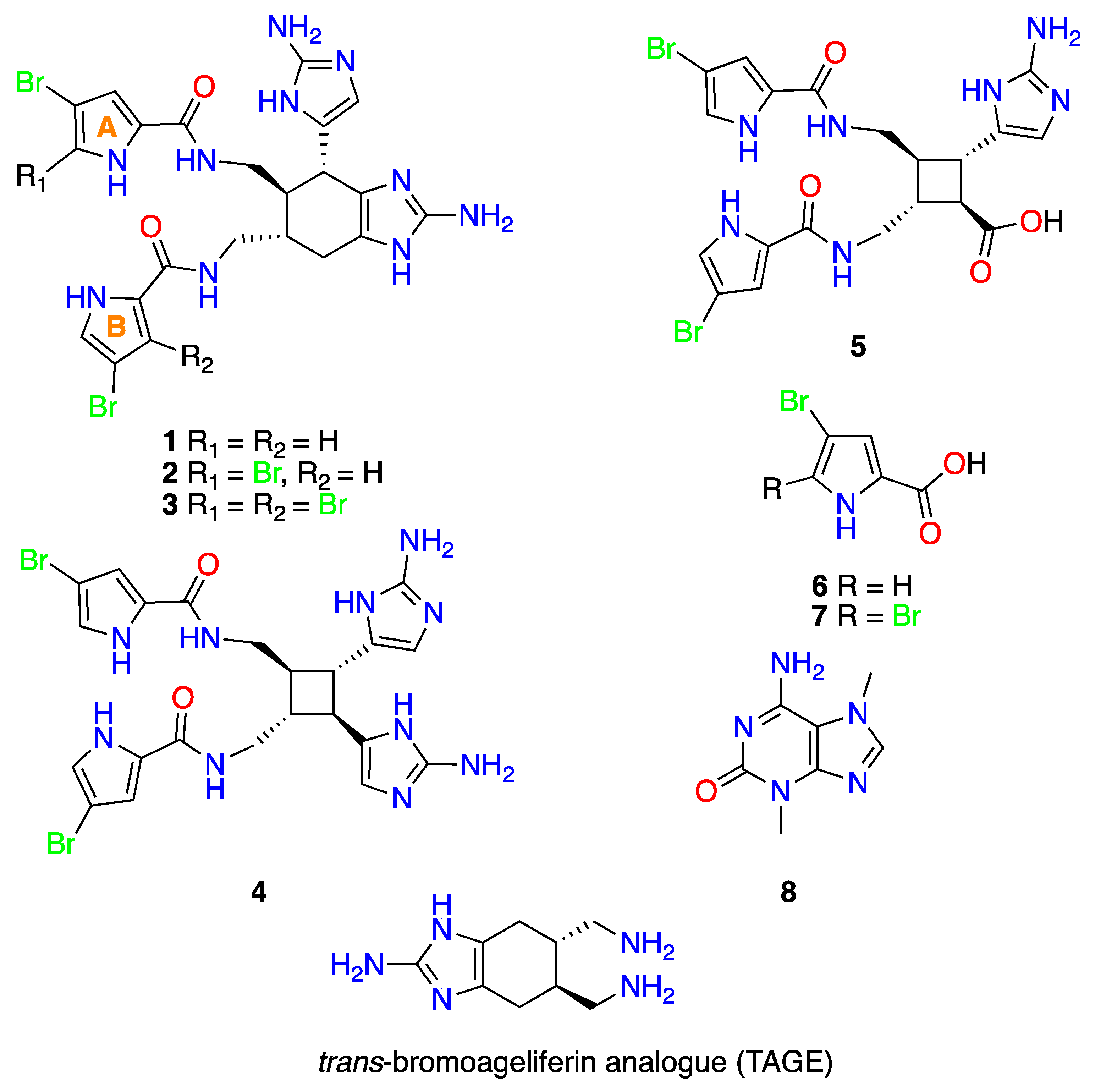
2.1. Extraction, Isolation and Structural Elucidation

Specimens of the sponge A. dilatata, collected from Cozumel Island, state of Quintana Roo, Yucatan Peninsula (Mexico), were extracted several times with a 1:1 mixture of CH3OH/CH2Cl2 to yield an organic extract which displayed antibacterial activity in a bioassay against three Gram-negative pathogens. The extract was then partitioned between water and solvents of increasing polarity to produce hexane, n-butanol, dichloromethane and a final aqueous methanolic fraction. Bioassay-guided fractionation allowed us to select the n-butanol and aqueous methanolic bioactive fractions enriched with bromopyrrole alkaloids from the oroidin family. The core skeleton of these compounds was deduced by MS analysis, which revealed the typical bromine isotopic peak clusters, and by 13C NMR studies, which showed the characteristic carbon chemical shifts at δC ~160 (CO), ~125 (C), ~124 (CH), ~118 (CH) and ~98 (C-Br) [19]. The n-BuOH fraction was subjected to solid phase extraction (SPE)-C18 (H2O/CH3OH/CH2Cl2 gradient system), and the resulting fractions were separated by reversed-phase HPLC to yield compounds 1, 2 and 48. In addition, the aqueous methanolic fraction was subjected to reversed-phase HPLC, yielding compounds 13 and 57 (Figure 1).
Comparison of the NMR spectra (1D and 2D NMR) and MS data for 18 with previously reported data allowed us to determine the structures of the compounds (See Figure 1 and Figures S1–S24 in Suplementary Material. We were thus able to identify five oroidin derivatives, three of which were the ageliferins resulting from putative 4π + 2π (Diels-Alder) cyclization [20,21,22]: ageliferin (1), bromoageliferin (2), and dibromoageliferin (3); another two resulting from putative 2π + 2π cyclization: sceptrin (4) [23] and nakamuric acid (5) [24]; two pyrrole derivatives: 4-bromo-1H-pyrrole-2-carboxylic acid (6) [25] and 4,5-dibromopyrrole-2-carboxylic acid (7) [26], and finally, the nitrogenated base 3,7-dimethylisoguanine (8) [27]. Although 18 have been isolated from other sponges in the genus Agelas, this is first time that they have been isolated from A. dilatata. Once the chemical structures were determined, the alkaloids were subjected to several biological studies.

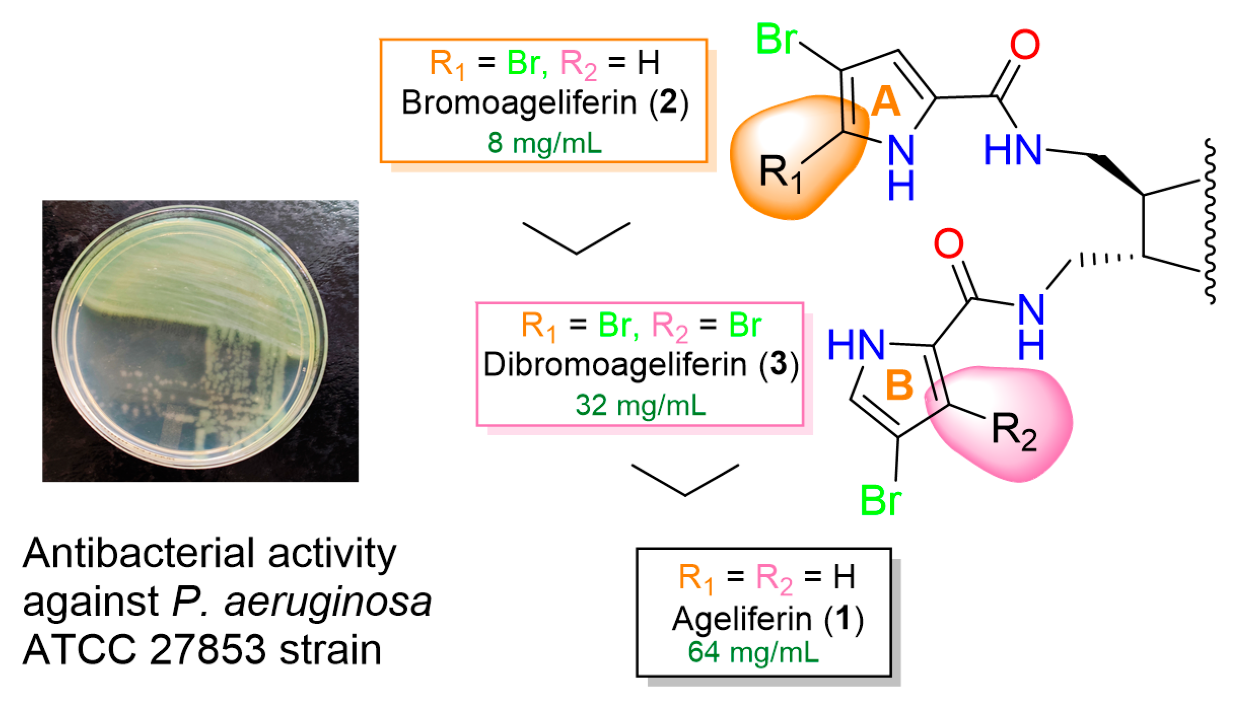
2.2. Antimicrobial Susceptibility Testing

Minimum inhibitory concentration (MIC) assays were performed with all of the alkaloids (18) against reference bacterial strains and clinical isolates (see Table 1). Four categories of antibacterial activity were established: high activity (MIC ≤ 8 mg/L); moderate activity (MIC = 16–32 mg/L), low activity (MIC = 64 mg/L) and no activity (MIC ≥ 128 mg/L). Compounds 2 and 3 displayed the highest activity against the Gram-negative bacterium P. aeruginosa. Higher MICs were obtained for the remaining purified compounds extracted from A. dilatata in tests with the pathogens.
Bromoageliferin (2) proved to be the most active against P. aeruginosa of the alkaloids isolated from A. dilatata. The MIC values for 2 and P. aeruginosa strains indicated high activity against ATCC 27853 (8 mg/L) and moderate activity against PAO1 (32 mg/L). To further study the activity of 2 against P. aeruginosa, 4 clinical isolates were included in the study. Bromoageliferin (2) displayed moderate activity against P. aeruginosa clinical isolates 29-200 SV, 30-127 VI, 30-223 SV and 30-230 SV, with a MIC value of 32 mg/L, while the corresponding MIC value for imipenem was 2 mg/L. Low concentrations of bromoageliferin (2) did not inhibit the growth of the tested strains of A. baumannii or K. pneumoniae. Additionally, 2 has also been reported to display antibacterial against Gram-positive bacteria, including Micrococcus luteus and Bacillus subtilis [22,28] and the human pathogen methicillin-resistant Staphylococcus aureus (MRSA) [29], and also against the Gram-negative bacteria Escherichia coli [22,28] and Rhodospirillum salexigens (a marine bacterium known to form biofilms) [30]. This work represents the first assessment of the antibacterial activity of bromoageliferin (2) against the multidrug resistant pathogen P. aeruginosa.
Also noteworthy is the activity of dibromoageliferin (3) against ATCC 27853 and PAO1 reference strains of P. aeruginosa (MIC, 32 mg/L) and against the two strains of K. pneumoniae included and the A. baumannii strain RYC 52763/97 (MIC, 64 mg/L). Ageliferin (1) showed a similar ability to inhibit growth against K. pneumoniae and P. aeruginosa (MIC, 64 mg/L). A. baumannii displayed greater resistance to these ageliferin-derived compounds.
Although 1 and 3 have previously been reported to display antibacterial activity against M. luteus [28], B. subtilis [22,24,28], S. aureus [24] and E. coli [22,24,28], this is the first report of an assessment of the antibacterial activity of 1 and 3 against the human pathogenic bacteria A. baumannii, K. pneumoniae and P. aeruginosa.
Isolation of the three ageliferins (13), which show slight structural differences, and assessment of their antibacterial activity allowed us to identify a relationship between the structure of the compounds and their activity against the P. aeruginosa ATCC 27853 strain. The structural comparison of the three ageliferins (13) in relation to the MIC values for this strain indicated that the presence of a second bromine atom at C-2 of pyrrol A ring increases the antibacterial activity. Thus, 1, which does not contain the atom, was less active than 2 and 3, in which the atom does occur (1 was eight times less active than 2 and two times less active than 3). However, the presence of a second bromine atom at C-2 of pyrrol B ring decreased the antibacterial activity because 2, which bears a hydrogen at C-2 of the pyrrol B ring was more active than 3, which bears a bromine atom at that position (2 was four times more active than 3) (Figure 2).
By contrast, the antibacterial analysis revealed that compounds 48 were moderately active or not active against all A. baumannii, K. pneumoniae and P. aeruginosa strains. However, these compounds have previously been shown to possess some antibacterial activity. Thus, 4 was reported to display antibacterial activity against A. baumannii [29], P. aeruginosa [23,27], M. luteus [28], S. aureus [23,24,27,29], B. subtilis [22,23,24,27,28], B. cereus [27], Streptococcus faecalis [27], Salmonella typhi [27] and E. coli. [22,24,27,28], but not against S. aureus (MRSA), Mycobacterium intracellulare or M. tuberculosis [31]. Compound 5 has been reported to be active against B. subtilis but not against S. aureus or E. coli [24]. Compound 6 did not display antimicrobial activity against S. aureus, E. coli or Proteus vulgaris [32]. Compound 7 has been reported to display activity against S. aureus, E. coli, Serratia marcescens and Micrococcus sp. [33], but not against B. megaterium [34], B. subtilis [35], S. aureus [35], E. coli [34,35] or Mycobacterium smegmatis [36]. Moreover, 7 did not display enzyme inhibitory activity in analogous FabI enzymes from M. tuberculosis (MtFabI, InhA) and E. coli (EcFabI) [37]. Compound 8 was reported inactive as it did not display antifouling activity and was unable to inhibit biofilm formation in the marine bacterial species Pseudoalteromonas spp. and Paracoccus sp. [38]. However, this is the first report of the evaluation of the antibacterial activity of 58 against A. baumannii, K. pneumoniae and P. aeruginosa and of 4 against K. pneumoniae.
Taking into account the remarkable MIC values observed for bromoageliferin (2) against P. aeruginosa, which is particularly problematic in serious infections such as cystic fibrosis, we wished to gain further insight into the antibacterial activity of this compound by performing biofilm biomass inhibition analysis and a survival assay with Galleria mellonella.

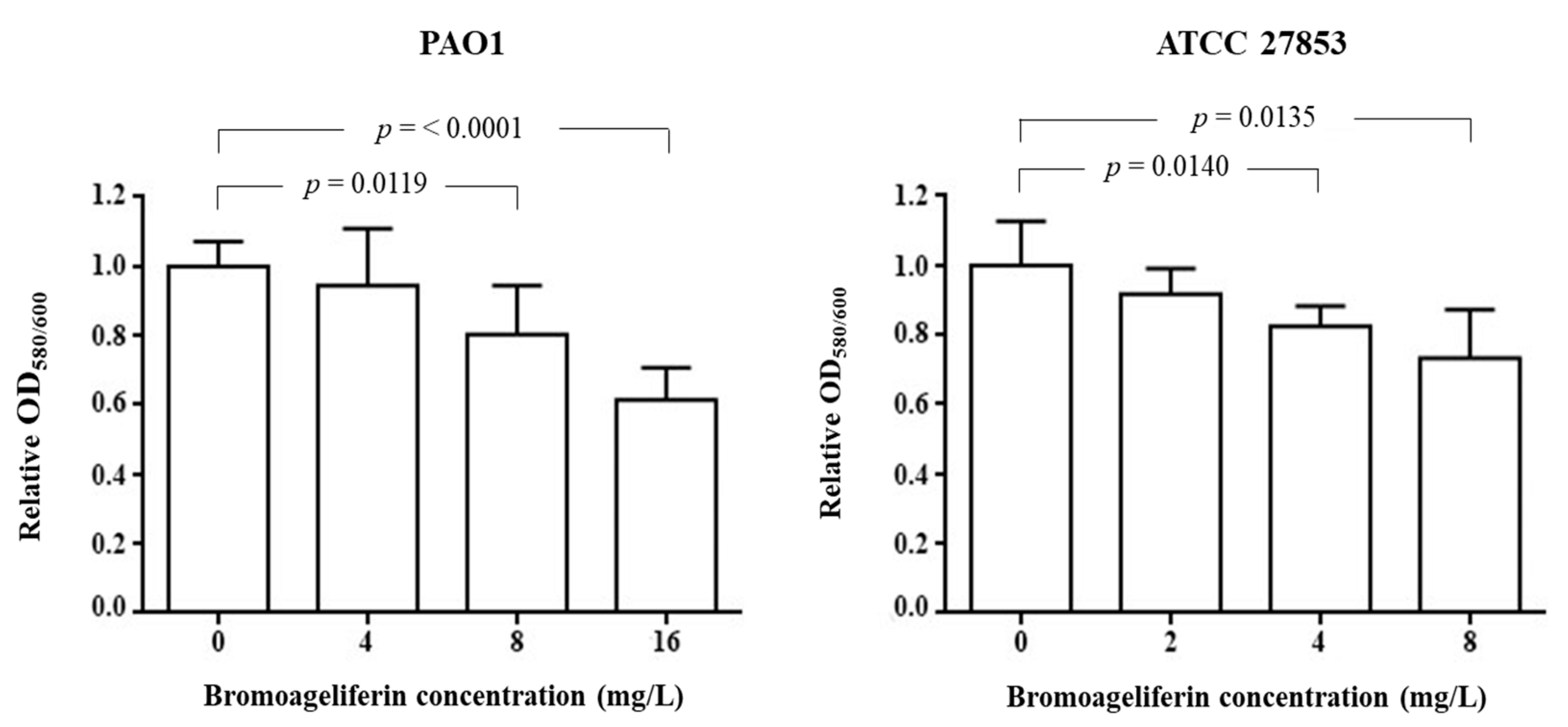
2.3. Analysis of Biofilm Biomass Inhibition

Bromoageliferin (2) has been reported to possess anti-biofilm activity against the marine R-proteobacterium R. salexigens [30]. Identification of a 2-aminoimidazole (2-AI) subunit in these bioactive brominated pyrrol alkaloids led to the suggestion that this structural motif, in tandem with the bicyclic core of bromoageliferin, may be the key pharmacophore that imparts biological activity [39]. For this reason, bromoageliferin (2) has been used as template for designing a library of simplified bromoageliferin scaffolds, such as trans-bromoageliferin analogue (TAGE, see Figure 1), which has proven to be very effective in inhibiting biofilm formation in strains of P. aeruginosa [39], A. baumannii, Bordetella bronchiseptica and S. aureus [40]. Furthermore, some of these simplified analogues suppress resistance of multiple antibiotic classes across a broad-spectrum of clinically important bacteria [40,41,42]. The parent natural product, bromoageliferin (2), was later reported to inhibit biofilm formation in two representative human pathogens, A. baumannii and S. aureus [29]. In the present study, we wished to evaluate, for the first time, the anti-biofilm activity of bromoageliferin (2) in P. aeruginosa strains.
The concentration-dependence of the effect of 2 on biofilm reduction formation was detected with both the PAO1 and ATCC 27853 reference strains (Figure 3). A significant decrease in the ability of P. aeruginosa PAO1 strain to generate biofilm was observed in the presence of bromoageliferin (2) at concentrations of 8 mg/L (p = 0.0119) and 16 mg/L (p ≤ 0.0001), relative to the control without compound. Regarding strain ATCC 27853, significant differences were found after addition of respectively 4 mg/L of 2 (p = 0.0140) or 8 mg/L (p = 0.0135) to the culture, relative to the control.
The data obtained regarding the inhibition of biofilm production by bromoageliferin (2) are consistent with those previously obtained with two simplified synthetic analogues of 2 against P. aeruginosa [39]. Indeed, a concentration of 100–200 µM of these analogues was required to inhibit 50% of biofilm production, while for 2 we observed 30–40% biofilm inhibition at concentrations of 8 or 16 mg/L (11.45 or 22.9 µM), depending on the P. aeruginosa strain used. Furthermore, inhibition of bacterial growth in the presence of the simplified synthetic analogues of bromoageliferin was evaluated by means of growth curves and was found to occur at 400–500 µM. Thus, the original compound 2 appears to have a greater capacity to inhibit bacterial growth, with MICs of 8–32 mg/L (11.45–45.83 µM) obtained in the present study.
Little is known about the antimicrobial mechanism of action of bromoageliferin (2). Although 2 displays high activity against P. aeruginosa, the growth inhibition is not as significant with the other pathogens tested, i.e., A. baumannii and K. pneumoniae. However, the inhibitory effect on biofilm production in different bacteria seems to be demonstrated. The genes encoding structural subunits of fimbriae fimA and mfa1 of the oral pathogen Porphyromonas gingivalis show altered expression when the bacteria is grown in the presence of small molecules of bromoageliferin-derivates [43]. These molecules prevented P. gingivalis from binding to Streptococcus gordoni to form a mixed species biofilm community. The possible targets (fimA and mfa1) are involved in attachment and biofilm formation, which may partly explain the anti-virulence effect of bromoageliferin (2) observed in this study.

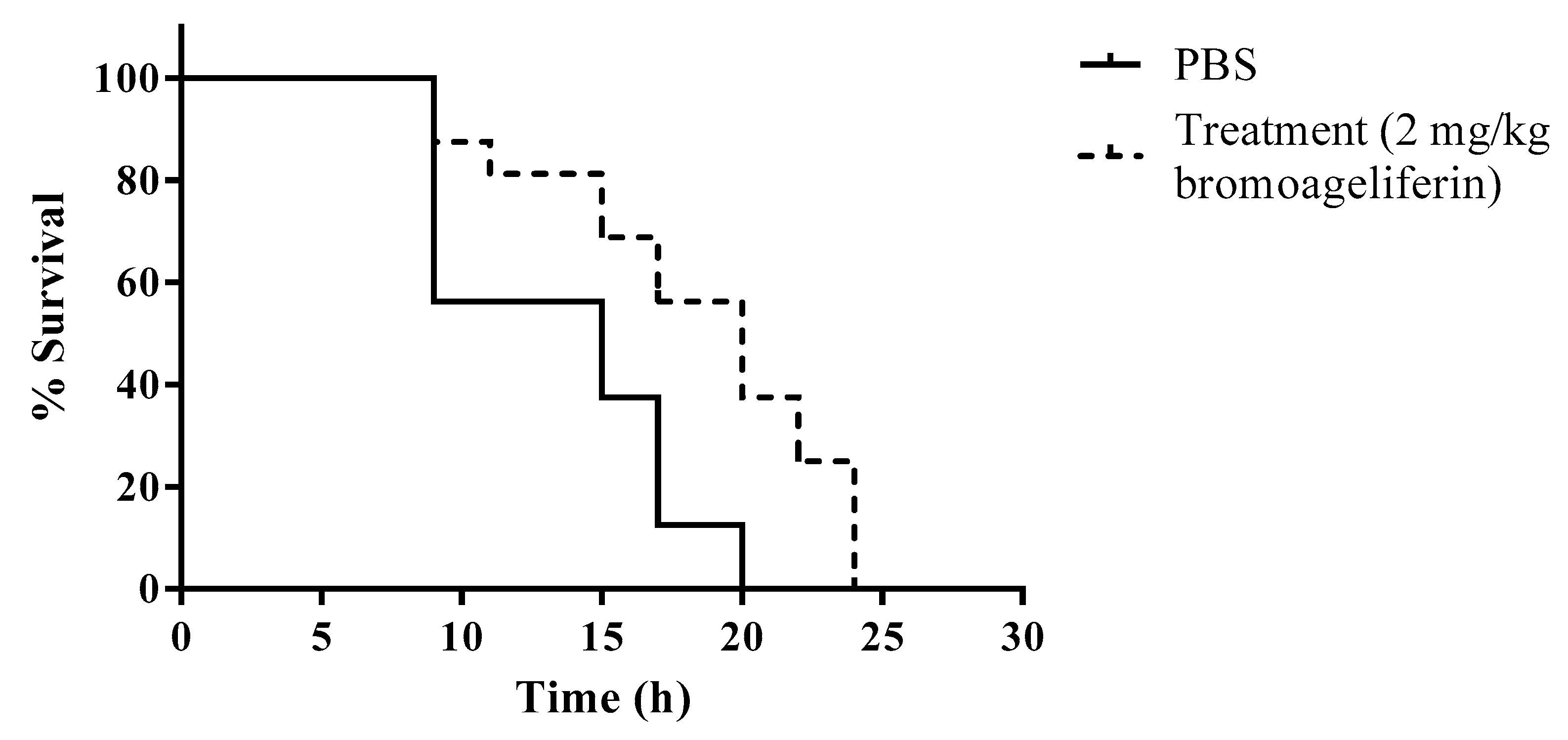
2.4. In Vivo Efficacy of Bromoageliferin against P. aeruginosa

Mammalian animal models are considered the gold standard for screening new drugs. However, they have important ethical and administrative restrictions and are costly. The in vivo Galleria mellonella (wax moth) model is suitable for studying P. aeruginosa infections and the results obtained correlate well with those obtained in mammals. Although the wax moth does not have an adaptive immune system, it does possess an immune system analogous to the innate immune system in humans, and the model is therefore suitable for studying acute infections [44,45]. We therefore decided to test the in vivo efficacy of bromoageliferin (2) in a G. mellonella survival assay, in larvae infected with the P. aeruginosa ATCC 27853 strain (Figure 4).
Although the survival rate to end point did not increase in larvae treated with bromoageliferin (2) relative to untreated larvae, a delay in death of the treated larvae was observed throughout the experiment. The mean survival time of larvae in the treated group was 18.3 h, compared with 13.5 h in the untreated larvae. Interestingly, at 20 h, once all untreated larvae were dead, a survival rate of 37.5% was observed in those treated with bromoageliferin (2). Therefore, significant differences were observed in mean survival time between treated and untreated larvae (p = 0.0035). Higher survival rates were not observed with higher concentrations of bromoageliferin (2) (5 and 20 mg/kg) (data not shown).

2.5. Additional Reported Activities for 18

In order to summarize the broad range of activities observed for 18, we list here other previously reported biological activities.
Ageliferin (1) has previously been reported to act as an antiviral agent (Herpes simplex virus-type 1 and Vesicular stomatitis) [22], antifouling agent (Balanus amphitrite amphitrite) [22] and potent actomyosin ATPase activator [21], and also to display activity against the somatostatin receptor and vasoactive intestinal peptide (VIP) receptor [46]. By contrast, compound 1 did not display antifungal (Penicillium atrovenetum and Saccharomyces cerevisiae) [22], cytotoxic (Artemia salina [47] and monkey kidney cells [22] or antifouling activity (Barnacle improvisus) [47] and yielded a negative response in a biochemical prophage induction assay (PIA) [22].
Bromoageliferin (2) has previously been reported to act as an antiviral agent (H. simplex-type 1 and V. stomatitis) [22], potent actomyosin ATPase activator [21], inhibitor of voltage-operated, but not store-operated calcium entry in PC12 cells [48] and as a potent feeding deterrent (Thalassoma bifasciatum) [49]. Other biological studies of compound 2 report no antifungal activity (P. atrovenetum and S. cerevisiae) [22], cytotoxic activity (monkey kidney cells) [22] or activity in the biochemical prophage induction (BIA) assay [22] and also no antitumoral activity against three human tumor cell lines (A549 lung cancer cells, HT29 colonic cancer cells and MDA-MB-231 breast cancer cells) [50]. Compound 3 has been reported to display antiviral activity (H. simplex virus-type 1 and V. stomatitis) [22], potent actomyosin ATPase activity [21], to inhibit voltage-operated, but not store-operated calcium entry in PC12 cells [48], and to display potent feeding deterrent activity (T. bifasciatum) [49]. Dibromoageliferin (3) did not display antifungal activity (P. atrovenetum and S. cerevisiae) [22], cytotoxic activity (monkey kidney cells) [22], antitumoral activity against three human tumor cell lines (A549 lung cancer cells, HT29 colonic cancer cells and MDA-MB-231 breast cancer cells) [50] or activity in a biochemical prophage induction assay [22]. Sceptrin (4) displayed antiviral activity (H. simplex virus-type 1 and V. stomatitis) [22], activity in a biochemical prophage induction assay [22], antifouling activity (B. amphitrite amphitrite) [22], inhibitory activity against protein phosphatase type 2A [28], potent feeding deterrent activity (T. bifasciatum) [49], reduced voltage dependent calcium elevation in PC12 cells [48], antifungal activity (Cryptococcus neoformans) [31], fungicidal activity (Phytophthora infestans [51], Candida albicans [23], Alternaria sp. [23] and Cladosporium cucumerinum [23], and also inhibited cell motility in a variety of cancer cell lines (HeLa cells, metastatic breast cancer cell line (MDA-MB-231), lung cancer cell line (A549) and mouse fibroblasts (3T3) [52]. Sceptrin (4) also displayed antiparasitic activity (Trypanosoma brucei rhodesiense and Plasmodium falciparum) [53], activity at the somatostatin receptor and vasoactive intestinal peptide (VIP) receptor [46], antihistaminic activity (guinea pig ileum) [54], anti-muscarinic activity (muscarinic acetylcholine receptors (mAChR)) [55] and in the interaction with bacterial MreB protein [56]. However, sceptrin (4) did not display antifungal activity (C. albicans, Aspergillus fumigatus [31], Stagonospora nodorum, Fusarium culmorum, Pyricularia grisei and Puccinia recondita [51]), antimalarial activity (P. falciparum, D6 and W2 clone) [31], antiparasitic activity (Leishmania donovani) [31], antiviral activity (HIV-1 in PBM cells) [31], antifungal activity (P. atrovenetum and S. cerevisiae) [22], insecticidal activity (Diabrotica virgifera virgifera, Heliothis virescens and Lygus hesperus) [51], herbicidal activity (Agrostis stolonifera and Nicotiana tabacum) [51], cytotoxic activity (A. salina) [22], activity against monkey kidney cells [22], KB cell line [57], L6 cells [53], L929, KB-31, MCF-7, and FS4-LTM) [58], inhibitory activity in P. falciparum enzymes (PfFabI, PfFabG and PfFabZ) [53], antiparasitic activity (Trypanosoma cruzi and L. donovani [31,53] or antifouling activity (B. improvisus) [47]. Nakamuric acid (5) reduced the inhibition of cell motility in a variety of HeLa cancer cells lines [52]. 4-Bromo-1H-pyrrole-2-carboxylic acid (6) showed feeding deterrent activity (T. bifasciatum) [49] but no activity in reducing voltage dependent calcium elevation in PC12 cells [48], and also no cytotoxic (HL-60, K562, A549, and HCT-116 tumor cell lines) [32], no antitumoral activity against three human tumor cell lines (A549 lung cancer cells, HT29 colonic cancer cells and MDA-MB-231 breast cancer cells) [50] and no antimicrobial activity against C. albicans [32]. 4,5-Dibromopyrrole-2-carboxylic acid (7) displayed feeding deterrent activity (T. bifasciatum) [25,49,59], reduced voltage dependent calcium elevation in PC12 cells [48,60], enzyme inhibitory activity (PfFabI) [37], antiprotozoal activity (P. falciparum, T. brucei rhodesiense, T. cruzi and L. donovani) [37], immunosuppressive activity [61] and antifouling activity (B. amphitrite) [62]. However, compound 7 was not cytotoxic against rat skeletal myoblasts (L6 cells) [37], mouse lymphoma (L5178Y) [35], rat brain cancer (PC12) [35] or human cervix cancer cells (HeLa) [35] and did not display antitumoral activity against three human tumour cell lines (A549 lung cancer cells, HT29 colonic cancer cells and MDA-MB-231 breast cancer cells) [50], fungicidal activity (Ustilago violacea, Mycotypha microspora, Eurotium repens, Fusarium oxysporum [34], S. cerevisiea, C. cucumerinum, and C. herbarum [35], algicidal activity (Chlorella fusca) [34], activity in the protein kinase inhibition assays (cyclin-dependent kinase-1, cyclin-dependent kinase-5 and glycogen synthase kinase-3) [35], or inhibitory activity against 2,2-diphenyl-1-picrylhydrazyl (DPPH) radical scavenging, acetylcholinesterase (AChE) [63] and protein tyrosine phosphatase 1B (PTP1B) [36].

3. Materials and Methods

3.1. General Experimental Chemical Procedures

Optical rotations were measured in a JASCO DIP-1000 polarimeter (JASCO, Tokyo, Japan), with a Na (589 nm) lamp and filter. 1H, 13C and 2D NMR spectra were recorded in a Bruker Avance 500 spectrometer, at 500 and 125 MHz, respectively, with CD3OD and D2O as solvents. HRESIMS experiments were performed in an Applied Biosystems QSTAR Elite system or a Thermo MAT95XP spectrometer. HPLC separations were performed in the Agilent 1100 liquid chromatography system equipped with a solvent degasser, quaternary pump, and diode array detector (Agilent Technologies, Waldbronn, Germany) with a semipreparative reversed phase column (Luna C18: 5 μ, 100 Å, 250 × 10 mm, Phenomenex, Lane Cove, Australia). Precoated silica gel plates (Merck, Kieselgel 60 F254, 0.25 mm, Merck Millipore, Merck KGaA, Darmstadt, Germany) were used for TLC analysis and the spots were visualized under a UV light (254 nm) or by heating the plate pretreated with H2SO4/H2O/AcOH (1:4:20).

3.2. Sponge Collection

The sponge A. dilatata was collected by SCUBA from the waters surrounding Cozumel Island, Quintana Roo (20°43′55.03″ N/87°00′24.70″ W), at depths ranging from 10 to 15 m, in October 2016. The sponges were frozen immediately after collection. A voucher specimen E25-1 was deposited in the Phylum Porifera Gerardo Green National Collection of the Institute of Marine Sciences and Limnology (ICMyL) at the National Autonomous University of Mexico (UNAM), Mexico City.

3.3. Extraction and Isolation

Sliced bodies of A. dilatata (wet weight, 431.2 g; dry weight, 113.0 g) were exhaustively extracted with CH3OH-CH2Cl2 (1:1, 3 × 1.5 L) at room temperature. The combined extracts were concentrated under reduced pressure to yield 20.0 g of a crude residue that was first partitioned between CH2Cl2 and H2O (1:1 v/v). The resulting aqueous portion was extracted with n-butanol (200 mL) to yield the n-butanol fraction (3.25 g). The organic phase was concentrated under reduced pressure and partitioned between 10% aqueous CH3OH (400 mL) and hexane (2 × 400 mL) to produce 227.4 mg of the hexane fraction, after removal of the solvent under reduced pressure. The H2O content (% v/v) of the methanolic fraction was adjusted to 50% aqueous CH3OH, and this mixture was extracted with CH2Cl2 (100 mL) to yield 109.4 mg of the CH2Cl2 fraction and 150.4 mg of the remaining aqueous methanolic fraction, after removal of the solvent under reduced pressure.
The n-butanol fraction was subjected to Solid Phase Extraction (SPE) with RP-18 (Merck KGaA) using a stepped gradient from H2O to CH3OH and then CH2Cl2. For separation, the fraction was eluted with H2O/CH3OH (2:1, 520 mg) by RP-HPLC with a mobile phase consisting of a gradient from 40% to 60% of CH3OH in H2O for 3 min (v/v, each containing 0.04% trifluoroacetic acid) followed by isocratic elution at 60% of CH3OH for 13 min and, finally, a gradient from 60% to 100% of CH3OH in H2O at a flow rate of 2.0 mL/min for 9 min, which yielded 3,7-dimethylisoguanine (8) (30.0 mg; tR = 6.1 min), sceptrin (4) (5.8 mg; tR = 10.2 min), ageliferin (1) (6.5 mg; tR = 15.1 min), 4-bromo-1H-pyrrole-2-carboxylic acid (6) (10.0 mg; tR = 16.6 min) and bromoageliferin (2) (2.3 mg; tR = 23.8 min).
Separation of the fraction eluted with H2O/CH3OH (1:1, 250.6 mg) by RP-HPLC with a mobile phase consisting of a gradient from 40% to 50% of CH3OH in H2O (v/v, each containing 0.04% trifluoroacetic acid) for 2 min, followed by isocratic elution at 50% of CH3OH for 8 min, followed by a gradient from 50% to 60% of CH3OH in H2O for 9 min, isocratic elution at 60% of CH3OH for 8 min and, finally, a gradient from 60% to 100% of CH3OH in H2O at a flow rate of 2.0 mL/min for 13 min, which yielded 4-bromo-1H-pyrrole-2-carboxylic acid (6) (7.3 mg; tR = 24.7 min), nakamuric acid (5) (3.0 mg; tR = 26.5 min), bromoageliferin (2) (2.5 mg; tR = 34.7 min) and 4,5-dibromopyrrole-2- carboxylic acid (7) (12.2 mg; tR = 36.5 min).
Separation of the fraction eluted with H2O/CH3OH (1:2, 212.7 mg) by RP-HPLC with a mobile phase consisting of isocratic elution at 50% CH3OH in H2O (v/v, each containing 0.04% trifluoroacetic acid) for 5 min, followed by a gradient from 50% to 60% of CH3OH in H2O for 10 min, followed by isocratic elution at 60% of CH3OH for 10 min and, finally, a gradient from 60% to 100% of CH3OH in H2O at a flow rate of 2.0 mL/min for 15 min, which yielded 4-bromo-1H-pyrrole-2-carboxylic acid (6) (2.4 mg; tR = 19.7 min), nakamuric acid (5) (1.8 mg; tR = 20.8 min), bromoageliferin (2) (1.7 mg; tR = 22.8 min) and 4,5-dibromopyrrole-2- carboxylic acid (7) (17.7 mg; tR = 32.6 min).
The aqueous methanolic fraction (150.4 mg) was subjected to RP-HPLC separation with a mobile phase consisting of isocratic elution at 50% CH3OH in H2O (v/v, each containing 0.04% trifluoroacetic acid) for 5 min, followed by a gradient from 50% to 60% of CH3OH in H2O for 10 min, followed by a gradient from 60% to 65% of CH3OH in H2O and for 15 min, finally a gradient from 65% to 100% of CH3OH in H2O at a flow rate of 2.0 mL/min for 10 min, which yielded ageliferin (1) (2.6 mg; tR = 17.3 min), 4-bromo-1H-pyrrole-2-carboxylic acid (6) (3.3 mg; tR = 18.2 min), nakamuric acid (5) (3.6 mg; tR = 19.3 min), bromoageliferin (2) (3.2 mg; tR = 23.8 min), 4,5-dibromopyrrole-2- carboxylic acid (7) (4.2 mg; tR = 29.2 min) and dibromoageliferin (3) (1.2 mg; tR = 33.0 min).

3.4. Structural Characterization

  • Ageliferin (1). [ α ] D 25 +15.1° (c 0.2, CH3OH); 1H and 13C NMR see SM; (−)-HRESIMS m/z 617.0380 [M−H] (calcd. for C22H2379Br2N10O2, 617.0378).
  • Bromoageliferin (2). [ α ] D 25 +9.1° (c 0.2, CH3OH); 1H and 13C NMR see SM; (−)-HRESIMS m/z 694.9482 [M−H] (calcd. for C22H2279Br3N10O2, 694.9483).
  • Dibromoageliferin (3). [ α ] D 25 +4.0° (c 0.2, CH3OH); 1H and 13C NMR see SM; (−)-HRESIMS m/z 772.8582 [M−H] (calcd. for C22H2179Br4N10O2, 772.8588).
  • Sceptrin (4). [ α ] D 25 −13.8° (c 0.2, CH3OH); 1H and 13C NMR see SM; (+)-HRESIMS m/z 619.0531 [M−H]+ (calcd. for C22H2579Br2N10O2, 619.0523).
  • Nakamuric acid (5). [ α ] D 25 − 9.5° (c 0.2, CH3OH); 1H and 13C NMR see SM; (−)-HRESIMS m/z 579.9945 [M−H] (calcd. for C20H2079Br2N7O4, 579.9949).
  • 4-bromo-1H-pyrrole-2-carboxylic acid (6). 1H and 13C NMR see SM; (−)-HRESIMS m/z 187.9353 [M−H] (calcd. for C5H379BrNO2, 187.9353).
  • 4,5-dibromopyrrole-2- carboxylic acid (7). 1H and 13C NMR see SM; (−)-HRESIMS m/z 265.8455 [M−H] (calcd. for C5H279Br2NO2, 265.8458).
  • 3,7-dimethylisoguanine (8). 1H and 13C see SM; (+)−HRESIMS m/z 180.0881 [M+H]+ (calcd. for C7H10N5O, 180.0880).

3.5. Bacterial Strains and Culture Media

Reference strains and clinical isolates of three Gram-negative pathogens A. baumannii (ATCC 17978 and RYC 52763/97 strains), K. pneumoniae (ATCC 700603 and KP 1803 strains) and P. aeruginosa (ATCC 27853, PAO1, 29-200 SV, 30-127 VI, 30-223 SV, and 30-230 SV strains) included in the study are listed in Table 2. Bacterial strains were frozen in Luria-Bertani (LB) with 10% glycerol and stored at −80 °C until analysis, when they were grown at 37 °C in LB medium.

3.6. Microdilution Method: Minimum Inhibitory Concentration

The minimum inhibitory concentrations (MIC) of 18 were evaluated against bacterial strains by the microdilution method, as described by Clinical and Laboratory Standards Institute (CLSI), with some modifications [67]. Dimethylsulfoxide (DMSO) was used to dissolve the crude extracts, at a maximum concentration of 1.2% v/v in the well with the highest concentration of the plate (128 mg/L). Briefly, the strains were cultured overnight in Mueller Hinton II (MH) agar plates (Becton Dickinson) at 37 °C, and the turbidity of the bacterial suspensions was standardized at 0.5 on the McFarland scale to prepare the inocula. Wells were inoculated with approximately 1 × 10⁶ colony forming units/mL. Two-fold serial dilutions of compounds were performed in 96-wells microplates, in Mueller Hinton II broth medium (Sigma, St. Louis, MO, USA). The range of extract concentrations used for MIC analysis was 0.5–128 mg/L. One well was used in each row as positive growth control, composed of growth media and bacterial suspension, and another well, used as a negative control, consisted of medium without bacterial inoculum. Solvent controls of DMSO were included to determine whether the used concentration interfered with bacterial growth. The β-lactam antibiotic imipenem, which displays a broad spectrum of activity against Gram-negative bacteria, was used as a control for the microdilution methodology. The minimum inhibitory concentration was determined after incubation for 20–24 h at 37 °C and was established as the lowest concentration of the compound in which the bacterial strains did not grow. All extracts were tested in triplicate.

3.7. Biofilm Inhibition Assay

P. aeruginosa strains ATCC 27853 and PAO1 were cultivated on MH agar for 18 h at 37 °C and used to inoculate 5 mL of MH broth. These cultures were, in turn, grown overnight at 37 °C with shaking. A 1:100 dilution of each strain (initial inoculum of approx. 1 × 10⁷ CFU/mL) was then incubated for 24 h in 24-well plates. Assays were performed in the presence of sub-MICs of bromoageliferin.
Bacterial growth was then measured at OD600nm, in an Epoch 2 Microplate Spectrophotometer (BioTek Instruments, VT, USA), to determine the total cell biomass. Afterward, medium with bacteria was removed from the wells and then, they were washed with phosphate-buffered saline (PBS). Biofilm formation was determined by staining with a final concentration of 10% crystal violet per well, washing vigorously with PBS and solubilizing in 30% (v/v) acetic acid. The OD580nm/OD600nm ratio was calculated to normalize the amount of produced biofilm to the total cell biomass, thus avoiding variations due to different culture conditions. A minimum of 5 replicates were analyzed per condition. A Student’s t-test was carried out with GraphPad Prism (GraphPad Software, San Diego, CA, USA), in order to evaluate the statistical significance of observed differences (p ≤ 0.05).

3.8. Galleria Mellonella Treatment

The efficacy of bromoageliferin (2) treatment was evaluated in a G. mellonella survival assay, as previously described [68]. The larvae were obtained from BioSystems Technology. Briefly, P. aeruginosa ATCC 27853 was grown to an OD600nm of 0.7 at 37 °C, centrifuged, washed and resuspended in sterile PBS. Two groups of 15 larvae were injected with 10 µL of bacterial suspension containing 5 × 10² CFU/mL. The treatment evaluated was bromoageliferin (2 mg/kg), and the same volume of sterile PBS was used as control. Higher bromoageliferin concentrations (5 and 20 mg/kg) were also tested. The groups were incubated at 37 °C in darkness, and survival was monitored during a period of 30 h. The resulting survival curves were plotted using the Kaplan-Meier method and analysed using the log-rank (Mantel-Cox) test.

4. Conclusions

We describe the isolation and structural characterization of eight known alkaloids 18, most of which are bromopyrrols, isolated from specimens of the sponge A. dilatata collected on Cozumel Island (Yucatan Peninsula, Mexico), in the first chemical assessment of the secondary metabolites from this sponge. Although the antibacterial activity of these compounds has previously been reported, the activity of most of the compounds was evaluated for the first time in relation to problematic multidrug resistant pathogens, i.e., the Gram-negative bacteria K. pneumoniae, P. aeruginosa and A. baumannii. Bromoageliferin (2) and dibromoageliferin (3) display both moderate antibacterial activity against the P. aeruginosa PAO1 strain while they show high and moderate antibacterial activity, respectively, against the P. aeruginosa ATCC27853 strain. Compound 2 inhibited growth and biofilm production in P. aeruginosa, and increased the survival time of larvae in the in vivo G. mellonella assay. Moreover, a relationship between structure and activity was deduced from the antibacterial analysis of the three isolated ageliferins with similar structures (13). In summary, in vitro and in vivo findings for the multidrug resistant pathogen P. aeruginosa indicate bromoageliferin (2) as a promising lead for optimization in the design of new antimicrobial therapies.

Supplementary Materials

The following are available online at https://www.mdpi.com/1660-3397/18/6/326/s1, Figures S1–S24: NMR spectra in CD3OD and D2O, and HRESIMS of compounds 18.

Author Contributions

Conceptualization, D.P.-P. and C.J. Collection of samples and chemistry (extraction, isolation and structural elucidation), D.P.-P. and M.P.-P.; Biological assays, (MICs, biofilm production and G. mellonella model) M.M.-G., C.L.-M. and J.C.V.-U. Resources, J.R., A.B. and C.J. Writing original draft, D.P.-P. and C.J.; Writing review and editing, D.P.-P., J.R., A.B. and C.J. Data curation, G.B. All authors have read and agreed to the published version of the manuscript.

Funding

This work was supported by Grants RTI2018-093634-B-C22 (AEI/FEDER, EU) from the State Agency for Research (AEI) of Spain, both co-funded by the FEDER Programme from the European Union and BLUEBIOLAB (0474_BLUEBIOLAB_1_E), Programme INTERREG V A of Spain-Portugal (POCTEP). The study was also funded by projects GRC2018/039 and Agrupación Estratégica CICA-INIBIC ED431E 2018/03 (Consellería de Educación, Universidades e Formación Profesional) from the Xunta de Galicia (autonomous government of the region). This work was also funded by Projects PI17/01482 awarded to AB and PI15/00860 to GB, all within in the National Plan for Scientific Research, Development and Technological Innovation 2013-2016 and funded by the ISCIII-General Subdirection of Assessment and Promotion of the Research-European Regional Development Fund (FEDER) “A way of making Europe” and operative program Intelligent Growth 2014-2020. The study was also funded by project IN607A 2016/22 (GAIN- Agencia Gallega de Innovación - Consellería de Economía, Emprego e Industria) awarded to GB. D.P.-P. received a fellowship from the program National Council of Science and Technology (CONACYT) of Mexico and the Secretariat of Research, Innovation and Higher Education (SIIES) of Yucatan. Mexico and the Secretariat of Research, Innovation and Higher Education (SIIES) of Yucatan. JCVU was financially supported by the pFIS Program (FI18/00315), MMG by a Clara Roy grant (SEIMC) and CLM by IN606A-2019/029.

Acknowledgments

We gratefully acknowledge help from our colleagues, Daniel Catzim Pech, Gabriel González Mapen, Melissa Llanes López and Rodrigo Garcia Uribe, in collecting the marine samples. We thank Patricia Gomez (ICMyL-UNAM) for taxonomic identification of the sponges.

Conflicts of Interest

The authors declare no conflict of interest.

References

  1. Dolgin, E. Sequencing of superbugs seen as key to combating their spread. Nat. Med. 2010, 16, 1054. [Google Scholar] [CrossRef]
  2. Dadgostar, P. Antimicrobial resistance: Implications and costs. Infect. Drug Resist. 2019, 12, 3903–3910. [Google Scholar] [CrossRef] [Green Version]
  3. World Health Organization (WHO). Antimicrobial Resistance-Global Report on Surveillance. 2014. Available online: http://apps.who.int/iris/bitstream/10665/112642/1/9789241564748_eng.pdf (accessed on 23 May 2014).
  4. Donlan, R.M.; Costerton, J.W. Biofilms: Survival mechanisms of clinically relevant microorganisms. Clin. Microbiol. Rev. 2002, 15, 167–193. [Google Scholar] [CrossRef] [Green Version]
  5. Newman, D.J.; Cragg, G.M. Natural Products as Sources of New Drugs from 1981 to 2014. J. Nat. Prod. 2016, 79, 629–661. [Google Scholar] [CrossRef] [Green Version]
  6. Jiménez, C. Marine Natural Products in Medicinal Chemistry. ACS Med. Chem. Lett. 2018, 9, 959–961. [Google Scholar] [CrossRef] [PubMed] [Green Version]
  7. Lindel, T. Chemistry and Biology of the Pyrrole–Imidazole Alkaloids. Alkaloids Chem. Biol. 2017, 77, 117–219. [Google Scholar] [CrossRef]
  8. Zhang, H.; Dong, M.; Chen, J.; Wang, H.; Tenney, K.; Crews, P. Bioactive secondary metabolites from the marine sponge genus Agelas. Mar. Drugs 2017, 15, 351. [Google Scholar] [CrossRef] [Green Version]
  9. Sharma, G.M.; Burkholder, P.R. Structure of dibromophakellin, a new bromine-containing alkaloid from the marine sponge Phakellia flabellata. J. Chem. Soc. D Chem. Commun. 1971, 151–152. [Google Scholar] [CrossRef]
  10. Rane, R.; Sahu, N.; Shah, C.; Karpoormath, R. Marine Bromopyrrole Alkaloids: Synthesis and Diverse Medicinal Applications. Curr. Top. Med. Chem. 2014, 14, 253–273. [Google Scholar] [CrossRef]
  11. Forte, B.; Malgesini, B.; Piutti, C.; Quartieri, F.; Scolaro, A.; Papeo, G. A submarine journey: The pyrrole-imidazole alkaloids. Mar. Drugs 2009, 7, 705–753. [Google Scholar] [CrossRef] [Green Version]
  12. Santafé, G.; Paz, V.; Rodríguez, J.; Jiménez, C. Novel cytotoxic oxygenated C29 sterols from the Colombian marine sponge Polymastia tenax. J. Nat. Prod. 2002, 65, 1161–1164. [Google Scholar] [CrossRef] [PubMed]
  13. Anta, C.; González, N.; Santafé, G.; Rodríguez, J.; Jiménez, C. New xenia diterpenoids from the Indonesian soft coral Xenia sp. J. Nat. Prod. 2002, 65, 766–768. [Google Scholar] [CrossRef] [PubMed]
  14. Pech-Puch, D.; Rodríguez, J.; Cautain, B.; Sandoval-Castro, C.A.; Jiménez, C. Cytotoxic Furanoditerpenes from the Sponge Spongia tubulifera Collected in the Mexican Caribbean. Mar. Drugs 2019, 17, 416. [Google Scholar] [CrossRef] [PubMed] [Green Version]
  15. Pech-Puch, D.; Pérez-Povedano, M.; Lenis-Rojas, O.A.; Rodríguez, J.; Jiménez, C. Marine Natural Products from the Yucatan Peninsula. Mar. Drugs 2020, 18, 59. [Google Scholar] [CrossRef] [PubMed] [Green Version]
  16. Hochmuth, T.; Niederkrüger, H.; Gernert, C.; Siegl, A.; Taudien, S.; Platzer, M.; Crews, P.; Hentschel, U.; Piel, J. Linking chemical and microbial diversity in marine sponges: Possible role for poribacteria as producers of methyl-branched fatty acids. ChemBioChem 2010, 11, 2572–2578. [Google Scholar] [CrossRef]
  17. Parra-Velandia, F.J.; Zea, S.; Van Soest, R.W.M. Reef sponges of the Genus Agelas (Porifera: Desmospongiae) from the Greater Caribbean. Zootaxa 2014, 3794, 301–343. [Google Scholar] [CrossRef]
  18. Shrivastava, S.R.; Shrivastava, P.S.; Ramasamy, J. World Health Organization releases global priority list of antibiotic-resistant bacteria to guide research, discovery, and development of new antibiotics. J. Med. Soc. 2018, 32, 76–77. [Google Scholar] [CrossRef]
  19. Jiménez, C.; Crews, P. Mauritamide A and accompanying oroidin alkaloids from the sponge Agelas mauritiana. Tetrahedron Lett. 1994, 35, 1375–1378. [Google Scholar] [CrossRef]
  20. Rinehart, K.L. Biologically Active Marine Natural Products. Pure Appl. Chem. 1989, 61, 525–528. [Google Scholar] [CrossRef]
  21. Kobayashi, J.; Tsuda, M.; Murayama, T.; Nakamura, H.; Ohizumi, Y.; Ishibashi, M.; Iwamura, M.; Ohta, T.; Nozoe, S. Ageliferins, potent actomyosin ATPase activators from the Okinawan marine sponge Agelas sp. Tetrahedron 1990, 46, 5579–5586. [Google Scholar] [CrossRef]
  22. Keifen, P.A.; Schwartz, R.E.; Koker, M.E.S.; Hughes, R.G.; Rittschof, D.; Rinehart, K.L. Bioactive Bromopyrrole Metabolites from the Caribbean Sponge Agelas conifera. J. Org. Chem. 1991, 56, 2965–2975. [Google Scholar] [CrossRef]
  23. Walker, R.P.; John Faulkner, D.; Van Engen, D.; Clardy, J. Sceptrin, an Antimicrobial Agent from the Sponge Agelas sceptrum. J. Am. Chem. Soc. 1981, 103, 6772–6773. [Google Scholar] [CrossRef]
  24. Eder, C.; Proksch, P.; Wray, V.; Van Soest, R.W.M.; Ferdinandus, E.; Pattisina, L.A. Sudarsono New bromopyrrole alkaloids from the Indopacific sponge Agelas nakamurai. J. Nat. Prod. 1999, 62, 1295–1297. [Google Scholar] [CrossRef] [PubMed]
  25. Chanas, B.; Pawlik, J.R.; Lindel, T.; Fenical, W. Chemical defense of the Caribbean sponge Agelas clathrodes (Schmidt). J. Exp. Mar. Bio. Ecol. 1997, 208, 185–196. [Google Scholar] [CrossRef]
  26. Forenza, S.; Minale, L.; Riccio, R.; Fattorusso, E. New bromo-pyrrole derivatives from the sponge Agelas oroides. J. Chem. Soc. D Chem. Commun. 1971, 1129–1130. [Google Scholar] [CrossRef]
  27. Cafieri, F.; Fattorusso, E.; Mangoni, A.; Taglialatela-scafati, O. Longamide and 3,7-dimethylisoguanine, Two Novel Alkaloids from the Marine Sponge Agelas longissima. Tetrahedron Lett. 1995, 36, 7893–7896. [Google Scholar] [CrossRef]
  28. Endo, T.; Tsuda, M.; Okada, T.; Mitsuhashi, S.; Shima, H.; Kikuchi, K.; Mikami, Y.; Fromont, J.; Kobayashi, J. Nagelamides A-H, new dimeric bromopyrrole alkaloids from marine sponge Agelas species. J. Nat. Prod. 2004, 67, 1262–1267. [Google Scholar] [CrossRef]
  29. Melander, R.J.; Liu, H.B.; Stephens, M.D.; Bewley, C.A.; Melander, C. Marine sponge alkaloids as a source of anti-bacterial adjuvants. Bioorganic Med. Chem. Lett. 2016, 26, 5863–5866. [Google Scholar] [CrossRef] [Green Version]
  30. Yamada, A.; Kitamura, H.; Yamaguchi, K.; Fukuzawa, S.; Kamijima, C.; Yazawa, K.; Kuramoto, M.; Wang, G.Y.S.; Fujitani, Y.; Uemura, D. Development of Chemical Substances Regulating Biofilm Formation. Bull. Chem. Soc. Jpn. 1997, 70, 3061–3069. [Google Scholar] [CrossRef]
  31. Mohammed, R.; Peng, J.; Kelly, M.; Hamann, M.T. Cyclic heptapeptides from the Jamaican sponge Stylissa caribica. J. Nat. Prod. 2006, 69, 1739–1744. [Google Scholar] [CrossRef] [Green Version]
  32. Chu, M.J.; Tang, X.L.; Qin, G.F.; Sun, Y.T.; Li, L.; de Voogd, N.J.; Li, P.L.; Li, G.Q. Pyrrole Derivatives and Diterpene Alkaloids from the South China Sea Sponge Agelas nakamurai. Chem. Biodivers. 2017, 14, e1600446. [Google Scholar] [CrossRef] [PubMed]
  33. Barrow, R.A.; Capon, R.J. Brominated pyrrole carboxylic acids from an australian marine sponge, Axinella sp. Nat. Prod. Lett. 1993, 1, 243–250. [Google Scholar] [CrossRef]
  34. König, G.M.; Wright, A.D.; Linden, A. Antiplasmodial and cytotoxic metabolites from the maltese sponge Agelas oroides. Planta Med. 1998, 64, 443–447. [Google Scholar] [CrossRef] [PubMed]
  35. Hassan, W.; Elkhayat, E.S.; Edrada, R.A.; Ebel, R.; Proksch, P. New Bromopyrrole Alkaloids from the Marine Sponges Axinella damicornis and Stylissa flabelliformis. Nat. Prod. Commun. 2007, 2, 1149–1154. [Google Scholar] [CrossRef]
  36. Abdjul, D.B.; Yamazaki, H.; Kanno, S.I.; Tomizawa, A.; Rotinsulu, H.; Wewengkang, D.S.; Sumilat, D.A.; Ukai, K.; Kapojos, M.M.; Namikoshi, M. An anti-mycobacterial bisfunctionalized sphingolipid and new bromopyrrole alkaloid from the Indonesian marine sponge Agelas sp. J. Nat. Med. 2017, 71, 531–536. [Google Scholar] [CrossRef]
  37. Tasdemir, D.; Topaloglu, B.; Perozzo, R.; Brun, R.; O’Neill, R.; Carballeira, N.M.; Zhang, X.; Tonge, P.J.; Linden, A.; Rüedi, P. Marine natural products from the Turkish sponge Agelas oroides that inhibit the enoyl reductases from Plasmodium falciparum, Mycobacterium tuberculosis and Escherichia coli. Bioorganic Med. Chem. 2007, 15, 6834–6845. [Google Scholar] [CrossRef]
  38. Pénez, N.; Culioli, G.; Pérez, T.; Briand, J.F.; Thomas, O.P.; Blache, Y. Antifouling properties of simple indole and purine alkaloids from the Mediterranean gorgonian Paramuricea clavata. J. Nat. Prod. 2011, 74, 2304–2308. [Google Scholar] [CrossRef]
  39. Huigens, R.W.; Richards, J.J.; Parise, G.; Ballard, T.E.; Zeng, W.; Deora, R.; Melander, C. Inhibition of Pseudomonas aeruginosa biofilm formation with bromoageliferin analogues. J. Am. Chem. Soc. 2007, 129, 6966–6967. [Google Scholar] [CrossRef]
  40. Huigens, R.W.; Ma, L.; Gambino, C.; Moeller, P.D.R.; Basso, A.; Cavanagh, J.; Wozniak, D.J.; Melander, C. Control of bacterial biofilms with marine alkaloid derivatives. Mol. Biosyst. 2008, 4, 614–621. [Google Scholar] [CrossRef] [PubMed]
  41. Yeagley, A.A.; Su, Z.; McCullough, K.D.; Worthington, R.J.; Melander, C. N-Substituted 2-aminoimidazole inhibitors of MRSA biofilm formation accessed through direct 1,3-bis(tert-butoxycarbonyl)guanidine cyclization. Org. Biomol. Chem. 2013, 11, 130–137. [Google Scholar] [CrossRef]
  42. Majik, M.S.; Tilvi, S.; Mascarenhas, S.; Kumar, V.; Chatterjee, A.; Banerjee, M. Construction and screening of 2-aryl benzimidazole library identifies a new antifouling and antifungal agent. RSC Adv. 2014, 4, 28259–28264. [Google Scholar] [CrossRef]
  43. Wright, C.J.; Wu, H.; Melander, R.J.; Melander, C.; Lamont, R.J. Disruption of heterotypic community development by Porphyromonas gingivalis with small molecule inhibitors. Mol. Oral Microbiol. 2014, 29, 185–193. [Google Scholar] [CrossRef] [PubMed] [Green Version]
  44. Harding, C.R.; Schroeder, G.N.; Collins, J.W.; Frankel, G. Use of Galleria mellonella as a model organism to study Legionella pneumophila infection. J. Vis. Exp. 2013, e50964. [Google Scholar] [CrossRef] [Green Version]
  45. Champion, O.L.; Wagley, S.; Titball, R.W. Galleria mellonella as a model host for microbiological and toxin research. Virulence 2016, 7, 840–845. [Google Scholar] [CrossRef] [PubMed] [Green Version]
  46. Vassas, A.; Bourdy, G.; Paillard, J.J.; Lavayre, J.; Païs, M.; Quirion, J.C.; Debitus, C. Naturally occurring somatostatin and vasoactive intestinal peptide inhibitors. Isolation of alkaloids from two marine sponges. Planta Med. 1996, 62, 28–30. [Google Scholar] [CrossRef]
  47. Ortlepp, S.; Sjögren, M.; Dahlström, M.; Weber, H.; Ebel, R.; Edrada, R.A.; Thoms, C.; Schupp, P.; Bohlin, L.; Proksch, P. Antifouling activity of bromotyrosine-derived sponge metabolites and synthetic analogues. Mar. Biotechnol. 2007, 9, 776–785. [Google Scholar] [CrossRef]
  48. Bickmeyer, U.; Drechsler, C.; Köck, M.; Assmann, M. Brominated pyrrole alkaloids from marine Agelas sponges reduce depolarization-induced cellular calcium elevation. Toxicon 2004, 44, 45–51. [Google Scholar] [CrossRef]
  49. Assmann, M.; Lichte, E.; Pawlik, J.R.; Köck, M. Chemical defenses of the Caribbean sponges Agelas wiedenmayeri and Agelas conifera. Mar. Ecol. Prog. Ser. 2000, 207, 255–262. [Google Scholar] [CrossRef] [Green Version]
  50. Regalado, E.L.; Laguna, A.; Mendiola, J.; Thomas, O.P.; Nogueiras, C. Bromopyrrole alkaloids from the Caribbean sponge Agelas cerebrum. Quim. Nova 2011, 34, 289–291. [Google Scholar] [CrossRef] [Green Version]
  51. Peng, J.; Shen, X.; El Sayed, K.A.; Dunbar, D.C.; Perry, T.L.; Wilkins, S.P.; Hamann, M.T.; Bobzin, S.; Huesing, J.; Camp, R.; et al. Marine natural products as prototype agrochemical agents. J. Agric. Food Chem. 2003, 51, 2246–2252. [Google Scholar] [CrossRef] [Green Version]
  52. Cipres, A.; O’Malley, D.P.; Li, K.; Finlay, D.; Baran, P.S.; Vuori, K. Sceptrin, a marine natural compound, inhibits cell motility in a variety of cancer cell lines. ACS Chem. Biol. 2010, 5, 195–202. [Google Scholar] [CrossRef] [PubMed]
  53. Scala, F.; Fattorusso, E.; Menna, M.; Taglialatela-Scafati, O.; Tierney, M.; Kaiser, M.; Tasdemir, D. Bromopyrrole alkaloids as lead compounds against protozoan parasites. Mar. Drugs 2010, 8, 2162–2174. [Google Scholar] [CrossRef] [PubMed] [Green Version]
  54. Cafieri, F.; Carnuccio, R.; Fattorusso, E.; Taglialatela-Scafati, O.; Vallefuoco, T. Anti-histaminic activity of bromopyrrole alkaloids isolated from Caribbean Agelas sponges. Bioorganic Med. Chem. Lett. 1997, 7, 2283–2288. [Google Scholar] [CrossRef]
  55. Rosa, R.; Silva, W.; de Motta, G.E.; Rodríguez, A.D.; Morales, J.J.; Ortiz, M. Anti-Muscarinic Activity of a Family of C11N5 Compounds Isolated from Agelas Sponges. Experientia 1992, 48, 885–887. [Google Scholar] [CrossRef]
  56. Rodríguez, A.D.; Lear, M.J.; La Clair, J.J. Identification of the binding of sceptrin to MreB via a bidirectional affinity protocol. J. Am. Chem. Soc. 2008, 130, 7256–7258. [Google Scholar] [CrossRef] [PubMed]
  57. Tilvi, S.; Moriou, C.; Martin, M.T.; Gallard, J.F.; Sorres, J.; Patel, K.; Petek, S.; Debitus, C.; Ermolenko, L.; Al-Mourabit, A. Agelastatin E, agelastatin F, and benzosceptrin C from the marine sponge Agelas dendromorpha. J. Nat. Prod. 2010, 73, 720–723. [Google Scholar] [CrossRef]
  58. Muñoz, J.; Köck, M. Hybrid Pyrrole-Imidazole Alkaloids from the Sponge Agelas sceptrum. J. Nat. Prod. 2016, 79, 434–437. [Google Scholar] [CrossRef]
  59. Lindel, T.; Hoffmann, H.; Hochgürtel, M.; Pawlik, J.R. Structure-activity relationship of inhibition of fish feeding by sponge- derived and synthetic pyrrole-imidazole alkaloids. J. Chem. Ecol. 2000, 26, 1477–1496. [Google Scholar] [CrossRef]
  60. Bickmeyer, U.; Assmann, M.; Köck, M.; Schütt, C. A secondary metabolite, 4,5-dibromopyrrole-2-carboxylic acid, from marine sponges of the genus Agelas alters cellular calcium signals. Environ. Toxicol. Pharmacol. 2005, 19, 423–427. [Google Scholar] [CrossRef] [Green Version]
  61. Gunasekera, S.P.; Cranick, S.; Longley, R.E. Immunosuppressive compounds from a deep water marine sponge, Agelas flabelliformis. J. Nat. Prod. 1989, 52, 757–761. [Google Scholar] [CrossRef]
  62. Ponasik, J.A.; Conova, S.; Kinghorn, D.; Kinney, W.A.; Rittschof, D.; Ganem, B. Pseudoceratidine, a marine natural product with antifouling activity: Synthetic and biological studies. Tetrahedron 1998, 54, 6977–6986. [Google Scholar] [CrossRef]
  63. Orhan, I.E.; Ozcelik, B.; Konuklugil, B.; Putz, A.; Kaban, U.G.; Proksch, P. Bioactivity Screening of the Selected Turkish Marine Sponges and Three Compounds from Agelas oroides. Rec. Nat. Prod. 2012, 6, 356–367. [Google Scholar]
  64. Bou, G.; Oliver, A.; Martinez-Beltran, J. OXA-24, a novel class D beta-lactamase with carbapenemase activity in an Acinetobacter baumannii clinical strain. Antimicrob. Agents Chemother. 2000, 44, 1556–1561. [Google Scholar] [CrossRef] [PubMed] [Green Version]
  65. Gato, E.; Álvarez-Fraga, L.; Vallejo, J.A.; Rumbo-Feal, S.; Martínez-Guitián, M.; Beceiro, A.; Poza, M.; Bou, G.; Pérez, A. Draft Genome Sequences of Two Epidemic OXA-48-Producing Klebsiella pneumoniae Clinical Strains Isolated during a Large Outbreak in Spain. Genome Announc. 2018, 6, e00026-18. [Google Scholar] [CrossRef] [Green Version]
  66. Perez, A.; Gato, E.; Perez-Llarena, J.; Fernandez-Cuenca, F.; Gude, M.J.; Oviano, M.; Pachon, M.E.; Garnacho, J.; Gonzalez, V.; Pascual, A.; et al. High incidence of MDR and XDR Pseudomonas aeruginosa isolates obtained from patients with ventilator-associated pneumonia in Greece, Italy and Spain as part of the MagicBullet clinical trial. J. Antimicrob. Chemother. 2019, 74, 1244–1252. [Google Scholar] [CrossRef]
  67. Weinstein, M.P. Methods for Dilution Antimicrobial Susceptibility Tests for Bacteria that Grow Aerobically; National Committee for Clinical Laboratory Standards: Wayne, PA, USA, 2018. [Google Scholar]
  68. Martínez-Guitián, M.; Vázquez-Ucha, J.C.; Álvarez-Fraga, L.; Conde-Pérez, K.; Bou, G.; Poza, M.; Beceiro, A. Antisense inhibition of lpxB gene expression in Acinetobacter baumannii by peptide-PNA conjugates and synergy with colistin. J. Antimicrob. Chemother. 2020, 75, 51–59. [Google Scholar] [CrossRef]
Figure 1. Stuctures of alkaloids 18, isolated from the sponge A. dilatata, and of TAGE.
Marinedrugs 18 00326 g001
Figure 2. Influence of the presence of bromine atoms in A and B pyrrol rings in the antibacterial activity of compounds 13 against P. aeruginosa ATCC 27853 strain.
Marinedrugs 18 00326 g002
Figure 3. Quantification of biofilm formation after 24 h by P. aeruginosa strains PAO1 and ATCC 27853 in the presence of different concentrations of bromoageliferin (2).
Marinedrugs 18 00326 g003
Figure 4. Survival of G. mellonella larvae (n = 15 per group) following infection with P. aeruginosa strain ATCC 27853 untreated (PBS) and treated with bromoageliferin (2) (2 mg/kg).
Marinedrugs 18 00326 g004
Table 1. Minimum inhibitory concentrations (mg/L) (MICs) of 18 and imipenem as control for reference strains of Gram-negative bacteria.
CompoundAcinetobacter baumanniiKlebsiella pneumoniaePseudomonas aeruginosa
ATCC 17978RYC 52763/97ATCC 700603KP 1803 ATCC 27853PAO1
Ageliferin (1)≥128≥12864646464
Bromoageliferin (2)≥128≥128≥128≥128832
Dibromoageliferin (3)≥1286464643232
Sceptrin (4)≥128≥1286464128128
Nakamuric acid (5)≥128≥128≥128≥128≥128≥128
4-Bromo-1H-pyrrole-2-carboxylic acid (6)≥128≥128≥128≥12864≥128
4,5-Dibromopyrrole-2- carboxylic acid (7)≥128≥12864≥12864≥128
3,7-Dimethylisoguanine (8)≥128≥128646464128
Imipenem (control)0.5160.25222
Table 2. Reference strains and clinical isolates used in this work.
Bacterial StrainDescriptionSource/References
A. baumannii
ATCC 17978Reference strain, completely sequenced. ATCC a
RYC 52763/97Clinical isolate from respiratory tract.Outbreak in Ramón y Cajal Hospital, Madrid, Spain [64]
K. pneumoniae
ATCC 700603Reference strain, completely sequenced. ATCC
KP 1803Clinical isolate from urinary tract, completely sequenced.Outbreak in A Coruña Hospital, Spain [65]
P. aeruginosa
ATCC 27853Reference strain, completely sequenced.ATCC
PAO1Reference strain, completely sequenced.ATCC
29-200 SVClinical isolate from respiratory tract. MagicBullet clinical trial [66]
30-127 VIClinical isolate from respiratory tract.MagicBullet clinical trial [66]
30-223 SVClinical isolate from respiratory tract.MagicBullet clinical trial [66]
30-230 SVClinical isolate from respiratory tract.MagicBullet clinical trial [66]
a American Type Culture Collection.

