Article

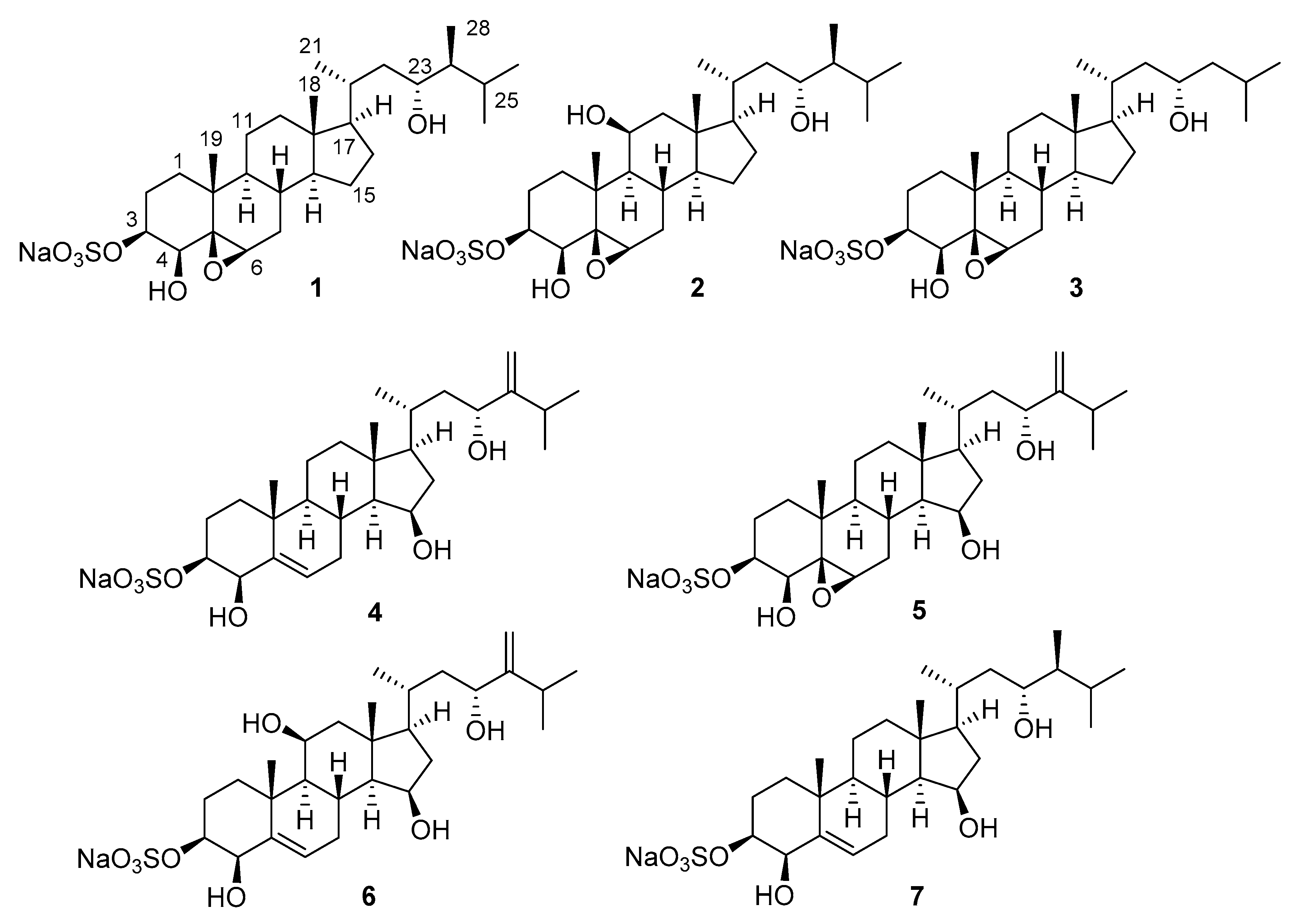
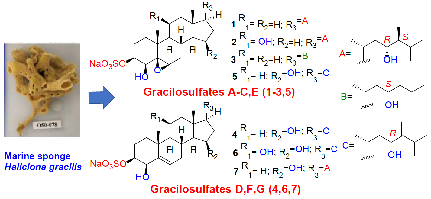
Gracilosulfates A–G, Monosulfated Polyoxygenated Steroids from the Marine Sponge Haliclona gracilis

by
Larisa K. Shubina
1,
Tatyana N. Makarieva
1,*,
Vladimir A. Denisenko
1,
Roman S. Popov
1,
Sergey A. Dyshlovoy
1,2,3,
Boris B. Grebnev
1,
Pavel S. Dmitrenok
1,
Gunhild von Amsberg
2,3 and
Valentin A. Stonik
1
1
G.B. Elyakov Pacific Institute of Bioorganic Chemistry, Far Eastern Branch of the Russian Academy of Sciences, Pr. 100-let Vladivostoku 159, 690022 Vladivostok, Russia
2
Department of Oncology, Hematology and Bone Marrow Transplantation with Section Pneumology, Hubertus Wald-Tumorzentrum, University Medical Center Hamburg-Eppendorf, 20251 Hamburg, Germany
3
Martini-Klinik, Prostate Cancer Center, University Hospital Hamburg-Eppendorf, 20251 Hamburg, Germany
*
Author to whom correspondence should be addressed.
Mar. Drugs 2020, 18(9), 454; https://doi.org/10.3390/md18090454
Submission received: 21 July 2020 / Revised: 21 August 2020 / Accepted: 27 August 2020 / Published: 30 August 2020
(This article belongs to the Collection Marine Compounds and Cancer)

Abstract

:
Seven new polyoxygenated steroids belonging to a new structural group of sponge steroids, gracilosulfates A–G (17), possessing 3β-O-sulfonato, 5β,6β epoxy (or 5(6)-dehydro), and 4β,23-dihydroxy substitution patterns as a common structural motif, were isolated from the marine sponge Haliclona gracilis. Their structures were determined by NMR and MS methods. The compounds 1, 2, 4, 6, and 7 inhibited the expression of prostate-specific antigen (PSA) in 22Rv1 tumor cells.

Graphical Abstract

1. Introduction

Marine organisms are known as a rich source of unique bioactive sulfate-containing metabolites [1]. Sulfated derivatives of different chemical classes (aliphatic compounds, steroids, terpenoids, carotenoids, aromatic compounds, alkaloids, carbohydrates, etc.) have been identified from them [1,2,3,4]. Marine invertebrates such as starfishes, ophiuroids, and ascidians contain mainly mono- and disulfated polyoxygenated steroids [1], which are almost exclusively marine secondary metabolites [1], while terrestrial sulfated polyoxygenated steroids are relatively rare. In fact, there is only one report concerning isolation of sulfated polyoxygenated steroid from plants [5].
Sulfated steroids represent one of the most numerous classes of sponge metabolites [1,2,3] Marine sponges provide a great structural diversity of bioactive sulfated polyoxygenated steroids, including nitrogen-containing [6], halogenated [7,8], monosulfated [9,10,11,12,13,14,15,16,17], disulfated [18], and trisulfated [7,8] steroids, as well as tetra- [19] or pentasulfated dimeric steroid derivatives [20]. The monosulfated polyoxygenated steroids account for only a part of these metabolites. Some of them show antimicrobial [13] and/or antifungal [10,13,15] and cytotoxic [10] activities or enhance glucose uptake via the AMPK signaling pathway [9].
During the search for bioactive compounds from the Northwestern Pacific deep-water marine invertebrates [21,22], we collected the pale orange sponge Haliclona gracilis near Shikotan Island, Russia, whose extract exhibited hemolytic and antifungal activities.
The genus Haliclona (order Haplosclerida, family Halinidae) is represented by more than 600 species [23]. Marine sponges of Haliclona genus have been extensively examined, and more than 200 various bioactive metabolites including steroids, alkaloids, macrolides, polyketides, cyclic peptides, long-chain sphingoid bases, merohexaprenoids, and cyclic bis-1,3-dialkylpyridinium salts have been isolated, and different activities, including cytotoxic and antitumor effects, have been reported [1].
Sponges of Haliclona genus have provided very few sulfated steroids [1]. Thus, only two trisulfated steroids have been isolated in one Indo-Pacific Haliclona sponge [17], while monosulfated polyoxygenated steroids have never been isolated from this genus. Moreover, thus far, the sponge H. gracilis has not been chemically investigated.
The 1H NMR analysis of the fractions obtained after diverse chromatographic separations suggested the presence of polar metabolites, inspiring our extensive investigation. Here, we report the details of the isolation and structure determination of compounds 17, belonging to a new group of naturally occurring monosulfated polyoxygenated steroids with a 3β-O-sulfonato, 5β,6β-epoxy (or 5(6)-dehydro), or 4β,23-dihydroxy substitution pattern as a common structural motif. Additionally, anticancer activities of 1, 2, 4, 6, and 7 were evaluated.

2. Results and Discussion

The concentrated EtOH extract of the sponge was partitioned between n-BuOH and H2O. The organic extract was concentrated and the obtained residue was fractionated by flash chromatography on a YMC gel column. Further separation using reversed-phase HPLC resulted in the isolation of seven new steroids, gracilosulfates A–G (17, Figure 1).
Compound 1 was isolated as a white, amorphous solid. The molecular formula of 1 was determined to be C28H47NaO7S from the [M − Na] ion peak at m/z 527.3045 in the (−)HRESIMS spectrum. The fragment ion peak at m/z 97.9606 in the (−)HRESIMS/MS spectrum and absorption band at 1213 cm−1 in the IR spectrum revealed the presence of a sulfate group in 1.
The 1H NMR spectrum of 1 (Table 1) showed signals attributable to six methyl groups at δH 1.16 (s), 0.94 (d), 0.91 (d), 0.82 (d), 0.74 (d), and 0.70 (s); four oxygen-bearing methine protons at δH 4.27 (d), 3.55 (br.d), 3.53 (br.d), and 3.17 (br.d); and a series of other methine and methylene multipletes. The 13C NMR (Table 2) and DEPT spectra of 1 revealed the presence of 28 signals, corresponding to 6 methyls, 8 methylenes, 11 methines, and 3 nonprotonated carbons (one bearing oxygen atom). These data evidenced a C-28 steroidal skeleton. Structure determination of 1 began with HMBC correlations from CH3-19 to C-1, C-5, C-9, and C-10. The COSY correlations (Figure 2) delineated the spin system H2-1 to H-4, which included protons of oxygenated methines at C-3 and C-4 based on their characteristic chemical shifts. The sequences of protons from H-6 to H-8, H-8 to H2-12, H-8 to H3-27, and H-24 to H3-28 were also established from COSY correlations and indicated the third oxymethine group at C-23. The cross peaks H-4/OH and H-23/OH in the COSY spectrum recorded in DMSO−d6 (Figure S11) and the 13C chemical shifts for C-4 (δC 77.7) and C-23 (δC 71.7) implied OH substitution, while the chemical shift for C-3 at δC 80.1 was more consistent with a sulfate half-ester O(SO3)Na [14]. The 13C NMR signals at δC 64.2 (CH) and 66.4 (C) and 1H NMR signal at δH 3.17 indicated the presence of trisubstituted epoxy ring [24]. The epoxy group was placed at C-5 and C-6 on the basis of HMBC correlations from H2-1, H-4, and H-19 to C-5 and from H-6 to C-4, C-8, and COSY correlations H-6/H2-7.
The large coupling constant of H-3 with H-2ax (J = 11.5 Hz) and small coupling constant of H-4 with H-3 (J = 3.0 Hz) pointed to β orientations for the 3-O(SO3)Na and 4-OH groups. The configuration of the 5β,6β-epoxy group was established by the NOESY correlations H-6/H-4. The same evidence was earlier used for β-orientation of epoxide group in a steroid from the soft coral Dendronephthya gigantea [25]. The key NOESY correlations H3-19/H-1β, H-2β, H-8, H-11β; H3-18/H-20, H-8, H-11β; H-9/H-1α, H-14; H-1α H-3α; H-4α/H-6; and H-17/H-14α, H3-21 confirmed the 3β,4β,5β,6β configurations of the oxygenated carbons and H-8β, H-9α, H-14α, and H-17α configurations of the ring portion in 1. The 20R configuration was demonstrated by the NOESY cross-peak H3-18/H-20 and chemical shift value of CH3-21 at δH 0.94 [26].
The absolute configuration at C-23 was assigned by application of the Mosher’s method. Esterification of 1 with (R)- and (S)-α-methoxy-α-(trifluoromethyl)-phenylacetyl chlorides (MTPACl) yielded the 23-MTPA adducts 1S and 1R, respectively, while C-4 hydroxy group was not modified. Interpretation of 1H NMR chemical shift differences Δδ between 1S and 1R (Figure 3) revealed that the absolute configuration of C-23 is R. The JH23/H24 coupling constant was 7.3 Hz, which indicated anti relationship of the H-23 and H-24 protons [27] (Figure 4). The NOESY cross peak for H2-22/H3-28 suggested the gauche relationship between the C-22 methylene and C-28 methyl groups, as shown in Figure 3. These data allowed us to determine the 24S absolute configuration.
Thus, the structure of 1 was defined as (20R,23R,24S)-4β,23-dihydroxy-5β,6β-epoxy-24- methylcholest-3β yl sulfate and was named gracilosulfate A.
The molecular formula of the second isolated compound, gracilosulfate B (2), was determined as C28H47NaO8S (m/z 543.3002 [M − Na]) on the basis of the negative ion HRESIMS analysis. The 1H NMR data of 2 resembled those of 1, except for the presence of an oxymethine group in 2H 4.14) instead of a methylene group for 1. Analysis of 1D and 2D NMR (COSY, HSQC, and HMBC) spectra allowed us to assign all the observed 1H and 13C signals for 2 (Table 1 and Table 2). The localization of the additional hydroxy group at C-11 followed from the HMBC correlations between H-11, C-8, and C-13 (Figure S16) and COSY data. The equatorial disposition of H-11 was evident from the small 3JHH vicinal coupling to H-9 and H2-12 (Table 1) and confirmed by the relatively low field shift of H-8 (Table 1) caused by the 1,3-diaxial relationship of this proton to the hydroxy group at C-11. The configurations of other stereogenic centers of the ring portion were assigned using similar principles used for 1. The similarity of the NMR data of the side chains of steroids 2 and 1 suggested the same (20R,23R,24S) configuration. Thus, gracilosulfate B was defined as (20R,23R,24S)-4β,11β,23-trihydroxy-5β,6β-epoxy-24-methylcholest-3β yl sulfate.
The molecular formula of gracilosulfate C (3), determined as C27H45NaO7S from HRESIMS data (m/z 513.2891[M–Na]), was one methylene unit less than that of 1. The spectroscopic properties of 3 were similar to those of 1 and differed only by the signals of steroid side chain (Table 1 and Table 2). A combination of 2D NMR data showed the lack of a C-28 methyl group, while the remaining portion of the molecule was intact in 1. The configuration of the ring moiety of 3 was assumed to be the same as that of 1 on the basis of the complete overlapping of proton and carbon resonances in NMR spectra. The configuration of the stereogenic center at C-23 was determined by the MTPA method as 23S (Figure 3). Thus, the gracilosulfate C was defined to be 24-demethyl derivative of gracilosulfate A (1), namely, (20R,23S)-4β,23-dihydroxy-5β,6β-epoxycholest-3β yl sulfate.
Gracilosulfate D (4) with a molecular formula C28H45NaO7S, confirmed by HRESIMS, was isolated as an optically active white amorphous solid. In addition to the signals relative to 3β-O-sulfonato-4β,23-dihydroxy structure, the 1H and 13C NMR spectra of 4 (Table 1 and Table 2) revealed signals of trisubstituted double bond (δH 5.70 and δC 144, 130.0), oxygenated methine group (δH 4.15 and δC 70.9), and terminal methylene group (δH 4.84, 5.03, and δC 106.8). The HMBC correlations between H3-19 and C-5 (δC 130.0), and between olefinic proton at δH 5.70 and C-4 (δC 77.6), were consistent with a double bond at C-5/C-6 position. The HMBC correlations from the oxymethine proton at δH 4.15 to C-13 and C-17 (Figure S32), in addition to COSY data (Figure S30), allowed placement of a hydroxy group at C-15 position, whereas the HMBC correlations between H2-28 and C-23, C-24, and C-25 confirmed the position of terminal methylene group at C-24. The coupling pattern associated with H-15 (ddd, J = 7.9, 5.8, 2.2 Hz) indicated that the hydroxy group at C-15 is β-positioned [28]. The configurations of other stereocenters of the steroid nucleus were assigned by NOESY (Figure 2) and coupling constants data (Table 1).
The absolute configuration at C-23 was deduced by theMTPA method. Treatment of 4 with (R)- and (S)- MTPACl yielded the corresponding 4,23-bis-MTPA adducts 4S and 4R, respectively. The Δδ values around the C-23 stereocenter between the adducts 4S and 4R (Figure 3) indicated the 23S configuration and, therefore, the structure of 4 was assigned as (20R, 23S)-4β,15β,23-trihydroxy-24-methylenecholest-5(6)-en-3βyl sulfate.
The molecular formula of C28H45NaO8S was assigned by HRESIMS to gracilosulfate E (5). The 1D (Table 1 and Table 2) and 2D NMR analysis showed that gracilosulfate E (5) differs from 4 in the 5,6-epoxy group, replacing trisubstituted double bond. The configurations of the ring moiety were assigned on the basis of the analyses of proton–proton coupling constants (Table 1) and NOESY data. The absolute configuration of the side chain of 5 was determined to be the same as in 4 by comparison of 1H and 13C chemical shifts. Thus, gracilosulfate E (5) was determined to be (20R,23R)-4β,15β,23-trihydroxy-5β,6β-epoxy-24-methylenecholest-3β yl sulfate.
Gracilosulfate F (6) of molecular formula C28H45NaO8S was a close analogue of gracilosulfate D (4) showing only an additional oxygen atom. Inspection of 1D (Table 1 and Table 2) and 2D NMR data allowed placement of an additional hydroxy group at C-11. The configuration at C-11 was deduced from NOESY correlation of H-11 to axial proton H-1 and small vicinal coupling constant of H-11 (Table 1), which is consistent with an equatorial disposition for this proton, thereby placing the hydroxy group in an axial position. The configurations of remaining stereogenic centers of the ring portion were the same as those of 4, as established on the basis of analyses of proton–proton coupling constants (Table 1 and Table 2) and NOESY data. The absolute configuration of the side chain was determined to be the same as that of 4 by comparison of 1H and 13C chemical shifts, and finally the structure of 6 was established as (20R, 23R)-4β,11β,15β,23-tetrahydroxy-24-methylenecholest-5(6)-en-3βyl sulfate.
Gracilosulfate G (7) showed the molecular formula C28H47NaO7S as determined by HRESIMS. On the basis of the results of the 1D NMR spectra, we were able to assign a trisubstituted double bond and four oxygen-bearing methine groups. The same steroid core constitution and configurations as in gracilosulfate D (4) were inferred from 1D (Table 1 and Table 2) and 2D NMR analysis. The proton and carbon resonances attributable to the side chain of 7 were coincident with those of 1 and 2 (Table 1 and Table 2). Thus, gracilosulfate G was defined as (20R, 23R, 24S)-4β,15β,23-trihydroxy-24-methylcholest-5(6)-en-3βyl sulfate.
Next, antitumor activity of compounds 1, 2, 4, 6, and 7 were determined in human prostate cancer cells 22Rv1. Of note, this cell line reveals resistance to androgen receptor (AR)-targeted therapy due to the expression of AR-V7 (AR transcript variant V7), which lacks the androgen-binding site [29,30]. The compounds exhibited moderate cytotoxic activity in the cancer cells after 48 h of treatment. Thus, compound 7 exhibited IC50 = 64.4 ± 14.9 µM, while the other tested compounds had IC50 > 100 µM (docetaxel was used as a positive control and exhibited IC50 = 17.3 ± 6.3 nM). However, all compounds were able to effectively inhibit the expression of PSA (prostate-specific antigen) in 22Rv1 cells (Figure 5). Earlier, only two monosulfated polyoxygenated steroids have been shown to exert cytotoxic activity on human cancer cell lines [10]. On the other hand, non-sulfated polyoxygenated steroid aragusterol with potent antitumor activities was isolated from a sponge of the genus Xestospongia [31]. Interestingly, for compounds 6 and 7, this effect was already detected at a concentration of 10 µM. PSA is a well-known downstream target of AR signaling. Thus, suppression of PSA expression may indicate an inhibition of this pathway. AR signaling is essential for the growth and survival of prostate cancer cells, with its targeting playing a central role in the modern therapy of advanced prostate cancer. The ability of the isolated compounds to suppress AR signaling can be explained by the similarity of their structures to androgen ligands, which may result in a binding to androgen receptors and therefore blocking of AR-mediated signaling in prostate cancer cells.

3. Materials and Methods

3.1. General Procedures

Optical rotations were measured using a PerkinElmer 343 polarimeter (Waltham, MA, USA). IR spectra were recorded using spectrophotometer Equinox 55 (Bruker, Ettlingen, Germany). The 1H and 13C NMR spectra were obtained using Bruker Avance III-700 and Bruker Avance III HD-500 spectrometers (Bruker, Ettlingen, Germany). Chemical shifts were referenced with Me4Si as an internal standard. ESI mass spectra (including HRESIMS) were measured using Bruker maXis Impact II mass spectrometer (Bruker Daltonics, Bremen, Germany). Low-pressure column liquid chromatography was performed using YMC Gel ODS-A (YMC Co., Ltd., Kyoto, Japan). HPLC was performed using Shimadzu Instrument equipped with RID-10A refractive index detector (Shimadzu Corporation, Kyoto, Japan) and YMC-Pack ODS-A (250 × 10 mm) column (YMC Co., Ltd., Kyoto, Japan).

3.2. Animal Material

Specimens of Haliclona gracilis were collected off the coast of Shikotan Island (43°28′0 N; 146°48′9 E) by dredging at 145 m depth on June 2017, and identified by Grebnev B. B. using the morphology of skeleton and spicules. Comparison the data of #050-078 with the corresponding characteristics of Haliclona gracilis and their complete coincidence supported the sponge identification as Haliclona gracilis [32]. A voucher specimen is deposited under registration number 050-078 in the collection of marine invertebrates of the Pacific Institute of Bioorganic Chemistry (Vladivostok, Russia).

3.3. Extraction and Isolation

The freshly collected specimens were immediately frozen and stored at −18 °C until use. Animal material (dry weight 20 g) were crushed and extracted with EtOH (2 × 1 L). The EtOH extract after evaporation in vacuo was partitioned between H2O and n-BuOH. The n-BuOH-soluble materials were partitioned with aqueous EtOH and n-hexane. The EtOH-soluble layer was fractioned by flash column chromatography on YMC gel ODS-A (75 μm), eluting with a step gradient of H2O – EtOH (100:0 − 20:80) with monitoring by HPLC. The fractions that eluted with 40% EtOH were further purified by repeated reversed-phase HPLC (YMC ODS-A column (250 × 10 mm), 1.5 mL/min, H2O-EtOH, 40:60 +1% AcONH4) to afford, in order of elution, compounds 6 (2 mg), 2 (3 mg), 4 (6 mg), 5 (1 mg), 3 (1 mg), 7 (4 mg), and 1 (8 mg) with retention times (tR) of 14.0, 17.5, 18.4, 22.5, 26.2, 32.5, and 36.1 min, respectively.

3.4. Compound Characterization Data

Gracilosulfate A (1): white, amorphous solid; [α] D 20 +6 (c 0.2, EtOH); IR (KBr) νmax 3467, 2957, 1457, 1242, 1002, 939 cm−1; 1H, 13C NMR, Table 1 and Table 2; HRESIMS m/z 527.3045 [M−Na] (calcd for C28H47O7S, 527.3048).
Gracilosulfate B (2): white, amorphous solid; [α] D 20 +22 (c 0.2, EtOH); IR (KBr) νmax 3446, 2947, 1457, 1242, 937 cm−1; 1H, 13C NMR, Table 1 and Table 2; HRESIMS m/z 543.3002 [M−Na] (calcd for C28H47O8S, 543.2997).
Gracilosulfate C (3): white, amorphous solid; [α] D 20 ≈0 (c 0.1, EtOH); IR (KBr) νmax 3465, 2960, 1450, 1240 cm−1; 1H, 13C NMR, Table 1 and Table 2; HRESIMS m/z 513.2891 [M−Na] (calcd for C27H45O7S, 513.2891).
Gracilosulfate D (4): white, amorphous solid; [α] D 20 −40 (c 0.2, EtOH); IR (KBr) νmax 3436, 2956, 1457, 1242, 1065, 998 cm−1; 1H, 13C NMR, Table 1 and Table 2; HRESIMS m/z 525.2890 [M−Na] (calcd for C28H45O7S, 525.2891).
Gracilosulfate E (5): white, amorphous solid; [α] D 20 ~0 (c 0.1, EtOH); IR (KBr) νmax 3440, 2938, 1457, 1241, 936 cm−1; 1H, 13C NMR, Table 1 and Table 2; HRESIMS m/z 541.2845 [M−Na] (calcd for C28H45O8S, 541.2841).
Gracilosulfate F (6): white, amorphous solid; [α] D 20 −17 (c 0.2, EtOH); IR (KBr) νmax 3456, 2942, 1457, 1242, 998 cm−1; 1H, 13C NMR, Table 1 and Table 2; HRESIMS m/z 541.2840 [M−Na] (calcd for C28H45O8S, 541.2841).
Gracilosulfate G (7): white, amorphous solid; [α] D 20 −32 (c 0.1, EtOH); IR (KBr) νmax 3440, 2932, 1653, 1457, 1240 cm−1; 1H, 13C NMR, Table 1 and Table 2; HRESIMS m/z 527.3057 [M − Na] (calcd for C28H47O7S, 527.3048).

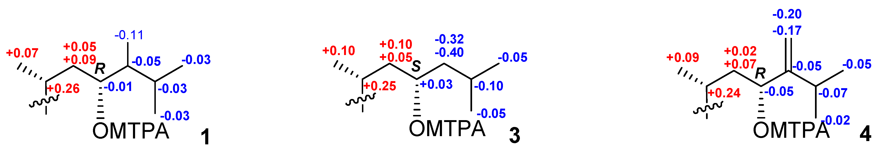
Preparation of MTPA esters of compounds 1, 3, and 4

To duplicate solutions of compound 1 (2 mg each) in 100 µL of anhydrous pyridine, we added (R)- or (S)-MTPACl (10 μL). After stirring for 30 min at rt, the reaction mixtures were concentrated under reduced pressure and separated by HPLC (YMC ODS-A column (250 × 10 mm), H2O-EtOH, 24:76 + 1% AcONH4) to afford the (S)- or (R)-MTPA esters of 1. The (S)- or (R)-MTPA derivatives of 3 and 4 were also prepared in a similar manner.
(S)-MTPA ester of 1 (1S): white, amorphous solid; 1H NMR (CD3OD, 500 MHz) δH 5.35 (1H, dd, J = 11.2, 4.7 Hz, H-23), 1.76 (1H, m, H-22), 1.52 (1H, m, H-24), 1.47 (1H, m, H-25), 1.39 (1H, m, H-20), 1.13 (1H, m, H-22), 0.99 (3H, d, J = 6.7 Hz, H-21), 0.94 (3H, d, J = 6.6 Hz, H-27), 0.86 (3H, d, J = 6.6 Hz, H-26), 0.76 (3H, d, J = 6.7 Hz, H-28), 0.62 (3H, s, H-18). HRESIMS m/z 779.3210 [M + Cl] (calcd for C38H55ClF3O9S, 779.3213).
(R)-MTPA ester of 1 (1R): white, amorphous solid; 1H NMR (CD3OD, 500 MHz) δH 5.36 (1H, dd, J = 11.2, 4.7 Hz, H-23), 1.69 (1H, m, H-22), 1.57 (1H, m, H-24), 1.50 (1H, m, H-25), 1.13 (1H, m, H-20), 1.04 (1H, m, H-22), 0.97 (3H, d, J = 6.6 Hz, H-27), 0.92 (3H, d, J = 6.7 Hz, H-21), 0.89 (3H, d, J = 6.6 Hz, H-26), 0.87 (3H, d, J = 6.7 Hz, H-28), 0.45 (3H, s, H-18). HRESIMS m/z 779.3210 [M + Cl] (calcd for C38H55ClF3O9S, 779.3213).
(S)-MTPA ester of3 (3S): white, amorphous solid; 1H NMR (CD3OD, 500 MHz) δH 5.33 (1H, m, H-23), 1.78 (1H, m, H-22), 1.49 (1H, septet, J = 6.6 Hz,, H-25), 1.41 (1H, m, H-20), 1.30 (1H, m, H-24), 1.19 (1H, m, H-22), 0.99 (3H, d, J = 6.5 Hz, H-21), 0.98 (1H, m, H-24), 0.90 (3H, d, J = 6.6 Hz, H-27), 0.87 (3H, d, J = 6.6 Hz, H-26), 0.63 (3H, s, H-18). HRESIMS m/z 765.3060 [M + Cl] (calcd for C37H53ClF3O9S, 765.3056).
(R)-MTPA ester of 3 (3R): white, amorphous solid; 1H NMR (CD3OD, 500 MHz) δH 5.30 (1H, m, H-23), 1.62 (1H, m, H-24), 1.68 (1H, m, H-22), 1.38 (1H, m, H-24), 1.59 (1H, septet, J = 6.6 Hz,, H-25), 1.16 (1H, m, H-20), 1.14 (1H, m, H-22), 0.89 (3H, d, J = 6.5 Hz, H-21), 0.95 (3H, d, J = 6.6 Hz, H-27), 0.92 (3H, d, J = 6.6 Hz, H-26), 0.42 (3H, s, H-18). HRESIMS m/z 765.3060 [M + Cl] (calcd for C37H53ClF3O9S, 765.3056).
Bis(S)-MTPA ester of 4 (4S): white, amorphous solid; 1H NMR (CD3OD, 500 MHz) δH 6.02 (1H, dd, J = 3.3, 1.1 Hz, H-4), 5.47 (1H, brd, J = 11.1 Hz, H-23), 4.87 (1H, t, J = 1.2 Hz, H-28), 4.84 (1H, brs, H-28), 1.91 (1H, m, H-22), 2.25 (1H, septet, J = 6.6 Hz, H-25), 1.67 (1H, m, H-20), 1.28 (1H, m, H-22), 1.10 (3H, d, J = 6.6 Hz, H-27), 1.03 (6H, d, J = 6.6 Hz, H-21, 26), 0.93 (3H, s, H-18). HRESIMS m/z 957.3675 [M − Na] (calcd for C48H59F6O11S, 957.3688).
Bis(R)-MTPA ester of 4 (4R): white, amorphous solid; 1H NMR (CD3OD, 500 MHz) δH 5.91 (1H, dd, J = 3.3, 1.1 Hz, H-4), 5.52 (1H, brd, J = 11.1 Hz, H-23), 5.07 (1H, t, J = 1.2 Hz, H-28), 5.01 (1H, brs, H-28), 2.32 (1H, septet, J = 6.6 Hz, H-25), 1.89 (1H, m, H-22), 1.43 (1H, m, H-20), 1.21 (1H, m, H-22), 0.94 (3H, d, J = 6.5 Hz, H-21), 1.12 (3H, d, J = 6.6 Hz, H-27), 1.08 (3H, d, J = 6.6 Hz, H-26), 0.55 (3H, s, H-18). HRESIMS m/z 957.3675 [M − Na] (calcd for C48H59F6O11S, 957.3688).

3.5. Bioactivity Assay

3.5.1. Reagents

The MTT reagent (thiazolyl blue tetrazolium bromide) was purchased from Sigma (Taufkirchen, Germany).

3.5.2. Cell Lines and Culture Conditions

The human prostate cancer cell line 22Rv1 was purchased from ATCC (Manassas, VA, USA). Cells were cultured according to the manufacturer’s instructions in RPMI media containing 10% FBS (Invitrogen, Carlsbad, USA). Cells were continuously kept in culture for a maximum of 3 months, and were routinely examined for stable phenotype and mycoplasma contamination.

3.5.3. In Vitro MTT-Based Drug Sensitivity Assay

The in vitro cytotoxicity of individual substances was evaluated using a MTT-based assay, which was performed as previously described [33]. Treatment time was 48 h.

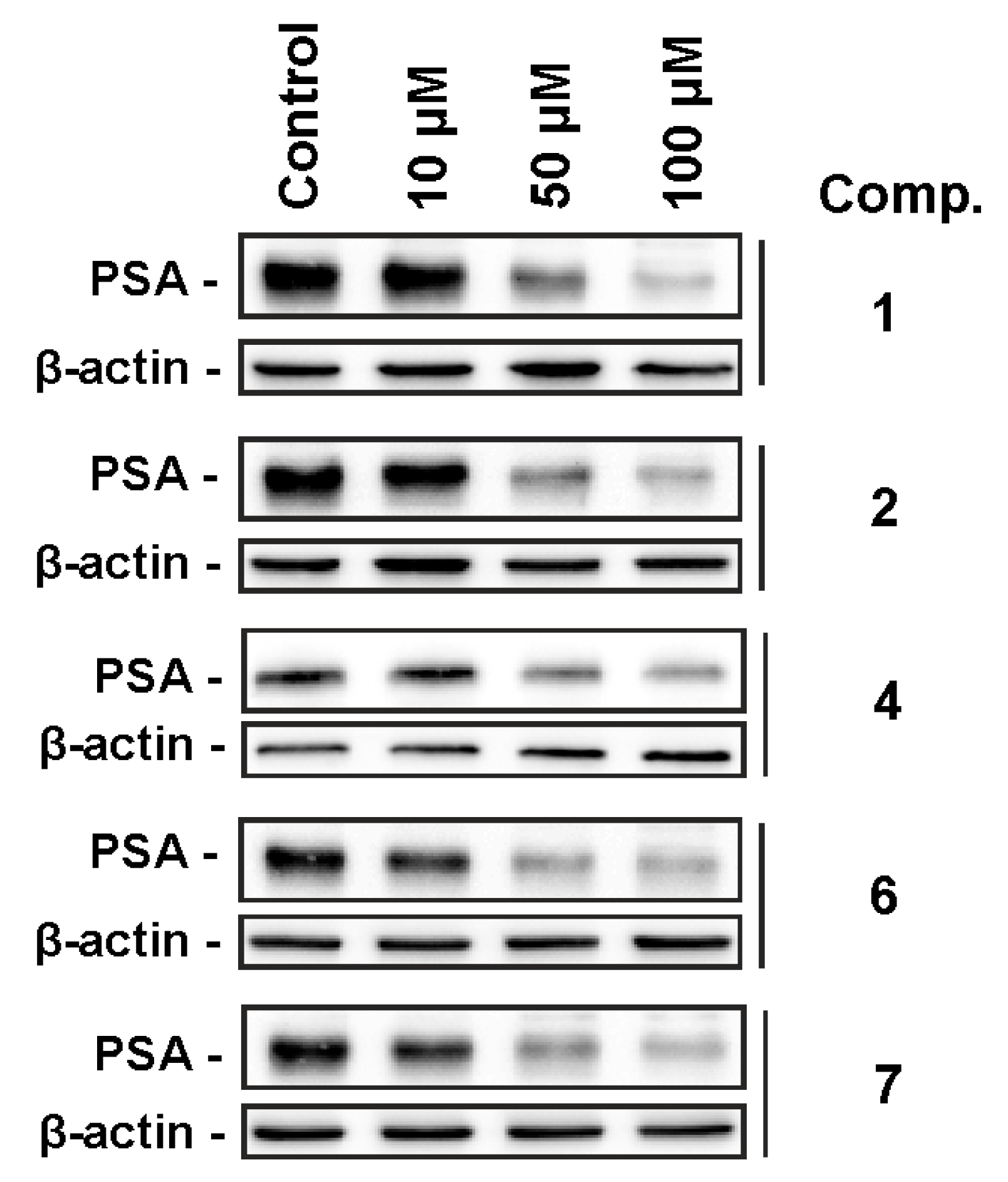
3.5.4. Western Blotting

Preparation of protein extracts and Western blotting were performed as described previously [34]. For the detection of PSA, expression the anti-PSA/KLK3 antibodies was used (Cell Signaling, #5365, 1:1000). Treatment time was of 24 h.

4. Conclusions

In summary, we isolated gracilosulfates A-G, new steroids from the marine sponge H. gracilis, possessing a rare 3β-O-sulfonato, 4β-hydroxy moiety [1]. To date, only one pregnane steroid [35] and two polyhydroxy steroids [36] with such a fragment have been isolated from the sponge Stylopus australis and the starfish Coscinasterias tenuispina, respectively. In addition, the 5β,6β epoxy fragment is unprecedented in sulfated steroids [1]. Finally, the combination of 3β-O-sulfonato, 5β,6β-epoxy (or 5(6)-dehydro), and 4β,23-dihydroxy moieties is unprecedented, taking into account structures of all previously known natural sulfated steroids. Interestingly, these compounds are able to inhibit PSA expression in human hormone-independent prostate cancer cells, suggesting inhibition of AR signaling, a central target for the treatment of advanced prostate cancer.

Supplementary Materials

The following are available online at https://www.mdpi.com/1660-3397/18/9/454/s1, Copies of HRESIMS, and 1D- and 2D-NMR spectra of 17, and photo of the marine sponge Haliclona gracilis (#050-078).

Author Contributions

L.K.S. isolated the metabolites; T.N.M. and L.K.S. elucidated structures; S.A.D. performed the bioactivity assays; V.A.D. performed the NMR spectra; R.S.P. and P.S.D. performed the mass spectra; B.B.G. performed species identification of the sponge; G.v.A. and V.A.S. assisted the results discussion; T.N.M., L.K.S., and V.A.S. wrote the paper, which was revised and approved by all the authors. All authors have read and agreed to the published version of the manuscript.

Funding

Isolation and establishment of chemical structures were partially supported by the RSF grant #20-14-00040 (Russian Science Foundation). Search for bioactive compounds from the Northwestern Pacific deep-water marine invertebrates was partially supported by the Grant of the Ministry of Science and Higher Education of the Russian Federation, grant #2020-1902-01-006.

Acknowledgments

The authors are thankful to Ms. Jessica Hauschild (University Medical Center Hamburg-Eppendorf) for assistance in the biological experiments. The study was carried out on the equipment of the Collective Facilities Center “The Far Eastern Center for Structural Molecular Research (NMR/MS) PIBOC FEB RAS.”

Conflicts of Interest

The authors declare no conflict of interest.

References

  1. Carroll, A.R.; Copp, B.R.; Davis, R.A.; Keyzers, R.A.; Prinsep, M.R. Marine Natural Products. Nat. Prod. Rep. 2020, 37, 175–223. [Google Scholar] [CrossRef]
  2. Carvalhal, F.; Correia-da-Silva, M.; Sousa, E.; Pinto, M.; Kijjoa, A. Sources and biological activities of marine sulfated steroids. J. Mol. Endocrinol. 2018, 61, T211–T231. [Google Scholar] [CrossRef] [PubMed] [Green Version]
  3. Kornprobst, J.M.; Sallenave, C.; Barnathan, G. Sulfated compounds from marine organisms. Comp. Biochem. Physiol. 1998, 119B, 1–51. [Google Scholar]
  4. Kellner Filho, L.C.; Picao, B.W.; Silva Marcio, L.A.; Cunha, W.R.; Pauletti, P.M.; Dias, G.M.; Copp, B.R.; Bertanha, C.S.; Januario, A.H. Bioactive aliphatic sulfates from marine invertebrates. Mar. Drugs 2019, 17, 527. [Google Scholar] [CrossRef] [PubMed] [Green Version]
  5. Xu, Y.-M.; Marron, M.T.; Seddon, E.; McLaughlin, S.P.; Ray, D.T.; Whitesell, L.; Gunatilaka, A.A.L. 2,3-Dihydrowithaferin A-3β-O-sulfate, a new potential prodrug of withaferin A from aeroponically grown Withania somnifera. Bioorg. Med. Chem. 2009, 17, 2210–2214. [Google Scholar] [CrossRef]
  6. Morinaka, B.I.; Pawlik, J.R.; Molinski, T.F. Amaranzoles B-F, imidazole-2-carboxy steroids from the marine sponge Phorbasam aranthus. C24-N- and C24-O-analogues from a divergent oxidative biosynthesis. J. Org. Chem. 2010, 75, 2453–2460. [Google Scholar] [CrossRef] [Green Version]
  7. Guzii, A.G.; Makarieva, T.N.; Denisenko, V.A.; Dmitrenok, P.S.; Burtseva, Y.V.; Krasokhin, V.B.; Stonik, V.A. Topsentiasterol sulfates with novel iodinated and chlorinated side chains from a marine sponge Topsentia sp. Tetrahedron Lett. 2008, 49, 7191–7193. [Google Scholar] [CrossRef]
  8. Tabakmakher, K.M.; Makarieva, T.N.; Denisenko, V.A.; Popov, R.S.; Dmitrenok, P.S.; Dyshlovoy, S.A.; Grebnev, B.B.; Bokemeyer, C.; von Amsberg, G.; Cuong, N.X. New trisulfated steroids from the vietnamese marine sponge Halichondria vansoesti and their PSA expression and glucose uptake inhibitory activities. Mar. Drugs 2019, 17, 445. [Google Scholar] [CrossRef] [Green Version]
  9. Woo, J.-K.; Ha, T.K.Q.; Oh, D.-C.; Oh, W.-K.; Oh, K.-B.; Shin, J. Polyoxygenated steroids from the sponge Clathria gombawuiensis. J. Nat. Prod. 2017, 80, 3224–3233. [Google Scholar] [CrossRef]
  10. Boonlarppradab, C.; Faulkner, D.J. Eurysterols A and B, cytotoxic and antifungal steroidal sulfates from a marine sponge of the genus Euryspongia. J. Nat. Prod. 2007, 70, 846–848. [Google Scholar] [CrossRef]
  11. Zhang, H.J.; Sun, J.B.; Lin, H.W.; Wang, Z.L.; Tang, H.; Cheng, P.; Chen, W.S.; Yi, Y.H. A new cytotoxic cholesterol sulfate from marine sponge Halichondria rugosa. Nat. Prod. Res. 2007, 21, 953–958. [Google Scholar] [CrossRef] [PubMed]
  12. Makarieva, T.N.; Stonik, V.A.; D’yachuk, O.G.; Dmitrenok, A.S. Annasterol sulfate, a novel marine sulfated steroid, inhibitor of glucanase activity from the deep-water sponge Poecillastra laminaris. Tetrahedron Lett. 1995, 36, 129–132. [Google Scholar] [CrossRef]
  13. Tsukamoto, S.; Matsunaga, S.; Fusetani, N.; Van Soest, R.W.M. Acanthosterol sulfates A−J:  Ten new antifungal steroidal sulfates from a marine sponge Acanthodendrilla sp. J. Nat. Prod. 1998, 61, 1374–1378. [Google Scholar] [CrossRef] [PubMed]
  14. Gabant, M.; Schmitz-Afonso, I.; Gallard, J.-F.; Menou, J.-L.; Laurent, D.; Debitus, C.; Al-Mourabit, A. Sulfated steroids: Ptilosteroids A−C and ptilosaponosides A and B from the Solomon Islands marine sponge Ptilocaulis spiculifer. J. Nat. Prod. 2009, 72, 760–763. [Google Scholar] [CrossRef]
  15. DiGirolamo, J.A.; Li, X.-C.; Jacob, M.R.; Clark, A.M.; Ferreira, D. Reversal of fluconazole resistance by sulfated sterols from the marine sponge Topsentia sp. J. Nat. Prod. 2009, 72, 1524–1528. [Google Scholar] [CrossRef] [Green Version]
  16. Radwan, M.M.; Manly, S.P.; Ross, S.A. Two new sulfated sterols from the marine sponge Lendenfeldia dendyi. Nat. Prod. Commun. 2007, 2, 901–904. [Google Scholar] [CrossRef] [Green Version]
  17. Sperry, S.; Crews, P. Haliclostanone sulfate and halistanol sulfate from an Indo-Pacific Haliclona sponge. J. Nat. Prod. 1997, 60, 29–32. [Google Scholar] [CrossRef]
  18. Fusetani, N.; Matsunaga, S.; Konosu, S. Bioactive marine metabolites II. Halistanol sulfate, an antimicrobial novel steroid sulfate from the marine sponge halichondria cf. moorei bergquist. Tetrahedron Lett. 1981, 22, 1985–1988. [Google Scholar] [CrossRef]
  19. Cheng, J.-F.; Lee, J.-S.; Sun, F.; Jares-Erijman, E.A.; Cross, S.; Rinehart, K.L. Hamigerols A and B, unprecedented polysulfate sterol dimers from the mediterranean sponge Hamigera hamigera. J. Nat. Prod. 2007, 70, 1195–1199. [Google Scholar] [CrossRef]
  20. Ushiyama, S.; Umaoka, H.; Kato, H.; Suwa, Y.; Morioka, H.; Rotinsulu, H.; Losung, F.; Mangindaan, R.E.P.; de Voogd, N.J.; Yokosawa, H.; et al. Manadosterols A and B, sulfonated sterol dimers inhibiting the ubc13-uev1a interaction, isolated from the marine sponge Lissodendryx fibrosa. Nat. Prod. 2012, 75, 1495–1499. [Google Scholar] [CrossRef]
  21. Guzii, A.G.; Makarieva, T.N.; Denisenko, V.A.; Gerasimenko, A.V.; Udovenko, A.A.; Popov, R.S.; Dmitrenok, P.S.; Golotin, V.A.; Fedorov, S.N.; Grebnev, B.B.; et al. Guitarrins A-E and Aluminumguitarrin A: 5-azaindoles from the northwestern pacific marine sponge Guitarra fimbriata. J. Nat. Prod. 2019, 82, 1704–1709. [Google Scholar] [CrossRef] [PubMed]
  22. Shubina, L.K.; Makarieva, T.N.; von Amsberg, G.; Denisenko, V.A.; Popov, R.S.; Dmitrenok, P.S.; Dyshlovoy, S.A. Monanchoxymycalin C with anticancer properties, new analogue of crambescidin 800 from the marine sponge Monanchora pulchra. Nat. Prod. Res. 2019, 33, 1415–1422. [Google Scholar] [CrossRef] [PubMed]
  23. Appeltans, W.; Boucher, P.; Boxshall, G.A.; De Broyer, C.; de Voogd, N.J.; Gordon, D.P.; Hoeksema, B.W.; Horton, T.; Kennedy, M.; Mees, J.; et al. (Eds.) World Register of Marine Species. 2012. Available online: http://www.marinespecies.org (accessed on 20 November 2019).
  24. Santafe´, G.; Paz, V.; Rodrıguez, J.; Jimenez, C. Novel cytotoxic oxygenated C29 sterols from the Colombian marine sponge Polymastia tenax. J. Nat. Prod. 2002, 65, 1161–1164. [Google Scholar] [CrossRef] [PubMed]
  25. Wu, J.; Xi, Y.; Huang, L.; Li, G.; Mao, Q.; Fang, C.; Shan, T.; Jiang, W.; Zhao, M.; He, W.; et al. A Steroid-type antioxidant targeting the Keap1/Nrf2/ARE signaling pathway from the soft coral Dendronephthya gigantea. J. Nat. Prod. 2018, 81, 2567–2575. [Google Scholar] [CrossRef]
  26. Vanderah, D.J.; Djerassi, C. Marine natural products. Synthesis of four naturally occurring 20β-H cholanic acid derivatives. J. Org. Chem. 1978, 43, 1442–1448. [Google Scholar] [CrossRef]
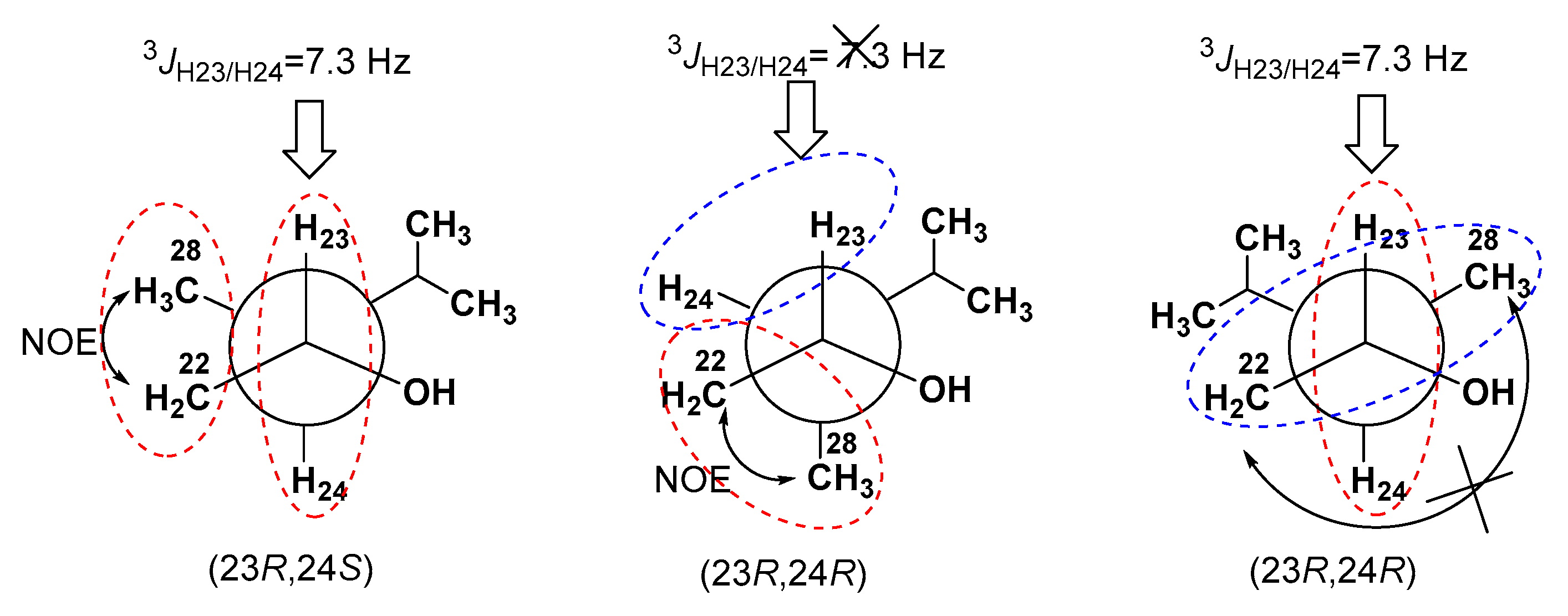
  27. Matsumori, N.; Kaneno, D.; Murata, M.; Nakamura, H.; Tachibana, K. Stereochemical determination of acyclic structures based on carbon-proton spin-coupling constants. A method of configuration analysis for natural products. J. Org. Chem. 1999, 64, 866–876. [Google Scholar] [CrossRef]
  28. Ivanchina, N.V.; Kicha, A.A.; Kalinovsky, A.I.; Dmitrenok, P.S.; Dmitrenok, A.S.; Chaikina, E.L.; Stonik, V.A.; Gavagnin, M.; Cimino, G. Polar steroidal compounds from the far eastern starfish Henricia Leviuscula. J. Nat. Prod. 2006, 69, 224–228. [Google Scholar] [CrossRef]
  29. Sampson, N.; Neuwirt, H.; Puhr, M.; Klocker, H.; Eder, I.E. In vitro model systems to study androgen receptor signaling in prostate cancer. Endocr. Relat. Cancer 2013, 20, R49–R64. [Google Scholar] [CrossRef] [Green Version]
  30. Nelson, P.S. Targeting the androgen receptor in prostate cancer—A resilient foe. N. Engl. J. Med. 2014, 371, 1067–1069. [Google Scholar] [CrossRef] [Green Version]
  31. Iguchi, K.; Fujita, M.; Nagaoka, H.; Mitome, H.; Yamada, Y. Aragusterol Aa potent antitumor marine steroid from the Okinawan sponge of the genus Xestospongia. Tetrahedron Lett. 1993, 34, 6277–6280. [Google Scholar] [CrossRef]
  32. Koltun, V.M. Demospongian Sponges of the Northern and Far-Eastern Seas; Publisher of USSR Academy of Sciences: Moscow, Russia; Leningrad, Russia, 1959; pp. 1–236. (In Russian) [Google Scholar]
  33. Dyshlovoy, S.A.; Venz, S.; Hauschild, J.; Tabakmakher, K.M.; Otte, K.; Madanchi, R.; Walther, R.; Guzii, A.G.; Makarieva, T.N.; Shubina, L.K.; et al. Antimigrating activity of marine alkaloid monanchocidin A, proteome-based discovery and confirmation. Proteomics 2016, 16, 1590–1603. [Google Scholar] [CrossRef] [PubMed]
  34. Dyshlovoy, S.A.; Tabakmakher, K.M.; Hauschild, J.; Shchekaleva, R.K.; Otte, K.M.; Guzii, A.G.; Makarieva, T.N.; Kudryashova, E.K.; Shubina, L.K.; Bokemeyer, C.; et al. Anticancer activity of eight rare guanidine alkaloids isolated from marine sponge Monanchora pulchra. Mar. Drugs 2016, 14, 133. [Google Scholar] [CrossRef] [PubMed]
  35. Prinsep, M.R.; Blunt, J.W.; Munro, M.H.G. A new sterol sulfate from the marine sponge Stylopus australis. J. Nat. Prod. 1989, 52, 657–659. [Google Scholar] [CrossRef]
  36. Riccio, R.; Iorizzi, M.; Minale, L. Starfish saponins XXX. Isolation of sixteen steroidal glycosides and three polyhydroxysteroids from the mediterranean starfish Coscinasterias tenuispina. Bull. Soc. Chim. Belg. 1986, 95, 869–893. [Google Scholar] [CrossRef]
Figure 1. The structures of 17.
Marinedrugs 18 00454 g001
Figure 2. Key COSY and HMBC correlations for 1, and NOESY correlations for 1 and 4.
Marinedrugs 18 00454 g002
Figure 3. Δδ values (δSδR) for 23(S)- and 23(R)-MTPA esters of compounds 1, 3, and 4.
Marinedrugs 18 00454 g003
Figure 4. J-based configuration analysis and NOESY data of compound 1.
Marinedrugs 18 00454 g004
Figure 5. Effects of the compounds on PSA expression in 22Rv1 cells. The cells were treated with the compounds for 24 h, then the proteins were extracted and examined using Western blotting. β-actin was used as a loading control.
Marinedrugs 18 00454 g005
Table 1. 1H NMR data for compounds 17 in CD3OD.
Position1234567
δH (J in Hz)δH (J in Hz)δH (J in Hz)δH (J in Hz)δH (J in Hz)δH (J in Hz)δH (J in Hz)
1.39, m1.37, m1.37, m1.15, m1.37, m1.31, m1.15, m
1.05, m2.10, m2.06, m1.90, m2.07, m2.12, dt (13.3, 3.3)1.89, dt (13.3, 3.3)
1.82, m1.84, m1.82, m1.81, m1.83, m1.84, m1.82, m
2.10, m2.12, m2.10, m2.11, m2.11, m2.21, m2.12, m
34.27, ddd (11.5, 4.1, 3.0)4.29, ddd (11.5, 4.1, 3.0)4.27, ddd (11.5, 4.1, 3.0)4.18, ddd (12.2, 4.3, 3.3)4.28, ddd (11.5, 4.1, 3.0)4.19, ddd (12.0, 3.9, 3.1)4.18, ddd (11.7, 4.0, 3.1)
43.55, br d (3.0)3.58, br d (3.0)3.56, br d (3.0)4.42, dd (3.3, 1.3)3.57, br d (3.0)4.40, dd (3.3, 1.3)4.42, dd (3.3, 1.3)
5
63.17, br d (2.7)3.12, br d (2.7)3.17, br d (2.5)5.70, dd (5.0, 2.4)3.19, br d (2.5)5.59, dd (4.2, 3.0)5.70, dd (5.0, 2.4)


1.29, m
 
 
2.08, m
1.35, m
 
 
2.19, m
1.27, m
 
 
2.08, m
1.68, ddd (18.2, 10.3, 2.3)
 
2.40, m
 
1.32, m
 
 
2.39, m
1.80, ddd (18.0, 9.8, 2.6)
2.53, ddd (18.8, 6.6, 4.3)
1.68, ddd (18.0, 10.3, 2.3)
2.39, m
81.43, m1.79, m1.42, m1.99, dd (10.8, 5.7)1.85, m2.42, m1.99, m
90.68, dd (12.0, 4.5)0.78 dd, (11.5, 3.0)0.68, dd, (11.6, 4.7)0.98, m0.74, m1.01, m0.98, m
11α
11 β
1.40, m
1.43, m
4.14, br q, (3.0)1.40, m
1.44, m
1.50, m
1.52, m
1.38, m
1.43, m
4.29, br q, (3.4)1.49, m
1.51, m
12α1.14, m1.30, m1.12, m1.18, m1.10, m1.36, m1.18, m
12 β2.02, m2.23, dd, (13.3, 3.0)2.02, m2.03, dt (12.6, 3.6)1.97, dt (12.3, 3.7)2.22, m2.03, dt (12.5, 3.3)
13
140.95, m0.94, m0.94, m0.90, m0.78, dd (11.3, 5.7)0.89, m0.90, m
151.64, m
1.05, m
1.65, m
1.15, m
1.64, m
1.06, m
4.15, ddd (7.9, 5.8, 2.2)4.15, ddd (8.1, 5.7, 2.3)4.18, ddd (7.8, 5.7, 2.2)4.15, ddd (7.7, 5.6, 2.0)
161.86, m
1.36, m
1.83, m
1.39, m
1.86, m
1.35, m
2.40, m
1.41, ddd (14.3, 10.4, 2.3)
2.36, m
1.37, m
2.37, m
1.41, m
2.40, m
1.39, m
171.08, m1.03, m1.08, m1.05, m1.02, m1.00, m1.07, m
180.70, s0.93, s0.69, s1.00, s0.94, s1.20, s0.99, s
191.16, s1.43, s1.16, s1.24, s1.19, s1.49, s1.24, s
201.73, m1.72, m1.72, m1.88, m1.86, m1.88, m1.87, m
210.94, d (6.7)0.96, d (6.7)0.95, d (6.7)1.02, d (6.7)0.99, d (6.7)1.04, d (6.7)0.97, d (6.7)
221.41, m
1.04, m
1.39, m
1.03, m
1.48, m
0.98, m
1.59, ddd (13.7, 10.3, 2.3)
1.11, m
1.57, ddd (14.1, 10.5, 2.7)
1.10, m
1.58, ddd (14.1, 10.5, 2.5)
1.11, m
1.43,
1.07, m
233.53, ddd (9.1, 7.3, 2.0)3.53, ddd (9.4, 7.3, 2.0)3.70, m4.13, br d (10.5)4.11, br d (10.5)4.13, br d (10.5)3.55, ddd (9.3, 7.1, 2.0)
241.29, m1.28, m1.38, m
1.14, m
1.31, m
251.91, m1.91, m1.75, m2.26, septet (6,7)2.24, septet (6,7)2.25, septet (6,7)1.93, m
260.82, d (6.6)0.82, d (6.6)0.90, d (6.8)1.06, d (6.9)1.05, d (6.8)1.06, d (6.8)0.83, d (6.9)
270.91, d (6.6)0.91, d (6.6)0.91, d (6.8)1.08, d (6.9)1.07, d (6.8)1.08, d (6.8)0.92, d (6.9)
280.74, d (6.8)0.74, d (6.8) 5.03, t (1.2)
4.84, br s
5.03, t (1.2)
4.84, br s
5.03, t (1.2)
4.84, br s
0.75, d
Table 2. 13C NMR dataa for compounds 1–7 in CD3OD.
Position1234567
δC, TypeδC, TypeδC, TypeδC, TypeδC, TypeδC, TypeδC, Type
139.2, CH240.2, CH239.2, CH239.2, CH239.2, CH238.2, CH239.4, CH2
223.7, CH224.1, CH223.7, CH224.6, CH223.7, CH224.0, CH224.4, CH2
380.1, CH79.9, CH80.1, CH82.5, CH80.1, CH82.0, CH82.1, CH
477.7, CH78.0, CH77.7, CH77.6, CH77.7, CH76.9, CH77.6, CH
566.4, C66.2, C66.4, C144.1, C66.3, C145.4, C144.1, C
664.2, CH63.3, CH64.2, CH130.0, CH64.2, CH129.5, CH130.0, CH
734.2, CH233.6, CH234.2, CH233.0, CH233.6, CH233.2, CH233.0, CH2
831.7, CH28.8, CH31.7, CH29.4, CH27.4, CH26.4, CH29.5, CH
953.9, CH57.8, CH53.9, CH52.9, CH54.3, CH56.2, CH59.2, CH
1036.9, C37.9, C36.9, C38.0, C36.9, C38.6, C38.1, C
1123.1, CH269.5, CH23.1, CH222.1, CH23.1, CH269.5, CH22.2, CH
1241.8, CH251.1, CH241.8, CH243.0, CH243.1, CH252.3, CH243.0, CH2
1344.1, C43.7, C44.1, C44.0, C43.9, C43.4, C44.0, C
1458.1, CH61.2, CH58.1, CH63.5, CH62.8, CH65.6, CH63.6, CH
1525.9, CH225.7, CH225.9, CH271.3, CH70.9, CH71.3, CH71.2, CH
1629.8, CH229.7, CH229.8, CH242.7, CH242.6, CH242.3, CH242.6, CH2
1759.0, CH59.8, CH58.9, CH59.1, CH59.0, CH59.9, CH59.4, CH
1812.8, CH316.1, CH312.8, CH315.7, CH315.4, CH318.1, CH315.6, CH3
1919.1, CH321.8, CH319.1, CH322.0, CH319.0, CH325.3, CH321.9, CH3
2034.1, CH34.2, CH34.1, CH34.2, CH34.2, CH34.3, CH33.9, CH
2119.5, CH319.5, CH319.7, CH319.6, CH319.5, CH319.5, CH319.7, CH3
2241.7, CH241.8, CH246.2, CH245.0, CH245.1, CH245.1, CH241.9, CH2
2371.7, CH71.8, CH68.1, CH72.5, CH72.5, CH72.6, CH71.8, CH
2447.4, CH47.4, CH49.5, CH162.4, C162.3, C162.4, C47.4, CH
2529.6, CH29.6, CH26.4, CH32.2, CH32.1, CH32.2, CH29.6, CH
2622.5, CH322.5, CH324.4, CH324.4, CH324.4, CH324.4, CH322.5, CH3
2718.3, CH318.4, CH323.3, CH323.6, CH323.7, CH323.7, CH318.4, CH3
2811.3, CH311.3, CH3 106.8, CH2106.8, CH2106.8, CH211.4, CH3
a Assignments were confirmed by HSQC and HMBC (8Hz) data.

