Article

Antifouling Activity of Halogenated Compounds Derived from the Red Alga Sphaerococcus coronopifolius: Potential for the Development of Environmentally Friendly Solutions

1
Laboratoire des Sciences de l’Environnement Marin (LEMAR), Université de Brest, CNRS, IRD, Ifremer, F-29280 Plouzané, France
2
Section of Pharmacognosy and Chemistry of Natural Products, Department of Pharmacy, National and Kapodistrian University of Athens, Panepistimiopolis Zografou, 15771 Athens, Greece
3
Institute of Applied Biosciences, Centre for Research & Technology, Hellas (CERTH), 570 01 Thessaloniki, Greece
*
Authors to whom correspondence should be addressed.
Mar. Drugs 2022, 20(1), 32; https://doi.org/10.3390/md20010032
Submission received: 2 December 2021 / Revised: 23 December 2021 / Accepted: 23 December 2021 / Published: 28 December 2021
(This article belongs to the Special Issue Marine Natural Products with Antifouling Activity, 2nd Edition)

Abstract

:
Nowadays, biofouling is responsible for enormous economic losses in the maritime sector, and its treatment with conventional antifouling paints is causing significant problems to the environment. Biomimetism and green chemistry approaches are very promising research strategies for the discovery of new antifouling compounds. This study focused on the red alga Sphaerococcus coronopifolius, which is known as a producer of bioactive secondary metabolites. Fifteen compounds, including bromosphaerol (1), were tested against key marine biofoulers (five marine bacteria and three microalgae) and two enzymes associated with the adhesion process in macroalgae and invertebrates. Each metabolite presented antifouling activity against at least one organism/enzyme. This investigation also revealed that two compounds, sphaerococcinol A (4) and 14R-hydroxy-13,14-dihydro-sphaerococcinol A (5), were the most potent compounds without toxicity towards oyster larvae used as non-target organisms. These compounds are of high potential as they are active towards key biofoulers and could be produced by a cultivable alga, a fact that is important from the green chemistry point of view.

1. Introduction

In the marine environment, all untreated man-made submerged surfaces are colonized by fouling organisms, such as bacteria, algae and invertebrates, including barnacles and mussels [1,2]. This phenomenon is defined as biofouling [3,4]. It is a natural process by which species whose life stage requires a fixed mode will adhere to surfaces in either a reversible or irreversible way. Biofouling is the focus of many studies, particularly to understand the attachment processes of invertebrate larvae or algal spores/zygotes. Actually, in the context of increasing mariculture’s activities, understanding these processes will allow for the improvement in sustainability of the aquaculture sector [5,6]. In the marine environment, there is very strong ecological pressure and competition during the adhesion phases, as the number of organisms that need to adhere is much higher than the available surfaces. Thus, many marine organisms have developed the mechanisms to adhere not only to natural substrates but also to artificial surfaces. In the latter case, unwanted colonization leads to deterioration and corrosion of these surfaces, resulting in significant economic losses [1,7]. Thus, biofouling has been the subject of continuous multidisciplinary research, particularly in maritime fields related to transportation (protection of hulls), marine energy (protection of structures), aquaculture (protection of nets and cages), infrastructure (jetties, dinghies, harbors) and water intakes and cooling circuits of thermal power plants [8,9].
The colonization of ship hulls has been extensively studied. Thus, marine biofoulers are classified in three categories, based on their impact on the increase of frictional drag (FD) of man-made immersed surfaces: (i) microfoulers which are responsible for 1–2% increase in FD (bacterial, fungal and microalgal biofilms); (ii) soft macrofoulers which are responsible for up to 10% in FD (macroalgae); and (iii) hard macrofoulers leading to up to 40% increase in FD (barnacles, etc) [1].
To prevent the settlement and growth of biofoulers and/or to reduce their impact on man-made surfaces, antifouling coatings have been extensively used. These paint matrices are formulated with heavy metals (copper and zinc mainly) and co-biocides. In the 1960s, the chemical industry developed efficient antifouling paints based on tributyltin (TBT) and triphenyltin (TPT). Unfortunately, these two chemicals appeared highly toxic for aquatic organisms and have been proven to bioaccumulate through the food chain and be persistent in the environment and as such were banned in 2008 [10]. Since then, TBT-based paints were replaced by new formulations containing copper or biocides, such as diuron and irgarol [10,11,12,13]. To increase their activity, inorganic zinc is often added to the formulations [14]. However, even if these new formulations were initially claimed to be environmentally friendly, there is now evidence of a widespread use of these compounds leading to significantly high concentrations measured in marinas and harbors [15,16].
Nowadays, it is an imminent necessity to develop non-toxic antifouling formulations, eliminating or minimizing the use of heavy metals and organic biocides. A fascinating concept of scientific inspiration is the combination of biomimetic and green chemistry approaches. Indeed, many marine organisms that live attached and lack physical defenses have evolved chemical defenses that allow them to protect their surfaces from epibionts. Thus, secondary metabolites with significant levels of anti-epibiosis activity have been isolated and could be incorporated in antifouling paints [17,18,19,20]. These molecules have the advantage of exhibiting targeted activities and acting at very low concentrations, and thus their use in paint compositions is promising from this point of view [2,21,22,23,24,25]. The ideal solution would be to use natural products to prevent the settlement of fouling organisms rather than inhibiting their growth [2,21,22,23,24,25]. Settlement inhibitors are not considered as biocides, and thus regulations for their market authorization are less restrictive, allowing for faster development and deployment. Developing the use of natural products is also an important aspect of green chemistry, allowing the reduction of the environmental and health impacts of chemical production. It is essential to strive to increase the use of renewable raw materials, to design antifouling paints based on safe and environmentally friendly substances, allowing for the reduction of by-products and degradation products to avoid pollution.
In search of new natural compounds, the marine environment appears as a prolific source for novel bioactive natural products. Actually, marine macro- and microorganisms produce a wide and diverse range of secondary metabolites with promising bioactivities [26,27,28]. Among them, algae are well-known for their capacity to produce molecules with anticancer, antifouling, antibacterial or anti-inflammatory activity [29,30,31].
We have been working for many years on the chemistry and chemical ecology of macroalgae, and we are particularly interested in the defense molecules against epibiosis. Among these biotechnologically interesting algae, the cosmopolitan red alga Sphaerococcus coronopifolius is a prolific source of secondary metabolites, mainly diterpenoids. Among these diterpenes, bromosphaerol (1) has been proven as a promising antifoulant, surpassing the strictest standards [32]. In these studies, bioassays conducted on the cirriped crustacean Amphibalanus amphitrite showed that bromosphaerol (1) exerted significant anti-settlement activity with an EC50 value of 0.23 mg/L, combined with extremely low toxicity (LC50 > 100 mg/L), with an astonishing therapeutic ratio (TRC = LC50/EC50) of 434.78.
The aim of this study was to further investigate the antifouling potential of bromosphaerol (1) and other major metabolites isolated from the red alga S. coronopifolius against prominent marine biofoulers (five marine bacteria and three microalgae) and two enzymes associated with adhesion process in macroalgae and invertebrates.

2. Results and Discussion

In order to broaden the knowledge on the mode of action and ecological roles of molecules isolated from S. coronopifolius, we studied the epibiosis defense activities of 15 secondary metabolites isolated from this alga (115) on biofouling model organisms. This fundamental research can have important applications in the marine biotechnology sector with the discovery of new molecules of interest in the field of antifouling paints’ formulation.

2.1. Isolation and Identification of Metabolites from Sphaerococcus coronopifolius

A series of chromatographic separations of the CH2Cl2 / MeOH extract of the red alga S. coronopifolius, collected in Agios Giannis bay off Parga in Greece, resulted in the isolation of 14 diterpenes (114) and one sesquiterpene (15) as the major constituents. By comparison of their spectroscopic and physical characteristics with those reported in the literature, the isolated metabolites were identified as bromosphaerol (1) [33,34], 12S-hydroxy-bromosphaerol (2) [35,36], 12R-hydroxy-bromosphaerol (3) [36,37], sphaerococcinol A (4) [38,39], 14R-hydroxy-13,14-dihydro-sphaerococcinol A (5) [36], 8-methoxy-dihydro-sphaerococcenol (6) [40], 2S-hydroxy-isobromosphaerol (7) [41], bromosphaerol B (8) [36], bromosphaerodiol (9) [42], 1S-hydroperoxy-12S-hydroxy-bromosphaerodiol B (10) [36], 1S-hydroperoxy-12R-hydroxy-bromosphaerodiol B (11) [36], 1S-hydroxy-1,2-dihydro-bromosphaerol (12) [41,43], bromotetrasphaerol (13) [37], 12R-hydroxy-bromocorodienol (14) [44] and allo-aromadendrene (15) [45] (Figure 1).

2.2. Evaluation of Antifouling Activity

Antifouling tests were conducted on bacteria, microalgae and enzymes related to macroalgae and invertebrates’ adhesion [46]. It is essential to work on the three biological compartments (microorganisms, macroalgae and invertebrates) because despite established installation sequences (in chronological order, microorganisms, macroalgae and then invertebrates), it has been previously highlighted that the inhibition of a single type of organism does not prevent the colonization of surfaces by other biofoulers. It is therefore essential to have an integrated approach, in terms of both complementary bioassays and types of molecules. Indeed, marine organisms use combinations of antifouling defenses that act synergistically, making it important to evaluate the activity of molecules as many as possible isolated from the same organism [47].
The choice of biotests (target organisms and types of bioassays) is an essential step in bioprospecting projects and has a crucial impact on the analysis of results [48]. Thus, it is important to carefully think about the desired mode of action and, if necessary, to work on the development of innovative methods to evaluate the activity. Within the framework of the development of new antifouling solutions that are eco-friendly and in order to avoid secondary effects on non-target organisms as much as possible, the biological activity sought is not a biocidal activity (as it was usually done) but an adhesion-inhibitory or deterrent activity [2,21,22,23,24,25]. This approach is also interesting in the context of marketing authorization applications. Indeed, these are very long, strict and complex processes in the case of biocides (same legislation as for pesticides) but much simpler in the case of adhesion inhibitors. Thus, for this work, we focused on the evaluation of modes of action involving adhesion inhibition. We also favored an ethical approach by minimizing tests on living organisms. To do so, biochemical assays targeting enzymes involved in the synthesis of adhesives by algae and invertebrates were chosen, i.e., bromoperoxidase and phenoloxidase, respectively.

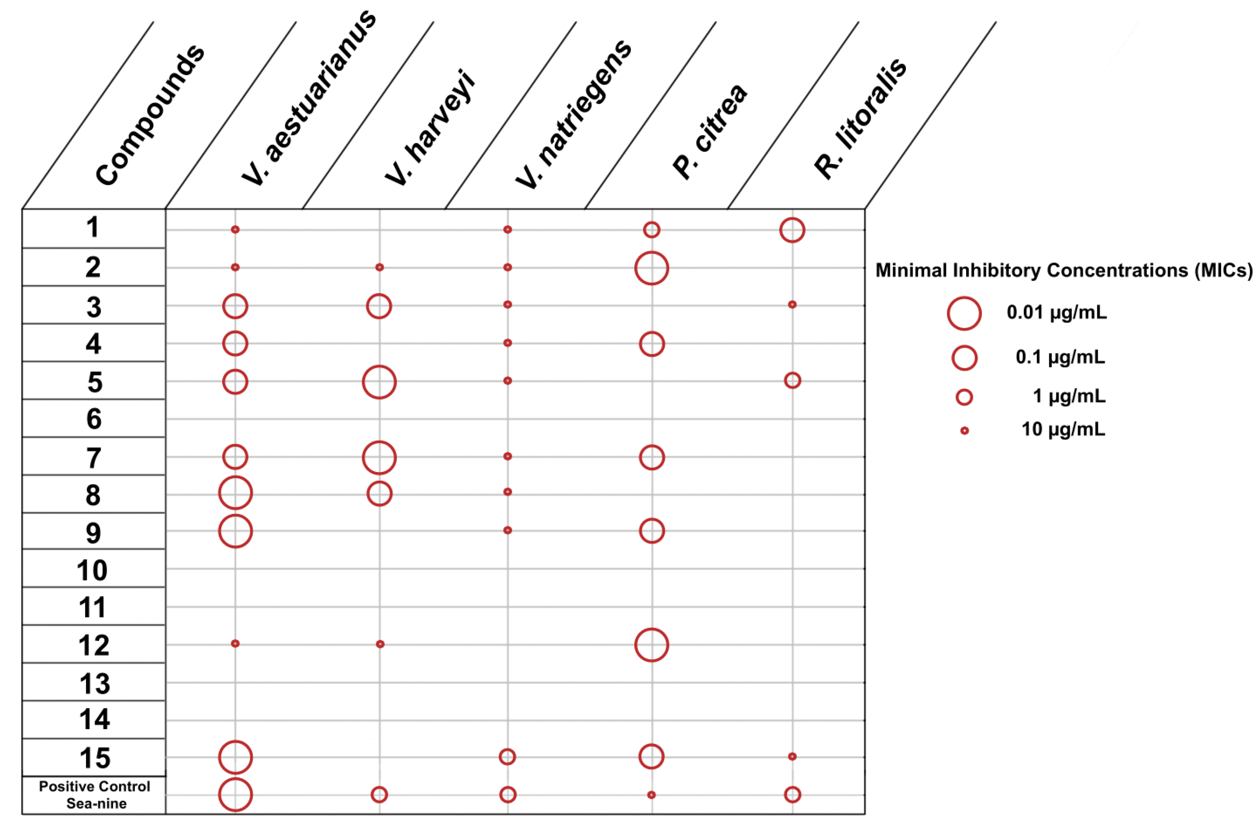
2.2.1. Inhibition of Adhesion and Growth of Marine Bacteria and Microalgae

The initial set of experiments was designed to evaluate the inhibition of adhesion of five marine bacteria, namely Vibrio aestuarianus, Vibrio harveyi, Vibrio natriegiens, Pseudoalteromonas citrea and Roseobacter litoralis, and three microalgae, namely Cylindrotheca closterium, Exanthemachrysis gayraliae and Halamphora coffeaeformis. The obtained results were expressed as Minimal Inhibitory Concentrations (MICs) (Figure 2 and Figure 3).
Concerning the evaluation of compounds 115 against marine bacteria, among the 15 tested compounds, only compound 11 did not show activity. All other compounds inhibited at least two marine bacteria, with one (2), three (1, 6, 12), five (3, 5, 8, 9, 13) and five (4, 7, 10, 14, 15) molecules inhibiting two, three, four and all five marine bacteria, respectively (Figure 2). Compounds 4, 7, 10, 14 and 15, which showed inhibition of adhesion against all five marine pathogens were active only at the highest concentration (10 µg/mL). In contrast, compounds 1, 6 and 12, which inhibited the adhesion of three biofoulers could be considered as the most effective since most of the MIC values observed were below 0.1 µg/mL. R. litoralis, which faced the least impact on its growth, appeared the most sensitive, with 13 molecules inhibiting its adhesion. It is interesting to note that SeaNine is less efficient towards adhesion inhibition than the most active compounds and was not active at all against P. citrea and R. littoralis.
Among the 15 compounds tested for the inhibition of adhesion of microalgae, only compound 10 showed activity against all three microalgae (Figure 3). Only 14 responses affecting the adhesion of microalgae (compounds exhibiting activity x tested microalgae) were observed, of which half (for 2, 4, 7, 911) were recorded with MIC values of 10 µg/mL. E. gayraliae was the least impacted microalga, as only compound 10 was active towards its adhesion. SeaNine was very effective towards the inhibition of adhesion of microalgae, but it is known that the mode of activity is through toxicity.
The same set of metabolites was evaluated for their activity against the growth of the same five marine bacteria and three microalgal species (Figure 4 and Figure 5).
According to the results (Figure 4), compounds 6, 10, 11, 13 and 14 did not show any activity against the growth of any of the tested marine bacteria. However, compounds 13, 5, 7 and 15 showed activity against four out of the five marine bacteria tested, while compounds 4, 8, 9 and 12 inhibited three out of five marine bacteria. The most potent compounds with the lowest MIC values were compounds 5, 7 and 15. Among marine bacteria, R. litoralis was the most resistant, with its growth affected by compounds 3 and 15 at high concentrations (10 µg/mL) and compounds 1 and 5 at low concentrations (0.1 and 1 µg/mL, respectively). On the contrary, V. aestuarianus and V. natriegens were the most impacted, being susceptible to 10 and 9 compounds, respectively. Among them, V. aestuarianus appeared more sensitive than V. natriegens, since seven metabolites were active with MIC values ≤ 0.1 µg/mL. It is interesting to note that some compounds were more active than the positive control, which confirms the great potential of the algal metabolites for inhibition of bacterial growth.
Furthermore, all tested compounds inhibited the growth of at least one microalga with three (1, 9, 15), six (2, 4, 6, 1214) and six (3, 5, 78, 1011) molecules inhibiting one, two and all three microalgae, respectively (Figure 5). Among the 33 responses affecting growth of microalgae (compounds exhibiting activity x tested microalgae), 9 were recorded with MIC values of 0.1 µg/mL and 17 with MIC values of 0.01 µg/mL. Compounds 5 and 10 appeared to be the most effective in inhibiting the growth of the tested microalgae, closely followed by compound 4. SeaNine toxicity towards microalgae was confirmed. The level of inhibition towards all the strains tested demonstrated that this compound is extremely deleterious to all photosynthetic organisms and that it is urgent to find alternative compounds with a more specific mode of action.

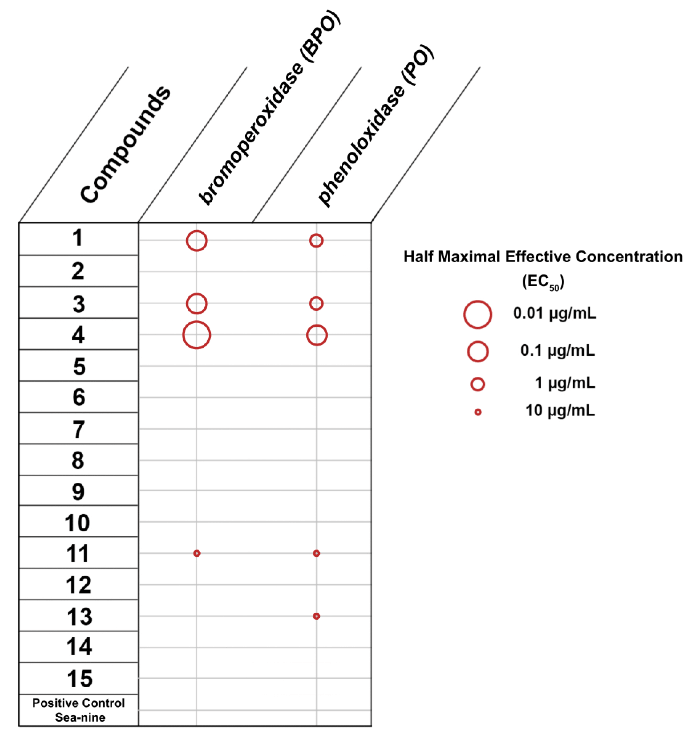
2.2.2. Inhibition of Bromoperoxidase and Phenoloxidase

Colonization, which is one of the most fundamental and precarious processes in the life history of benthic marine algae and invertebrates, regulates the distribution and abundance of organisms and therefore determines the community structure.
When working on bioprospection, the choice of test is crucial. Colonization by invertebrates has been extensively studied, and the barnacle A. amphitrite has been used as a model species for many years. Nevertheless, in previous investigations, we have demonstrated that, in the context of studying the potential of marine natural products for invertebrate growth inhibition, most marine species investigated developed targeted compounds that were active against their primary colonizers. Thus, it is rare to discover compounds with broad-spectrum activity but without toxicity [48]. However, an exception was highlighted for invasive organisms that often have a very elaborate, broad-spectrum chemical defense arsenal. In order to evaluate the activity of compounds 115 for their anti-adhesion efficacy in marine invertebrates but without the constraint of carrying out biological tests on a whole panel of larvae, we decided to work on the inhibition of phenoloxidase, an enzyme conserved in many invertebrates [49].
Algae are understudied in the context of biofouling despite the fact that they are very successful in settling on a wide range of artificial substrata, such as oil and gas platforms, pilings, wrecks, mariculture facilities (nets and cages), ships, buoys and pontoons, and can cause significant damage, e.g., corrosion, decrease of the ships speed, technical problems in aquaculture systems, fishnets and power plant cooling systems [50]. Over the last decades, the occurrence and intensity of algal blooms has increased significantly due to anthropogenic activities and global climate changes. The phenomenon of global warming could deeply influence the evolution of fouling communities; a potential impact would be the reduction of calciferous species (e.g., barnacles, oysters) in favor of macroalgae [51]. The pressure of macroalgal fouling may be very high in the future. All these facts corroborate the idea that the search for inhibitors of algal fixation and adhesion should be a priority in order to be proactive regarding environmental changes. Nowadays, the main difficulty regarding antifouling assays using macroalgae is that they are highly time-consuming, season-dependent and species-dependent. In order to skip these major constraints, we have developed a new approach based on the use of oxidases (enzymes) which is one of the opportunistic strategies developed by numerous marine organisms, including macroalgae, for adhesion [1]. Testing compounds for their inhibitory activities towards oxidase-activated mechanisms represents a fast, reliable and targeted anti-algal assay [50].
Algal bioadhesives are constituted of a complex mixture of various organic compounds, including proteins, carbohydrates, glycoproteins, polyhydroxyphenols and metal ions, mutually interacting through cross-linked bonds as well as electrostatic forces and metal ion bridge complexes [52,53,54]. A model of oxidative cross-linking of secreted phenolics mediated via the catalysis of a bromoperoxidase (BPO) was highlighted in adherent spores of algae [55]. Hardening with alginate and/or calcium is essential for high cohesive strength as shown in shear-lap tests that measured the adhesion strength of this assemblage [52]. These data implicate BPO to be the key reagent in controlling the cross-linking of phenolic polymers for the assembly of algal adhesives [56]. This is why we decided to use BPO inhibition as a probe for anti-adhesion assay.
In invertebrates, such as mussels, oysters and barnacles, substrate attachment is established through adhesives whose formation is catalyzed by a copper-depending phenoloxidase (PO), which oxidizes phenolic residues, such as tyrosine, to catechols, such as 3,4-dihydroxy-l-phenylalanin (l-DOPA) [57]. By studying the enzymatic kinetics of PO, a specific antifouling test has been developed [57].
Compounds 115 were evaluated for their ability to inhibit bromoperoxidase (BPO) and phenoloxidase (PO). SeaNine was not active at all towards these enzymes (this is due to the fact that its mode of action is based on toxicity rather than on specificity). According to the obtained results, expressed as EC50 values (Figure 6) towards BPO and PO, compounds 2, 4, 5 and 12 were active in inhibiting both enzymes, with the highest activity observed for compound 5, closely followed by compounds 2 and 4. Compound 14 was active only towards PO. These results are very promising as macroalgae and invertebrates are very difficult to inhibit specifically and without toxicity. In addition, these tests have the advantage of targeting the adhesion process of many species of algae and invertebrates, and therefore the active compounds offer protection against a wide range of colonizing species using an oxidase-mediated process for adhesion; this is a great advantage because very often when an alga is inhibited, it is replaced by another species.

2.2.3. Toxicity Evaluation of the Compounds

At the concentrations tested, none of the compounds was toxic towards oyster larvae. For all compounds, LC50 values were above 100 µg/mL. These results are very promising and demonstrate the great potential of S. coronopifolius as a producer of non-toxic compounds active for the regulation of epibiosis.

2.3. Overall Antifouling Potential

Bromosphaerol (1) is an active molecule that has already been described as potential cancer stem-cell-targeting and antifouling agent [32]. In this study, thirteen bromosphaerol-related compounds (214) and the major sesquiterpene isolated from S. coronopifolius (15) were evaluated through five specific antifouling tests and demonstrated several interesting activities. Of the tested conditions, 70% and 69% resulted in positive hits against bacterial adhesion and microalgal growth, respectively. Less than 50% of tested conditions exhibited antifouling activity against bacterial growth, and less than 30% concerning algal adhesion and BPO/PO. These levels of inhibition are very interesting in terms of chemical ecology: indeed, the organisms that are the most inhibited correspond to the microbial colonizers, which are the most abundant in the marine environment and that pose constant threats to algae. In the case of S. coronopifolius, we can see that a very efficient arsenal of compounds is produced to inhibit biofilm formation. Fewer compounds were active against oxidase, but the level of activity recorded was very high (low EC50 values); this can reflect an induced type of defense, when targeted compounds are produced with a very specific mode of action (rather than acting by toxicity mechanisms, such as for growth inhibition). Among the tested metabolites, the most potent were compounds 35, 7, 10 and 15, since they showed antifouling activity in more than 65% of the performed assays at different concentrations.
In 45% of the tests, compounds were proven inactive at the concentrations tested (up to 10 µg/mL). In 26% of the tests, compounds were active at 10 µg/mL, while in 4% they were active at 1.0 µg/mL. More importantly, in 12% and 13% of the bioassays, the compounds tested were active at 0.1 and 0.01 µg/mL, respectively. The compounds showing the most potent activity towards bacteria, microalgae and enzymes associated with macroorganisms at low concentrations were compounds 4, 7, 10 and 12, and they appeared to be promising candidates for future studies, as performed in Alves et al. (2020) [58].
As far as diterpenes 114 are concerned, they share relatively similar oxygenated carbocycles decorated with at least one Br atom that is possibly important for the exhibition of activity on epibiotic organisms. Nonetheless, it is evident that the presence of the bromomethylene moiety enhances the compounds‘ activity, as evidenced by the significantly lower activity values exhibited by 13 and 14.

3. Materials and Methods

3.1. General Experimental Procedures

1D and 2D NMR spectra were recorded on a Bruker DRX 400 (Bruker BioSpin GmbH, Rheinstetten, Germany) spectrometer, using standard Bruker pulse sequences. Chemical shifts internally referenced to residual solvent signals are given on a δ (ppm) scale. Column chromatography separations were performed with Kieselgel Si 60 (Merck, Darmstadt, Germany). HPLC separations were conducted on a CECIL 1100 Series liquid chromatography pump (Cecil Instruments Ltd., Cambridge, UK) equipped with a GBC LC-1240 refractive index detector (GBC Scientific Equipment, Braeside, VIC, Australia), using a Kromasil 100-7-C18 (250 × 10 mm, Akzonobel, Eka Chemicals AB, Separation Products, Bohus, Sweden) for reversed-phase HPLC or a 250 mm × 10 mm i.d. Kromasil 100-10-SIL (Akzonobel, Eka Chemicals AB, Separation Products, Bohus, Sweden) for normal-phase HPLC. TLC was performed on Kieselgel 60 F254 (0.2 mm) precoated aluminum or glass plates (Merck, Darmstadt, Germany), and spots were visualized after spraying with H2SO4 in MeOH (20% v/v) reagent and heating at 100 °C for 1 min.

3.2. Biological Material

Specimens of S. coronopifolius were collected by scuba diving in Agios Giannis bay off Parga in Greece, at a depth of 4–20 m in July 2018. A voucher specimen of the alga has been deposited at the Herbarium of Section of Pharmacognosy and Chemistry of Natural Products, Department of Pharmacy, National and Kapodistrian University of Athens (ATPH/MP0601).

3.3. Extraction and Isolation

Algal specimens of S. coronopifolius were initially freeze-dried and then exhaustively extracted with mixtures of CH2Cl2/MeOH (3:1) at room temperature. The combined extracts were concentrated to give a dark green residue, which was later subjected to VCC on silica gel, using cHex with increasing amounts of EtOAc and finally MeOH as mobile phase to afford 12 fractions (1–12). Fraction 6 (50% EtOAc in cHex) was fractionated by gravity column chromatography, using a 5% step gradient of cHex/EtOAc. Fraction 6j (30% EtOAc in cHex) was further separated by normal-phase HPLC using cHex/EtOAc (82:18) as the mobile phase to afford compounds 1, 4, 9 and 15. The CH3CN soluble part of fraction 7 (60% EtOAc in cHex) was subjected to reversed-phase HPLC, using CH3CN 100% as mobile phase and further purified by reversed-phase HPLC with MeCN/H2O (90:10) to yield compounds 2, 57 and 1013. A final purification by normal-phase HPLC with cHex/CHCl3 (45:55) led to the isolation of compounds 3, 8 and 14.

3.4. Antifouling Evaluation Assays

Bioassays were conducted by employing key marine biofoulers: bacteria, microalgae, macroalgae and invertebrates. For all bioassays, compounds were assessed at concentrations of 0.01, 0.1, 1 and 10 µg/mL and on two batches of microorganisms and enzymes. For all bioassays, as a relevant positive control, data for the commercial antifouling booster Sea-nine™ are included (tested at concentrations from 0.01 to 10 µg/mL) [59].
These bioassays were performed to highlight the potency of the 15 compounds to inhibit the adhesion or growth of microorganisms and activity of enzymes of macroalgae and invertebrates involved in adhesion (bromoperoxidase and phenoloxidase, respectively).
Five marine bacteria known as model biofoulers and/or as marine pathogens were selected for this study: Vibrio aesturianus (DSM 19606), Vibrio harveyi (DSM 19623), Vibrio natriegens (DSM 759), Pseudoalteromonas citrea (DSM 8771) and Roseobacter litoralis (DSM 6996). Compounds were dissolved in methanol as a carrier solvent and, after evaporation, were incubated at the concentration of 0.01, 0.1, 1.0 and 10 µg/mL with bacteria (2 × 10⁸ cells/mL) in 96-well plates (MERCK) in 100 µL of MB medium (Marine Broth, Difco 2216) at 25 °C for 24 h. The measurement of growth was carried out by reading the optical density at 630 nm [33]. Results are expressed as the minimal inhibitory concentration (MIC) values.
For the anti-adhesion test, wells were emptied and rinsed with 100 µL of sterile seawater to remove the non-attached cells. Wells were air-dried at room temperature and then the biofilm was stained with 100 µL of 0.1% of crystal violet for 30 min. Then, the previous steps were repeated, and the dried biofilms were resuspended and homogenized in 100 µL ethanol 80%. The control of adhesion was carried out by reading the optical density at 595 nm [60]. Results are expressed as MIC values.
Three microalgae characterized as the most common colonizers and known to be highly involved in biocorrosion processes were selected: Cylindrotheca closterium (AC 170), Exanthemachrysis gayraliae (AC 15) and Halamphora coffeaeformis (AC 713). Compounds were dissolved in methanol as a carrier solvent and after evaporation were incubated at the concentration of 0.01, 0.1, 1.0 and 10 µg/mL with algae in 96-well plates (MERCK) in 100 µL of f/2 medium at 20 °C during 5 days in constant light (incident irradiance: 140 µmol/m2/s). The microalgal concentration was estimated by correlation with chlorophyll a (Chl a) concentration [61]. The control of adhesion was carried out by reading the fluorescence density at 485 nm (excitation) and 645 nm (emission). Results are expressed as MIC values [60].
To evaluate the cell’s adhesion inhibition, the same conditions were used as stated above. After 5 days of incubation, 96 well plates were emptied. One hundred microliters of 100% methanol was added to liberated Chl a. The control of Chl a concentration (growth) was carried out by reading the fluorescence density at 485 nm (excitation) and 645 nm (emission). Results are expressed as MIC values [60].
In order to evaluate the anti-adhesion activity against macroalgae and invertebrates, oxidase enzymes were used as a probe. Thus, assays were performed using bromoperoxidase, an enzyme involved in adhesive-production by macroalgae, and phenoloxidase, an enzyme involved in adhesive production by mussels, barnacles and oysters.
Bromoperoxidase (BPO) inhibition assay: BPO activity, as a probe for macroalgae antifouling adhesion assay, was measured spectrophotometrically. The pure enzyme was purchased from Sigma (CAS no. 69279-19-2). For the enzymatic activity evaluation, an aliquot of the pure enzyme (corresponding to 0.1 unit/mL) was added to 0.375 mL of KBr 0.1 M (substrate of BPO) in 1.5 mL plastic UV-cuvettes, and production of bromophenol blue was monitored at 278 nm for 2 h. The results are presented as the concentration of compounds that reduces enzyme velocity by 50% (IC50 values). IC50 values were calculated using MINITAB.
Phenoloxidase (PO) inhibition assay: PO activity, as a probe for invertebrate antifouling adhesion assay, was measured spectrophotometrically as described earlier [62]. The pure enzyme (EC1.14.18.1) was incubated at 25 °C with 10 mM L-DOPA in a 50 mM phosphate buffer of pH 6.8. PO activity was determined by monitoring the increase in absorbance at 475 nm. One unit of enzyme activity was defined as the amount of enzyme that catalyzes the formation of 1 μmol dopachrome per minute under the described experimental conditions. The tested compounds were added at concentrations of 0.01 up to 10 μg/mL. Aliquots of the pure enzyme were incubated for 2 h in the presence of the compounds, and then the enzyme activity was recorded with L-DOPA (10 mM) as substrate. The results are presented as the concentration of compounds that reduces enzyme velocity by 50% (IC50 values). IC50 values were calculated using MINITAB.

3.5. Toxicity Assessment

Toxicity tests were conducted as previously described [63]. In order to determine the toxicity of the compounds on the development of larvae of oysters, Crassostrea gigas larvae were obtained from Argenton Marine Station (Lemar laboratory, University of Brest) and were incubated with a gradient of compounds’ concentrations (0.01, 0.1, 1.0, 10, 25, 50 and 100 µg/mL). Higher concentrations than those used for the antifouling assays were used in order to mimic a worst-case exposure scenario. Six replicates and two batches of larvae were used in order to reduce the effect of the natural variability between individuals. A population control comprised larvae reared in seawater. After 24 h at 20 °C, larvae reached the D stage (confirmed by microscopic observation). Larval development was then stopped by addition of 100 mL of formol to the culture medium. LC50 values were assessed using a log-logistic model (Kooijman, 1981). The model was fitted to the experimental data with the help of a non-linear regression program (SGPLUS, Manugistics, Incorporated, Rockville, MD, USA).

4. Conclusions

Among the 15 tested metabolites (115), compounds 3, 5 and 15 were active against a wide range of biofoulers. They showed at least one positive response towards the panel of organisms tested, whether on the biofoulers’ growth or adhesion or on enzymes; they displayed a broad-spectrum type of activity. More precisely, compounds 110, 1215 were active against bacterial adhesion and compounds 15, 79, 12 and 15 against bacterial growth. Among them, compounds 1, 6 and 12 were the most active with activities at lower concentration against bacterial adhesion, as well as compounds 5, 7 and 15 against bacterial growth. Then, compounds 27, 911 and 15 were active against microalgal adhesion and all compounds against microalgal growth. Among them, compounds 10 was the most active against microalgal adhesion, and most of the compounds (except for compound 2, 4, 11) appeared active at concentration under 0.1 µg/mL against microalgal growth. Finally, compounds 1, 3, 4, 11 and 13 were active against BPO and/or PO. Among them, compound 4 appeared as the most potent with EC50 value < 0.1 µg/mL. Such metabolites have the potential to be used as booster compounds in antifouling formulations.
None of the compounds tested were inactive, which confirmed the significant antifoulant potential of bromosphaerol (1) and compounds 215 isolated from S. coronopifolius [32,41]. These observations corroborate the fact that marine organisms use a combination of antifouling defenses, some molecules with a broad range of activities and some others displaying very specific modes of action. Moreover, it also corroborates the fact that diterpenes produced by algae are highly bioactive and associated with defense mechanisms [64,65,66]. Depending on the concentrations of each of these compounds and their combination (induced defense vs. constitutive defense), S. coronopifolius is able to efficiently control epibiosis on its surface. Marine macroalgae are known to adapt their chemical defenses to environmental conditions. Thus, some defences are synthesized constitutively and others inductively in order to best respond to epibiosis pressure. This is of particular interest since S. coronopifolius is a cultivable species (unpublished results) and it will be possible to apply OSMAC-like (One Strain MAny Compounds) approaches to grow it under various stress conditions to improve the commercial exploitation of its natural products [25,26,27,67,68]. Such studies could lead to the identification of other natural bioactive compounds with biotechnological applications.
Further analysis by monitoring the production of these secondary metabolites throughout the year would permit the elucidation of the roles of these compounds and under which conditions their production is more pronounced. As a postulate, compounds 4, 7, 10 and 12 might be used by the red alga S. coronopifolius for its clean surface maintenance year-long, and other compounds could be produced as a response to an epibiosis-related stress. Additionally, in future applications, transcriptomic approaches would help to understand the biosynthetic pathways implicated in the production of such bioactive molecules, and then studied mixtures of these molecules to evaluate their biotechnological potential.

Author Contributions

Conceptualization, A.M.M., V.R., E.I. and C.H.; methodology, V.R., E.I. and C.H.; formal analysis, V.R., E.I. and C.H.; investigation, M.Q., S.K., M.F., Y.T., F.A., E.I. and C.H.; resources, V.R., E.I. and C.H.; writing—original draft preparation, M.Q., S.K., M.F., Y.T., F.A., V.R., E.I. and C.H.; writing—review and editing, M.Q., A.M.M., V.R., E.I. and C.H.; visualization, M.Q., S.K., E.I. and C.H.; supervision, V.R., E.I. and C.H.; project administration, V.R., E.I. and C.H.; funding acquisition, A.M.M., V.R., E.I. and C.H. All authors have read and agreed to the published version of the manuscript.

Funding

This research was supported by 3rd Joint Transnational Call of the ERA-Net COFASP in collaboration with ERA-NET MarineBiotech (project ANTIFOUL).

Institutional Review Board Statement

Not applicable.

Informed Consent Statement

Not applicable.

Acknowledgments

Access to the infrastructure obtained by the research project “Center for the study and sustainable exploitation of Marine Biological Resources” (CMBR, MIS 5002670) in the framework of the National Roadmap for Research Infrastructures is gratefully acknowledged. M.Q., M.F. and C.H. thank Biogenouest for support.

Conflicts of Interest

The authors declare no conflict of interest. The funders had no role in the design of the study; in the collection, analyses or interpretation of data; in the writing of the manuscript; or in the decision to publish the results.

References

  1. Maréchal, J.-P.; Hellio, C. Challenges for the development of new non-toxic antifouling solutions. Int. J. Mol. Sci. 2009, 10, 4623–4637. [Google Scholar] [CrossRef] [PubMed] [Green Version]
  2. Bovio, E.; Fauchon, M.; Toueix, Y.; Mehiri, M.; Varese, G.C.; Hellio, C. The Sponge-Associated Fungus Eurotium Chevalieri MUT 2316 and Its Bioactive Molecules: Potential Applications in the Field of Antifouling. Mar. Biotechnol. 2019, 21, 743–752. [Google Scholar] [CrossRef] [PubMed]
  3. Saha, M.; Goecke, F.; Bhadury, P. Minireview: Algal Natural Compounds and Extracts as Antifoulants. J. Appl. Phycol. 2018, 30, 1859–1874. [Google Scholar] [CrossRef] [PubMed] [Green Version]
  4. Antunes, J.; Leão, P.; Vasconcelos, V. Marine Biofilms: Diversity of Communities and of Chemical Cues. Environ. Microbiol Rep. 2019, 11, 287–305. [Google Scholar] [CrossRef] [PubMed]
  5. Fitridge, I.; Dempster, T.; Guenther, J.; de Nys, R. The Impact and Control of Biofouling in Marine Aquaculture: A Review. Biofouling 2012, 28, 649–669. [Google Scholar] [CrossRef]
  6. Bannister, J.; Sievers, M.; Bush, F.; Bloecher, N. Biofouling in Marine Aquaculture: A Review of Recent Research and Developments. Biofouling 2019, 35, 631–648. [Google Scholar] [CrossRef] [PubMed] [Green Version]
  7. Li, Y.; Ning, C. Latest Research Progress of Marine Microbiological Corrosion and Bio-Fouling, and New Approaches of Marine Anti-Corrosion and Anti-Fouling. Bioact. Mater. 2019, 4, 189–195. [Google Scholar] [CrossRef] [PubMed]
  8. Schultz, M.P.; Bendick, J.A.; Holm, E.R.; Hertel, W.M. Economic Impact of Biofouling on a Naval Surface Ship. Biofouling 2011, 27, 87–98. [Google Scholar] [CrossRef]
  9. Jadidi, P.; Zeinoddini, M. Influence of Hard Marine Fouling on Energy Harvesting from Vortex-Induced Vibrations of a Single-Cylinder. Renew. Energy 2020, 152, 516–528. [Google Scholar] [CrossRef]
  10. Amara, I.; Miled, W.; Slama, R.B.; Ladhari, N. Antifouling Processes and Toxicity Effects of Antifouling Paints on Marine Environment. A Review. Environ. Toxicol. Pharmacol. 2018, 57, 115–130. [Google Scholar] [CrossRef]
  11. Yebra, D.M.; Kiil, S.; Weinell, C.E.; Dam-Johansen, K. Presence and Effects of Marine Microbial Biofilms on Biocide-Based Antifouling Paints. Biofouling 2006, 22, 33–41. [Google Scholar] [CrossRef]
  12. Onduka, T.; Mizuno, K.; Shikata, T.; Mastubara, T.; Onitsuka, G.; Hamaguchi, M. Assessment of the Risk Posed by Three Antifouling Biocides to Pacific Oyster Embryos and Larvae in Hiroshima Bay, Japan. Environ. Sci Pollut Res. 2021, 1–12. [Google Scholar] [CrossRef] [PubMed]
  13. Ali, H.R.; Ariffin, M.M.; Omar, T.F.T.; Ghazali, A.; Sheikh, M.A.; Shazili, N.A.M.; Bachok, Z. Antifouling Paint Biocides (Irgarol 1051 and Diuron) in the Selected Ports of Peninsular Malaysia: Occurrence, Seasonal Variation, and Ecological Risk Assessment. Environ. Sci. Pollut. Res. 2021, 28, 52247–52257. [Google Scholar] [CrossRef] [PubMed]
  14. Watermann, B.T.; Daehne, B.; Sievers, S.; Dannenberg, R.; Overbeke, J.C.; Klijnstra, J.W.; Heemken, O. Bioassays and Selected Chemical Analysis of Biocide-Free Antifouling Coatings. Chemosphere 2005, 60, 1530–1541. [Google Scholar] [CrossRef] [PubMed]
  15. Kymenvaara, S.; Tegnér Anker, H.; Baaner, L.; Ekroos, A.; Gipperth, L.; Seppälä, J. Regulating Antifouling Paints for Leisure Boats—A Patchwork of Rules Across Three Baltic Sea Countries. Nord. Environ. Law J. 2017, 1, 7–32. Available online: http://nordiskmiljoratt.se/onewebmedia/Kymenvaara%20et%20al.pdf (accessed on 1 December 2021).
  16. Jones, B.; Bolam, T. Copper Speciation Survey from UK Marinas, Harbours and Estuaries. Mar. Pollut Bull. 2007, 54, 1127–1138. [Google Scholar] [CrossRef] [PubMed]
  17. Qian, P.-Y.; Li, Z.; Xu, Y.; Li, Y.; Fusetani, N. Mini-Review: Marine Natural Products and Their Synthetic Analogs as Antifouling Compounds: 2009-2014. Biofouling 2015, 31, 101–122. [Google Scholar] [CrossRef] [PubMed]
  18. Alemán-Vega, M.; Sánchez-Lozano, I.; Hernández-Guerrero, C.J.; Hellio, C.; Quintana, E.T. Exploring Antifouling Activity of Biosurfactants Producing Marine Bacteria Isolated from Gulf of California. Int. J. Mol. Sci. 2020, 21, 6068. [Google Scholar] [CrossRef] [PubMed]
  19. Sánchez-Lozano, I.; Hernández-Guerrero, C.J.; Muñoz-Ochoa, M.; Hellio, C. Biomimetic Approaches for the Development of New Antifouling Solutions: Study of Incorporation of Macroalgae and Sponge Extracts for the Development of New Environmentally-Friendly Coatings. Int. J. Mol. Sci. 2019, 20, 4863. [Google Scholar] [CrossRef] [PubMed] [Green Version]
  20. Gu, Y.; Yu, L.; Mou, J.; Wu, D.; Xu, M.; Zhou, P.; Ren, Y. Research Strategies to Develop Environmentally Friendly Marine Antifouling Coatings. Mar. Drugs 2020, 18, 371. [Google Scholar] [CrossRef] [PubMed]
  21. Carson, R.T.; Damon, M.; Johnson, L.T.; Gonzalez, J.A. Conceptual Issues in Designing a Policy to Phase out Metal-Based Antifouling Paints on Recreational Boats in San Diego Bay. J. Environ. Manag. 2009, 90, 2460–2468. [Google Scholar] [CrossRef]
  22. Dafforn, K.A.; Lewis, J.A.; Johnston, E.L. Antifouling Strategies: History and Regulation, Ecological Impacts and Mitigation. Mar. Pollut. Bull. 2011, 62, 453–465. [Google Scholar] [CrossRef] [PubMed]
  23. Blunt, J.W.; Carroll, A.R.; Copp, B.R.; Davis, R.A.; Keyzers, R.A.; Prinsep, M.R. Marine Natural Products. Nat. Prod. Rep. 2018, 35, 8–53. [Google Scholar] [CrossRef] [PubMed] [Green Version]
  24. Carroll, A.R.; Copp, B.R.; Davis, R.A.; Keyzers, R.A.; Prinsep, M.R. Marine Natural Products. Nat. Prod. Rep. 2020, 37, 175–223. [Google Scholar] [CrossRef] [PubMed]
  25. Levert, A.; Foulon, V.; Fauchon, M.; Tapissier-Bontemps, N.; Banaigs, B.; Hellio, C. Antifouling Activity of Meroterpenes Isolated from the Ascidian Aplidium Aff. Densum. Mar. Biotechnol. 2021, 23, 51–61. [Google Scholar] [CrossRef] [PubMed]
  26. Shinde, P.; Banerjee, P.; Mandhare, A. Marine Natural Products as Source of New Drugs: A Patent Review (2015–2018). Expert Opin. Ther. Pat. 2019, 29, 283–309. [Google Scholar] [CrossRef] [PubMed]
  27. Matulja, D.; Wittine, K.; Malatesti, N.; Laclef, S.; Turks, M.; Markovic, M.K.; Ambrožić, G.; Marković, D. Marine Natural Products with High Anticancer Activities. Curr. Med. Chem. 2020, 27, 1243–1307. [Google Scholar] [CrossRef]
  28. Blunt, J.W.; Copp, B.R.; Keyzers, R.A.; Munro, M.H.G.; Prinsep, M.R. Marine Natural Products. Nat. Prod. Rep. 2015, 32, 116–211. [Google Scholar] [CrossRef] [PubMed] [Green Version]
  29. Fernando, I.P.S.; Nah, J.-W.; Jeon, Y.-J. Potential Anti-Inflammatory Natural Products from Marine Algae. Environ. Toxicol. Pharmacol. 2016, 48, 22–30. [Google Scholar] [CrossRef] [PubMed]
  30. Martínez Andrade, K.A.; Lauritano, C.; Romano, G.; Ianora, A. Marine Microalgae with Anti-Cancer Properties. Mar. Drugs 2018, 16, 165. [Google Scholar] [CrossRef] [PubMed] [Green Version]
  31. O’Neill, E. Mining Natural Product Biosynthesis in Eukaryotic Algae. Mar. Drugs 2020, 18, 90. [Google Scholar] [CrossRef] [Green Version]
  32. Piazza, V.; Roussis, V.; Garaventa, F.; Greco, G.; Smyrniotopoulos, V.; Vagias, C.; Faimali, M. Terpenes from the red alga Sphaerococcus coronopifolius inhibit the settlement of barnacles. Mar. Biotechnol. 2011, 13, 764–772. [Google Scholar] [CrossRef]
  33. Fattorusso, E.; Magno, S.; Santacroce, C.; Sica, D.; Di Blasio, B.; Pedone, C.; Impellizzeri, G.; Mangiafico, S.; Oriente, G.; Piattelli, M.; et al. Bromosphaerol, a new bromine containing diterpenoid from the red alga Sphaerococcus coronopifolius. Gazz. Chim. Ital. 1976, 106, 779–783. [Google Scholar]
  34. Cafieri, F.; Ciminiello, P.; Fattorusso, E.; Mangoni, A. Two novel bromoditerpenes from the red alga Sphaerococcus coronopifolius. Gazz. Chim. Ital. 1990, 120, 139–142. [Google Scholar]
  35. Cafieri, F.; Ciminiello, P.; Fattorusso, E.; Santacroce, C. 12S-Hydroxybromosphaerol, a new bromoditerpene from the red alga Sphaerococcus coronopifolius. Experientia 1982, 38, 298–299. [Google Scholar] [CrossRef]
  36. Smyrniotopoulos, V.; Quesada, A.; Vagias, C.; Moreau, D.; Roussakis, C.; Roussis, V. Cytotoxic bromoditerpenes from the red alga Sphaerococcus coronopifolius. Tetrahedron 2008, 64, 5184–5190. [Google Scholar] [CrossRef]
  37. Cafieri, F.; De Napoli, L.; Fattorusso, E.; Santacroce, C. Diterpenes from the red alga Sphaerococcus coronopifolius. Phytochemistry 1987, 26, 471–473. [Google Scholar] [CrossRef]
  38. Fenical, W.; Finer, J.; Clardy, J. Sphaerococcenol A, a new rearranged bromo-diterpene from the red alga Sphaerococcus coronopifolius. Tetrahedron Lett. 1976, 17, 731–734. [Google Scholar] [CrossRef]
  39. De Rosa, S.; De Stefano, S.; Scarpelli, P.; Zavodnik, N. Terpenes from the red alga Sphaerococcus coronopifolius of the North Adriatic Sea. Phytochemistry 1988, 27, 1875–1878. [Google Scholar] [CrossRef]
  40. Smyrniotopoulos, V.; de Andrade Tomaz, A.C.; Vanderlei de Souza, M.F.; da Cunha, E.V.L.; Kiss, R.; Mathieu, V.; Ioannou, E.; Roussis, V. Halogenated diterpenes with in vitro antitumor activity from the red alga Sphaerococcus coronopifolius. Mar. Drugs 2020, 18, 29. [Google Scholar] [CrossRef] [PubMed] [Green Version]
  41. Smyrniotopoulos, V.; Vagias, C.; Rahman, M.M.; Gibbons, S.; Roussis, V. Brominated diterpenes with antibacterial activity from the red alga Sphaerococcus coronopifolius. J. Nat. Prod. 2008, 71, 1386–1392. [Google Scholar] [CrossRef]
  42. Cafieri, F.; De Napoli, L.; Fattorusso, E.; Impellizzeri, G.; Piattelli, M.; Sciuto, S. Bromosphaerodiol, a minor bromo compound from the red alga Sphaerococcus coronopifolius. Experientia 1977, 33, 1549–1550. [Google Scholar] [CrossRef]
  43. Cafieri, F.; Ciminiello, P.; Santacroce, C.; Fattorusso, E. (1S)-1,2-Dihydro-1-hydroxybromosphaerol, a minor bromoditerpene from the red alga Sphaerococcus coronopifolius. Phytochemistry 1982, 21, 2412–2413. [Google Scholar] [CrossRef]
  44. Smyrniotopoulos, V.; Vagias, C.; Bruyere, C.; Lamoral-Theys, D.; Kiss, R.; Roussis, V. Structure and in vitro antitumor activity evaluation of brominated diterpenes from the red alga Sphaerococcus coronopifolius. Bioorg. Med. Chem. 2010, 18, 1321–1330. [Google Scholar] [CrossRef] [PubMed]
  45. Faure, R.; Ramanoelina, A.R.P.; Rokotonirainy, O.; Bianchini, J.P.; Gaydou, E. Two-dimensional nuclear magnetic resonance of sesquiterpenes. Application to complete assignment of 1H and 13C NMR spectra of some aromadendrane derivatives. Mag. Reson. Chem. 1991, 29, 969–971. [Google Scholar] [CrossRef]
  46. Almeida, J.R.; Correia-da-Silva, M.; Sousa, E.; Antunes, J.; Pinto, M.; Vasconcelos, V.; Cunha, I. Antifouling Potential of Nature-Inspired Sulfated Compounds. Sci. Rep. 2017, 7, 42424. [Google Scholar] [CrossRef] [Green Version]
  47. Ralston, E.; Swain, G. Bioinspiration--the Solution for Biofouling Control? Bioinspir. Biomim. 2009, 4, 015007. [Google Scholar] [CrossRef] [PubMed]
  48. Marechal, J.-P.; Hellio, C. Antifouling Activity against Barnacle Cypris Larvae: Do Target Species Matter (Amphibalanus Amphitrite versus Semibalanus Balanoides)? Int. Biodeterior. Biodegrad. 2010, 65, 92–101. [Google Scholar] [CrossRef] [Green Version]
  49. Niemann, H.; Hagenow, J.; Chung, M.-Y.; Hellio, C.; Weber, H.; Proksch, P. SAR of Sponge-Inspired Hemibastadin Congeners Inhibiting Blue Mussel PhenolOxidase. Mar. Drugs 2015, 13, 3061–3071. [Google Scholar] [CrossRef] [PubMed] [Green Version]
  50. Lebret, K.; Thabard, M.; Hellio, C. Algae as Marine Fouling Organisms: Adhesion Damage and Prevention. Adv. Mar. Antifouling Coat. Tech. 2009, 1, 80–112. [Google Scholar] [CrossRef]
  51. Dobretsov, S.; Coutinho, R.; Rittschof, D.; Salta, M.; Ragazzola, F.; Hellio, C. The Oceans Are Changing: Impact of Ocean Warming and Acidification on Biofouling Communities. Biofouling 2019, 35, 585–595. [Google Scholar] [CrossRef]
  52. Bitton, R.; Ben-Yehuda, M.; Davidovich, M.; Balazs, Y.; Potin, P.; Delage, L.; Colin, C.; Bianco-Peled, H. Structure of Algal-Born Phenolic Polymeric Adhesives. Macromol. Biosci. 2006, 6, 737–746. [Google Scholar] [CrossRef]
  53. Chiovitti, A.; Heraud, P.; Dugdale, T.M.; Hodson, O.M.; Curtain, R.C.A.; Dagastine, R.R.; Wood, B.R.; Wetherbee, R. Divalent Cations Stabilize the Aggregation of Sulfated Glycoproteins in the Adhesive Nanofibers of the Biofouling Diatom Toxarium Undulatum. Soft. Matter. 2008, 4, 811–820. [Google Scholar] [CrossRef] [PubMed]
  54. Dimartino, S.; Savory, D.M.; Fraser-Miller, S.J.; Gordon, K.C.; McQuillan, A.J. Microscopic and Infrared Spectroscopic Comparison of the Underwater Adhesives Produced by Germlings of the Brown Seaweed Species Durvillaea Antarctica and Hormosira Banksii. J. Royal Soc. Interface 2016, 13, 20151083. [Google Scholar] [CrossRef] [PubMed] [Green Version]
  55. Vreeland, V.; Waite, J.; Epstein, L. Minireview—Polyphenols and Oxidases in Substratum Adhesion by Marine Algae and Mussels. J. Phycol. 1998, 34, 1–8. [Google Scholar] [CrossRef]
  56. La Barre, S.; Potin, P.; Leblanc, C.; Delage, L. The Halogenated Metabolism of Brown Algae (Phaeophyta), Its Biological Importance and Its Environmental Significance. Mar. Drugs 2010, 8, 988–1010. [Google Scholar] [CrossRef] [PubMed] [Green Version]
  57. Hellio, C.; Bourgougnon, N.; Gal, Y.L. Phenoloxidase (E.C. 1.14.18.1) from the Byssus Gland of Mytilus Edulis: Purification, Partial Characterization and Application for Screening Products with Potential Antifouling Activities. Biofouling 2000, 16, 235–244. [Google Scholar] [CrossRef]
  58. Alves, C.; Serrano, E.; Silva, J.; Rodrigues, C.; Pinteus, S.; Gaspar, H.; Botana, L.M.; Alpoim, M.C.; Pedrosa, R. Sphaerococcus Coronopifolius Bromoterpenes as Potential Cancer Stem Cell-Targeting Agents. Biomed. Pharm. 2020, 128, 110275. [Google Scholar] [CrossRef]
  59. Moodie, L.W.K.; Cervin, G.; Trepos, R.; Labriere, C.; Hellio, C.; Pavia, H.; Svenson, J. Design and Biological Evaluation of Antifouling Dihydrostilbene Oxime Hybrids. Mar. Biotechnol. 2018, 20, 257–267. [Google Scholar] [CrossRef] [Green Version]
  60. Hellio, C.; Trepos, R.; Aguila-Ramírez, R.N.; Hernández-Guerrero, C.J. Protocol for Assessing Antifouling Activities of Macroalgal Extracts. Methods Mol. Biol. 2015, 1308, 421–435. [Google Scholar] [CrossRef] [PubMed]
  61. Chambers, L.D.; Hellio, C.; Stokes, K.R.; Dennington, S.P.; Goodes, L.R.; Wood, R.J.K.; Walsh, F.C. Investigation of Chondrus Crispus as a Potential Source of New Antifouling Agents. Int. Biodeterior. Biodegrad. 2011, 7, 939–946. [Google Scholar] [CrossRef]
  62. Hellio, C.; De La Broise, D.; Dufossé, L.; Le Gal, Y.; Bourgougnon, N. Inhibition of Marine Bacteria by Extracts of Macroalgae: Potential Use for Environmentally Friendly Antifouling Paints. Mar. Environ. Res. 2001, 52, 231–247. [Google Scholar] [CrossRef]
  63. Hellio, C.; Bremer, G.; Pons, A.M.; Le Gal, Y.; Bourgougnon, N. Inhibition of the Development of Microorganisms (Bacteria and Fungi) by Extracts of Marine Algae from Brittany, France. Appl. Microbiol. Biotechnol. 2000, 54, 543–549. [Google Scholar] [CrossRef]
  64. Chen, J.; Li, H.; Zhao, Z.; Xia, X.; Li, B.; Zhang, J.; Yan, X. Diterpenes from the Marine Algae of the Genus Dictyota. Mar. Drugs 2018, 16, 159. [Google Scholar] [CrossRef] [PubMed] [Green Version]
  65. Zhang, X.-Y.; Hao, H.-L.; Lau, S.C.K.; Wang, H.-Y.; Han, Y.; Dong, L.-M.; Huang, R.-M. Biodiversity and Antifouling Activity of Fungi Associated with Two Soft Corals from the South China Sea. Arch. Microbiol. 2019, 201, 757–767. [Google Scholar] [CrossRef]
  66. Tobal, I.E.; Roncero, A.M.; Moro, R.F.; Díez, D.; Marcos, I.S. Antibacterial Natural Halimanes: Potential Source of Novel Antibiofilm Agents. Molecules 2020, 25, 1707. [Google Scholar] [CrossRef] [PubMed] [Green Version]
  67. Pan, R.; Bai, X.; Chen, J.; Zhang, H.; Wang, H. Exploring Structural Diversity of Microbe Secondary Metabolites Using OSMAC Strategy: A Literature Review. Front. Microbiol. 2019, 10, 294. [Google Scholar] [CrossRef] [Green Version]
  68. Navarri, M.; Jégou, C.; Meslet-Cladière, L.; Brillet, B.; Barbier, G.; Burgaud, G.; Fleury, Y. Deep Subseafloor Fungi as an Untapped Reservoir of Amphipathic Antimicrobial Compounds. Mar. Drugs 2016, 14, 50. [Google Scholar] [CrossRef] [PubMed] [Green Version]
Figure 1. Chemical structures of compounds 115 isolated from Sphaerococcus coronopifolius.
Marinedrugs 20 00032 g001
Figure 2. Inhibition of adhesion of bacteria by compounds 115 (MICs in µg/mL).
Marinedrugs 20 00032 g002
Figure 3. Inhibition of adhesion of microalgae by compounds 1–15 (MICs in µg/mL).
Marinedrugs 20 00032 g003
Figure 4. Inhibition of growth of bacteria by compounds 115 (MICs in µg/mL).
Marinedrugs 20 00032 g004
Figure 5. Inhibition of growth of microalgae by compounds 115 (MICs in µg/mL).
Marinedrugs 20 00032 g005
Figure 6. Inhibition of the activity of the bromoperoxidase and phenoloxidase by compounds 115.
Marinedrugs 20 00032 g006
Publisher’s Note: MDPI stays neutral with regard to jurisdictional claims in published maps and institutional affiliations.

