

Ultrasound-Based Recovery of Anti-Inflammatory and Antimicrobial Extracts of the Acidophilic Microalga Coccomyxa onubensis
 , , , ,
Abstract
: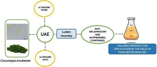
1. Introduction
2. Results and Discussion
2.1. Effects of the Ultrasonic Techniques on the Extraction Yield of C. onubensis
2.2. Effects of the Ultrasonic Techniques on the Anti-Inflammatory Carotenoids Profile of C. onubensis
2.3. Desirability Function
2.4. Antimicrobial Activity and Anti-Inflammatory Metabolite Identification
3. Materials and Methods
3.1. Biomass and Chemicals
3.2. Conventional Extraction
3.3. Ultrasonic Green Extraction Design
3.4. Quantification of Carotenoids
3.5. Quantification of the Bioactive Extracts Using Gas Chromatography–Mass Spectrometry (GC–MS)
3.6. Antimicrobial Activity Assay
3.7. Statistical Analysis and Multiple Response Optimization
4. Conclusions
Supplementary Materials
Author Contributions
Funding
Institutional Review Board Statement
Informed Consent Statement
Data Availability Statement
Conflicts of Interest
References
- Evangelista, V.; Barsanti, L.; Frassanito, A.M.; Passarelli, V.; Gualtieri, P. Algal Toxins: Nature, Occurrence, Effect and Detection; Springer Science & Business Media: Berlin/Heidelberg, Germany, 2008; pp. 353–391. [Google Scholar]
- Tan, J.S.; Lee, S.Y.; Chew, K.W.; Lam, M.K.; Lim, J.W.; Ho, S.H.; Show, P.L. A review on microalgae cultivation and harvesting, and their biomass extraction processing using ionic liquids. Bioengineered 2020, 11, 116–129. [Google Scholar] [CrossRef] [PubMed]
- Canganella, F.; Wiegel, J. Extremophiles: From abyssal to terrestrial ecosystems and possibly beyond. Naturwissenschaften 2011, 98, 253–279. [Google Scholar] [CrossRef]
- Mendes-Silva, T.d.C.D.; da Silva Andrade, R.F.; Ootani, M.A.; Mendes, P.V.D.; da Silva, M.R.F.; Souza, K.S.; dos Santos Correia, M.T.; da Silva, M.V.; de Oliveira, M.B.M. Biotechnological potential of carotenoids produced by extremophilic microorganisms and application prospects for the cosmetics industry. Adv. Microbiol. 2020, 10, 397–410. [Google Scholar] [CrossRef]
- Fuentes, J.-L.; Montero, Z.; Cuaresma, M.; Ruiz-Domínguez, M.-C.; Mogedas, B.; Nores, I.G.; González del Valle, M.; Vílchez, C. Outdoor large-scale cultivation of the acidophilic microalga Coccomyxa onubensis in a vertical close photobioreactor for lutein production. Processes 2020, 8, 324. [Google Scholar] [CrossRef]
- Vaquero, I.; Ruiz-Domínguez, M.C.; Márquez, M.; Vílchez, C. Cu-mediated biomass productivity enhancement and lutein enrichment of the novel microalga Coccomyxa onubensis. Process Biochem. 2012, 47, 694–700. [Google Scholar] [CrossRef]
- Bermejo, E.; Ruiz-Domínguez, M.C.; Cuaresma, M.; Vaquero, I.; Ramos-Merchante, A.; Vega, J.M.; Vílchez, C.; Garbayo, I. Production of lutein, and polyunsaturated fatty acids by the acidophilic eukaryotic microalga Coccomyxa onubensis under abiotic stress by salt or ultraviolet light. J. Biosci. Bioeng. 2018, 125, 669–675. [Google Scholar] [CrossRef]
- Navarro, F.; Forján, E.; Vázquez, M.; Toimil, A.; Montero, Z.; Ruiz-Domínguez, M.d.C.; Garbayo, I.; Castaño, M.Á.; Vílchez, C.; Vega, J.M. Antimicrobial activity of the acidophilic eukaryotic microalga Coccomyxa onubensis. Phycol. Res. 2017, 65, 38–43. [Google Scholar] [CrossRef]
- Navarro, F.; Toimil, A.; Ramírez, S.; Montero, Y.; Fuentes, J.L.; Perona, J.S.; Castaño, M.Á.; Pásaro, R.; Vega, J.M.; Vílchez, C. The acidophilic microalga Coccomyxa onubensis and atorvastatin equally improve antihyperglycemic and antihyperlipidemic protective effects on rats fed on high-fat diets. J. Appl. Phycol. 2020, 32, 3923–3931. [Google Scholar] [CrossRef]
- Navarro, F.; Forján, E.; Vázquez, M.; Montero, Z.; Bermejo, E.; Castaño, M.Á.; Toimil, A.; Chagüaceda, E.; García-Sevillano, M.Á.; Sánchez, M.; et al. Microalgae as a safe food source for animals: Nutritional characteristics of the acidophilic microalga Coccomyxa onubensis. Food Nutr. Res. 2016, 60, 30472. [Google Scholar] [CrossRef]
- Cardozo, K.H.M.; Guaratini, T.; Barros, M.P.; Falcão, V.R.; Tonon, A.P.; Lopes, N.P.; Campos, S.; Torres, M.A.; Souza, A.O.; Colepicolo, P.; et al. Metabolites from algae with economical impact. Comp. Biochem. Physiol. Toxicol. Pharmacol. CBP 2007, 146, 60–78. [Google Scholar] [CrossRef]
- Plaza, M.; Santoyo, S.; Jaime, L.; García-Blairsy Reina, G.; Herrero, M.; Señoráns, F.J.; Ibáñez, E. Screening for bioactive compounds from algae. J. Pharm. Biomed. Anal. 2010, 51, 450–455. [Google Scholar] [CrossRef]
- De Morais, M.G.; Vaz, B.d.S.; de Morais, E.G.; Costa, J.A.V. Biologically Active Metabolites Synthesized by Microalgae. BioMed Res. Int. 2015, 2015, 835761. [Google Scholar] [CrossRef]
- Suganya, T.; Varman, M.; Masjuki, H.; Renganathan, S. Macroalgae and microalgae as a potential source for commercial applications along with biofuels production: A biorefinery approach. Renew. Sustain. Energy Rev. 2016, 55, 909–941. [Google Scholar] [CrossRef]
- Poojary, M.; Barba, F.; Aliakbarian, B.; Donsì, F.; Pataro, G.; Dias, D.; Juliano, P. Innovative alternative technologies to extract carotenoids from microalgae and seaweeds. Mar. Drugs 2016, 14, 214. [Google Scholar] [CrossRef]
- Hu, Y.; Yan, B.; Chen, Z.S.; Wang, L.; Tang, W.; Huang, C. Recent technologies for the extraction and separation of polyphenols in different plants: A review. J. Renew. Mater. 2022, 10, 1471–1490. [Google Scholar] [CrossRef]
- Mason, T.; Chemat, F.; Vinatoru, M. The extraction of natural products using ultrasound or microwaves. Curr. Org. Chem. 2011, 15, 237–247. [Google Scholar] [CrossRef]
- Zheng, S.; Zhang, G.; Wang, H.; Long, Z.; Wei, T.; Li, Q. Progress in ultrasound-assisted extraction of the value-added products from microorganisms. World J. Microbiol. Biotechnol. 2021, 37, 71. [Google Scholar] [CrossRef]
- Santos, H.M.; Capelo, J.L. Trends in ultrasonic-based equipment for analytical sample treatment. Talanta 2007, 73, 795–802. [Google Scholar] [CrossRef] [PubMed]
- Juliano, P.; Augustin, M.A.; Xu, X.Q.; Mawson, R.; Knoerzer, K. Advances in high frequency ultrasound separation of particulates from biomass. Ultrason. Sonochem. 2017, 35, 577–590. [Google Scholar] [CrossRef] [PubMed]
- Roselló-Soto, E.; Galanakis, C.M.; Brnčić, M.; Orlien, V.; Trujillo, F.J.; Mawson, R.; Knoerzer, K.; Tiwari, B.K.; Barba, F.J. Clean recovery of antioxidant compounds from plant foods, by-products and algae assisted by ultrasounds processing. Modeling approaches to optimize processing conditions. Trends Food Sci. Technol. 2015, 42, 134–149. [Google Scholar] [CrossRef]
- Maligan, J.; Widayanti, V.; Zubaidah, E. Production and Identification of Antimicrobial Compounds from Marine Microalgae Tetraselmis chuii Using Ultrasound Assisted Extraction Method. In Proceedings of the 4th International Conference on Food Engineering and Biotechnology (ICFEB 2013), Copenhagen, Denmark, 19–20 May 2013. [Google Scholar]
- Kumar, K.; Srivastav, S.; Sharanagat, V.S. Ultrasound assisted extraction (UAE) of bioactive compounds from fruit and vegetable processing by-products: A review. Ultrason. Sonochem. 2021, 70, 105325. [Google Scholar] [CrossRef] [PubMed]
- Sivaramakrishnan, R.; Incharoensakdi, A. Microalgae as feedstock for biodiesel production under ultrasound treatment—A review. Bioresour. Technol. 2018, 250, 877–887. [Google Scholar] [CrossRef] [PubMed]
- Liu, Y.; Liu, X.; Cui, Y.; Yuan, W. Ultrasound for microalgal cell disruption and product extraction: A review. Ultrason. Sonochem. 2022, 87, 106054. [Google Scholar] [CrossRef]
- Pingret, D.; Fabiano-Tixier, A.-S.; Chemat, F. Degradation during application of ultrasound in food processing: A review. Food Control 2013, 31, 593–606. [Google Scholar] [CrossRef]
- Vintila, A.C.N.; Vlaicu, A.; Radu, E.; Ciltea-Udrescu, M.; Enascuta, E.C.; Banu, I.; Oprescu, E.-E. Evaluation of ultrasound assisted extraction of bioactive compounds from microalgae. J. Food Meas. Charact. 2022, 16, 2518–2526. [Google Scholar] [CrossRef]
- Vaquero, I.; Mogedas, B.; Ruiz-Domínguez, M.C.; Vega, J.M.; Vílchez, C. Light-mediated lutein enrichment of an acid environment microalga. Algal Res. 2014, 6, 70–77. [Google Scholar] [CrossRef]
- Bezerra, M.A.; Santelli, R.E.; Oliveira, E.P.; Villar, L.S.; Escaleira, L.A. Response surface methodology (RSM) as a tool for optimization in analytical chemistry. Talanta 2008, 76, 965–977. [Google Scholar] [CrossRef]
- Lou, Z.; Wang, H.; Zhang, M.; Wang, Z. Improved extraction of oil from chickpea under ultrasound in a dynamic system. J. Food Eng. 2010, 98, 13–18. [Google Scholar] [CrossRef]
- Zhang, Z.-S.; Wang, L.-J.; Li, D.; Jiao, S.-S.; Chen, X.D.; Mao, Z.-H. Ultrasound-assisted extraction of oil from flaxseed. Sep. Purif. Technol. 2008, 62, 192–198. [Google Scholar] [CrossRef]
- Carrera, C.; Ruiz-Rodríguez, A.; Palma, M.; Barroso, C.G. Ultrasound assisted extraction of phenolic compounds from grapes. Anal. Chim. Acta 2012, 732, 100–104. [Google Scholar] [CrossRef]
- Ruiz-Domínguez, M.C.; Medina, E.; Salinas, F.; Bugueño, W.; Fuentes, J.-L.; Vílchez, C.; Garbayo, I.; Cerezal-Mezquita, P. Methodological Optimization of Supercritical Fluid Extraction of Valuable Bioactive Compounds from the Acidophilic Microalga Coccomyxa onubensis. Antioxidants 2022, 11, 1248. [Google Scholar] [CrossRef] [PubMed]
- Deenu, A.; Naruenartwongsakul, S.; Kim, S.M. Optimization and economic evaluation of ultrasound extraction of lutein from Chlorella vulgaris. Biotechnol. Bioprocess Eng. 2013, 18, 1151–1162. [Google Scholar] [CrossRef]
- Sun, Y.; Ma, G.; Ye, X.; Kakuda, Y.; Meng, R. Stability of all-trans-β-carotene under ultrasound treatment in a model system: Effects of different factors, kinetics and newly formed compounds. Ultrason. Sonochem. 2010, 17, 654–661. [Google Scholar] [CrossRef] [PubMed]
- Molina Grima, E.; Belarbi, E.H.; Acién Fernández, F.G.; Robles Medina, A.; Chisti, Y. Recovery of microalgal biomass and metabolites: Process options and economics. Biotechnol. Adv. 2003, 20, 491–515. [Google Scholar] [CrossRef] [PubMed]
- Aligiannis, N.; Kalpoutzakis, E.; Mitaku, S.; Chinou, I.B. Composition and Antimicrobial Activity of the Essential Oils of Two Origanum Species. J. Agric. Food Chem. 2001, 49, 4168–4170. [Google Scholar] [CrossRef] [PubMed]
- Ördög, V.; Stirk, W.A.; Lenobel, R.; Bancířová, M.; Strnad, M.; van Staden, J.; Szigeti, J.; Németh, L. Screening microalgae for some potentially useful agricultural and pharmaceutical secondary metabolites. J. Appl. Phycol. 2004, 16, 309–314. [Google Scholar] [CrossRef]
- Saeed Niazi, V.; Behboudi, H.; Tavakoli, S.; Aminian, F.; Ranjbar, R. Antimicrobial Potential of the Green Microalgae Isolated from the Persian Gulf. Iran. J. Public Health 2022, 51, 1134–1142. [Google Scholar] [CrossRef]
- Wiegand, I.; Hilpert, K.; Hancock, R.E. Agar and broth dilution methods to determine the minimal inhibitory concentration (MIC) of antimicrobial substances. Nat. Protoc. 2008, 3, 163–175. [Google Scholar] [CrossRef]
- Syukriah, A.R.N.; Liza, M.S.; Harisun, Y.; Fadzillah, A.A.M. Effect of solvent extraction on antioxidant and antibacterial activities from Quercus infectoria (Manjakani). Int. Food Res. J. 2014, 21, 1031–1037. [Google Scholar]
- Franco, D.; Sineiro, J.; Rubilar, M.; Sánchez, M.; Jerez, M.C.D.; Pinelo, M.; Costoya, N.; Núñez, M.J. Polyphenols from Plant Materials: Extraction and Antioxidant power. Electron. J. Environ. Agric. Food Chem. 2008, 7, 3210–3216. [Google Scholar]
- Luo, X.; Su, P.; Zhang, W. Advances in Microalgae-Derived Phytosterols for Functional Food and Pharmaceutical Applications. Mar. Drugs 2015, 13, 4231–4254. [Google Scholar] [CrossRef] [PubMed]
- Kilic, N.K.; Erdem, K.; Donmez, G. Bioactive compounds produced by Dunaliella species, antimicrobial effects and optimization of the efficiency. Turk. J. Fish. Aquat. Sci. 2019, 19, 923–933. [Google Scholar] [CrossRef]
- Kusmiati; Ningsih, E.B.; Ramadhani, I.; Amir, M. Antibacterial and antioxidant activity test of crude lutein extracted from sunflower (Helianthus annuus L.). AIP Conf. Proc. 2021, 2331, 41594. [Google Scholar] [CrossRef]
- Lopes, G.; Sousa, C.; Valentão, P.; Andrade, P.B. Sterols in Algae and Health. In Bioactive Compounds from Marine Foods: Plant and Animal Sources; Hernández-Ledesma, B., Herrero, M., Eds.; Wiley: Hoboken, NJ, USA, 2013; pp. 173–191. [Google Scholar] [CrossRef]
- Sasso, S.; Pohnert, G.; Lohr, M.; Mittag, M.; Hertweck, C. Microalgae in the postgenomic era: A blooming reservoir for new natural products. FEMS Microbiol. Rev. 2012, 36, 761–785. [Google Scholar] [CrossRef]
- Fernandes, T.; Cordeiro, N. Microalgae as Sustainable Biofactories to Produce High-Value Lipids: Biodiversity, Exploitation, and Biotechnological Applications. Mar. Drugs 2021, 19, 573. [Google Scholar] [CrossRef] [PubMed]
- Del Castillo, E.; Montgomery, D.C.; McCarville, D.R. Modified desirability functions for multiple response optimization. J. Qual. Technol. 1996, 28, 337–345. [Google Scholar] [CrossRef]
- Costa, N.R.; Lourenço, J.; Pereira, Z.L. Desirability function approach: A review and performance evaluation in adverse conditions. Chemom. Intell. Lab. Syst. 2011, 107, 234–244. [Google Scholar] [CrossRef]


| Run | T (°C) | Time (min) | Extraction Yield (% w/w) | Lutein Recovery (% w/w) |
|---|---|---|---|---|
| 1 | −1 | −1 | 13.05 | 51.81 |
| 2 | 0 | −1 | 21.50 | 85.17 |
| 3 | 1 | −1 | 27.98 | 127.81 |
| 4 | −1 | 0 | 21.15 | 128.51 |
| 5 | 0 | 0 | 31.27 | 108.68 |
| 6 | 1 | 0 | 35.15 | 129.93 |
| 7 | −1 | 1 | 17.82 | 88.29 |
| 8 | 0 | 1 | 26.00 | 101.53 |
| 9 | 1 | 1 | 31.48 | 134.27 |
| 10 | 0 | 0 | 28.45 | 125.81 |
| 11 | 0 | 0 | 29.01 | 123.20 |
| Run | Pulse * (s/s) | Time (min) | Extraction Yield (% w/w) | Lutein Recovery (% w/w) |
|---|---|---|---|---|
| 1 | −1 | −1 | 15.43 | 37.85 |
| 2 | 0 | −1 | 14.57 | 40.69 |
| 3 | 1 | −1 | 14.98 | 69.88 |
| 4 | −1 | 0 | 17.17 | 51.44 |
| 5 | 0 | 0 | 15.71 | 58.94 |
| 6 | 1 | 0 | 16.12 | 97.85 |
| 7 | −1 | 1 | 16.97 | 65.36 |
| 8 | 0 | 1 | 16.78 | 54.19 |
| 9 | 1 | 1 | 17.34 | 54.93 |
| 10 | 0 | 0 | 14.97 | 60.64 |
| 11 | 0 | 0 | 15.24 | 57.90 |
| Bacteria | Biocidal Effect (µg/mL) | ||||
|---|---|---|---|---|---|
| Gram-Negative | Gram-Positive | ||||
| P. aeruginosa | E. coli | S. aureus | E. hirae | B. subtilis | |
| Extraction method | |||||
| Conventional | 192/96 | n.e./n.e. | n.e./n.e. | 192/96 | 192/96 |
| Ultrasonic bath | 552/276 | 552/276 | 138/69 | 9/4 | 9/4 |
| Ultrasonic probe | 262/131 | 262/131 | 262/131 | 4/2 | 131/65 |
| Bioactive Compound | Retention Time (min) | Molecular Ion (m/z) M+ | Fragments Profile |
|---|---|---|---|
| Neophytadiene | 9.444 | 278 | 123, 96, 83, 70, 69, 67, 58, 55, 43 |
| Phytol | 10.545 | 296 | 123, 95, 81, 72, 69, 68, 58, 55, 43, 41 |
| Campesterol | 16.875 | 400 | 145, 107, 105, 95, 81, 57, 55, 44, 41 |
| Stigmasterol | 17.250 | 440 | 91, 81, 79, 69, 67, 55, 44, 43, 41 |
| Biomolecule | Chemical Structure | Physiological Role | Bioactivity |
|---|---|---|---|
| Phytol | Diterpenoid | Antioxidant biosynthesis precursor | Anti-inflammatory, antimicrobial |
| Lutein | Xanthophyll | Light absorption and antioxidant activity against ROS | Anti-inflammatory, antimicrobial, antioxidant |
| Neophytadiene | Diterpene | Cell defense against stress | Anti-inflammatory, antimicrobial, anxiolytic-like, antidepressant-like, anticonvulsant |
| Campesterol Stigmasterol | Sterol | Cell defense against oxidative stress, cell membrane fluidity regulation | Anti-inflammatory, antimicrobial, antioxidant, Anticancer |
Disclaimer/Publisher’s Note: The statements, opinions and data contained in all publications are solely those of the individual author(s) and contributor(s) and not of MDPI and/or the editor(s). MDPI and/or the editor(s) disclaim responsibility for any injury to people or property resulting from any ideas, methods, instructions or products referred to in the content. |
© 2023 by the authors. Licensee MDPI, Basel, Switzerland. This article is an open access article distributed under the terms and conditions of the Creative Commons Attribution (CC BY) license (https://creativecommons.org/licenses/by/4.0/).
Share and Cite
Ruiz-Domínguez, M.C.; Robles, M.; Martín, L.; Beltrán, Á.; Gava, R.; Cuaresma, M.; Navarro, F.; Vílchez, C. Ultrasound-Based Recovery of Anti-Inflammatory and Antimicrobial Extracts of the Acidophilic Microalga Coccomyxa onubensis. Mar. Drugs 2023, 21, 471. https://doi.org/10.3390/md21090471
Ruiz-Domínguez MC, Robles M, Martín L, Beltrán Á, Gava R, Cuaresma M, Navarro F, Vílchez C. Ultrasound-Based Recovery of Anti-Inflammatory and Antimicrobial Extracts of the Acidophilic Microalga Coccomyxa onubensis. Marine Drugs. 2023; 21(9):471. https://doi.org/10.3390/md21090471
Chicago/Turabian StyleRuiz-Domínguez, Mari Carmen, María Robles, Lidia Martín, Álvaro Beltrán, Riccardo Gava, María Cuaresma, Francisco Navarro, and Carlos Vílchez. 2023. "Ultrasound-Based Recovery of Anti-Inflammatory and Antimicrobial Extracts of the Acidophilic Microalga Coccomyxa onubensis" Marine Drugs 21, no. 9: 471. https://doi.org/10.3390/md21090471