Article

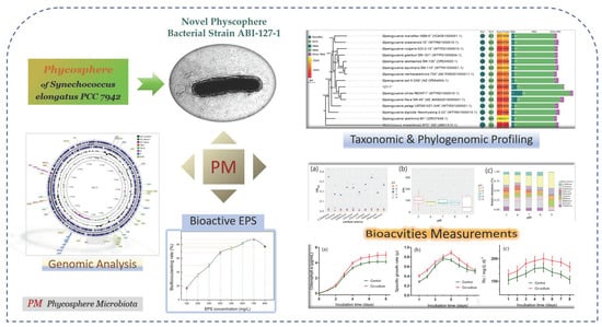
Taxonomic, Phylogenomic and Bioactivity Profiling of Novel Phycosphere Bacterium from Model Cyanobacterium Synechococcus elongatus PCC 7942

1
Department of Marine Chemistry, College of Marine Science and Technology, Zhejiang Ocean University, Zhoushan 316022, China
2
Cooperative Innovation Center of Industrial Fermentation (Ministry of Education & Hubei Province), Key Laboratory of Fermentation Engineering (Ministry of Education), National “111” Center for Cellular Regulation and Molecular Pharmaceutics, College of Bioengineering, Hubei Key Laboratory of Industrial Microbiology, Hubei University of Technology, Wuhan 430068, China
3
ABI Group, Laboratory of Phycosphere Microbiology, Zhejiang Ocean University, Zhoushan 316021, China
4
Zhejiang Provincial Key Laboratory of Petrochemical Pollution Control, Zhejiang Ocean University, Zhoushan 316022, China
5
Donghai Laboratory, Zhoushan 316022, China
6
East China Sea Fisheries Research Institute, Chinese Academy of Fishery Sciences, Shanghai 200090, China
7
State Key Laboratory of Swine and Poultry Breeding Industry, Institute of Animal Science, Guangdong Academy of Agricultural Sciences, Guangzhou 510640, China
*
Author to whom correspondence should be addressed.
Mar. Drugs 2024, 22(1), 36; https://doi.org/10.3390/md22010036
Submission received: 9 November 2023 / Revised: 14 December 2023 / Accepted: 29 December 2023 / Published: 7 January 2024

Abstract

:
Phycosphere niches host rich microbial consortia that harbor dynamic algae–bacteria interactions with fundamental significance in varied natural ecosystems. Hence, culturing the uncultured microbial majority of the phycosphere microbiota is vital for deep understanding of the intricate mechanisms governing the dynamic interactions, and also to provide novel and rich microbial resources, and to discover new natural bioactive metabolites. Synechococcus elongatus PCC 7942 is a robust model cyanobacterium widely used in environment, synthesis biology, and biotechnology research. To expand the number of novel phycosphere species that were brought into culture and to discover the natural bioactivities, we presented a new yellow-pigmented bacterium named ABI-127-1, which was recovered from the phycosphere of PCC 7942, using an optimized bacterial isolation procedure. Combined polyphasic taxonomic and phylogenomic characterization was performed to confidently identify the new isolate as a potential novel species belonging to the genus Qipengyuania. The observed bioactivity of strain ABI-127-1 with promoting potential towards the growth and CO2 fixation efficiency of the host microalgae was measured. Additionally, the bacterial production of active bioflocculant exopolysaccharides was evaluated after culture optimization. Thus, these findings revealed the potential environmental and biotechnological implications of this new microalgae growth-promoting bacterium isolated from the phycosphere microenvironment.

Graphical Abstract

1. Introduction

Phytoplankton are key primary producers in the ocean and freshwater ecosystems [1,2]. Among them, Synechococcus is one of the unicellular photosynthetic cyanobacteria with wild distribution, which is responsible for a large part of global oceanic primary production and carbon cycling [3,4]. Different cyanobacterial components (e.g., pigments, proteins, polysaccharides, and lipids) display varied biological availability during the decomposition process. However, the majority of Synechococcus-derived organic matter is easily labile for use by the heterotrophic bacterial community [5]. Thus, through varied interactions, marine phytoplanktons and their heterotrophic bacterial communities compose a closely related regulator alliance in the oceans by acting as the dominant primary producers (phytoplankton) and biogeochemical cycle drivers (bacteria), respectively [6]. The kinds of algae–bacteria interactions (ABI) usually span mutualism, commensalism, antagonism, parasitism, and competition, and thus underpin most functions of marine ecological processes [7]. Thus, the phycosphere niches host rich microbial consortium, and thus are regarded as the boundary of phytoplankton holobionts and the ecological interface for complex and dynamic algae–bacteria interactions [8]. Presently, it is becoming increasingly clear that these complex associations usually exert profound influences on fundamental processes including primary production, biogeochemical cycles, phycotoxins biosynthesis, and microbial loops in the oceans [9]. Within this microscopic interface, exopolysaccharides or extracellular polysaccharides (EPS) secreted by both interactive sides are regarded as one vital component of the biological matrix, which embeds the proliferating cells and promotes colony formation during algae–bacteria interactions [10,11,12].
Previous studies have confirmed that the family Erythrobacteraceae in the order Sphingomonadales within the class Alphaproteobacteria is one of the predominant bacterial communities among various phycosphere microbiota found in nature [13,14,15]. Among the 19 currently validated members of Erythrobacteraceae, the genus Qipengyuania was first described in 2015, with the species type Qipengyuania sediminis [16]. Currently, the genera comprises 31 valid type species (https://lpsn.dsmz.de/genus/qipengyuania, accessed on 6 June 2023) isolated from various habitats [17,18,19]. It may indicate that this genus may be widely distributed in coastal environments like the Roseobacter lineage [20]. In recent years, members of Qipengyuania have attracted increasing attention due to their diverse potential applications in agriculture, biotechnology, and industry [21].
Still, only a minority of marine microorganisms can be cultured in the laboratory and the enormous bioprospecting potential of uncultured diversity remains unexplored [22]. To improve our understanding of uncultured microbial diversity, it is essential to increase our capacity for bringing microorganisms from the environment into culture [23,24,25]. During the execution of our Phycosphere Microbiome Project (PMP) conveying the microbial diversity of various phycosphere microbiota recovered from the ocean and freshwater ecosystems [26,27,28,29,30], a bioactive yellow-pigmented bacterium named ABI-127-1 was isolated from the phycosphere of Synechococcus elongatus PCC 7942, which is a versatile and robust model cyanobacterial strain widely used in the environment, such as in carbon fixation and blue carbon, capturing and acclimation, synthesis biology, and biotechnology research such as green biofuel production [31,32,33,34]. In this study, we proposed a new isolate, strain ABI-127-1, to represent a potential novel species of the genus Qipengyuania within the family Erythrobacteraceae based on combined polyphasic taxonomic and phylogenomic characterizations. Moreover, various bioactivities of strain ABI-127-1 either acting as new microalgae-growth-promoting bacterium (MGPB), or the bacterial bioflocculanting EPS, as well as the bacterial enhanced carbon fixation efficiency via microalgae, were also measured.

2. Results and Discussion

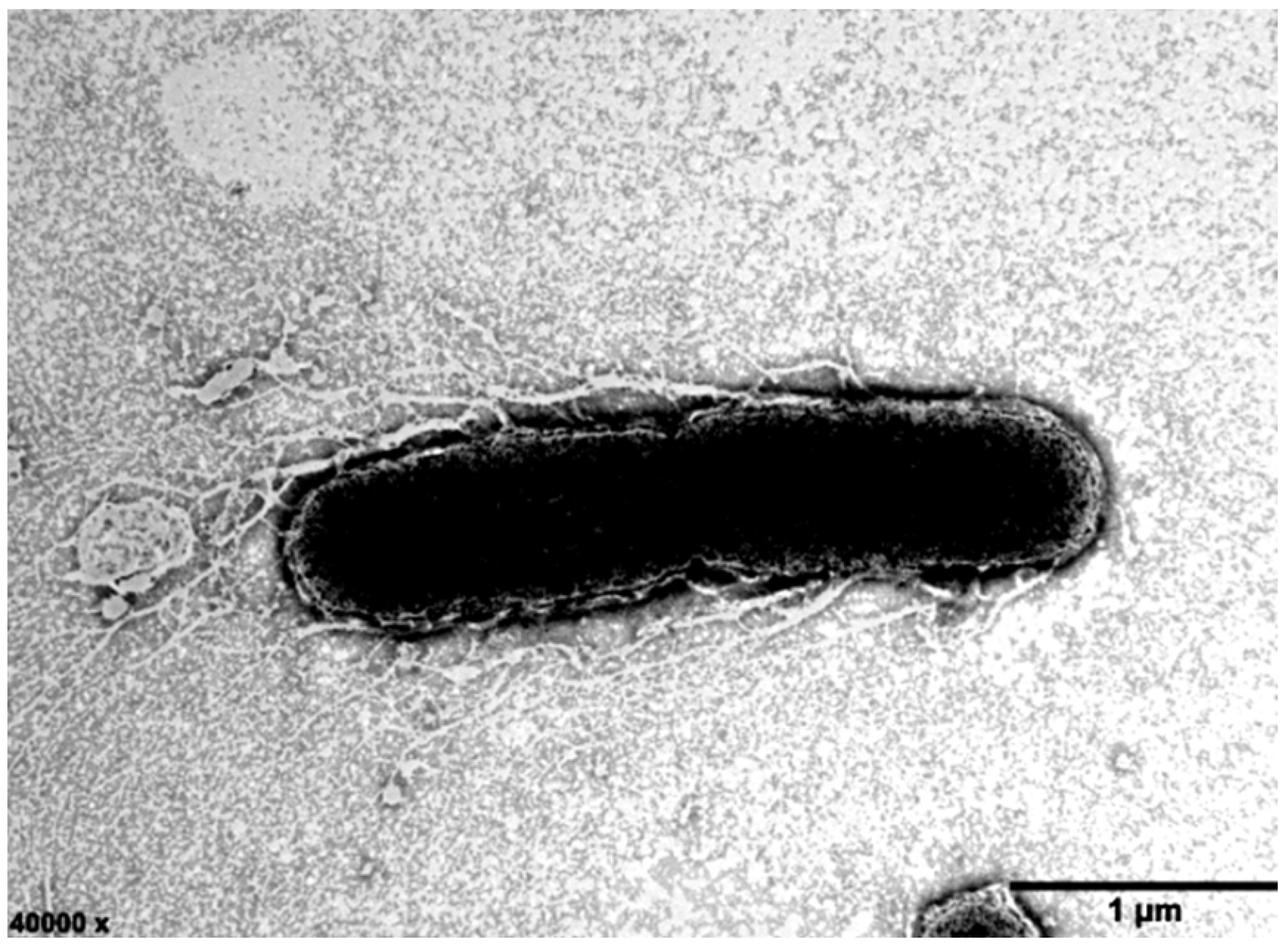
2.1. Morphological and Biochemical Characterization of Strain ABI-127-1

Strain ABI-127-1 was observed to grow well on MA, R2A, nutrient agar (NA), and trypticase soy agar (TSA), and the optimum medium was MA. Colonies of this strain were faint yellow, smooth, and circular when they were grown on MA. Cells of variable size (0.8–1.1 × 2.1–2.9 µm) were observed (Figure 1). Cells were rod-shaped, Gram-stain negative, aerobic, oxidase and catalase positive, and non-motile. Bacterial growth occurred at pH 5.0–10.0 (optimum, 7.0–8.0) and 15–35 °C (optimum, 28–30 °C) with 1–10% (w/v) NaCl (optimum, 2.5%) in MA. Differential physiological and biochemical characteristics between strain ABI-127-1 and close relatives in the genus Qipengyuania were shown in Table 1. Some unique properties such as the utilization of glucose, fructose, and lactate can be used to distinguish strain ABI-127-1 from close relatives. Also, the new isolate strain ABI-127-1 has been deposited in two counties, with certificate deposition numbers of CCTCC AB 2022122 (China Center for Type Culture Collection, CCTCC in China) or KCTC 92508 (Korean Collection for Type Cultures, KCTC in Republic of Korea), respectively.

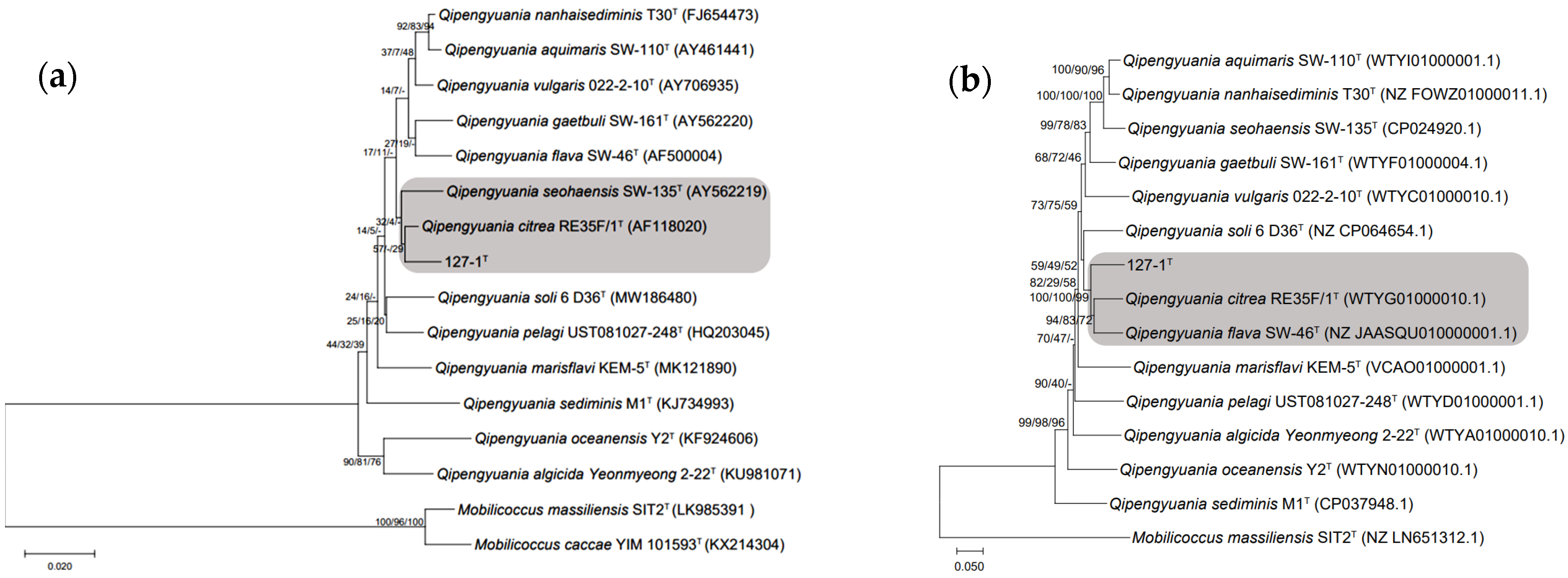
2.2. Phylogenetic Characteristics of Strain ABI-127-1

The almost complete 16S rRNA gene sequence (1474 bp) of strain ABI-127-1 was obtained and submitted to GenBank with the accession number of ON276358. It was found that it is identical to the gene sequence from the genomic sequence. The similarity comparison of 16S rRNA gene sequences showed the highest similarity values to Q. citrea RE35F/1T (98.6%), Q. aquimaris SW-110T (98.3%) and Q. nanhaisediminis CGMCC 1.7715T (97.8%), respectively. The constructed phylogenetic tree based on bacterial 16S rRNA gene sequences indicated that strain ABI-127-1 formed a distinct separate phylogenetic line and clustered with type strain Q. citrea RE35F/1T (Figure 2a). Additionally, the MLSA phylogenetic analysis based on three housekeeping genes (dnaA, ropA, and pheS) also showed that strain ABI-127-1 occupied a separate phylogenetic branch and clustered with two strains of Q. flava and Q. citrea types (Figure 2b).

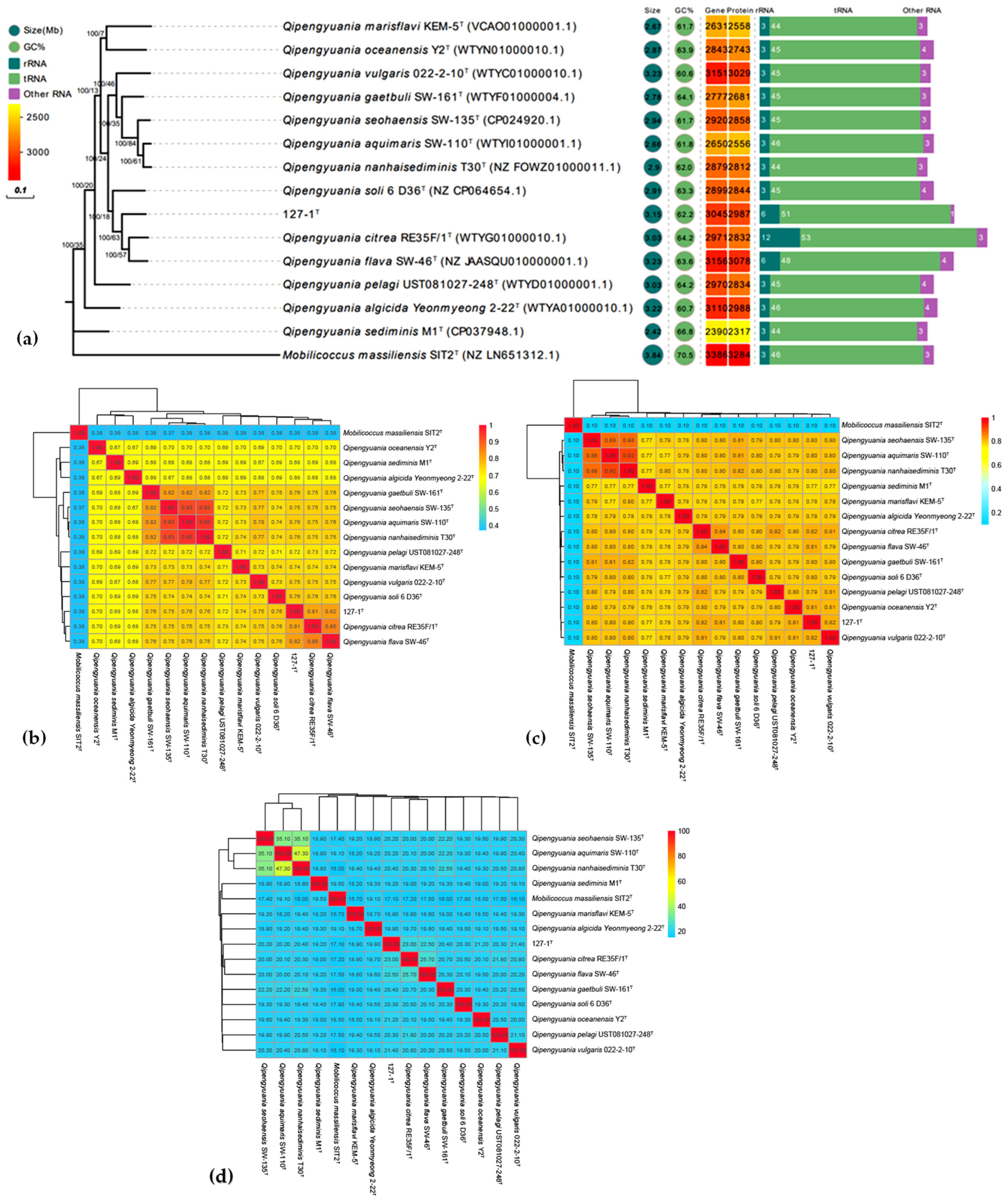
2.3. Phylogenomic Inference

The length of the draft genome sequence of strain ABI-127-1 (GenBank accession number JALJCR000000000) was approximately 3.15 Mb. The genomic DNA G+C content was calculated to be 62.15 mol%. To further infer the phylogenetic relationship between strain ABI-127-1 and closely related type strains with available genome sequences, the phylogenomic analysis was performed using an up-to-date bacterial core gene set (UBCG) [27,28,29]. As shown in Figure 3a, strain ABI-127-1 formed a separate branch in the constructed phylogenetic tree and clustered with Q. citrea RE35F/1T and Q. flava SW-46T. This finding was consistent with the result obtained using the MLSA analysis (Figure 2b).
Additionally, in order to fully resolve the taxonomic status of this new isolate, three measures of phylogenomic similarity including the average nucleotide identity (ANI), average amino acid identity (AAI), and digital DNA–DNA hybridization (dDDH) were performed. The calculated ANI, AAI, and dDDH values between strain ABI-127-1 and close relatives in the genus Qipengyuania were 77.1–81.2%, 64.1–75.6%, and 18.5–22.2%, respectively (Figure 3). Three phylogenomic similarities were far below the threshold values for novel species delineation [35]. Therefore, it is proposed that strain ABI-127-1 represents a potential novel species of the genus Qipengyuania.

2.4. Genomic Sequencing and Annotation of Strain ABI-127-1

The obtained draft genome of strain ABI-127-1 consisted of six contigs with a genome size of 3,151,676 bp and an N50 value of 1,937,448 bp with an over 360× genome coverage. A circular representation of the draft genome is shown in Figure 4. Based on the genome annotation, the total number of predicted genes was 3089, and 3068 were identified as protein-coding genes. It also had 57 RNA genes including 51 tRNA and 6 rRNA genes. A total of 2215 genes (73.7%) were assigned to 26 functional categories based on the COG database, and 850 genes participated in the pathway of metabolism, which was classified as the primary function of the chromosome. Additionally, a total of 217 genes were not assigned to any known function, accounting for 9.8% of the total genes. KEGG analysis revealed that a total of 1550 proteins were classified into six pathways, and 42.8% were involved in different metabolism categories. GO analysis assigned total 2177 proteins into three functional groups, and the top five subgroups were assigned to the molecular function, biological process, cellular process, catalytic activity, and metabolic process, respectively.
Due to the close phylogenomic relationship between strains strain ABI-127-1 and Q. flava SW-46T and Q. citrea RE35F/1T, the genomes of the three type strains were compared. For Q. citrea RE35F/1T, the genomic length was calculated as 3,031,321 bp consisting of 2971 predicted coding genes with a DNA G+C content of 64.2 mol%, whereas the genome size of strain Q. flava SW-46T was 3,229,668 bp in which the DNA G+C contents of both strains were 63.6 mol% and 3156 predicted coding genes. The DNA G+C contents of three type strains all fell within the range of 60.5–66.8 mol% for members of the genus Qipengyuania [16,17,18,19].

2.5. Biosynthetic Gene Prediction for Active Metabolites

Based on genomic annotation, a bioinformatics prediction of the biosynthetic genes in strain ABI-127-1 was performed. It was found that strain ABI-127-1 had genes which were responsible for biosynthetic modules for various co-factors and biotin B12 (Figure 4 and Figure 5), which are essential vitamins for algal growth and proliferation [33,34,36]. It indicated that strain ABI-127-1 owned the genetic basis to synthesize some interactive metabolites and exchanged them with the algal hosts during algae–bacteria interactions. In addition, two core genes (epsL and epsN) which were responsible for bacterial EPS biosynthesis were also found [37,38]. EPSs are considered to be the fundamental intermediates during algae–bacteria interactions thar occur within microscale phycosphere niches [10,39,40,41].
It is also noteworthy that the biosynthetic genes for the nucleotide sugar precursor dTDP-L-rhamnose were only found in strain ABI-127-1 (Figure 5) when compared with three phylogenetic neighbors including Q. citrea (from seawater), Q. soli (from mangrove soil), and Q. flava (from seawater). These genes are responsible for the production of structurally similar rhamnose polysaccharides in the bacterial cell wall, which is critical for maintaining cell shape, bacterial physiology, and virulence, and also for host interactions as vital chemical intermediates [35,42]. This finding may indicate the unique characteristic of the phycosphere niche that strain ABI-127-1 inhabits [39]. Also, the cobalamin biosynthesis pathway, the process by which bacteria and archea make cobalamin vitamin B12, was only found for strain Q. citrea isolated from the marine environment.

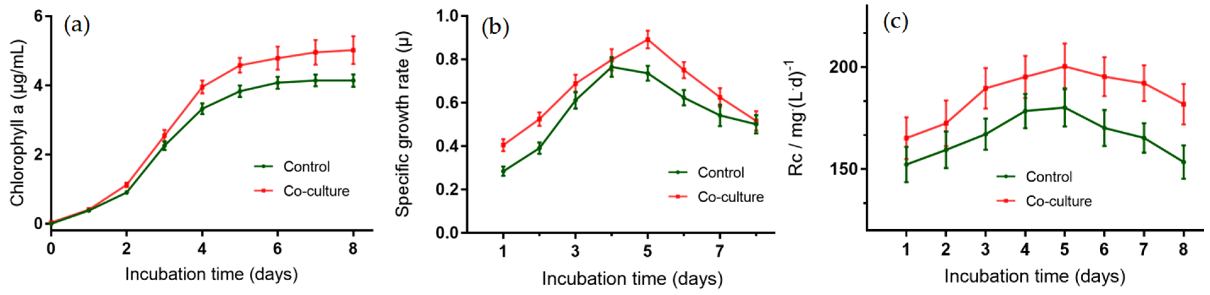
2.6. Growth-Promoting Potential of Strain ABI-127-1 towards Algal Host

Our previous studies showed that some species in the phycosphere bacteria community demonstrated growth-promoting potential towards algal hosts [10,12,28,34]. Based on microalgae growth-promoting (MGP) bioactivity assaying, the total numbers of algal cells of PCC 7942 significantly increased when co-inoculated with strain ABI-127-1 with at least 16.5 ± 3.8% promotion efficiency (Figure 6). This indicates that the strain ABI-127-1 has obvious MGP activity. This finding may indicate that the possible association between strain ABI-127-1 and algal host, in spite of the nature of the detailed mechanism, remains unsolved. The current ongoing exploration of the potential associations in the co-culture circumstances based on multi-omics analysis is believed to offer insights into the mechanism, governing the interactions between strain ABI-127-1 and algal host PCC 7942 [6,7,8,43,44,45].

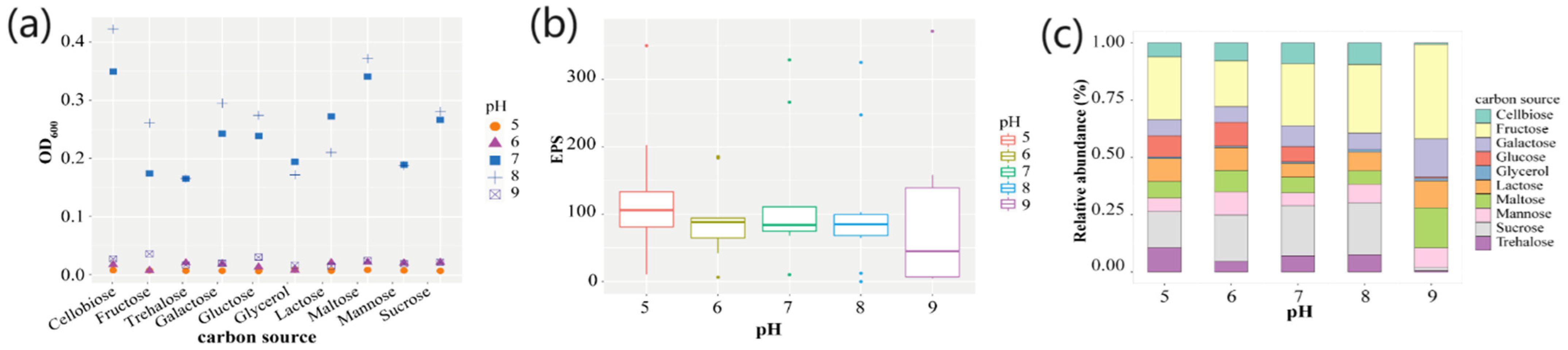
2.7. Culture Optimization for Bacterial EPS Production by Strain ABI-127-1

A preliminary investigation showed that two factors, the pH and carbon source in the media, were the main factors influencing the EPS accumulation in strain ABI-127-1. In this study, ten selected carbon sources including the cellbiose, fructose, galactose, glucose, glycerol, lactose, maltose, mannose, sucrose, and trehalose, and a pH ranging from 5.0 to 9.0, were selected and used for culture optimization for bacterial ESP production (Figure 7a). For the pH test, it was found that strain ABI-127-1 grew better at pH values ranging from 7.0 to 9.0. Moreover, it achieved the fastest growth rate when the cellobiose was used as the single carbon source when cultured at pH 8.0 (Figure 7b). However, according to the EPS accumulation measurements, the higher EPS yield was achieved when strain ABI-127-1 was cultured at pH 9.0 (Figure 7b). Thus, under the optimized conditions, the highest EPS yield of 118.2 ± 15.8 μg/mL (means ± SD) was obtained when cellobiose (10 g/L) was used as the carbon source and cultured at 30 °C at pH 9.0 (Figure 7c).

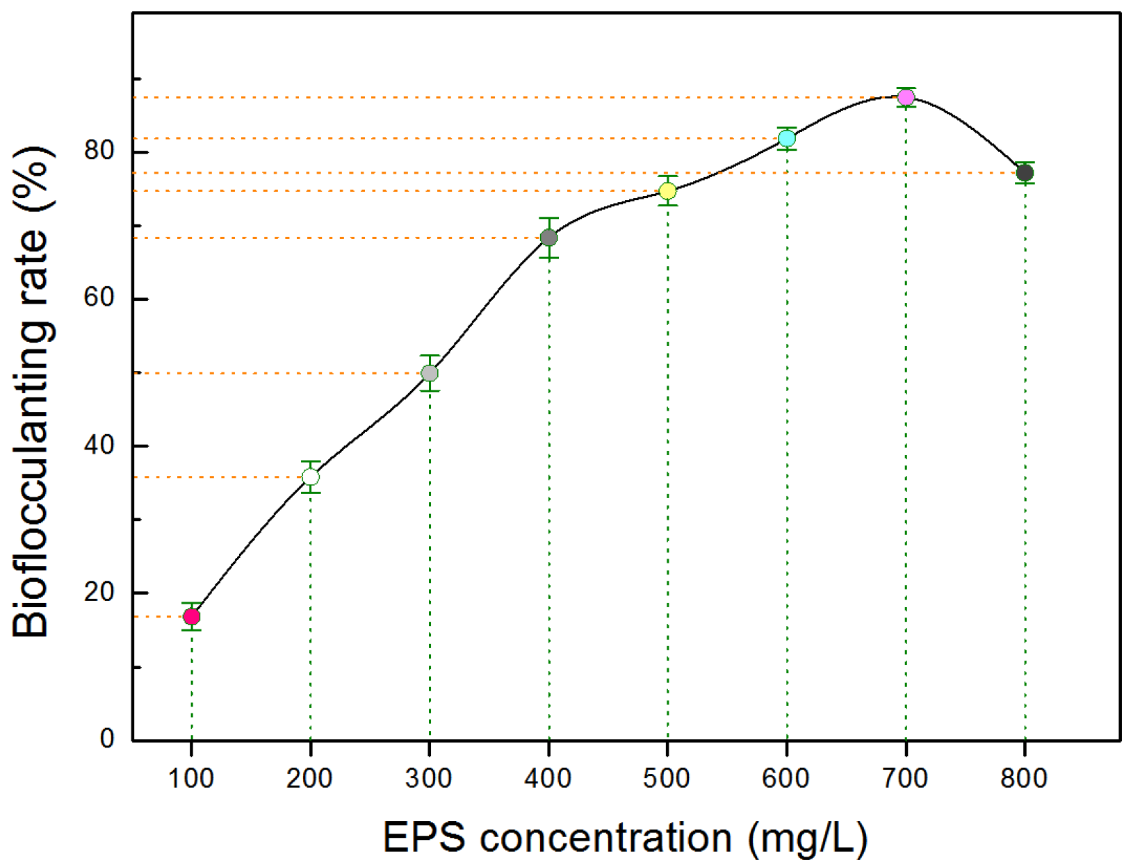
2.8. Bioflocculant Bioactivity of EPS Produced by Strain ABI-127-1

To measure the production capacity of bacterial bioflocculants, the extracted EPSs produced by strain ABI-127-1 were subjected to bioflocculant activity evaluation [10,33,43]. A comparison of the bioflocculant effect under a series of EPS concentrations was performed. As shown in Figure 8. it can be seen that the bioflocculant efficiency of EPS produced by strain ABI-127-1 showed a concentration-dependent manner, and it reached a maximum of 86.4 ± 3.9% (means ± SD) when the EPS concentration was set as 0.70 g/L. It exhibited a higher bioflocculant capacity compared with other marine bacterial strains which were also isolated from the phycosphere microenvironments as previously reported [10,33,44]. The finding indicated that strain ABI-127-1 may serve as a new bacterial candidate with natural potential for the production of promising microbial bioflocculants derived from marine phycosphere niches. Furthermore, the ongoing chemical elucidation of the produced bacterial EPSs is believed to uncover the chemical nature of the natural bioflocculant instinct contributed by strain ABI-127-1.

2.9. Bacterial Enchantment of Strain ABI-127-1 for Algal CO2 Fixation Efficiency

In a co-culture system, the cells of PCC 7942 were cultivated with strain ABI-127-1 in the optimized medium and aerated with 30 mL/min of 15% CO2 to determine the biomass productivity and CO2 fixation efficiency. As shown in Figure 9a, based on the chlorophyll a measurements, strain ABI-127-1 showed an obvious promoting effect on the microalgae biomass productivity with at least 20.8 ± 5.4% promotion efficiency. Chlorophyll a production reached the maximum value (17.6 ± 0.8%) by the sixth day. Figure 9b showed that, under co-culture circumstances, the peak growth rate of the PCC 7942 cells was increased to 0.892 g/L·day (0.715 g/L·day with the sole PCC 7942). Moreover, the time of peak growth rate was observed to be delayed from day 4 to day 5. The CO2 fixation efficiency by PCC 7942 cells when co-cultured with strain ABI-127-1 was increased from 2.25 ± 0.24% to 4.36 ± 0.39% within the first 2 days with increasing values from 152.3 ± 15.8 to 165.2 ± 17.3 mg/d, and then stabilized to over 10% within the following six days, and the average CO2 fixation efficiency under co-culture circumstance was 46.8 ± 5.6% during the whole experiment period (Figure 9c). This clearly shows the enhancing effect of the strain ABI-127-1 on algal CO2 fixation efficiency.

3. Materials and Methods

3.1. Bacterial Isolation and Culture Conditions

The model cyanobacterium Synechococcus elongatus PCC 7942 was purchased from the Freshwater Algae Culture Collection at the Institute of Hydrobiology (FACHB, Wuhan, China). For algal cultivation, PCC 7942 cells were grown in BG11 medium shaken at 150 rpm and at 30 °C under constant white-light illumination (30 μmol·m2−1·s−1) [31]. Bacterial strains were isolated from PCC 7942 cells using the dilution-to-extinction method as previously reported [33]. The isolation procedure was performed by spreading algal cells on marine 2216 agar (MA, BD, Franklin Lakes, NJ, USA) supplemented with an algal extract of PCC 7942 cells at a final concentration of 0.023 mg/L [35]. The isolated bacteria were routinely cultured on marine 2216 agar and preserved as 20 % (v/v) glycerol suspensions and stored at −80 °C.
For the comparative analyses, five type reference bacterial strains were purchased and used, including Qipengyuania citrea RE35F/1T (=DSM 14432T) and Qipengyuania seohaensis SW-135T (=DSM 16221T) obtained from the Deutsche Sammlung von Mikroorganismen und Zellkulturen GmbH (DSMZ, Braunschweig, Germany), Qipengyuania nanhaisediminis T30T (=CGMCC 1.7715T) obtained from the China General Microbiological Culture Collection Center (CGMCC, Beijing, China), Qipengyuania aquimaris SW-110T (=KCCM 41818T) obtained from the Korean Culture Center of Microorganisms (KCCM, Seoul, South Korea), and Qipengyuania pelagi UST081027-248T (=NRRL 59511T) obtained from the Agricultural Research Service Culture Collection (NRRL, Peoria, IL, USA), respectively.

3.2. Phenotypic and Physiological Characterization

For the comparative analyses, all biochemical and physiological tests were performed where ABI-127-1 and five reference strains were routinely cultivated on MA at 30 °C following different experimental requirements. Bacterial growth was investigated on R2A agar (BD), trypticase soy agar (BD), MacConkey (BD), nutrient agar (BD), and marine agar (BD) after incubation at 30 °C for 2 days [43]. Colony color and morphology were observed. Morphological and cultural characteristics were investigated with cells grown on MA. Cell morphology and motility were observed using light microscopy (BH-2; Olympus, Tokyo, Japan) and transmission electron microscopy (JEM-1200; JEOL, Tokyo, Japan), respectively, after bacterial incubation at 30 °C for 48 h. The gram reaction was tested using the bioMérieux Gram 2 Kit (bioMérieux, Marcy-l’Étoile, France) according to the manufacturer’s instructions. Oxidase activity was determined with a 1% (w/v) solution of tetramethyl-p-phenylenediamine. Catalase activity was tested with a 3% (w/v) H2O2 solution [37]. Growth at different temperatures (4, 10, 20, 25, 30, 37, 40, 45, and 50 °C) and pH values (4.0–11.0, at intervals of 0.5 pH units) was determined after culture in MB at 30 °C for up to 10 days [38]. Growth at various NaCl concentrations (0–15.0%, w/v, with the interval of 0.5%) was investigated in MB and cultured in a rotary shaker for 7 days.

3.3. Phylogenetic Analysis

For the phylogenetic analysis, the genomic DNA extraction, PCR amplification of the bacterial 16S rRNA gene using the universal primers of 27F/1492R, and the sequencing of the PCR products were performed as described previously [46,47]. The identification of the phylogenetic neighbors and calculation of the 16S rRNA gene sequence similarities were performed using the sequences of the type strains available from GenBank and Ez-Taxon using the BLAST program and online Ez-Taxon database (https://www.ezbiocloud.net, accessed on 26 June 2023). The 16S rRNA gene sequences were aligned using the Clustal_W program, and the phylogenetic tree was constructed via the neighbor-joining (NJ), maximum likelihood (ML), and maximum parsimony (MP) algorithms implemented in the MEGA software (version 7.0) using the default settings [48]. Bootstrap confidence analysis was carried out with 1000 replicates for evaluating the robustness of the tree topologies.

3.4. Genome Sequencing, Assembly, and Annotation

Strain ABI-127-1 was cultured in marine 2216 (MB) (Difco) at 30 °C and the cells were harvested in the mid-logarithmic phase. The genome DNA was extracted and purified using the QIAamp DNA Mini Kit (Qiagen, Hilden, Germany), according to the manufacturer’s instructions, and sequenced by the PacBio RS II Single Molecule Real Time (SMRT) sequencing platform (Pacific Biosciences, Menlo Park, CA, USA) as described previously [10]. For PacBio sequencing, the genomic DNA was sheared to 10 kb using g-TUBE kit (Covaris, Woburn, MA, USA) and converted into the proprietary SMRTbellTM library format using the PacBio RS DNA Template Preparation Kit. The constructed library was then sequenced on the SMRT platform. A total of 1078 Mb filtered high-quality reads with average 360-fold genome coverage were assembled into one contig. The constructed contig was then connected into one chromosome using SOAPdenovo software (version 2.0) [49]. Based on the genome sequence of strain ABI-127-1, the protein-coding regions were predicted by Glimmer (Version 3.02), and all the gene functions were annotated by aligning to the GO (https://geneontology.org/, accessed on 10 July 2023), COG (https://www.ncbi.nlm.nih.gov/COG, accessed on 10 July 2023), and KEGG (http://www.kegg.jp, accessed on 12 July 2023) database, respectively.

3.5. Phylogenomic Calculations

The DNA G+C content was calculated based on the obtained genome of strain ABI-127-1. Three phylogenomic measures of similarity including the average nucleotide identity (ANI), average amino acid identity (AAI), and digital DNA–DNA hybridization (dDDH) [50] were used. The values were calculated using an online EzAAI tool (http://leb.snu.ac.kr/ezaai, accessed on 7 July 2023), and GGDC tool (http://ggdc.dsmz.de/distcalc2.php, accessed on 10 July 2023) using the default parameters [51], respectively. The genome-based phylogenetic tree was constructed using an up-to-date bacterial core gene set (UBCG) consisting of the bacterial core of 92 genes as described previously [52].

3.6. Multilocus Sequence Analysis (MLSA)

For the multilocus sequence analysis (MLSA), three bacterial housekeeping genes including dnaA, ropA, and pheS were used [53]. A total of 13 genome sequences of the closely related reference type strains in the genus Qipengyuania were retrieved from the GenBank database (https://www.ncbi.nlm.nih.gov/genome, accessed on 10 July 2023). Three housekeeping genes were extracted from the genome sequence using PhyloSuite software (version 1.2.2) [54]. Phylogenetic trees were reconstructed by MEGA_X (version 4.0) using three algorithms and using bootstrap analysis based on 1000 replications.

3.7. Bacterial Growth and EPS Production Measurements

For the culture optimization procedure, 1 mL of 24 h fresh bacterial culture of strain ABI-127-1 was taken and mixed with 24 mL of fresh 2216 medium. Then, a 150 μL of the mixture was added into the wells of the 96-well microplate containing different carbon sources and cultured with shaking at 60 rpm/min for 72 h [55]. During the culture period, the bacterial growth rate was monitored with the measurement of the optical density change recorded at OD600 nm every three hours. Ten carbon sources including cellbiose, fructose, galactose, glucose, glycerol, lactose, maltose, mannose, sucrose, and trehalose, which were added with the final concentration of 10 g/L and a pH range of 5.0–9.0 with 1.0 as the interval, were used for the measurements [56]. The extraction and the quantification analysis of the produced EPS were performed using the phenol sulfuric acid method, as reported previously [57].

3.8. Measurements of Bioflocculant and MGP Bioactivities

The evaluation of the bioflocculant activity of the EPS was performed according to procedures reported previously [10,33,58]. The prepared EPS substances were dissolved in the distilled water for a further bioactivity assay. Measurements using kaolin clay suspension flocculation assay calculated and expressed as bioflocculation rate (%) were used and were performed using 96-well microplates with at least six replicates [59]. The microalgae growth-promoting (MGP) activity of strain ABI-127-1 toward PCC 7942 cells in a co-culture system was evaluated as reported previously [60] and performed in a SpectraMax M2 model 96-well microplate reader (Molecular Devices, LLC, San Jose, CA, USA). All the obtained results were expressed as means ± SD.

3.9. Measurement of Algal Carbon Fixation Efficiency in Co-Culture System

During the co-culture, the chlorophyll was extracted from microalgae cells via soaking in DMSO/acetone solution (1/2, v/v) and then measured as previously reported [61]. In total, 20 mL of the samples was centrifuged at 6000× g for 10 min and dried at 65 °C for 24 h to obtain the dried biomass weight. Biomass concentration (g/L) was calculated from the microalgae dry weight produced per liter [62]. The growth rate (GR, g/L·day) was calculated using the following formula, GR = (M1 − M2)/(t1 − t2), where M1 was the biomass concentration at the time of t1 and M2 was the biomass concentration at the time of t2. The carbon content of dry algae cells (Xcbm, %) was analyzed using a CHN element analyzer (Vario Macro Cube, Elementar, Germany), as previously reported [63]. The algal CO2 fixation efficiency (mg/L·d) was calculated according to the following formula, Rc = Px·Xcbm·MCO2/M, where the Rc was for the CO2 fixation rate (g/L·d), Pxwas was for the growth rate (g/L·d), Xcbm was for the carbon content of dry algae cells (%), MCO2 was for the molecular mass of CO2, and Mc was for the molecular mass of carbon, respectively.

4. Conclusions

Based on a phylogenetic analysis using the bacterial 16S rRNA gene alignment, the yellow-pigmented bacterial strain ABI-127-1, which was isolated from the cultivable phycosphere microbiota of model cyanobacterium, Synechococcus elongatus PCC 7942, was shown to belong to the genus Qipengyuania. Additional phylogenomic analysis obtained the unequivocally separated strain ABI-127-1 from close relatives and further confirmed the phylogenetic position of this isolate to represent a potential novel species of this genus. This new isolate demonstrated obvious promoting potential towards both algal growth and CO2 fixation efficiency. Additionally, the bacterial production of active bioflocculant exopolysaccharides was also presented. The current study reveals the potential environmental and biotechnological implications of this new microalgae growth-promoting bacterium recovered from unique phycosphere microenvironment.

Author Contributions

Conceptualization, Q.Y.; methodology, J.X., J.D., L.F. and X.T; software, X.Z., J.X., L.F. and X.T.; validation, X.Z., L.Z., L.F. and Q.Y.; formal analysis, X.Z., J.X., J.D., L.Z. and X.T.; investigation, X.Z., J.X., J.D. and Q.Y.; resources, X.Z. and Q.Y.; data curation, X.Z. and X.T.; writing—original draft preparation, X.Z. and Q.Y.; writing—review and editing, X.Z. and Q.Y.; visualization, X.Z. and X.T.; supervision, Q.Y.; project administration, X.Z. and Q.Y.; funding acquisition, X.Z. and Q.Y. All authors have read and agreed to the published version of the manuscript.

Funding

This work was supported by Open Project Funding of the Key Laboratory of Fermentation Engineering of Ministry of Education (202209FE04), Natural Science Foundation of Zhejiang (LY23D060005), Project from Municipal Science and Technology Bureau of Zhoushan (2022C41018), Open Project of State Key Laboratory of Swine and Poultry Breeding Industry (2023GZ18), Open Project of Zhejiang Provincial Key Laboratory of Petrochemical Pollution Control (2022Y03), Open Project of Key Laboratory of Prevention and Control of Livestock and Poultry Disease of Guangdong (YDWS202108), NSF of China (41876114), Central Public-interest Scientific Institution Basal Research Fund of CAFS (2023TD73), and Science Foundation of Donghai Laboratory (DH-2022KF0218).

Informed Consent Statement

Not applicable.

Data Availability Statement

All relevant data are included in the manuscript.

Conflicts of Interest

The authors declare no conflicts of interest.

References

  1. Giovannoni, S.J.; Nemergut, D. Microbes ride the current. Science 2014, 345, 1246. [Google Scholar] [CrossRef] [PubMed]
  2. Landry, Z.C.; Vergin, K.; Mannenbach, C.; Block, S.; Yang, Q.; Blainey, P.; Carlson, C.; Giovannoni, S.J. Optofluidic Single-Cell Genome Amplification of Sub-Micron Bacteria in the Ocean Subsurface. Front. Microbiol. 2018, 9, 1152. [Google Scholar] [CrossRef]
  3. Li, Z.; Li, S.; Chen, L.; Sun, T.; Zhang, W. Fast-growing cyanobacterial chassis for synthetic biology application. Crit. Rev. Biotechnol. 2023, 26, 1–15. [Google Scholar] [CrossRef]
  4. Doré, H.; Leconte, J.; Guyet, U.; Breton, S.; Farrant, G.K.; Demory, D.; Ratin, M.; Hoebeke, M.; Corre, E.; Pitt, F.D.; et al. Global Phylogeography of Marine Synechococcus in Coastal Areas Reveals Strong Community Shifts. mSystems 2022, 7, e0065622. [Google Scholar] [CrossRef]
  5. Díez, J.; López-Lozano, A.; Domínguez-Martín, M.A.; Gómez-Baena, G.; Muñoz-Marín, M.C.; Melero-Rubio, Y.; García-Fernández, J.M. Regulatory and metabolic adaptations in the nitrogen assimilation of marine picocyanobacteria. FEMS Microbiol. Rev. 2023, 47, fuac043. [Google Scholar] [CrossRef] [PubMed]
  6. Amin, S.A.; Parker, M.S.; Armbrust, E.V. Interactions between diatoms and bacteria. Microbiol. Mol. Biol. Rev. 2012, 76, 667–684. [Google Scholar] [CrossRef] [PubMed]
  7. Kouzuma, A.; Watanabe, K. Exploring the potential of algae/bacteria interactions. Curr. Opin. Biotechnol. 2015, 33, 125–129. [Google Scholar] [CrossRef] [PubMed]
  8. Seymour, J.R.; Amin, S.A.; Raina, J.B.; Stocker, R. Zooming in on the phycosphere: The ecological interface for phytoplankton-bacteria relationships. Nat. Microbiol. 2017, 2, 17065. [Google Scholar] [CrossRef]
  9. Cirri, E.; Pohnert, G. Algae-bacteria interactions that balance the planktonic microbiome. New Phytol. 2019, 223, 100–106. [Google Scholar] [CrossRef]
  10. Yang, Q.; Ge, Y.M.; Iqbal, N.M.; Yang, X.; Zhang, X.L. Sulfitobacter alexandrii sp. nov.; a new microalgae growth-promoting bacterium with exopolysaccharides bioflocculanting potential isolated from marine phycosphere. Antonie Van Leeuwenhoek 2021, 114, 1091–1106. [Google Scholar] [CrossRef]
  11. Ramanan, R.; Kang, Z.; Kim, B.H.; Cho, D.H.; Jin, L.; Oh, H.M.; Kim, H.S. Phycosphere bacterial diversity in green algae reveals an apparent similarity across habitats. Algal Res. 2015, 8, 140–144. [Google Scholar] [CrossRef]
  12. Zhu, W.Z.; Ge, Y.M.; Dai, J.; Zhang, X.L.; Yang, Q. Alexandriicola marinus gen. nov.; sp. nov.; a new member of the family Rhodobacteraceae isolated from marine phycosphere. Antonie Van Leeuwenhoek 2022, 115, 473–486. [Google Scholar] [CrossRef] [PubMed]
  13. Schwenk, D.; Nohynek, L.; Rischer, H. Algae-bacteria association inferred by 16S rDNA similarity in established microalgae cultures. MicrobiologyOpen 2014, 3, 356–368. [Google Scholar] [CrossRef] [PubMed]
  14. Zhang, X.L.; Tian, X.Q.; Ma, L.Y.; Feng, B.; Liu, Q.H.; Yuan, L.D. Biodiversity of the symbiotic bacteria associated with toxic marine dinoflagellate Alexandrium tamarense. J. Biosci. Med. 2015, 3, 23–28. [Google Scholar] [CrossRef]
  15. Zhang, X.L.; Ma, L.Y.; Tian, X.Q.; Huang, H.L.; Yang, Q. Biodiversity study of intracellular bacteria closely associated with paralytic shellfish poisoning dinoflagellates Alexandrium tamarense and A. minutum. Int. J. Environ. Resour. 2015, 4, 23–27. [Google Scholar] [CrossRef]
  16. Feng, X.M.; Mo, Y.X.; Han, L.; Nogi, Y.; Zhu, Y.H.; Lv, J. Qipengyuania sediminis gen. nov.; sp. nov.; a member of the family Erythrobacteraceae isolated from subterrestrial sediment. Int. J. Syst. Evol. Microbiol. 2015, 65, 3658–3665. [Google Scholar] [CrossRef] [PubMed]
  17. Ko, S.R.; Le, V.V.; Srivastava, A.; Kang, M.; Oh, H.M.; Ahn, C.Y. Algicidal activity of a novel bacterium, Qipengyuania sp. 3-20A1M, against harmful Margalefidinium polykrikoides: Effects of its active compound. Mar. Pollut. Bull. 2023, 186, 114397. [Google Scholar] [CrossRef] [PubMed]
  18. Oren, A.; Garrity, G.M. Notification list. Notification that new names and new combinations have appeared in volume 70, part 8 of the IJSEM. Int. J. Syst. Evol. Microbiol. 2020, 70, 5602–5608. [Google Scholar]
  19. Zhao, Z.Y.; Xia, T.T.; Jiao, J.Y.; Liu, L.; Su, Q.Y.; Li, M.M.; Lv, A.P.; Ouyang, Y.T.; Li, W.J.; Ming, H. Qipengyuania thermophila sp. nov.; isolated from a Chinese hot spring. Arch. Microbiol. 2022, 204, 305. [Google Scholar] [CrossRef]
  20. Buchan, A.; González, J.M.; Moran, M.A. Overview of the marine roseobacter lineage. Appl. Environ. Microbiol. 2005, 71, 5665–5677. [Google Scholar] [CrossRef]
  21. Liu, Y.; Pei, T.; Du, J.; Yao, Q.; Deng, M.R.; Zhu, H. Comparative Genomics Reveals Genetic Diversity and Metabolic Potentials of the Genus Qipengyuania and Suggests Fifteen Novel Species. Microbiol. Spectr. 2022, 10, 0126421. [Google Scholar] [CrossRef] [PubMed]
  22. Rinke, C.; Schwientek, P.; Sczyrba, A.; Ivanova, N.N.; Anderson, I.J.; Cheng, J.F.; Darling, A.; Malfatti, S.; Swan, B.K.; Gies, E.A.; et al. Insights into the phylogeny and coding potential of microbial dark matter. Nature 2013, 499, 431–437. [Google Scholar] [CrossRef]
  23. Li, S.; Liu, Q.; Duan, C.; Li, J.; Sun, H.; Xu, L.; Yang, Q.; Wang, Y.; Shen, X.; Zhang, L. c-di-GMP inhibits the DNA binding activity of H-NS in Salmonella. Nat. Commun. 2023, 14, 7502. [Google Scholar] [CrossRef] [PubMed]
  24. Dance, A. The search for microbial dark matter. Nature 2020, 582, 301–303. [Google Scholar] [CrossRef] [PubMed]
  25. Zhang, L.; Li, S.; Liu, X.; Wang, Z.; Jiang, M.; Wang, R.; Xie, L.; Liu, Q.; Xie, X.; Shang, D.; et al. Sensing of autoinducer-2 by functionally distinct receptors in prokaryotes. Nat. Commun. 2020, 11, 5371. [Google Scholar] [CrossRef] [PubMed]
  26. Zhang, X.L.; Li, G.X.; Ge, Y.M.; Iqbal, N.M.; Yang, X.; Cui, Z.D.; Yang, Q. Sphingopyxis microcysteis sp. nov.; a novel bioactive exopolysaccharides-bearing Sphingomonadaceae isolated from the Microcystis phycosphere. Antonie Van Leeuwenhoek 2021, 114, 845–857. [Google Scholar] [CrossRef] [PubMed]
  27. Ren, C.Z.; Gao, H.M.; Dai, J.; Zhu, W.Z.; Xu, F.F.; Ye, Y.; Zhang, X.L.; Yang, Q. Taxonomic and Bioactivity Characterizations of Mameliella alba Strain LZ-28 Isolated from Highly Toxic Marine Dinoflagellate Alexandrium catenella LZT09. Mar. Drugs 2022, 20, 321. [Google Scholar] [CrossRef] [PubMed]
  28. Yang, Q.; Feng, Q.; Zhang, B.P.; Gao, J.J.; Sheng, Z.; Xue, Q.P.; Zhang, X.L. Marinobacter alexandrii sp. nov.; a novel yellow-pigmented and algae growth-promoting bacterium isolated from marine phycosphere microbiota. Antonie Van Leeuwenhoek 2021, 114, 709–718. [Google Scholar] [CrossRef]
  29. Yang, Q.; Jiang, Z.; Zhou, X.; Zhang, R.; Xie, Z.; Zhang, S.; Wu, Y.; Ge, Y.; Zhang, X. Haliea alexandrii sp. nov.; isolated from phycosphere microbiota of the toxin-producing dinoflagellate Alexandrium catenella. Int. J. Syst. Evol. Microbiol. 2020, 70, 1133–1138. [Google Scholar] [CrossRef]
  30. Yang, Q.; Jiang, Z.; Zhou, X.; Zhang, R.; Wu, Y.; Lou, L.; Ma, Z.; Wang, D.; Ge, Y.; Zhang, X.; et al. Nioella ostreopsis sp. nov.; isolated from toxic dinoflagellate, Ostreopsis lenticularis. Int. J. Syst. Evol. Microbiol. 2020, 70, 759–765. [Google Scholar] [CrossRef]
  31. Sawa, N.; Tatsuke, T.; Ogawa, A.; Hirokawa, Y.; Osanai, T.; Hanai, T. Modification of carbon metabolism in Synechococcus elongatus PCC 7942 by cyanophage-derived sigma factors for bioproduction improvement. J. Biosci. Bioeng. 2019, 127, 256–264. [Google Scholar] [CrossRef] [PubMed]
  32. Liang, Y.; Zhang, M.; Wang, M.; Zhang, W.; Qiao, C.; Luo, Q.; Lu, X. Freshwater Cyanobacterium Synechococcus elongatus PCC 7942 Adapts to an Environment with Salt Stress via Ion-Induced Enzymatic Balance of Compatible Solutes. Appl. Environ. Microbiol. 2020, 86, e02904-19. [Google Scholar] [CrossRef] [PubMed]
  33. Helliwell, K.E.; Scaife, M.A.; Sasso, S.; Araujo, A.P.; Purton, S.; Smith, A.G. Unraveling vitamin B12-responsive gene regulation in algae. Plant Physiol. 2014, 165, 388–397. [Google Scholar] [CrossRef] [PubMed]
  34. Croft, M.T.; Lawrence, A.D.; Raux-Deery, E.; Warren, M.J.; Smith, A.G. Algae acquire vitamin B12 through a symbiotic relationship with bacteria. Nature 2005, 438, 90–93. [Google Scholar] [CrossRef]
  35. Guérin, H.; Kulakauskas, S.; Chapot-Chartier, M.P. Structural variations and roles of rhamnose-rich cell wall polysaccharides in Gram-positive bacteria. J. Biol. Chem. 2022, 298, 102488. [Google Scholar] [CrossRef] [PubMed]
  36. Wang, H.; Tomasch, J.; Jarek, M.; Wagner-Döbler, I. A dual-species co-cultivation system to study the interactions between Roseobacters and dinoflagellates. Front. Microbiol. 2014, 5, 311. [Google Scholar] [CrossRef]
  37. Islam, S.T.; Lam, J.S. Synthesis of bacterial polysaccharides via the Wzx/Wzy-dependent pathway. Can. J. Microbiol. 2014, 60, 697–716. [Google Scholar] [CrossRef]
  38. Pérez-Burgos, M.; García-Romero, I.; Jung, J.; Schander, E.; Valvano, M.A.; Søgaard-Andersen, L. Characterization of the Exopolysaccharide Biosynthesis Pathway in Myxococcus xanthus. J. Bacteriol. 2020, 202, e00335-20. [Google Scholar] [CrossRef]
  39. Zhang, X.L.; Qi, M.; Li, Q.H.; Cui, Z.D.; Yang, Q. Maricaulis alexandrii sp. nov.; a novel active bioflocculants-bearing and dimorphic prosthecate bacterium isolated from marine phycosphere. Antonie Van Leeuwenhoek 2021, 114, 1195–1203. [Google Scholar] [CrossRef]
  40. Bruckner, C.G.; Rehm, C.; Grossart, H.P.; Kroth, P.G. Growth and release of extracellular organic compounds by benthic diatoms depend on interactions with bacteria. Environ. Microbiol. 2011, 13, 1052–1063. [Google Scholar] [CrossRef]
  41. Daly, G.; Perrin, E.; Viti, C.; Fondi, M.; Adessi, A. Scaling down the microbial loop: Data-driven modelling of growth interactions in a diatom-bacterium co-culture. Environ. Microbiol. Rep. 2021, 13, 945–954. [Google Scholar] [CrossRef] [PubMed]
  42. Mistou, M.Y.; Sutcliffe, I.C.; van Sorge, N.M. Bacterial glycobiology: Rhamnose- containing cell wall polysaccharides in Gram-positive bacteria. FEMS Microbiol. Rev. 2016, 40, 464–479. [Google Scholar] [CrossRef] [PubMed]
  43. Yan, Z.; Chen, B.; Yang, Y.; Yi, X.; Wei, M.; Ecklu-Mensah, G.; Buschmann, M.M.; Liu, H.; Gao, J.; Liang, W.; et al. Multi-omics analyses of airway host-microbe interactions in chronic obstructive pulmonary disease identify potential therapeutic interventions. Nat. Microbiol. 2022, 7, 1361–1375. [Google Scholar] [CrossRef] [PubMed]
  44. Liu, J.; Yang, J.; Ge, Y.; Yang, Q.; Sun, J.; Yu, X. Acute effects of CH3NH3PbI3 perovskite on Scenedesmus obliquus and Daphnia magana in aquatic environment. Ecotoxicol. Environ. Saf. 2021, 208, 111677. [Google Scholar] [CrossRef] [PubMed]
  45. Barott, K.L.; Rohwer, F.L. Unseen players shape benthic competition on coral reefs. Trends Microbiol. 2012, 20, 621–628. [Google Scholar] [CrossRef] [PubMed]
  46. Yang, Q.; Jiang, Z.; Zhou, X.; Xie, Z.; Wang, Y.; Wang, D.; Feng, L.; Yang, G.; Ge, Y.; Zhang, X. Saccharospirillum alexandrii sp. nov.; isolated from the toxigenic marine dinoflagellate Alexandrium catenella LZT09. Int. J. Syst. Evol. Microbiol. 2020, 70, 820–826. [Google Scholar] [CrossRef] [PubMed]
  47. Duan, Y.; Jiang, Z.; Wu, Z.; Sheng, Z.; Yang, X.; Sun, J.; Zhang, X.; Yang, Q.; Yu, X.; Yan, J. Limnobacter alexandrii sp. nov.; a thiosulfate-oxidizing, heterotrophic and EPS-bearing Burkholderiaceae isolated from cultivable phycosphere microbiota of toxic Alexandrium catenella LZT09. Antonie Van Leeuwenhoek 2020, 13, 1689–1698. [Google Scholar] [CrossRef] [PubMed]
  48. Zhang, X.L.; Yang, X.; Wang, S.J.; Jiang, Z.W.; Xie, Z.X.; Zhang, L. Draft Genome Sequences of Nine Cultivable Heterotrophic Proteobacteria Isolated from Phycosphere Microbiota of Toxic Alexandrium catenella LZT09. Microbiol. Resour. Announc. 2020, 9, e00281-20. [Google Scholar] [CrossRef]
  49. Zhou, X.; Zhang, X.; Jiang, Z.; Yang, X.; Zhang, X.; Yang, Q. Combined characterization of a new member of Marivita cryptomonadis, strain LZ-15-2 isolated from cultivable phycosphere microbiota of toxic HAB dinoflagellate Alexandrium catenella LZT09. Braz. J. Microbiol. 2021, 52, 739–748. [Google Scholar] [CrossRef]
  50. Gao, H.M.; Xie, P.F.; Zhang, X.L.; Yang, Q. Isolation, Phylogenetic and Gephyromycin Metabolites Characterization of New Exopolysaccharides-Bearing Antarctic Actinobacterium from Feces of Emperor Penguin. Mar. Drugs 2021, 19, 458. [Google Scholar] [CrossRef]
  51. Yang, X.; Jiang, Z.W.; Zhang, J.; Zhou, X.; Zhang, X.L.; Wang, L.; Yu, T.; Wang, Z.; Bei, J.; Dong, B. Mesorhizobium alexandrii sp. nov.; isolated from phycosphere microbiota of PSTs-producing marine dinoflagellate Alexandrium minutum amtk4. Antonie Van Leeuwenhoek 2020, 113, 907–917. [Google Scholar] [CrossRef] [PubMed]
  52. Jiang, Z.; Duan, Y.; Yang, X.; Yao, B.; Zeng, T.; Wang, X.; Feng, Q.; Qi, M.; Yang, Q.; Zhang, X.L. Nitratireductor alexandrii sp. nov.; from phycosphere microbiota of toxic marine dinoflagellate Alexandrium tamarense. Int. J. Syst. Evol. Microbiol. 2020, 70, 4390–4397. [Google Scholar] [CrossRef] [PubMed]
  53. Yang, Q.; Jiang, Z.W.; Huang, C.H.; Zhang, R.N.; Li, L.Z.; Yang, G.; Feng, L.J.; Yang, G.F.; Zhang, H.; Zhang, X.L. Hoeflea prorocentri sp. nov.; isolated from a culture of the marine dinoflagellate Prorocentrum mexicanum PM01. Antonie Van Leeuwenhoek 2018, 111, 1845–1853. [Google Scholar] [CrossRef] [PubMed]
  54. Kumar, S.; Stecher, G.; Tamura, K. MEGA7: Molecular evolutionary genetics analysis version 7.0 for bigger datasets. Mol. Biol. Evol. 2016, 33, 1870–1874. [Google Scholar] [CrossRef] [PubMed]
  55. Bankevich, A.; Nurk, S.; Antipov, D.; Gurevich, A.A.; Dvorkin, M.; Kulikov, A.S.; Lesin, V.M.; Nikolenko, S.I.; Pham, S.; Prjibelski, A.D.; et al. SPAdes: A new genome assembly algorithm and its applications to single-cell sequencing. J. Comput. Biol. 2012, 19, 455–477. [Google Scholar] [CrossRef]
  56. Philippot, L.; Andersson, S.G.; Battin, T.J.; Prosser, J.I.; Schimel, J.P.; Whitman, W.B.; Hallin, S. The ecological coherence of high bacterial taxonomic ranks. Nat. Rev. Microbiol. 2010, 8, 523–529. [Google Scholar] [CrossRef] [PubMed]
  57. Glaeser, S.P.; Kämpfer, P. Multilocus sequence analysis (MLSA) in prokaryotic taxonomy. Syst. Appl. Microbiol. 2015, 38, 237–245. [Google Scholar] [CrossRef] [PubMed]
  58. Zhang, D.; Gao, F.; Jakovlić, I.; Zou, H.; Zhang, J.; Li, W.X.; Wang, G.T. PhyloSuite: An integrated and scalable desktop platform for streamlined molecular sequence data management and evolutionary phylogenetics studies. Mol. Ecol. Resour. 2020, 20, 348–355. [Google Scholar] [CrossRef]
  59. Masuko, T.; Minami, A.; Iwasaki, N.; Majima, T.; Nishimura, S.; Lee, Y.C. Carbohydrate analysis by a phenol-sulfuric acid method in microplate format. Anal. Biochem. 2005, 339, 69–72. [Google Scholar] [CrossRef]
  60. Siddharth, T.; Sridhar, P.; Vinila, V.; Tyagi, R.D. Environmental applications of microbial extracellular polymeric substance (EPS): A review. J. Environ. Manag. 2021, 287, 112307. [Google Scholar] [CrossRef]
  61. Gonzalez, L.E.; Bashan, Y. Increased growth of the microalga Chlorella vulgaris when coimmobilized and cocultured in alginate beads with the plant-growth-promoting bacterium Azospirillum brasilense. Appl. Environ. Microbiol. 2000, 6, 1527–1531. [Google Scholar] [CrossRef] [PubMed]
  62. Zhang, L.; Zhang, W.; Li, Q.; Cui, R.; Wang, Z.; Wang, Y.; Zhang, Y.Z.; Ding, W.; Shen, X. Deciphering the Root Endosphere Microbiome of the Desert Plant Alhagi sparsifolia for Drought Resistance-Promoting Bacteria. Appl. Environ. Microbiol. 2020, 86, e02863-19. [Google Scholar] [CrossRef] [PubMed]
  63. Shangguan, L.; Zhang, H.; Liu, Z.; An, F.; Yang, Q.; Zhang, X.; Yao, L.; Yang, S.; Dai, J.; Chen, X. Improved Glutamic Acid Production Capacity of Corynebacterium glutamicum by the ARTP Mutagenesis Method. Fermentation 2023, 9, 599. [Google Scholar] [CrossRef]
Figure 1. Transmission electron micrograph of the cell of strain ABI-127-1 grown on MA for 48 h. Bar 1 μm.
Marinedrugs 22 00036 g001
Figure 2. The constructed phylogenetic trees based on bacterial 16S rRNA gene sequences (a), and the multilocus sequence analysis (MLSA) using three bacterial housekeeping genes (dnaA, ropA, and pheS) (b). Bootstrap values (≥50%) based on 1000 replications are shown at the branch points. Mobicoccus masssiliensis SIT2T was used as the outgroup for both trees.
Marinedrugs 22 00036 g002
Figure 3. Phylogenomic tree (a) of strain ABI-127-1 constructed based on the up-to-date bacterial core gene (UBCG) method. Five genomic characteristics including the genomic size, DNA G+C content (%), numbers of protein-coding genes (CDs), gene numbers of rRNA and tRNA, and phylogenomic trees based on three phylogenomic similarity indexes were shown, including the ANI (b), AAI (c) and dDDH (d) value calculations.
Marinedrugs 22 00036 g003
Figure 4. Circular representation of the genome of strain ABI-127-1. The scale of genome size is shown in the outer line. From the outer to inner circle: two outer circles showing protein-coding sequences (CDs) with different colors showing the genes in varied COG categories; the third circle shows the rRNA and tRNA genes (red); the fourth and inner fifth circles show DNA G+C content and G+C skew, respectively. Predicted biosynthetic gene clusters (BGCs) including biotin synthesis (VBH), vitamin B12 synthesis and transport (VB12), nitrogen (N) and phosphorus (P) metabolism, and EPS synthesis- and transport-related genes are also marked in the genome structure.
Marinedrugs 22 00036 g004
Figure 5. Comparison of the biosynthetic genes responsible for secondary metabolites, co-factors, and vitamin metabolisms between strain ABI-127-1 and three phylogenetic neighbors, Q. citrea, Q. soli, and Q. flava in the genus Qipengyuania.
Marinedrugs 22 00036 g005
Figure 6. The microalgae growth-promoting potential of strain ABI-127-1 on PCC 7942 cells.
Marinedrugs 22 00036 g006
Figure 7. Culture optimization of bacterial growth and EPS production of strain ABI-127-1. The effects of the different pH and carbon sources on bacterial growth recorded at OD600nm (a) and relative EPS production (b,c).
Marinedrugs 22 00036 g007
Figure 8. Bioflocculant activity of the bacterial EPS produced by strain ABI-127-1. The bioflocculant efficiency expressed with the relative bioflocculant rate (%).
Marinedrugs 22 00036 g008
Figure 9. Effects of the co-cultivated strain ABI-127-1 on algal growth based on the measurements of chlorophyll a production (a), the growth rate (b), and also the CO2 fixation efficiency (c) of the PCC 7942 cells.
Marinedrugs 22 00036 g009
Table 1. Characteristics differentiating strain ABI-127-1 from the phylogenetic neighbors within the genus Qipengyuania.
Characteristic123456
Isolation source marine phycosphereseawater seawater seawater seawater marine sediment
Colony colorYellowYellowOrangeOrangeOrangeOrange
Motility+
Nitrate reduction+++
Tolerance to NaCl (%, v/v, optimal)1.0–10.0 (2.5)1.0–10.0 (3.0)1.0–9.0 (2–3)0.5–15.0 (3.0)0.5–9.0 (2–3)0–10.0 (3.5)
Growth temperature (°C, optimal)15–35 (28–30)4–37 (25–30)10–40 (30–35)10–41 (30–35)12–40 (30–36)10-40 (30–35)
Growth pH range (optimal)5–10 (7–8)5–10 (7–8)5–10 (7–8)5–10 (7–8)5–10 (8–9)5–10 (7–8)
Utilization of
Glucose+++++
Fructose+
Lactate+
16S rRNA gene similarity (%)98.6497.7998.2997.2997.79
DNA G+C content (mol%)62.1562.0162.1862.1760.4059.51
Strains: 1, ABI-127-1; 2, Q. citrea RE35F/1T; 3, Q. seohaensis SW-135T; 4, Q. aquimaris SW-110T; 5, Q. pelagi UST081027-248T; 6, Q. nanhaisediminis T30T.
Disclaimer/Publisher’s Note: The statements, opinions and data contained in all publications are solely those of the individual author(s) and contributor(s) and not of MDPI and/or the editor(s). MDPI and/or the editor(s) disclaim responsibility for any injury to people or property resulting from any ideas, methods, instructions or products referred to in the content.

