Article

Sphaeroane and Neodolabellane Diterpenes from the Red Alga Sphaerococcus coronopifolius

Department of Pharmacognosy and Chemistry of Natural Products, School of Pharmacy, University of Athens, Panepistimiopolis Zografou, Athens 15771, Greece
*
Author to whom correspondence should be addressed.
Mar. Drugs 2009, 7(2), 184-195; https://doi.org/10.3390/md7020184
Submission received: 17 April 2009 / Revised: 4 May 2009 / Accepted: 18 May 2009 / Published: 19 May 2009

Abstract

:
Investigation of minor metabolites in the extracts of the red alga Sphaerococcus coronopifolius collected from the rocky coasts of Corfu Island in the Ionian Sea yielded two new diterpene alcohols, sphaerollanes I, and II (1, 2) featuring neodolabellane skeletons, and the new sphaeroane diterpene alcohol 16-hydroxy-9S*-acetoxy-8-epi-isosphaerodiene-2 (3), along with two previously reported metabolites 4, 5. The structures of the new natural products, as well as their relative stereochemistry, were elucidated on the basis of extensive spectral analysis, including 2D-NMR experiments.

Graphical Abstract

1. Introduction

All organisms biosynthesize secondary metabolites for certain essential physiological functions and exhibit complex chemical profiles, frequently including terpene metabolites. The numerous ways that the basic C5 units can be joined together and the different ecological pressures under which organisms have evolved justifies the enormous number and the diversity of the elaborated terpenoid structures [1].
Sphaerococcus coronopifolius has been the focus of previous chemical investigations, which showed the presence of interesting diterpenes with two, three and four ring carbon skeletons, most of which contain one or two bromine atoms [2,3].
In the course of our ongoing investigations toward the isolation of bioactive metabolites from marine organisms of the Greek seas [4,5], we recently studied the chemical composition of the red alga S. coronopifolius, collected from the west coast of Corfu Island. In this report we describe the isolation and structure elucidation of two new diterpenes 1, 2 with neodolabellane carbon skeletons, and one new sphaeroane diterpene 3, along with the already described metabolites presphaerol (4) [68] and isosphaerodiene-1 (5) [8,9] (Figure 1). The structures of the new metabolites were elucidated by extensive spectroscopic analyses and their relative stereochemistry was established by NOESY experiments. Moreover, detailed analyses of the 1D- and 2D-NMR spectra allowed full assignment of the 13C- and 1H- data which had not been previously reported for 4 and 5.

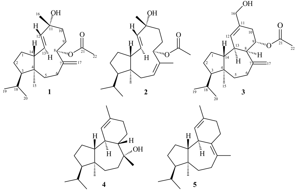
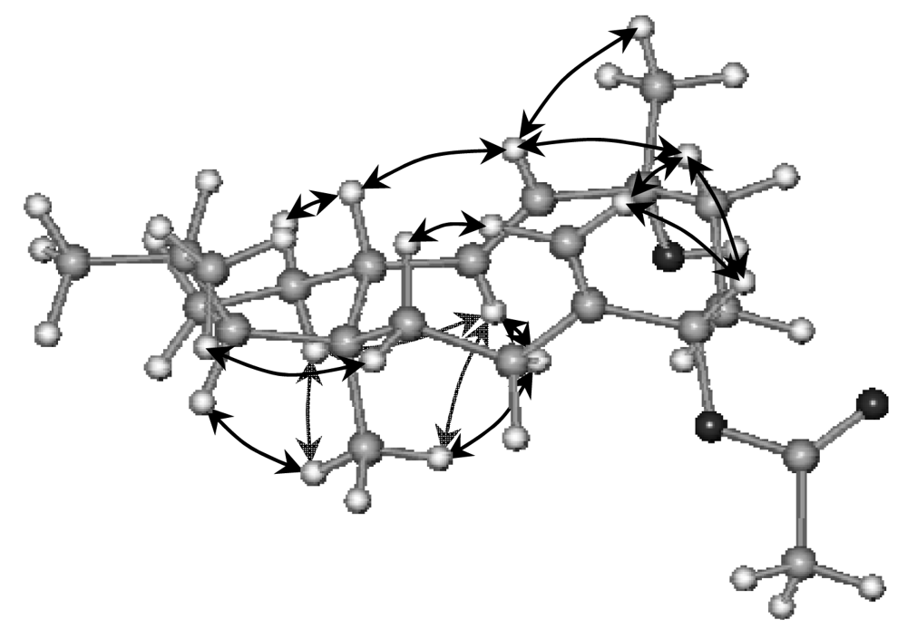
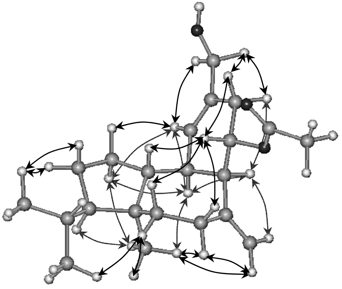
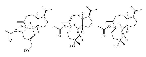
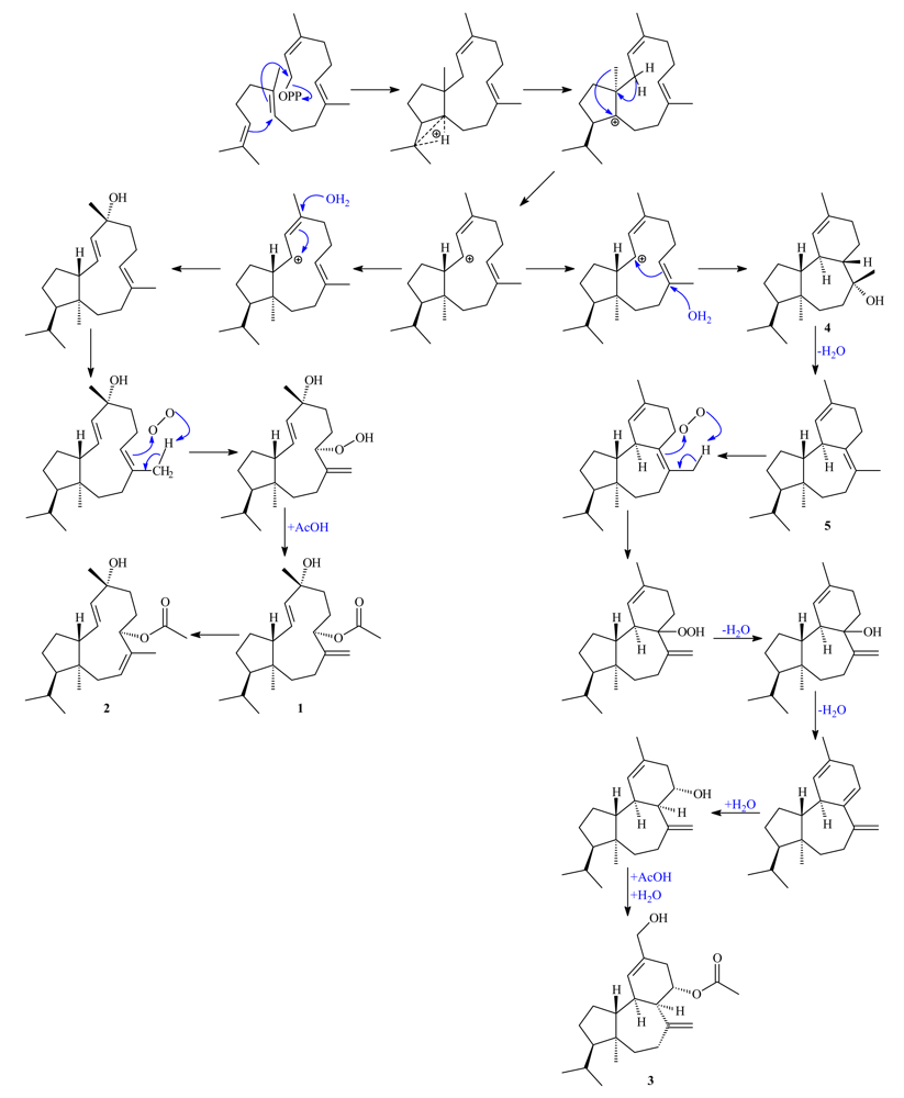
2. Results and Discussion

S. coronopifolius was collected in Palaiokastritsa Bay on the west side of Corfu Island and the CH2Cl2/MeOH extract of the freeze-dried alga was subjected to a series of column chromatography fractionations on silica gel, as well as normal, reversed and chiral phase high performance liquid chromatography (HPLC) separations to yield compounds 15 in pure form.
Sphaerollane I (1) was obtained as a colorless viscous oil. The molecular formula, C22H36O3, which was derived from HRMS and NMR data (Table 1), requires five degrees of unsaturation. The LREI-MS ions at m/z 330 [M-H2O]+, at m/z 288 [M-AcOH]+, and at m/z 270 [M-H2O-AcOH]+ indicated the presence of an acetate and a hydroxyl group. This was supported by the IR spectrum, which contained intense ester bands at 1,730 cm−1 (C=O stretch), 1,240 and 1,030 cm−1 (C–O stretch), and showed the presence of hydroxyl functionality (vmax 3,492 cm−1). Analysis of the NMR data (Table 1) revealed the presence of one acetate group (δH 2.06 s; δC 170.8 s, 21.2 q), a disubstituted double bond (δH 5.52 d and 5.66 dd; δC 137.3 d and 127.5 d), an olefinic methylene group (δH 4.74 br. t and 4.65 br. s; δC 149.8 s and 107.0 t), and a tertiary alcohol (δC 73.4 s). Interpretation of the 1H-1H COSY, 1H-1H TOCSY, HSQC, and HMBC data allowed the remaining two degrees of unsaturation to be assigned to a neodolabellane carbon skeleton. The H3-16 signal at δH 1.24 (s) showed HMBC correlations to C-10 (δC 38.2), C-11 (δC 73.4), and C-12 (δC 137.3), which placed the methyl on the quaternary oxygenated carbon adjacent to the 1,2-disubstituted double bond. Cross peaks in the COSY spectrum revealed couplings between H-10α (δH 1.66) and H-9b (δH 1.42), H-10β (δH 1.48) and H-9a (δH 1.89), and from both H2-9 to H-8 (δH 5.41). Furthermore, the H-8 methine signal showed HMBC correlations to C-9 (δC 28.3), C-10 (δC 38.2), C-7 (δC 149.8), C-17 (δC 107.0), and to the acetate signal at δC 170.8, confirming the position of methylene carbons C-9 and C-10 between the acetoxy-bearing carbon C-8 and the oxygenated carbon C-11. The correlations from H-5β (δH 1.28) and both H2-17 (δH 4.74 and 4.65) to C-6 (δC 28.4) required the presence of the exomethylene at C-7. The H3-15 signal at δH 0.77 (s) correlated with the C-3 (δC 55.4), C-4 (δC 47.1), C-5 (δC 37.0), and C-14 (δC 59.4) signals, allowing it to be placed at the C-4 bridge-head position. Homonuclear coupling between H-13 (δH 5.66) and H-14 (δH 2.18), and HMBC correlations of both H-1β (δH 1.47) and H-12 (δH 5.52) to C-14 (δC 59.4) completed the 11-membered ring by positioning the disubstituted double bond next to the ring junction. The correlations from both methyl signs H3-19 and H3-20 at δH 0.83 (d) and 0.95 (d), respectively, to C-3 (δC 55.4), C-18 (δC 30.3) clearly positioned the isopropyl group at C-3. The signal of H-1α at δH 1.36 (m), belonging to one of the remaining intercoupling methylene groups, showed HMBC correlation with carbon C-13 (δC 127.5). Thus, C-1 had to be connected to C-14, and the remaining C-2 methylene had to be connected to C-3, forming a cyclopentane ring.
The relative stereochemistry shown for 1 (Figure 2) was assigned by interpretation of 1H-NMR coupling constants and NOESY data. The E-geometry of the 12,13-olefin was assigned on the basis of a 15.3 Hz coupling constant. The NOE correlations observed between H-1β and H-12 with H-14, as well as the correlations between H-1α, H-13 and H3-15, required a trans ring fusion and indicated that protons H-1β, H-12 and H-14 were on the same (upper) side of the compound, while H-1α, H-13 and H-15 were on the opposite. The stereochemistry at C-3 was deduced by the NOE correlation between proton H-3 and methyl protons H3-15. The H-8 signal showed a correlation to the H-10β signal, which in turn showed a NOE to the H-12 signal, thereby establishing the stereochemistry at C-8. The correlation between H-12 and H3-16 indicated that methyl H3-16 is also on the upper side of the ring, resulting in the (3R*,4S*,8S*,11R*,12E,14R*)-geometry proposed for sphaerollane I (1).
Compound 2 proved to have the same molecular formula as 1, C22H36O3, which was derived from HRMS and NMR data (Table 1), and is an isomer of 1 that differs only in the location of one of the double bonds. The structural type and substitution pattern of 2 was elucidated by means of 1D- and 2D-NMR correlated spectroscopy including HSQC, HMBC and 1H-1H COSY. Sphaerollane II (2) contained a Δ6 trisubstituted double bond instead of the Δ7,17 olefinic methylene in 1. In the 1H-NMR spectrum (Table 1), both H2-5 signals at δH 2.52 (dd) and 2.28 (ddq) were coupled to the H-6 olefinic signal at δH 5.25 (ddq) that along with methylene proton H-5α (δH 2.28) showed long-range couplings to the H3-17 methyl sign at δH 1.61 (t). The Z geometry of Δ6olefinic bond was assigned on the basis of NOE correlations between H-6 and H3-17, and between H-5β and H-8. Further analysis of the NOESY spectrum revealed that the relative stereochemistry at C-3, C-4, C-8, C-11 and C-14 was the same as in 1 (Table 1, Figure 3).
(3R*,4S*,8R*,9S*,13R*,14R*)-16-Hydroxy-9-acetoxy-8-epi-isosphaerodiene-2 (3) was isolated as a colorless oil. The high-resolution mass measurement established the molecular formula as C22H34O3, requiring six unsaturation equivalents. The IR spectrum showed bands that were assigned to ester (vmax 1,733, 1,240, 1,024 cm−1) and hydroxyl (vmax 3,367 cm−1) functionalities, respectively These assignments were supported by the positive LRCI-MS ions at m/z 329 [M+H-H2O]+, at m/z 287 [M+H-AcOH]+, at m/z 269 [M+H-AcOH-H2O]+, and at m/z 255 [M+H-AcOH-CH2OH]+ indicating the presence of an acetate and a primary hydroxyl group. The presence of five sp2 carbon atoms in the molecule, as deduced from the 13C-NMR and DEPT spectra (Table 2), being for one carbon-oxygen and two carbon-carbon double bonds, indicated compound 3 to be tricyclic. The 1H- and 13C-NMR spectra (Table 2) contained resonances for one acetate group (δH 1.99 s; δC 170.8 s, 21.2 q), a trisubstituted olefinic bond (δH 5.68 br. dd; δC 133.5 s and 127.4 d), an exomethylene (δH 4.79 br. s, 2H; δC 149.4 s and 114.1 t), and a primary alcohol (δH 3.99 br. s, 2H; δC 66.8 t). Extensive analyses of the 2D-NMR data of 3 including COSY, HSQC and HMBC spectra led to the unambiguous assignment of all protons and carbons on the sphaeroane skeleton. The H2-16 oxygenated methylene protons at δH 3.99 (br. s) showed heteronuclear long range couplings to C-10 (δC 32.7), C-11 (δC 133.5), and C-12 (δC 127.4), which confirmed the presence of the primary allylic alcohol at C-11. Correlations from H-8 methine (δH 2.74) and both H2-10 (δH 2.59 and 2.04) to C-9 (δC 68.7), as well as from H-9 (δH 5.35) to the carbonyl signal at δC 170.8 required the acetyl group to be positioned at C-9. The olefinic proton H-12 (δH 5.68) showed HMBC correlations to C-8 (δC 51.3) and C-13 (δC 41.6), thus positioning both the double bond and the acetate group next to the 6-membered ring junctions. The H2-17 olefinic methylene protons at δH 4.79 (br. s) correlated with C-6 (δC 30.8), C-7 (δC 149.4), and C-8 (δC 51.3), had to be placed on C-7. Long range heteronuclear couplings from H2-6 (δH 2.23 and 2.22) to both C-4 (δC 46.4) and C-5 (δC 38.9), and from H2-5 (δH 1.87 and 1.48) to C-7 (δC 149.4) were observed in the HMBC spectrum. The H3-15 methyl signal at δH 0.76 showed correlations to C-3 (δC 58.0), C-4 (δC 46.4), C-5 (δC 38.9), and C-14 (δC 53.7), and was placed on the quaternary carbon C-4. The cycloheptane ring was completed by the HMBC correlation observed between H-8 (δH 2.74) and C-14 (δC 53.7). The correlation of both H3-19 and H3-20 (δH 0.82 and 0.92) with C-3 (δC 58.0) and C-18 (δC 29.8), required the presence of the isopropyl group at C-3. The correlations observed between H-1α (δH 1.25) and C-13 (δC 41.6), and between H-2b (δH 1.28) and C-18 (δC 29.8), confirmed the place of methylene carbons C-1 and C-2 in the 5-membered ring.
Stereochemical assignment of the chiral centers was facilitated by interpretation of 1H-NMR and NOESY spectra. NOE correlations between H3-15 and H-13 and H-1α required trans fusion between the 7-membered and the cyclopentane ring. Furthermore, the correlation between H3-15 and H-3 positioned both H-3 and H-13 co-facial to methyl H3-15. The strong NOEs observed between the pairs of protons H-9, H-14 and H-8, H-13 suggested the unusual for a sphaeroane skeleton cis-fusion of the 7-membered and the cyclohexene rings. Both C-8 and C-13 were assigned as R*, based on the NOE correlations of H3-15 with H2-17, of H2-17 with H-8, of the latter with H-10α, and of H3-15 with H-13, H-5α and H-6α. Moreover, the NOEs observed between H-14 and H-9, and between H-9 and H-5β, H-6β and H-10β, secured the relative stereochemistry at chiral centers C-9 and C-14, thereby defining the structure as (3R*,4S*,8R*,9S*,13R*,14R*)-16-hydroxy-9-acetoxy-8-epi-isosphaerodiene-2 (3) (Figure 4).
Along with the above described metabolites two previously reported compounds, presphaerol (4) [68] and isosphaerodiene-1 (5) [8, 9], were isolated and spectroscopically characterized. Complete 1H- and 13C-NMR assignments, based on extensive analyses of their 2D-NMR spectra, are now provided for the first time for 4 and 5. The co-occurrence of compounds 1, 2 and 3 with the previously described 4 and 5 in the same organism indicated the possibility that they all derive from a common precursor neodolabellane cation [10], as illustrated in the proposed biogenetic pathway (Scheme 1). Thus, successive nucleophilic substitution of a H2O molecule, oxidation of the trisubstituted olefinic bond [11], and acetylation can yield the neodolabellanes 1 and 2. Alternatively, nucleophilic attack of a H2O molecule on the trisubstituted Δ7 double bond can produce presphaerol (4) [10], which through water elimination generates isosphaerodiene-1 (5). Metabolite 3 can be derived from 5 by oxidation of Δ7 double bond and successive eliminations, additions of H2O molecules, and a final acetylation.
Diterpenes of the neodolabellane class have previously been isolated only from soft corals of the genera Cespitularia, Clavularia, and Lobophytum of the Indian and Pacific Ocean [1216], while diterpenes of the sphaeroane skeleton have been isolated, only from the red alga S. coronopifolius and the macromycete Mycena tintinnabulum [17].

3. Experimental Section

3.1. General Experimental Procedures

Optical rotations were measured on a Perkin-Elmer model 341 polarimeter with a 10 cm cell. UV spectra were acquired in spectroscopic grade CHCl3 on a Shimadzu UV-160A spectrophotometer. IR spectra were obtained using a Paragon 500 Perkin-Elmer spectrophotometer. NMR spectra were recorded using a Bruker AC 200 and Bruker DRX 400 spectrometers. Chemical shifts are given on a δ (ppm) scale using TMS as internal standard. The 2D-NMR experiments (1H-1H COSY, HSQC, HMBC, NOESY) were performed using standard Bruker microprograms. The structures in Figures 2, 3 and 4 were generated and optimised (energy: 43.50, 38.24 and 37.77 Kcal/mole, respectively) by “HyperChem 7.0” molecular modelling and simulation software (force field: MM+; optimisation algorithm: Polak-Ribiere). High-Resolution mass spectral data were provided by the University of Notre Dame, Department of Chemistry and Biochemistry, Indiana, USA. Low-Resolution Electron Impact or Chemical Ionisation MS data were recorded on a Thermo DSQ Mass Detector using Direct Exposure Probe (DEP) and methane as the CI gas. Vacuum Liquid Chromatography (VLC) separation was performed with Kieselgel 60 (Merck), gravity column chromatography (GCC) was performed with Kieselgel 60H (Merck), thin layer chromatography (TLC) was performed with Kieselgel 60 F254 aluminum support plates (Merck) and spots were detected after spraying with 15% H2SO4 in MeOH and charring. HPLC separations were conducted using an Agilent 1100 system equipped with refractive index detector and a SupelcoSil 5μ (250×10 mm) HPLC normal phase column or a Kromasil 100 C18 5μ (250×8 mm) HPLC reversed phase column or a Chiralcel OD 5μ (250×4.6 mm) analytical chiral column.

3.2. Plant Material

S. coronopifolius was collected by SCUBA diving in Palaiokastritsa Bay at the west coast of Corfu island, Greece, at a depth of 10–15 m in May of 2002. A specimen is kept at the Herbarium of the Laboratory of Pharmacognosy and Chemistry of Natural Products, University of Athens (ATPH/MO/201).

3.3. Extraction and Isolation

S. coronopifolius was initially freeze-dried (291.4 g dry weight) and then exhaustively extracted with mixtures of CH2Cl2/MeOH (3/1) at room temperature. The combined extracts were concentrated to give a dark green residue (8.20 g), which was later subjected to vacuum liquid chromatography on silica gel, using a 10% step gradient of cyclohexane-EtOAc elution sequence. Fraction F2 (4.01 g), eluted with 20% EtOAc in cyclohexane, was fractionated by gravity column chromatography, using a 2% step gradient of cyclohexane-EtOAc. Fraction F2.8 (524.5 mg), eluted with 8% EtOAc in cyclohexane, was further separated by silica gel GCC using isocratic 5% EtOAc in cyclohexane. Fraction F2.8.15 (53.3 mg) was subjected repeatedly to reverse phase HPLC using MeOH as mobile phase to afford pure 4 (1.9 mg) and 5 (1.8 mg). The CH3CN soluble portion (173.4 mg) of fraction F2.11 (50% EtOAc in cyclohexane) (199.6 mg) was subjected to reverse phase HPLC, using CH3CN as mobile phase. Peak F2.11.7 (retention time 17.9 min) (12.4 mg) was further purified by normal phase HPLC with 20% n-hexane in CHCl3. Compounds 1 (4.9 mg) and 2 (0.9 mg) were isolated by chiral HPLC with 0.5% i-PrOH in n-hexane as eluent, from peak F2.11.7.2 (retention time 9.0 min) (5.8 mg). The CH3CN soluble portion (323.4 mg) of fraction F5 (70% EtOAc in cyclohexane) (446.6 mg) was subjected to reverse phase HPLC, using CH3CN as mobile phase. Peak F5.12 (retention time 11.6 min) (51.6 mg) was further separated by normal HPLC with 30% n-hexane in CHCl3 to yield pure 3 (4.4 mg).

Sphaerollane-I (1)

Colorless oil; [α]D +81.8 (c 2.90, CHCl3); UV (CHCl3) λmax (log ɛ) 255 (0.72) nm; IR (CHCl3) νmax 3,492, 1,730, 1,240, 1,030, 970, 896 cm−1; NMR data (CDCl3), see Table 1; EIMS 70 eV, m/z (rel. int. %) 330 [M-H2O]+ (3), 306 [M-CH2CO]+ (29), 288 [M-AcOH]+ (37), 270 [M-AcOH-H2O]+ (44), 255 [M-AcOH-H2O-Me]+ (20), 245 [M-AcOH-iPr]+ (72), 227 [M-AcOH-H2O-iPr]+ (71), 199 (17), 171 (31), 133 (45), 91 (69), 43 [CH3CO]+ (100); HRFAB-MS (m/z): 349.2732 [M+H]+ (calcd for C22H37O3, 349.2743).

Sphaerollane-II (2)

Colorless oil; [α]D +5.5 (c 1.10, CHCl3); UV (CHCl3) λmax (log ɛ) 250 (1.76) nm; IR (CHCl3) νmax 3,422, 1,728, 1,240, 1,014, 973 cm−1; NMR data (CDCl3), see Table 1; CIMS m/z (rel. int. %) 331 [M+H-H2O]+ (1), 289 [M+H-AcOH]+ (11), 271 [M+H-AcOH-H2O]+ (38), 255 [M+H-AcOH-H2O-Me]+ (9), 245 [M+H-AcOH-iPrH]+ (14), 227 [M+H-AcOH-H2O-iPrH]+ (8), 173 (14), 147 (9), 119 (11), 105 (8), 60 [AcOH]+ (100); HRFAB-MS (m/z): 348.2674 [M]+ (calcd for C22H36O3, 348.2664).

16-Hydroxy-9S*-acetoxy-8-epi-sphaerodiene-2 (3)

Colorless oil; [α]D +10.3 (c 2.90, CHCl3); UV (CHCl3) λmax (log ɛ) 244 (1.95) nm; IR (CHCl3) νmax 3,367, 1,733, 1,240, 1,024, 888 cm−1; NMR data (CDCl3), see Table 2; CIMS m/z (rel. int. %) 347 [M+H]+ (1), 329 [M+H-H2O]+ (9), 287 [M+H-AcOH]+ (29), 269 [M+H-AcOH-H2O]+ (100), 255 [M+H-AcOH-CH2OH]+ (11), 243 [M+H-AcOH-iPrH]+ (8), 225 [M+H-AcOH-H2O-iPrH]+ (12), 213 [M+H-AcOH-CH2OH-iPr]+ (7), 171 (14), 145 (13), 137 (16), 105 (13), 60 [AcOH]+ (38); HRFAB-MS (m/z): 347.2586 [M+H]+ (calcd for C22H35O3, 347.2589).

Acknowledgements

This study was partially supported by the FP-39 EPAN program of the Greek Secretariat for Research and Technology.
  • Samples Availability: Available from the authors.

References

  1. Gershenzon, J; Dudareva, N. The Function of Terpene Natural Products in the Natural World. Nat Chem Biol 2007, 3, 408–414. [Google Scholar] [CrossRef] [PubMed]
  2. Smyrniotopoulos, V; Vagias, C; Rahman, MM; Gibbons, S; Roussis, V. Brominated Diterpenes with Antibacterial Activity from the Red Alga Sphaerococcus coronopifolius. J Nat Prod 2008, 71, 1386–1392. [Google Scholar] [CrossRef] [PubMed]
  3. Smyrniotopoulos, V; Quesada, A; Vagias, C; Moreau, D; Roussakis, C; Roussis, V. Cytotoxic Bromoditerpenes from the Red Alga Sphaerococcus coronopifolius. Tetrahedron 2008, 64, 5184–5190, and references cited therein. [Google Scholar] [CrossRef]
  4. Kladi, M; Vagias, C; Stavri, M; Rahman, MM; Gibbons, S; Roussis, V. C15 Acetogenins with Antistaphylococcal Activity from the Red Alga Laurencia glandulifera. Phytochem Lett 2008, 1, 31–36. [Google Scholar] [CrossRef]
  5. Kontiza, I; Stavri, M; Zloh, M; Vagias, C; Gibbons, S; Roussis, V. New Metabolites with Antibacterial Activity from the Marine Angiosperm Cymodocea nodosa. Tetrahedron 2008, 64, 1696–1702. [Google Scholar] [CrossRef]
  6. Cafieri, F; de Napoli, L; Fattorusso, E; Piattelli, M; Sciuto, S. Presphaerol, a New Rearranged Diterpene from the Alga Sphaerococcus coronopifolius. Tetrahedron Lett 1979, 20, 963–966. [Google Scholar] [CrossRef]
  7. Cafieri, F; Fattorusso, E; di Blasio, B; Redone, C. Diterpenes from the Red Alga Sphaerococcus coronopifolius. Structure of Sphaerodiene and Reassignment of Sstructure for Presphaerol. Tetrahedron Lett 1981, 22, 4123–4126. [Google Scholar] [CrossRef]
  8. Lee, J; Hong, J. First Synthesis and Structural Elucidation of (−)-Presphaerene. J Org Chem 2004, 69, 6433–6440. [Google Scholar] [CrossRef] [PubMed]
  9. Cafieri, F; de Napoli, L; Fattorusso, E; Santacroce, C. Diterpenes from the Red Alga Sphaerococcus coronopifolius. Phytochemistry 1987, 26, 471–473. [Google Scholar] [CrossRef]
  10. Cafieri, F; Fattorusso, E; Santacroce, C. Bromocorodienol, a Diterpenoid Based on a Novel Bicyclic Skeleton from the Red Alga Sphaerococcus coronopifolius. Tetrahedron Lett 1984, 25, 3141–3144. [Google Scholar] [CrossRef]
  11. Bowden, BF; Cusack, BJ; Dangel, A. 13-Epi-9-deacetoxyxenicin, a Cytotoxic Diterpene from the Soft Coral Asterospicularia laurae (Alcyonacea). Mar Drugs 2003, 1, 18–26. [Google Scholar]
  12. Bowden, BF; Coll, JC; Gulbis, JM; Mackay, MF; Willis, RH. Studies of Australian Soft Corals. XXXVIII. Structure Determination of Several Diterpenes Derived from a Cespitularia sp. (Coelenterata, Octocorallia, Xeniidae). Aust J Chem 1986, 39, 803–812. [Google Scholar] [CrossRef]
  13. Bowden, BF; Braekman, J; Mitchell, SJ. Studies of Australian Soft Corals. XX. A New Sesquiterpene Furan from Soft Corals of the Family Xeniidae and an Examination of Clavularia inflata from North Queensland Waters. Aust J Chem 1980, 33, 927–932. [Google Scholar] [CrossRef]
  14. Bowden, BF; Coll, JC; Mitchell, SJ; Stokie, GJ; Blount, JF. Studies of Australian Soft Corals. VIII. A Chemical and Crystallographic Study of a Novel Bicyclic Diterpene Alcohol with a Rearranged Skeleton from an Unknown Species of Soft Coral. Aust J Chem 1978, 31, 2039–2047. [Google Scholar] [CrossRef]
  15. Kobayashi, M; Son, BW; Fujiwara, T; Kyogoku, Y; Kitagawa, I. Neodolabelline, a Methyl Migrated Dolabellane-type Diterpene from the Okinawan Soft Coral Clavularia koellikeri. Tetrahedron Lett 1984, 25, 5543–5546. [Google Scholar] [CrossRef]
  16. Subrahmanyam, C; Venkateswara Rao, C; Anjaneyulu, V; Satyanarayana, P; Subba Rao, PV; Ward, RS; Pelter, A. New Diterpenes from a New Species of Lobophytum Soft Coral of the South Andaman Coast. Tetrahedron 1992, 48, 3111–3120. [Google Scholar] [CrossRef]
  17. Engler, M; Anke, T; Sterner, O. Tintinnadiol, a Sphaeroane Diterpene from Fruiting Bodies of Mycena tintinnabulum. Phytochemistry 1998. [Google Scholar]
Figure 1. Structures of compounds 15.
Marinedrugs 07 00184f1
Figure 2. Relative configurations and key NOE correlations for compound 1.
Marinedrugs 07 00184f2
Figure 3. Relative configurations and key NOE correlations for compound 2.
Marinedrugs 07 00184f3
Figure 4. Relative configurations and key NOE correlations for compound 3.
Marinedrugs 07 00184f4
Scheme 1. Proposed biogenetic origin of 15.
Marinedrugs 07 00184f5
Table 1. NMR dataa of compounds 12.
12

Pos.δHmult, JNOESYδCbHMBC (C→H)δHmult, JNOESYδCbHMBC (C→H)

1β 1.47
α 1.36
m
m
14
13, 15
27.9 t2, 3β 1.57
α 1.41
m
m
14
13, 15
28.2 t2β, 3
2a 1.85
b 1.81
m
m
27.1 t1α, 1β, 18α 1.87
β 1.30
m
m
20
14
28.1 t3
31.21m1555.4 d1α, 1β, 2a, 15, 18, 19, 201.29m1557.0 d15, 19, 20
447.1 s1α, 2a, 2b, 3, 5β, 1547.1 s14, 15
5α 2.07
β 1.28
m
m
20
17b
37.0 t3, 15β 2.52
α 2.28
dd 14.9, 12.0
ddq 14.9, 6.2, 1.2
8, 14
6, 19
36.3 t14, 15
6α 2.16
β 1.97
m
m
13, 1528.4 t5β, 17a, 17b5.25ddq 12.0, 6.2, 1.25α, 15, 17, 19127.1 d5α, 5β, 8, 17
7149.8 s5β, 8, 17a132.8 s5α, 17
85.41brd 7.910β, 17a75.7 d6β, 9b, 10α,10β, 17a, 17b5.86dd 9.1, 1.65β, 12, 9α, 10β73.8 d9α, 17, 10α, 10β
9a 1.89
b 1.42
m
m
28.3 t8, 10α, 10ββ 1.76
α 1.51
m
m
8, 1327.5 t8, 10α
10α 1.66
β 1.48
ddd 14.5, 7.9, 1.2
m
8, 12, 17a38.2 t8, 9a, 9b, 16α 1.63
β 1.49
m
m
8, 1238.3 t8, 9α, 16
1173.4 s9a, 9b, 10α, 12, 13, 1673.0 s12, 13, 16
125.52d 15.310β, 14, 16137.3 d10α, 13, 165.66d 15.38, 10β, 14, 16137.1 d10α, 13, 16
135.66dd 15.3, 10.01α, 6α, 15127.5 d1α, 125.49dd 15.3, 10.41α, 9α, 15129.1 d12
142.18m1β, 1259.4 d3, 5β, 152.22m1β, 2β, 5β, 1259.7 d5β, 12, 15
150.77s1α, 3, 6α, 1310.9 q3, 5β0.67s1α, 3, 6, 1312.5 q14
161.24s1230.7 q10α1.29s1230.3 q
17a 4.74
b 4.65
brt 1.2
brs
8, 10β
107.0 t6β, 81.61t 1.2617.6 q6, 8
181.56brhept
6.6
30.3 d3, 19, 201.53m31.0 d19, 20
190.83d 6.622.5 q3, 18, 200.98d 6.65α, 622.6 q20
200.95d 6.623.5 q3, 18, 190.85d 6.622.8 q19
21170.8 s8, 22171.3 s8, 22
222.06s21.2 q1.99s21.3 q
Table 2. NMR data of compounds 35.
3a4b5b

Pos.δHmult, JNOESYδCcHMBC (C→H)δHmult, JδCcδHmult, JδCc

1β 1.72
α 1.25
m
m
12
12, 13, 15
27.8 t14a 1.69
b 1.38
m
m
27.2 ta 1.34
b 1.09
m
m
26.4 t

2a 1.72
b 1.28
m
m
19
19
26.0 t3a 1.66
b 1.16
m
m
28.2 ta 1.65
b 1.35
m
m
25.9 t

31.15m1558.0 d1α, 2b, 5β, 15, 19, 201.06m59.4 d0.95m58.7 d

446.4 s1β, 2a, 3, 5α, 5β, 6α, 6β, 1545.3 s46.4 s
5α 1.87
β 1.48
ddd 13.7, 7.0, 5.8
m
15, 20
9
38.9 t6α, 6β, 14, 15a 1.50
b 1.49
m
m
37.8 ta 1.85
b 1.06
dd 12.8, 7.4
t 12.8
40.0 t
6α 2.23
β 2.22
m
m
15, 17
9
30.8 t5α, 5β, 17a 1.73
b 1.36
m
m
37.2 ta 2.46
b 1.72
m
m
30.3 t
7149.4 s5α, 5β, 6α, 6β, 1774.4 s129.0 s
82.74dd 11.2, 5.410α, 13, 1751.3 d6α, 6β, 9, 10β, 12, 171.67m48.2 d135.8 s
95.35ddd 11.2, 8.3, 6.25β, 6β, 10β, 1468.7 d8, 10α, 10βa 1.74
b 1.49
m
m
22.5 ta 1.59
b 1.11
ddd 13.7,4.2, 3.7
dt 13.7, 7.9
26.0 t
10β 2.59
α 2.04
dd 17.0, 6.2
dd 17.0, 8.3
9, 16
8, 16
32.7 t8, 12, 16a 1.91
b 1.89
m
m
30.8 ta 1.94
b 1.33
m
m
30.2 t
11133.5 s10α, 10β, 16132.4 s133.9 s
125.68brdd 2.9, 1.21α, 1β, 13, 16127.4 d10α, 10β, 13, 165.39dm 3.5126.8 d5.42brs124.6
d
132.28m1α, 8, 12, 1541.6 d1α, 8, 9, 12, 172.45dm 3.536.1 d2.99brs37.3 d
141.65m953.7 d5α, 8, 151.61m50.4 d1.35m53.0 d
150.76s1α, 3, 5α, 6α, 13, 1713.9 q3, 5β0.90s15.1 q0.77s13.0 q
16a 3.99
b 3.99
brs
brs
10α, 10β, 1266.8 t10β, 121.67brs23.7 q1.73brs24.1 q
17a 4.79
b 4.79
brs
brs
6α, 8, 15114.1 t6α, 6β, 81.10s29.6 q1.76brs20.7 q
181.53m29.8 d2b, 3, 19, 201.51m30.9 d1.49dhept
9.1, 6.6
30.9 d
190.82d 6.62a, 2b22.4 q18, 200.89d 6.722.9 q0.96d 6.623.5 q
200.92d 6.623.7 q18, 200.98d 6.723.6 q0.86d 6.623.2 q
21170.8 s9, 22
221.99s21.2 q

