Article

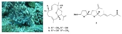
Cembranoids with 3,14-Ether Linkage and a Secocembrane with Bistetrahydrofuran from the Dongsha Atoll Soft Coral Lobophytum sp.

by
Mohamed Elamir F. Hegazy
1,
Jui-Hsin Su
2,3,
Ping-Jyun Sung
2,3 and
Jyh-Horng Sheu
1,4,*
1
Department of Marine Biotechnology and Resources, National Sun Yat-sen University, Kaohsiung 804, Taiwan
2
National Museum of Marine Biology & Aquarium, Pingtung 944, Taiwan
3
Graduate Institute of Marine Biotechnology, National Dong Hwa University, Pingtung 944, Taiwan
4
Division of Marine Biotechnology, Asia-Pacific Ocean Research Center, National Sun Yat-sen University, Kaohsiung 804, Taiwan
*
Author to whom correspondence should be addressed.
Mar. Drugs 2011, 9(7), 1243-1253; https://doi.org/10.3390/md9071243
Submission received: 23 May 2011 / Revised: 21 June 2011 / Accepted: 5 July 2011 / Published: 12 July 2011

Abstract

:
Four new cembranoids, lobophylins A–D (14), and one novel secocembrane, lobophylin E (5) were isolated from a soft coral Lobophytum sp. The structures of new metabolites were elucidated on the basis of extensive spectroscopic methods. Among these metabolites, 14 are rarely found cembranoids possessing a tetrahydrofuran moiety with a 3,14-ether linkage. In addition, 5 is the first secocembrane possessing two tetrahydrofuran moieties with 3,14- and 4,7-ether linkages.

Graphical Abstract

1. Introduction

Soft corals have proven to be important sources of secondary metabolites with interesting biological activities [1]. In the investigation of the secondary metabolites from soft corals in Taiwan waters, a series of bioactive cembranoids have been isolated from octocorals (Alcyonaceae) belonging to the genera Sinularia [27], Lobophytum [810], Sarcophyton [1116] and Pachyclavularia [17,18]. Some of these metabolites have been shown to exhibit significant cytotoxic activity against the growth of various cancer cell lines [10,1517], and/or anti-inflammatory activity [3,6,8,10,15,16]. Our previous chemical investigation on Dongha Atoll soft coral Lobophytum sarcophytoides has led to the isolation of bioactive cembranoids [19]. In our continuing search for bioactive metabolites from Dongsha Atoll soft corals of the genus Lobophytum, we investigated the chemical constituents of Lobophytum sp. and succeeded in the isolation of four new cembranoidal lobophylins A–D (14) and a novel secocembrane, lobophylin E (5) (Chart 1). The structures of these compounds have been established by extensive spectroscopic analysis. The cytotoxicity of compounds 15 against four human cancer cell lines was investigated, however, none of these was found to possess useful biological activity.

2. Results and Discussion

The new metabolite lobophylin A (1) exhibited a protonated molecule peak in the HRESIMS at m/z 343.2251 [M + Na]+, establishing the molecular formula C20H32O3 and five degrees of unsaturation. The IR spectrum suggested the presence of hydroxy group (νmax 3460 cm−1) in 1. The 13C NMR spectrum of 1 measured in CDCl3 (Table 1) showed the presence of twenty carbon signals, which were assigned by the assistance of DEPT spectrum to four methyls, six sp3 methylenes, one sp2 methylene, four sp3 methines (including three oxymethines), one sp2 methine, and two sp3 quaternary and two sp2 quaternary carbons. From the 1H NMR spectroscopic data of 1 (Table 2), the presence of two hydroxy protons resonating at δ 3.98 (dd, J = 9.6, 4.4 Hz) and 4.37 (ddd, J = 12.0, 3.6, 3.6 Hz) were observed. Moreover, the 1H NMR spectrum revealed the presence of two olefinic methylene protons at δ 4.87 (d, J = 1.6 Hz) and 4.81 (s) and one olefinic methine proton at δ 5.09 (t, J = 6.8 Hz). A proton signal appearing at δ 3.27 (1H, d, J = 6.8 Hz) and correlating with a carbon signal at δ 64.7 in the HMQC spectrum was due to the proton of the trisubstituted epoxide. The planar structure and all of the assignments of 1H and 13C NMR data of 1 were determined by the assistance of 2D NMR studies, including 1H-1H COSY and HMBC experiments (Figure 1). 1H-1H COSY spectrum revealed proton sequences from H-1 to H-3 and H-13 to H-1; H2-5 to H-7; H2-9 to H-11, as shown by the bold lines in Figure 1. Key HMBC correlations of H-3 to C-4; H-7 to C-8; H2-13 to C-11 and C-12; H2-16 to C-1 and C-15; H3-17 to C-1, C-15 and C-16; H3-18 to C-3, C-4 and C-5; H3-19 to C-7, C-8 and C-9; and H3-20 to C-11, C-12 and C-13, permitted the connection of the carbon skeleton. Furthermore, the HMBC cross-peak from H-14 to C-3 suggested that C-3 and C-14 were linked through an oxygen to form a tetrahydrofuran ring. Thus, 1 was revealed as a cembranoid possessing a 3,14-ether linked tetrahydrofuran ring, on the basis of the above analysis.
The relative configuration of 1 elucidated mainly by NOESY spectrum was compatible with that of 1 offered by using the MM2 force field calculations which suggested the most stable conformations as shown in Figure 2. In the NOESY spectrum, it was found that H-1 (δ 2.77, dt, J = 8.8, 8.0 Hz) showed NOE interactions with H-14 and H3-18 (δ 1.15, s); therefore, assuming the β-orientation of H-1, H-14 and H3-18 should also be positioned on the β face. One of the methylene protons at C-2 (δ 1.92) exhibited NOE correlations with H-1 and was characterized as H-2β, while the other (δ 2.16) was assigned as H-2α. NOE correlations observed between H-2α and H-3 (δ 3.98, dd, J = 9.6, 4.4 Hz), and H-3 and H-7 (δ 3.27, d, J = 6.8 Hz), reflected the α-orientations of both protons H-3 and H-7. Also, H3-19 was found to interact with H2-6, but not with H-7, revealing the trans geometry of the trisubstituted epoxide. Furthermore, the NOE correlations observed H3-20 and H-10 (δ 2.21), but not with H-11, reflected the E geometry of double bond at C-11. On the basis of the above findings and other detailed NOE correlations (Figure 2), the relative structure of 1 was determined.
HRESIMS analysis of lobophylin B (2) provided a molecular formula of C20H32O2 ([M + Na]+ m/z 327.2301). The 1H and 13C NMR spectroscopic data of 2 were very close to those of 1 (Tables 1 and 2), except for the replacement of the two carbon signals of the epoxide moiety in 1 by the signals of a trisubstituted double bond in 2 (δ 126.6, CH, C-7 and 132.8, C, C-8). This double bond was positioned at C-7/C-8 due to the 1H-1H COSY correlation found between the H-6 and H-7, the HMBC correlations observed from the olefinic methyl protons at δ 1.65 (3H, s) to C-7, C-8 and C-9. Furthermore, the E geometry of the 7,8-double bond was deduced from the NOE correlation of H3-19 with H2-6 and not with H-7. Thus, the structure of 2 was determined unambiguously. Literature review revealed a known compound similar to compound 2 but possessing a rare 3,13-bridged tetrahydropyran ring [20].
Lobophylin C (3) showed a protonated molecule peak [M + Na]+ at m/z 343.2248 in the HRESIMS, corresponding to the molecular formula C20H32O3 and five degrees of unsaturation. The IR spectrum showed the presence of hydroxy (3377 cm −1 ) group. 1H and 13C NMR spectroscopic data (Tables 1 and 2) of 3 showed the structural unit of a 3,14-oxa-bridged tetrahydrofuran, too. 1H-1H COSY and HMBC (Figure 1) further revealed that 3 possesses a 1,2-disubstituted double bond (δ 118.9 and 142.7, each CH) at C-6 and C-7 and a quaternary oxycarbon at C-8 (δ 73.6, C). On the basis of the above observations, and by the assistance of additional 2D NMR (1H-1H COSY and HMBC) correlations, it was possible to establish the planar structure of 3 as illustrated in Figure 1. The relative configurations of the five chiral centers at C-1, C-3, C-4, C-8 and C-14 in 3 were thus determined on the basis of NOE correlations (Figure 3). By careful inspection on the NOESY spectrum of 3, it was found that one proton (δ 2.40) of H2-5 showed NOE interaction with both H3-18 and H-7, and H-7 was NOE correlated with H3-19. Therefore, H3-18 and H3-19 are situated on the same β-face. Furthermore, NOESY spectrum showed correlation of H3-20 with one proton (δ 2.19) of CH2-10, but not with H-11, revealing the E-configurations of the 11,12-trisubstituted double bond. The above finding, together with J values for both H-6 (15.2 Hz) and H-7 (15.6 Hz), confirmed the E-configuration of the 6,7-double bond. Further NOE analysis revealed that 3 possessed the same configurations at C-1, C-3, C-4 and C-14, as in compound 1 (Figure 3). Based on the above results, the structure of 3 was established.
The HRESIMS spectrum of lobophylin D (4) showed a molecular formula of C20H32O3, the same as that of 3. By analysis 2D NMR spectra, including 1H-1H COSY, HMQC and HMBC, 4 was shown to possess the same molecular framework as that of 3. Furthermore, it was found that the NMR data of 4 were very similar to those of 3 (Tables 1 and 2), revealing that 4 might be an isomer of 3. However, the significant downfield shift at C-6 (ΔδC +2.9 ppm) and the upfield shift at C-7 (ΔδC −1.2 ppm), C-8 (ΔδC −1.0 ppm) and C-19 (ΔδC −1.3 ppm), relative to those of 3 (Table 2), suggested that 4 might be the C-8 epimer of 3. From NOESY spectrum, it was found that one proton (δ 2.56, m) of H2-10 of 4 showed NOE correlations with H-7 (δ 5.75, d, J = 15.5 Hz) and H3-20 (δ 1.70, s), while H-6 (5.51, ddd, J = 15.5, 10.0, 5.0 Hz) was NOE correlated with H3-19 (δ 1.37, s) (Figure 3). Therefore, both H-7 and H3-20 are situated on the β-face, and in contrast, H-6 and H3-19 should be positioned on the α-face. This inferred the R* configuration at C-8. Further analysis of other NOE interactions revealed that 4 possessed the same relative configurations at C-1, C-3, C-4 and C-14 as those of 3 (Figure 3). Therefore, 4 was found to be the C-8 epimer of 3.
Lobophylin E (5) was assigned a molecular formula of C21H34O4, according to the HRESIMS and NMR spectroscopic data (Tables 1 and 2). The IR absorption band at 3444 cm−1 revealed the presence of hydroxy group. By the analysis of 13C and DEPT spectroscopic data, the carbons signals were assigned into five methyls (including one methoxy methyl resonating at δC 54.3), six sp3 methylenes, one sp2 methylene, four sp3 methines (including two monooxygenated carbons resonating at δC 82.2 and 80.3 and an acetal carbon resonating at δC 105.6), one sp2 methine, one sp3 quaternary carbons and three sp2 quaternary carbons (including a normal ketone resonating at δC 208.9). From the 1H-1H COSY spectrum of 5, it was possible to identify three different structure units, which were assembled with the assistance of an HMBC experiment. Key HMBC correlations between H-3 to C-4; H2-9 and H2-10 to C-8 (carbonyl carbon); H-11 to C-13; H2-16 to C-1 and H3-17 to C-1, C-15 and C-16; H3-18 to C-3, C-4 and C-5; H3-19 to C-8 and C-9; and H3-20 to C-11, C-12 and C-13 permitted the connection of the carbon skeleton (Figure 1). Furthermore, the HMBC correlation observed from the methoxy protons (δ 3.34, 3H, s) to the carbon resonating at δ 105.6 positioned a methoxy group at C-7. In considering the degrees of unsaturation and molecular formula, two oxa-bridged ether linkages were placed between C-3/C-14 and C-4/C-7 by HMBC correlations from H-14 to C-3 and H-7 to C-4. The relative configuration of 5 was determined by the interpretation of the NOESY correlations (Figure 4). It was found that H3-18 showed NOE interactions with H-1, H-3 and methoxy protons (H3-21). Thus, by considering a molecular model as shown in Figure 4 and assuming the β-orientation of H3-18, all of H-1, H-3 and methoxy group should be positioned on the β face. The NOE correlation observed between H-1 and H-14 also reflected the β-orientation of H-14. Furthermore, NOESY spectrum showed NOE interaction of H3-20 with H-10, but not with H-11, revealing the E geometry of the C-11/C-12 double bond. From the above evidence and the other NOE correlations (Figure 4) the relative configurations at chiral centers of 5 was assumed to be 1R*, 3R*, 4R*, 7R* and 14S*. On the basis of the above analysis, the structure of 5 was established.
It is worth noting that metabolites 14 are rare cembranoids possessing a tetrahydrofuran moiety with a 3,14-ether linkage, which has been discovered previously in the soft coral Sinularia gibberosa [5,21]. In addition, 5 is the first secocembrane possessing two tetrahydrofuran moieties with 3,14- and 4,7-ether linkages. Our study thus adds the structure diversity of cembranoidal natural compounds.
The cytotoxicity of compounds 15 against the proliferation of a limited panel of cancer cell lines, including K562 (human chronic myelogenous leukemia), DLD-1 (human colon adenocarcinoma) and HepG2 and Hep3B (human liver carcinoma), was studied. The results showed that 15 are not cytotoxic toward the above cancer cells (IC50 > 20 μg/mL).

3. Experimental Section

3.1. General Experimental Procedures

The melting points were determined using a Fisher-Johns melting point apparatus. Optical rotation values were measured with a JASCO P-1010 digital polarimeter. IR spectra were recorded on a VARIAN DIGLAB FTS 1000 Fourier transform infrared spectrophotometer. The NMR spectra were recorded on a VARIAN MERCURY PLUS 400 FT-NMR (or Varian Unity INOVA 500 FT-NMR) instrument at 400 MHz (or 500 MHz) for 1H NMR and 100 MHz (or 125 MHz) for 13C NMR, respectively, in CDCl3. ESIMS were recorded on a Bruker APEX II mass spectrometer. Silica gel 60 (Merck, 230–400 mesh) was used for column chromatography. Precoated silica gel plates (Merck, Kieselgel 60 F254, 0.25 mm) and precoated RP-18 F254S plates (Merck, 1.05560) were used for TLC analysis. High-performance liquid chromatography (HPLC) was performed on a Hitachi L-7100 pump equipped with a Hitachi L-7400 UV detector at 210 nm. A semipreparative reversed-phase column (250 × 10 mm, 5 μm) and a preparative normal phase column (250 × 21.2 mm, 5 μm) was used for HPLC.

3.2. Animal Material

The soft coral Lobophytum sp. was collected by hand using SCUBA off the coast of Dongsha Atoll, in April, 2007, at a depth of 10 m, and stored in a freezer until extraction. A voucher specimen (Specimen No. DA2007-04-20) was deposited in the Department of Marine Biotechnology and Resources, National Sun Yat-sen University.

3.3. Extraction and Separation

The frozen soft coral (1.5 kg, fresh wt) was minced and extracted exhaustively with EtOAc (5 × 1 L). The organic extract was evaporated to yield a residue (21.9 g), which was fractionated by open column chromatography on silica gel using n-hexane–EtOAc and EtOAc–MeOH mixtures of increasing polarity to yield 16 fractions. Fraction 5, eluting with n-hexane–EtOAc (15:1), was further separated by silica gel column chromatography with gradient elution (n-hexane–EtOAc, 15:1 to 5:1) to yield five subfractions (5A–5E). Subfraction 5C was subjected to normal phase HPLC (n-hexane–EtOAc, 15:1) to obtain compound 2 (2.5 mg). Fractions 7 and 8, eluting with n-hexane–EtOAc (5:1), were combined and further separated over silica gel column chromatography (n-hexane–EtOAc, gradient elution, 5:1 to 1:1) to give four subfractions (7A–7D). Subfraction 7A was further purified by RP-18 HPLC (CH3CN–H2O, 3:2) to yield compound 5 (2.2 mg). In the same manner, compound 1 (4.2 mg) was obtained from subfraction 7B using RP-18 HPLC (CH3CN–H2O, 5:2). Fraction 11, eluting with n-hexane–EtOAc (1:1), was further separated by silica gel column chromatography with gradient elution (n-hexane–EtOAc, 1:1 to 1:5) to yield five subfractions (11A–11E). Subfraction 11C was further purified by RP-18 HPLC (CH3CN–H2O, 1:1) to yield compounds 3 (3.0 mg) and 4 (2.5 mg).
Lobophylin A (1): colorless oil; [α]D25 = −39 (c 0.3, CHCl3); IR (neat) νmax 3460, 2926, 1649, 1458, 1381 and 1215 cm−1; 1H and 13C NMR data, see Tables 1 and 2; ESIMS m/z 343 [100, (M + Na)+]; HRESIMS m/z 343.2251 (calcd for C20H32O3Na, 343.2249).
Lobophylin B (2): colorless oil; [α]D25 = −35 (c 0.3, CHCl3); IR (neat) νmax 3445, 2926, 1649, 1456, 1376 and 1265 cm−1; 1H and 13C NMR data, see Tables 1 and 2; ESIMS m/z 327 [100, (M + Na)+]; HRESIMS m/z 327.2301 (calcd for C20H32O2Na, 327.2300).
Lobophylin C (3): white powder; mp 76–78 °C; [α]D25 = +30 (c 0.1, CHCl3); IR (neat) νmax 3377, 2927, 1649, 1459, 1377 and 1269 cm−1; 1H and 13C NMR data, see Tables 1 and 2; ESIMS m/z 343 [100, (M + Na)+]; HRESIMS m/z 343.2248 (calcd for C20H32O3Na, 343.2249).
Lobophylin D (4): white powder; mp 68–70 °C; [α]D25 = +22 (c 0.2, CHCl3); IR (neat) νmax 3425, 2924, 1640, 1455, 1379 and 1240 cm−1; 1H and 13C NMR data, see Tables 1 and 2; ESIMS m/z 343 [100, (M + Na)+]; HRESIMS m/z 343.2246 (calcd for C20H32O3Na, 343.2249).
Lobophylin E (5): colorless oil; [α]D25 = +19 (c 0.2, CHCl3); IR (neat) νmax 3444, 2929, 1715, 1640, 1454, 1374 and 1214 cm−1; 1H and 13C NMR data, see Tables 1 and 2; ESIMS m/z 373 [100, (M + Na)+]; HRESIMS m/z 373.2356 (calcd for C21H34O4Na, 373.2355).

3.4. Cytotoxicity Testing

Cell lines were purchased from the American Type Culture Collection (ATCC). Cytotoxicity assays of compounds 15 were performed using the MTT [3-(4,5-dimethylthiazol-2-yl)-2,5-diphenyltetrazolium bromide] colorimetric method [22].

3.5. Molecular Mechanics Calculations

Implementation of the MM2 force filed in Chem3D Pro software from Cambridge Soft Corporation, Cambridge, MA, USA (ver. 9.0, 2005), was used to calculate molecular models.

Acknowledgements

This work was supported by grants from the National Science Council of Taiwan (NSC98-2113-M- 110-002-MY3) and Ministry of Education (98C031702) awarded to J.-H.S.
  • Samples Availability: Not available.

References

  1. Blunt, JW; Copp, BR; Munro, MHG; Northcote, PT; Prinsep, MR. Marine natural products. Nat. Prod. Rep 2011, 28, 196–268. [Google Scholar]
  2. Su, J-H; Ahmed, AF; Sung, P-J; Chao, C-H; Kuo, Y-H; Sheu, J-H. Manaarenolides A–I, diterpenoids from the soft coral Sinularia manaarensis. J. Nat. Prod 2006, 69, 1134–1139. [Google Scholar]
  3. Lu, Y; Huang, C-Y; Lin, Y-F; Wen, Z-H; Su, J-H; Kuo, Y-H; Chiang, MY; Sheu, J-H. Anti-inflammatory cembranoids from the soft corals Sinularia querciformis and Sinularia granosa. J. Nat. Prod 2008, 71, 1754–1759. [Google Scholar]
  4. Ahmed, AF; Tai, S-H; Wen, Z-H; Su, J-H; Wu, Y-C; Hu, W-P; Sheu, J-H. A C-3 methylated isocembranoid and 10-oxocembranoids from a Formosan soft coral, Sinularia grandilobata. J. Nat. Prod 2008, 71, 946–951. [Google Scholar]
  5. Ahmed, AF; Wen, Z-H; Su, J-H; Hsieh, Y-T; Wu, Y-C; Hu, W-P; Sheu, J-H. Oxygenated cembranoids from a Formosan soft coral Sinularia gibberosa. J. Nat. Prod 2008, 71, 179–185. [Google Scholar]
  6. Lu, Y; Su, J-H; Hsieh, C-H; Liu, Y-C; Kuo, Y-H; Wen, Z-H; Hsu, C-H; Sheu, J-H. Cembranoids from the soft corals Sinularia granosa and Sinularia querciformis. Chem. Pharm. Bull 2010, 58, 464–466. [Google Scholar]
  7. Chen, B-W; Chao, C-H; Su, J-H; Huang, C-Y; Dai, C-F; Wen, Z-H; Sheu, J-H. A novel symmetric sulfur-containing biscembranoid from the Formosan soft coral Sinularia flexibilis. Tetrahedron Lett 2010, 44, 5764–5766. [Google Scholar]
  8. Lin, S-T; Wang, S-K; Cheng, S-Y; Duh, C-Y. Unprecedented hemiketal cembranolides with anti-inflammatory activity from the soft coral Lobophytum durum. J. Nat. Prod 2009, 72, 152–155. [Google Scholar]
  9. Cheng, S-Y; Wen, Z-H; Wang, S-K; Chiou, S-F; Hsu, C-H; Dai, C-F; Chiang, MY; Duh, C-Y. Lobocrasol, a new diterpenoid from the soft coral Lobophytum crassum. Org. Lett 2009, 11, 3012–3014. [Google Scholar]
  10. Chao, C-H; Wen, Z-H; Wu, Y-C; Yeh, H-C; Sheu, J-H. Cytotoxic and anti-inflammatory cembranoids from the soft coral Lobophytum crassum. J. Nat. Prod 2008, 71, 1819–1824. [Google Scholar]
  11. Huang, H-C; Ahmed, AF; Su, J-H; Wu, Y-C; Chiang, MY; Sheu, J-H. Crassocolides A–F, cembranoids with a trans-fused lactone from the soft coral Sarcophyton crassocaule. J. Nat. Prod 2006, 69, 1554–1559. [Google Scholar]
  12. Huang, H-C; Chao, C-H; Kuo, Y-H; Sheu, J-H. Crassocolides G-M, cembranoids from the Formosan soft coral Sarcophyton crassocaule. Chem. Biodiv 2009, 6, 1232–1242. [Google Scholar]
  13. Cheng, Y-B; Shen, Y-C; Kuo, Y-H; Khalil, AT. Cembrane diterpenoids from the Taiwanese soft coral Sarcophyton stolidotum. J. Nat. Prod 2008, 71, 1141–1145. [Google Scholar]
  14. Cheng, S-Y; Wang, S-K; Chiou, S-F; Hsu, C-H; Dai, C-F; Chiang, MY; Duh, C-Y. Cembranoids from the octocoral Sarcophyton ehrenbergi. J. Nat. Prod 2010, 73, 197–203. [Google Scholar]
  15. Lin, W-Y; Su, J-H; Lu, Y; Wen, Z-H; Dai, C-F; Kuo, Y-H; Sheu, J-H. Cytotoxic and anti-inflammatory cembranoids from the Dongsha Atoll soft coral Sarcophyton crassocaule. Bioorg. Med. Chem 2010, 18, 1936–1941. [Google Scholar]
  16. Lin, W-Y; Lu, Y; Su, J-H; Wen, Z-H; Dai, C-F; Kuo, Y-H; Sheu, J-H. Bioactive cembranoids from the Dongsha Atoll soft coral Sarcophyton crassocaule. Mar. Drugs 2011, 9, 994–1006. [Google Scholar]
  17. Sheu, J-H; Wang, G-H; Sung, P-J; Duh, C-Y; Chiang, MY. Pachyclavulariolides G–L and secopachyclavulariaenone A, seven novel diterpenoids from the soft coral Pachyclavularia violacea. Tetrahedron 2001, 57, 7639–7648. [Google Scholar]
  18. Sheu, J-H; Wang, G-H; Duh, C-Y; Soong, K. Pachyclavulariolides M-R, six novel diterpenoids from a Taiwanese soft coral Pachyclavularia violacea. J. Nat. Prod 2003, 66, 662–666. [Google Scholar]
  19. Lu, Y; Lin, Y-C; Wen, Z-H; Su, J-H; Sung, P-J; Hsu, C-H; Kuo, Y-H; Chiang, MY; Sheu, J-H. Steroid and cembranoids from the Dongsha Atoll soft coral Lobophytum. Tetrahedron 2010, 66, 7129–7135. [Google Scholar]
  20. Ravi, BN; Faulkner, J. Cembranoid diterpenes from a South Pacific soft coral. J. Org. Chem 1978, 43, 2127–2131. [Google Scholar]
  21. Duh, C-Y; Hou, R-S. Cytotoxic cembranoids from the soft corals Sinularia gibberosa and Sarcophyton trocheliophorum. J. Nat. Prod 1996, 59, 595–598. [Google Scholar]
  22. Scudiero, DA; Shoemaker, RH; Paull, KD; Monks, A; Tierney, S; Nofziger, TH; Currens, MJ; Seniff, D; Boyd, MR. Evaluation of a soluble tetrazolium/formazan assay for cell growth and drug sensitivity in culture using human and other tumor cell lines. Cancer Res 1988, 48, 4827–4833. [Google Scholar]
Figure 1. Selected 1H-1H COSY ( Marinedrugs 09 01243ig1
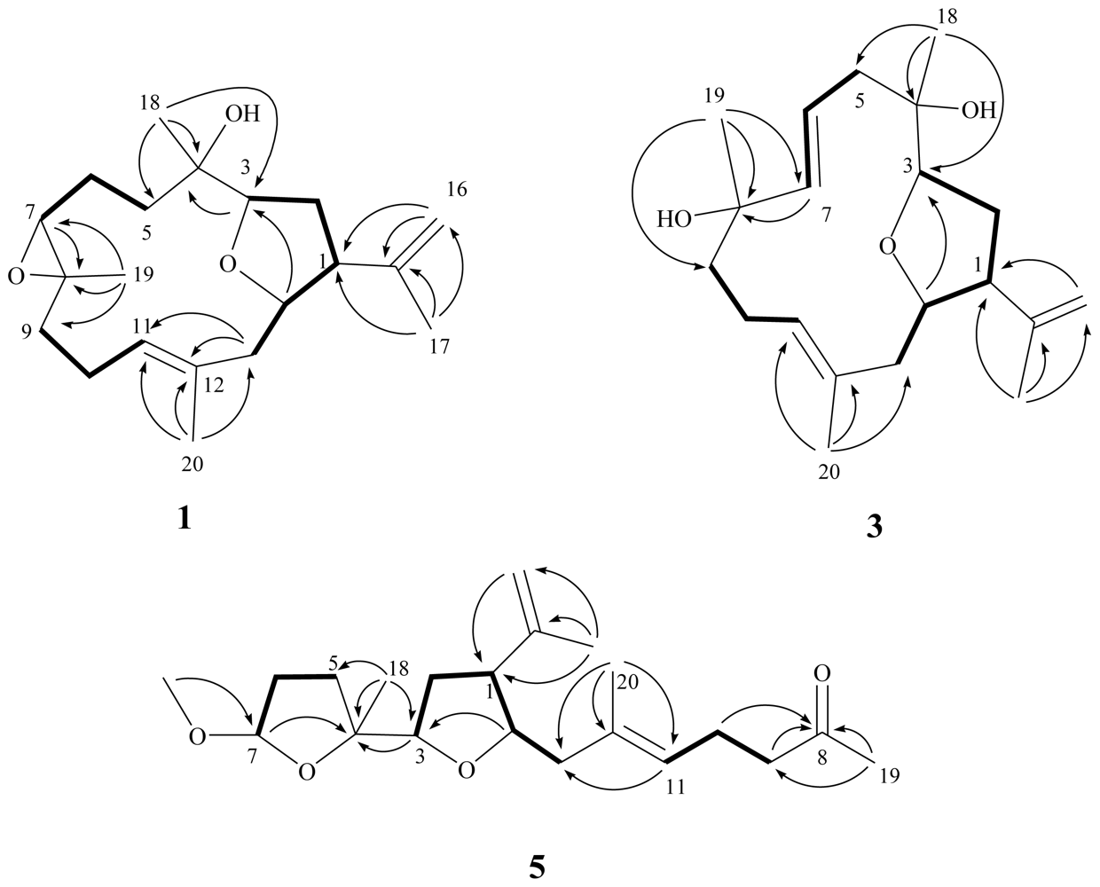
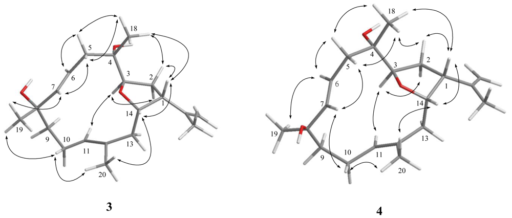
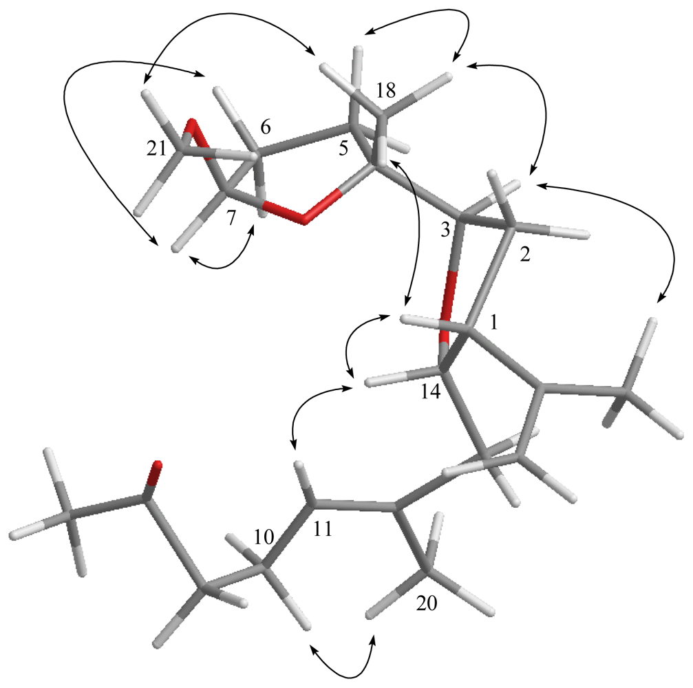
) and HMBC (→) correlations of 1, 3 and 5.
Marinedrugs 09 01243f1
Figure 2. Computer-generated model for 1 using MM2 force field calculations and key NOE correlations.
Marinedrugs 09 01243f2
Figure 3. Computer-generated model for 3 and 4 using MM2 force field calculations and key NOE correlations.
Marinedrugs 09 01243f3
Figure 4. Computer-generated model for 5 using MM2 force field calculations and key NOE correlations.
Marinedrugs 09 01243f4
Chart 1. Structures of metabolites 15.
Marinedrugs 09 01243f5
Table 1. 13C NMR data for compounds 15.
C#1 a2 a3 a4 b5 b
150.2 (CH) c49.0 (CH)49.3 (CH)49.3 (CH)49.8 (CH)
229.1 (CH2)27.4 (CH2)26.7 (CH2)26.7 (CH2)30.9 (CH2)
377.5 (CH)77.6 (CH)77.8 (CH)77.6 (CH)82.2 (CH)
474.5 (C)74.2 (C)74.6 (C)74.7 (C)86.6 (C)
539.1 (CH2)38.6 (CH2)42.5 (CH2)43.3 (CH2)31.9 (CH2)
623.8 (CH2)21.5 (CH2)118.9 (CH)121.8 (CH)33.3 (CH2)
764.7 (CH)126.6 (CH)142.7 (CH)141.5 (CH)105.6 (CH)
860.3 (C)132.8 (C)73.6 (C)72.6 (C)208.9 (C)
938.1 (CH2)38.2 (CH2)44.4 (CH2)43.7 (CH2)43.7 (CH2)
1023.9 (CH2)24.4 (CH2)23.5 (CH2)22.2 (CH2)22.5 (CH2)
11126.5 (CH)127.1 (CH)129.4 (CH)129.6 (CH)124.2 (CH)
12133.0 (C)131.9 (C)130.9 (C)130.8 (C)134.2 (C)
1340.2 (CH2)39.3 (CH2)38.9 (CH2)38.8 (CH2)39.7 (CH2)
1478.5 (CH)76.7 (CH)76.0 (CH)76.0 (CH)80.3 (CH)
15141.6 (C)142.4 (C)142.2 (C)142.3 (C)144.0 (C)
16111.3 (CH2)111.0 (CH2)111.2 (CH2)111.1 (CH2)112.2 (CH2)
1725.0 (CH3)23.5 (CH3)23.5 (CH3)23.5 (CH3)22.5 (CH3)
1824.6 (CH3)23.1 (CH3)21.6 (CH3)21.9 (CH3)24.2 (CH3)
1919.8 (CH3)16.3 (CH3)29.6 (CH3)28.3 (CH3)29.9 (CH3)
2017.3 (CH3)15.4 (CH3)15.4 (CH3)15.5 (CH3)16.5 (CH3)
OMe54.3 (CH3)
aSpectra recorded at 100 MHz in CDCl3;
bSpectra recorded at 125 MHz in CDCl3;
cAttached protons were deduced by DEPT experiments.
Table 2. 1H NMR data for compounds 15.
1 a2 a3 a4 b5 b
12.77 dt (8.8, 8.0) c2.73 dt (11.2, 7.2)2.73 dt (8.0, 8.8)2.74 dt (9.0, 8.5)2.78 dt (7.5, 8.5)
22.16 m; 1.92 m2.08 m; 1.90 m2.04 m; 1.86 m2.05 m; 1.86 m1.96 m; 1.91 m
33.98 dd (9.6, 4.4)3.97 dd (9.6, 4.5)3.82 dd (10.0, 4.8)3.82 dd (9.5, 4.5)3.98 dd (7.5, 7.5)
51.97 m; 1.70 m1.94 m; 1.53 m2.40 dd (14.0, 10.0); 2.05 m2.40 dd (14.0, 10.0); 2.10 m2.40 dd (14.0, 10.0); 1.94 m
62.05 m; 1.31 m2.25 m; 2.06 m5.60 ddd (15.2, 10.0, 5.2)5.51 ddd (15.5, 10.0, 5.0)2.02 m; 1.94 m
73.27 d (6.8)5.17 dd (6.0, 6.0)5.70 d (15.6)5.75 d (15.5)5.00 d (4.5)
91.86 m; 1.52 m2.14 m; 1.96 m1.92 m; 1.58 m1.95 m; 1.58 m2.45 dd (8.0, 7.0)
102.21 m; 1.88 m2.32 m; 2.04 m2.19 m; 2.10 m2.56 m; 1.96 m2.27 dd (7.5, 7.5)
115.09 t (6.8)4.89 d (8.0)4.96 d (9.6)4.94 d (10.0)5.12 dd (7.0, 6.5)
131.95 m; 1.68 m1.88 m; 1.72 m1.91 m; 1.64 m1.92 m; 1.64 m2.00 m; 1.97 m
144.37 ddd (12.0, 3.6, 3.6)4.36 ddd (11.6, 5.2, 4.8)4.33 ddd (11.6, 6.0, 5.2)4.33 ddd (12.0, 6.0, 5.5)4.14 ddd (9.0, 4.5, 3.5)
164.87 d (1.6); 4.81 s4.85 d (1.2); 4.78 s4.86 d (1.6); 4.80 s4.86 d (1.0); 4.80 s4.83 s; 4.72 s
171.77 s1.75 s1.76 s1.76 s1.75 s
181.15 s1.09 s1.11 s1.13 s1.28 s
191.24 s1.65 s1.28 s1.37 s2.13 s
201.61 s1.57 s1.67 s1.70 s1.65 s
OMe3.34 s
aSpectra recorded at 400 MHz in CDCl3;
bSpectra recorded at 500 MHz in CDCl3;
cJ values (in Hz) in parentheses.

