Article

Removal and Biodegradation of Nonylphenol by Four Freshwater Microalgae

1
Institute of Hydrobiology, Jinan University, Guangzhou 510632, China
2
Key Laboratory of Aquatic Eutrophication and Control of Harmful Algal Blooms, Guangdong Higher Education Institutes, Guangzhou 510632, China
3
Research Center of Offshore Marine Environment, South China Institute of Environmental Sciences, MEP, Guangzhou 510655, China
*
Author to whom correspondence should be addressed.
These authors contributed equally to this work.
Int. J. Environ. Res. Public Health 2016, 13(12), 1239; https://doi.org/10.3390/ijerph13121239
Submission received: 19 September 2016 / Revised: 1 December 2016 / Accepted: 8 December 2016 / Published: 14 December 2016

Abstract

:
The removal and biodegradation of nonylphenol (NP) by four freshwater microalgae, including three green algae (Scendesmus quadriauda, Chlorella vulgaris, and Ankistrodesmus acicularis) and one cyanobacterium (Chroococcus minutus) were studied in bacteria-free cultures exposed to different concentrations of NP for 5 days. All four algal species showed a rapid and high ability to remove NP (including bioaccumulation and biodegradation). Among these species, A. acicularis (Ankistrodesmus acicularis) had the highest NP removal rate (83.77%) at 120 h when exposed to different NP treatments (0.5–2.5 mg·L−1), followed by C. vulgaris (Chlorella vulgaris) (80.80%), S. quadriauda (Scendesmus quadriauda) (70.96%) and C. minutus (Chroococcus minutus) (64.26%). C. vulgaris had the highest NP biodegradation percentage (68.80%) at 120 h, followed by A. acicularis (65.63%), S. quadriauda (63.10%); and C. minutus (34.91%). The extracellular NP contents were lower than the intracellular NP contents in all tested algae. The ratio of the extracellular NP content and the intracellular NP content ranged from 0.04 to 0.85. Therefore, the removal of NP from the medium was mainly due to the algal degradation. These results indicate that A. acicularis and C. vulgaris are more tolerant to NP and could be used for treatment of NP contaminated aqueous systems effectively by bioremoval and biodegradation.

1. Introduction

Nonylphenol (NP) is a degradation product of the alkylphenol polyethoxylates (APEOs), an important class of nonionic surfactants employed in many detergent formulations for industrial and household use [1,2]. In the environment NP is both highly persistent and highly toxic, posing a serious threat to humans and other organisms due to its estrogenic properties [3,4]. Concentrations of 4-nonylphenol (NPs) as high as 325 μg·L−1 in surface water and up to 72 mg·kg−1 in sediment have been determined [5]. Conventional and advanced wastewater treatment methods are inefficient in removing NP [6,7], so new, cost-effective methods are needed to effectively remove NP from the contaminated environment.
Bioaccumulation and biodegradation of organic contaminants and even toxic pollutants has been reported. Those pollutants could be transformed into useful nutrients and growth-supporting substances by certain species of microalgae [8,9,10,11]. Green microalgae, such as Chlorella vulgaris and other Chlorella species have been used to remove organic matters, inorganic nutrients, heavy metals and even toxic organic contaminants from wastewater with both low cost and high efficiency [12,13,14,15]. Biotransformation of low-molecular weight phenols was reported in numerous species of microalgae, such as those isolated from olive-oil mill wastewaters [16]. Algae can have different interactions with aquatic contaminants, which include negative effects on algal growth and function and algal degradation of the contaminants. Growth of Scenedesmus obliquus was not affected by low NP concentrations (<1 mg·L−1), whereas algal growth was suppressed under high NP concentrations (>1 mg·L−1). In addition, more than 89% NP was removed by S. obliquus (Scenedesmus obliquus) due to biodegradation or biotransformation rather than the simple sorption on algal cell surface [17].
Reports have shown that species of microalgae harbor an attached bacterial flora, and also that various free-living bacteria coexist in algal cultures maintained in the laboratory [18,19]. These bacteria are an inherent part of the physical environment of algae and thus can be considered as symbionts [20,21]. Cultures of Alexandrium tamarense, as well as other dinoflagellates, often contain a considerable amount of bacteria from the original samples. These bacteria can produce substances which were either stimulatory or inhibitory to algae and thus change some characteristics of the algae, such as toxin production [22,23,24]. The elimination of bacteria did not affect the growth and toxin profile of Alexandrium lusitanicum and A. tamarense (Alexandrium tamarense), but it did affect the amount of toxins [25].
The bacterial communities of algal cultures interact with the algae, and thus, removal of bacteria from stock algal cultures would provide a simpler system for the study of NP removal and biodegradation by microalgae. The present study aimed to investigate the toxic effect of NP on Scendesmus quadriauda JNU39, Ankistrodesmus acicularis JNU14, Chlorella vulgaris JNU38 and Chroococcus minutus JNU17, and to evaluate the tolerance ability of the four different local freshwater microalgae to NP. The study also attempted to compare the NP removal and biodegradation ability among the four microalgae, and to identify the most effective species in an axenic medium. S. quadriauda, A. acicularis (Ankistrodesmus acicularis), C. vulgaris (Chlorella vulgaris) and C. minutus (Chroococcus minutus) are four microalgal species commonly used for the removal of wastewater-borne pollutants and they have a tolerance to some common pollutants found in wastewater, such as heavy metals, crude oil and polycyclic aromatic hydrocarbons [26,27,28,29].

2. Materials and Methods

2.1. Microalgal Species and Culture Conditions

Four freshwater microalgae, recorded as S. quadriauda JNU39, A. acicularis JNU14, C. vulgaris JNU38 and C. minutus JNU17, were tested in the present study. These species were isolated from NP-polluted water in Jinan University, Guangzhou, China. S. quadriauda was quadrate in shape with a dimension of 6.90 × 2.59 × 2.49 μm. A. acicularis was cylindrical in shape with diameter 2 μm and length 40 μm. C. vulgaris and C. minutus were spherical in shapes with diameters of 5.89 and 2.75 μm, respectively.
The microalgal culture of each species was cultivated in 2 L conical flasks containing 1000 mL BG11 medium, in an environmental chamber illuminated with cool white fluorescent tubes at a light intensity of 90 μmol·m−2·s−1, a diurnal cycle of 12 h light and 12 h dark and at a temperature of 25 ± 2 °C. Flasks were continuously shaken at 100 rpm. The components of basal culture medium were as follows: NaNO3 1.5 g·L−1, K2HPO4 40 mg·L−1, MgSO4·7H2O 75 mg·L−1, CaCl2·2H2O 36 mg·L−1, NaHCO3 20 mg·L−1, ferric ammonium citrate 6 mg·L−1, citric acid 6 mg·L−1. The trace metal solution contained: H3BO3 2.86 mg·L−1, MnCl2·4H2O 1.81 mg·L−1, ZnSO4·7H2O 222 mg·L−1, Na2MoO4·2H2O 390 mg·L−1, CuSO4·5H2O 79 mg·L−1, Co(NO3)2·6H2O 49.4 mg·L−1 [30].

2.2. Removal of Bacteria from Algal Cultures

Mid-exponential phase algal cultures (100 mL) were filtered through a 10 μm pore size membrane and subjected to the following treatments: the algal cells were suspended in 50 mL sterile BG11 medium before sequential centrifuging (1000× g, 10 min) and washing three times. The washed cells were suspended in 50 mL sterile BG11 medium containing 0.005% Tween-80 and 0.1 M EDTA (at 20 °C for 1 h) before lysozyme (0.5 mg·mL−1, 20 °C for 10 min) and SDS (Sodium Dodecyl Sulfate) (0.25%, 20 °C for 10 min) were added sequentially. The algal cells were centrifuged and washed twice to remove lysozyme and SDS and then resuspended in 50 mL sterile BG11 medium. The antibiotic cocktail containing 100 μg·mL−1 penicillin and 50 μg·mL−1 kanamycin were added to the treated algal cultures followed by incubation at 20 °C with a 12:12 light-dark cycle for 7 days. Assessment for bacterial presence was carried out after subculturing three times [31].
The algae were subjected to repeated washing, lysozyme/SDS and antibiotic treatment with a mixture of gentamycin, streptomycin, cephalothin and rifampicin. Axenic status was confirmed after subculturing three times in sterile BG11 medium without antibiotics. Bacteria could not be detected in the various media, both solid and liquid, nor by epifluorescence microscopy of both eubacteria and archaea. Bacterial presence was monitored throughout a full growth cycle and, following subculture, no bacteria were detected using the above methods [32].

2.3. NP Treatments

NP, purchased from Sigma-Aldrich (St. Louis, MO, USA) was dissolved in methanol as the stock solution at a concentration of 1 mg·mL−1. Microalgae cultures in the middle of the log phase of growth were decanted into 100 mL flasks containing 40 mL of medium at 25 ± 2 °C and illuminated with fluorescent lights (90 μmol·m−2·s−1 photon flux intensity) under a 12:12 h light/dark photoperiod. The cultures were initiated at 70.0 μg·L−1 chlorophyll a content, shaken periodically and used in triplicate. All solutions and experimental containers were autoclaved at 121 °C for 15 min. NP was added to the medium before inoculation at a concentration of 0, 0.5, 1.0, 1.5, 2.0 and 2.5 mg·L−1. Treatment with an equivalent amount of methanol (0.1%) was included as a control. The test lasted for 120 h.

2.4. Growth Analysis

The concentration of chlorophyll a in vivo was measured by TD-700 fluorometer (Turner Design, San Jose, CA, USA) every 24 h, which was calibrated with the standard solution of chlorophyll a. Prior to the detection, the test tubes were treated with dark adaption for 20 min at room temperature and shaken homogeneously for several times before determination. Content of chlorophyll a was measured using the excitation and emission wave lengths at 420 and 680 nm, respectively. Specific growth rate (μ) was calculated according to the following Equation (1):
μ = (lnXt − lnX0)/(Tt − T0)
where Xt and X0 are Chl a contents at times Tt and T0, respectively.
Optical density (OD) of the algae cultures was measured daily at 680 nm as the cell density and dry weight indicator using a BMG microplate reader (BMG Lab Technologies, Offenburg, Germany). The cell density was determined using a haemocytometer (Marienfeld, Lauda-Königshofen, Germany) under a light microscope. For cell dry weight measurement, a 20 mL aliquot of culture was filtered through pre-weighed 0.45 mm pore-size GF/F glass-fiber filter paper (Whatman, Maidstone, UK). The filter paper with algal cells was dried overnight in an oven at 60 °C till a constant weight was reached. The difference between the final weight and the weight before filtration was the dry weight of algal cells. The linear relationship between algal density (N, cells·mL−1), dry weight and OD680 is shown in the following equations:
JNU14 Cell density (104 cells·mL−1) = 737.22 × OD680 + 3.6148 R2 = 0.989
JNU17 Cell density (104 cells·mL−1) = 1013 × OD680 + 37.112 R2 = 0.9711
JNU38 Cell density (104 cells·mL−1) = 450.28 × OD680 + 1.5385 R2 = 0.9951
JNU39 Cell density (104 cells·mL−1) = 342.7 × OD680 + 7.4226 R2 = 0.9997
JNU14 Dry weight (g·L−1) = 0.9823 × OD680 + 0.0785 R2 = 0.9057
JNU17 Dry weight (g·L−1) = 1.3729 × OD680 + 0.0063 R2 = 0.9987
JNU38 Dry weight (g·L−1) = 1.0546 × OD680 − 0.0044 R2 = 0.9919
JNU39 Dry weight (g·L−1) = 1.4771 × OD680 − 0.0101 R2 = 0.9998

2.5. Determination of Residual NP

2.5.1. NP Concentration Dissolved in the Medium

At each sampling timepoint, 5 mL cultures were withdrawn from the flasks and cells were separated from the culture through centrifugation at 4500 g for 15 min at 4 °C. The supernatant was extracted with liquid-liquid microextraction (DLLME), as described by Rezaee et al. [32], with some modifications. In brief, a 5 mL of sample was injected with 0.2 mL mixture of chlorobenzene and acetone (1:2) in a 10 mL screw cap glass test tube with a conical bottom. After gently shaking, a milky cloudy solution (water/chlorobenzene) was formed in the test tube. The sample was then centrifuged for 5 min at 4500 g. The dispersed fine particles of extraction phase which settled in the bottom of the conical test tube were withdrawn using a 50 μL microsyringe (zero dead volume, cone tip needle). This extraction process was repeated three times and the sediment fractions were combined for further analysis with high performance liquid chromatograph (HPLC) (Agilent, Santa Clara, CA, USA). All the extraction was performed at room temperature (23 ± 2 °C).

2.5.2. NP Absorbed onto Cell Surface

The cell pellets from the above section were washed with 5 mL of 10 percent methanol and shaken for approximately 60 s, the NP contained in the water was considered as the surface adsorbed NP [33] and then extracted with DLLME, as described above and analyzed with HPLC.

2.5.3. NP Concentration Absorbed into Cells

After adding appropriate amount of anhydrous Na2SO4, the cell pellets obtained from the above section were mixed with dichloromethane-methanol (1:2 v/v, 3 mL); after sonication for 20 min, the sample was centrifuged for 5 min at 3500 g. The cell pellets were extracted two more times and the solvent fractions were combined for further analysis with HPLC [34].
Based on the measured concentrations, the removal efficiency (R) and biodegradation percentage (BDP) of NP by the algal biomass were calculated as previously described [35] with minor modifications according to the following equations:
R = 100 × (Ci − Cf)/Ci
where R is the dissolved NP removal rate (percent); Ci and Cf are the initial and final concentrations (mg·L−1) of NP in the solution, respectively, and:
BDP (%) = 100 × (Ci − Cr − Ca − Cd × Wa − Cc × Wa)/Ci
where Ci is the initial concentration (mg·L−1) of NP in the solution, Cr is the residual concentration (mg·L−1) in the solution, Ca is the concentration of abiotic removal (mg·L−1), Cd is the concentration (mg·g−1) dry weight of NP adsorbed on the cell wall, Cc is the concentration (mg·g−1 dry weight) of NP accumulated in algal cells, and Wa is the dry weight of algal biomass expressed in g·L−1.

2.5.4. Determination of NP

NP concentrations were analyzed by using an Agilent 1100 series high performance liquid chromatograph (HPLC) (Agilent, Santa Clara, CA, USA) coupled to a fluorescence detector. The elution was carried out under isocratic conditions with acetonitrile and Milli-Q water as the mobile phase. A XDB-C18 RS column (4.6 × 250 mm, 5 μm) was used and the volume ratio of acetonitrile to Milli-Q water was 80:20. The injection volume was 50 μL and the flow rate was set at 1 mL·min−1. The fluorescence detector was set at excitation and emission wavelengths of 230 and 305 nm, respectively. The retention time was 18 min. The limit of quantification for both NP was 5 μg·L−1. Results obtained here were compared to that obtained with a control group without nonylphenol.

2.6. Statistical Analysis

Statistical analysis was carried out using the SPSS16.0 package (SPSS Inc., Chicago, IL, USA). One Way-ANOVA followed by Tukey’s post hoc test was used to check the significance of treatments. Levels of significance used were 5% and 1%, described as “significant” and “highly significant”, respectively. Data are presented as mean ± standard deviation (mean ± SD) unless otherwise stated.

3. Results

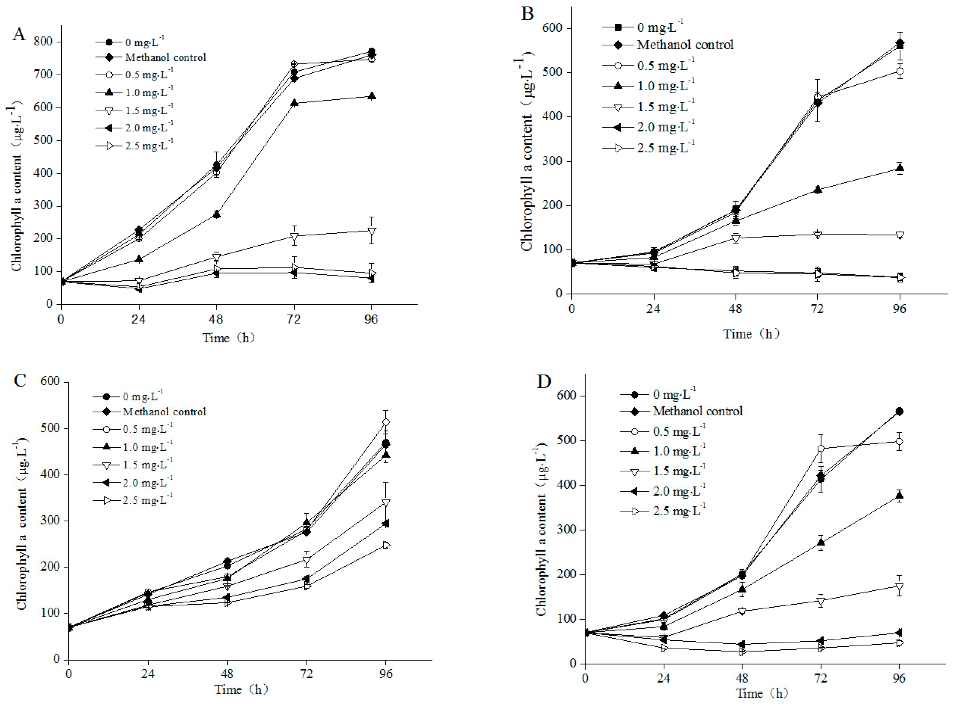
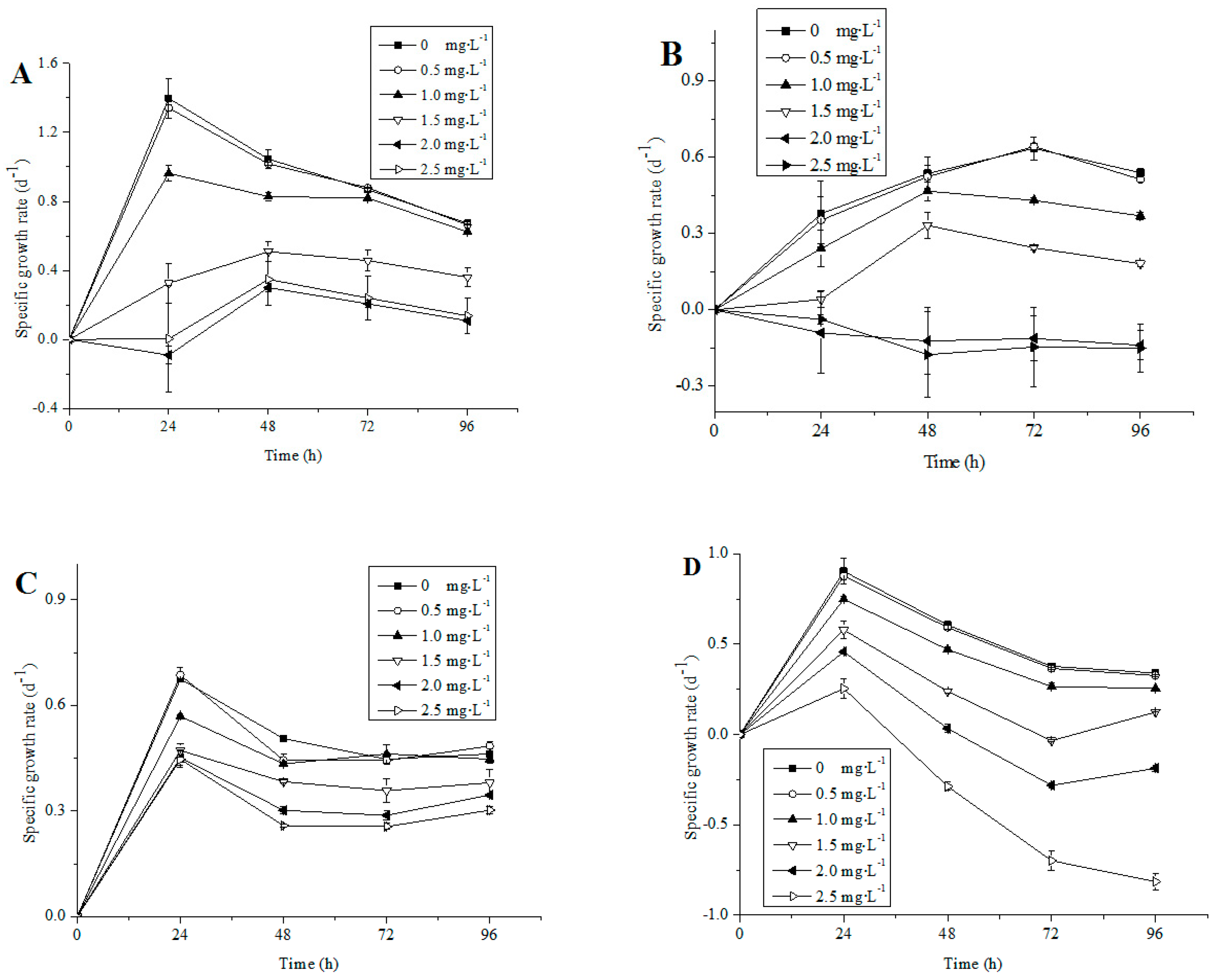
3.1. Growth of Different Microalgae Species Exposed to NP

The chlorophyll a contents of the four nontoxic freshwater microalgae strains were influenced by NP (Figure 1). The solvent (methanol) in the designated concentration in the study (0.1%) had no obvious effect on algal growth (Figure 1). A significant decrease in growth, in terms of chlorophyll a concentration was observed in all four species when exposed to 1.5–2.5 mg·L−1 NP, as compared with their corresponding control cultures (Figure 1). The specific growth rate of four microalgae were not different with control under lower NP concentrations (<0.5 mg·L−1) (p > 0.05), whereas significant growth inhibition was determined under higher NP concentrations (1–2.5 mg·L−1) (Figure 2). The growth patterns of A. acicularis, C. vulgaris and S. quadriauda were similar under low NP concentration (0.5–1 mg·L−1) exposure, however, significant differences were observed under high NP concentrations exposure (1.5–2.5 mg·L−1) (Figure 2A,C,D). In addition, the inhibitory effects were heightened with increasing concentrations of NP during exposure time. At 96 h of culturing, the specific growth rate of the four species decreased with increasing concentrations of NP. A total of 2.5 mg·L−1 NP completely inhibited cell growth of the C. minutus and S. quadriauda (Figure 1B,D and Figure 2B,D).
Our experimental results have shown that at more than 0.5 mg·L−1 NP, any of the four algae was inhibited. However, A. acicularis and C. vulgaris have a high tolerance to NP (0.5–1 mg·L−1, Figure 2A,C). Among four species, the growth of A. acicularis and C. vulgaris completely recovered to the control level at 96 h, whereas the growth of C. minutus and S. quadriauda was significantly inhibited at the end of the experiment (Figure 1B,D), when compared to that of their corresponding control, suggesting that A. acicularis and C. vulgaris were the most adaptive species to 0–1 mg·L−1 NP concentration from the four test algae.

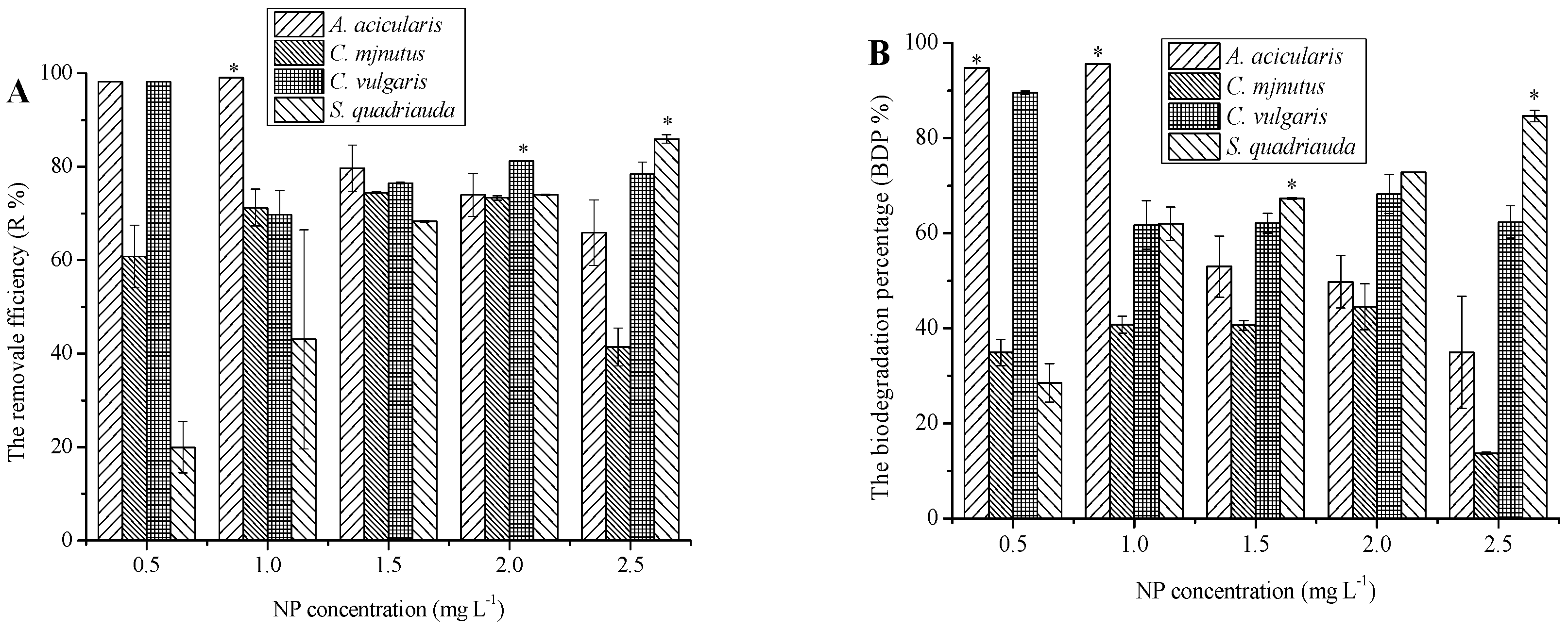
3.2. Removal of NP by Different Microalgae Species and Its Mechanisms

The residual concentrations of NP in the medium in the control flasks (without microalgal inoculation) did not show any significant changes during the 120 h experiments (data not shown), indicating that abiotic loss was negligible. The amount of NP remaining in the medium inoculated with microalgae species all decreased substantially within the first 24 h (Table 1). The decrease of NP was in a slower process, especially after a 72 h exposure (Table 2 and Table 3). The contents of NP in four algal cells under different treatments were measured (Table 1, Table 2 and Table 3) and it was observed that from the extra/intra ratios shown in Table 1, Table 2 and Table 3, the extracellular NP contents in all four species have been lower than the intracellular NP contents, with the ratios changing from 0.04 to 0.85.
As the results in Table 1, Table 2 and Table 3 demonstrated, biodegradation process was responsible for the removal of NP. The biodegradation percentages of NP by species decreased, whereas the removal efficiency percentages decreased with increasing NP bioconcentration (from 0.5 to 2.5 mg·L−1). Among all species, A. acicularis was the most effective species and removed more than 90% NP at low NP concentration exposure (0.5–1.0 mg·L−1) (Figure 3A). The process of removed NP by A. acicularis was mainly caused by biodegradation (Figure 3B, Table 3). At more than 1.5 mg·L−1 NP concentration, the specific growth rate and removal efficiency were decreased significantly (p < 0.05). Further, more than 90% of NP was removed by C. vulgaris at the end of the experiment under the 0.5 mg·L−1 NP treatment (Figure 3A). When the four species were exposed to 2.0–2.5 mg·L−1 NP, C. vulgaris and S. quadriauda removed more than 80% NP. For S. quadriauda, the NP degradation efficiency increased gradually with increasing concentrations of NP. However the NP degradation ability of three species decreased with increasing NP concentrations (Figure 3B). More than 60% of NP removed by S. quadriauda were attributed to biodegradation processes at 2.0–2.5 mg·L−1 NP. However, the growth of S. quadriauda was inhibited at more than 2.0 mg·L−1 NP. We supposed that some of the products by biodegradation may inhibit S. quadriauda growth. We were keeping research the specific biodegradation mechanism of S. quadriauda. In contrast, C. minutus displayed not only the lowest NP degradation ability, but also a more or less constant NP degradation efficiency during the experiment, with only 15%–40% NP biodegraded at the end of the experiment under different NP concentrations (Figure 3B). The highest of NP removed by C. minutus was 65% at the end of the experiment under the 1.5–2.0 mg·L−1 NP treatment. At more than 2.0 mg·L−1 NP concentration, the specific growth rate and removal efficiency of C. minutus were decrease significantly (p < 0.05) (Figure 1, Figure 2 and Figure 3).

4. Discussion

4.1. Influence of NP on Algal Growth

As an inherent part of the physical environment of dinoflagellates, bacteria could exist in the medium, attached to algal cell walls or even within the algal cells. This results in great difficulty in obtaining axenic cultures. A variety of procedures have been used to obtain bacteria-free algal cultures for the study of the relationship between toxic organic contaminants and algae [36]. The growth curves of the algae in the present study indicated that all algal cells were on the logarithmic phase of growth. When NP concentration was increased, growth of algal cells was inhibited in different degrees and the growth curves presented dose-effect relationship. The NP effect on growth inhibition in four algae (Figure 1) was in agreement with earlier reports [36,37] for freshwater algae, where a concentration of 500 μg·L−1 NP was able to reduce algal growth (EC50).
The present study clearly showed that the NP concentrations lower than 0.5 mg·L−1 did not markedly affect the algal growth of the four microalgae species, but when the concentrations were higher than 1.5 mg·L−1, the growth of the algal species decreased. A similar cell response was also observed in Microcystis aeruginosa and Chlorella species after NP exposure [38,39,40], and Chlorella fusca, Monoraphidium braunii and Stephanodiscus hantzschii after bisphenol-A (BPA) exposure [41,42,43]. The chlorophyll a curves showed that the A. acicularis, C. minutus and S. quadriauda had negative growth under 2.0–2.5 mg·L−1 NP exposure, whereas the C. vulgaris still grew well (Figure 1). Therefore, C. vulgaris were more resistant to NP than the other species at concentrations above 2 mg·L−1.
Microalgae possess several mechanisms for protecting themselves from the toxicity of organic contaminants. The cell wall, composed mainly of carbohydrates and proteins, serves as a barrier between organic pollutants and the cell interior [44,45]. In the present study, C. vulgaris might formed thicker cell walls than the other species to improve their adaptation to the stress caused by high concentrations of target compounds, and above results proved in some previous studies on this species exposed by the heavy metals zinc and copper [33,39,44,46]. Tsang et al. reported that C. vulgaris resisted tributyltin (TBT) by transforming TBT into a much less toxic metabolite [44]. Some microalgae species such as Chlorella fusca could metabolize BPA to an intermediate with no estrogenic activity [47]. Probably the phenolic organics were utilized by the algae as carbon source and assimilated by cell components for cell growth. No intermediates of NP were detected in the present study, metabolic product and its mechanism deserves further study.

4.2. Capacity of Algae for the Removal of Contaminants

The capability of freshwater micro- and macro-algae to adsorb pollutants was highly dependent on the cell biovolume and surface area, in particular, the ratio of surface area to volume [48]. However, the relationship between the amounts of NP uptake and the surface area/volume ratio in the present study was insignificant, as the NP removal efficiency among four algae species of different sizes and shapes were comparable. Tsezos and Bell found that the toxic organic pollutants removal capacity of the cell walls was less than that of the cell contents [49]. These results suggested that in addition to cell volume and shape, other properties, such as composition and structure of the cell, might also be important in determining NP biosorption. In this study, most of the NP accumulated inside the cells (74%–87%), whereas only 13%–26% was adsorbed by the cell walls. These high values were comparable to that reported for macroalgae, such as Cladophora [6], and were much higher than that for a marine microalga, Isochrysis galbana [34]. Due to the little accumulation of NP in the algal cells (Table 1, Table 2 and Table 3), the results from the present study showed that the removal of NP by algae was mainly caused by biodegradation by the algal cells rather than by simple sorption and accumulation in the cells. This is consistent with the results of previous studies on the BPA removal by some microalgae species [43,50], NP removal by C. vulgaris and Selenastrum capricornutum [39]. After adsorption and absorption, the target compounds NP were first accumulated and then metabolized by algal cells; thus the amounts of NP accumulated in algal cells were much less than the amounts biodegraded. Several freshwater microalgae were found to be able to glycosylate BPA by the action of glycosyl transferase [47]. S. obliquus might have a similar mechanism to metabolize NP because these two compounds also possess the requisite functional group (–OH) for direct metabolism.
Biodegradation of organic contaminants by algae has been demonstrated in previous and present studies. Tributyltin could be biodegradation by two Chlorella species [44] and some algae (such as Anabaena flosaquae and Microcysis aeruginosa) even were capable of producing di(n-butyl)phthalate (DBP) or mono(2-ethylhexyl)phthalate (MEHP) or both [51]. In the present study, NP showed an initial rapid removal phase during the first 24 h, followed by a slow dissipation phase (Table S1). Several processes might be involved in the dissipation processes, including sorption and biodegradation. It is expected that photolysis of NP induced by the presence of algae might occur, as demonstrated for the enhancement of BPA photodegradation by C. vulgaris and Anabaena cylindrical [52]. However, Table S1 clearly showed little variation in NP concentrations in the controls within 5 days. Therefore, the photolysis process played a small role in the dissipation of NP.
After the adsorption and absorption processes, NP was gradually degraded with 65.63%, 34.91%, 68.80% and 63.10% of the spiked NP degraded by A. acicularis, C. minutus, C. vulgaris and S. quadriauda at the end of the 120 h exposure under NP concentrations (0.5–2.5 mg·L−1), respectively. Such A. acicularis, C. vulgaris and S. quadriauda biodegradation was much faster than the previous results from two M. aeruginosa strains, which exhibited more than 60% NP degradation after 12 days of incubation, with different concentrations of NP [38] and microalgal species from other taxa [34]. These findings suggested that potential use of A. acicularis, C. vulgaris and S. quadriauda in the treatment of waste water containing the compound. The algae species A. acicularis, C. vulgaris and S. quadriauda used in the present study had nearly an equal removal capacity for NP as the green microalgae Selenastrum capricornutum and Chlorella species [39]. Therefore, algae used in the waste water treatment could not only remove heavy metals and inorganic substances such as nitrogen and phosphorus [46], but also remove organic substances such as NP [15,17,53,54]. Chlorella species, especially the commercial species C. vulgaris, were more capable of degrading NP than other algal genera, and the mechanisms involved two processes, a rapid initial passive physiochemical adsorption followed by active absorption, accumulation and degradation process.
The NP biodegradation pathway has been widely investigated in bacteria, but less so in plants, particularly in microalgae. Nevertheless, the metabolism of other phenolic compounds by microalgae displayed similar patterns as that in higher plants. Different freshwater microalgae were found to metabolize BPA to BPA glycosides, which were then released into the culture medium [47,55]. The metabolic pathway of p-chlorophenol (p-CP) in a marine microalga, Tetraselmis marina, involved glucosyl transfer followed by malonyl transfer [56]. The diatom Skeletonema costatum was able to detoxify 2,4-dichlorophenol by conjugation to glutathione catalyzed by glutathione S-transferase [57]. Further studies are needed to identify the major metabolic products and the biodegradation pathways of NP by microalgae.
The present and previous studies all showed that the NP might subsequently pose potential risks to organisms at higher trophic levels via biomagnification along food chains in aquatic ecosystems. Although the concentrations of NP used in the present study are unlikely to be detected in aquatic ecosystems, the algae A. acicularis, C. vulgaris and S. quadriauda demonstrated a high capability for the removal of the NP at mg·L−1 levels, indicating good prospects for their use in the treatment of wastewater.

5. Conclusions

All the four microalgae species investigated in the present study could efficiently remove NP at a low concentration of 0.5–1 mg·L−1 from water (close to the highest NP concentration detected in the environment), within a short exposure time (within 24 h), under photoautotrophic conditions. The mechanisms included initial rapid adsorption and absorption, followed by accumulation and biodegradation. In addition, the removal by the four algae species was mainly attributed to biodegradation or biotransformation process by the algal cells rather than to simple sorption and accumulation in the cells. The amounts of NP adsorbed on the algal cells were lower than those absorbed in algal cells. The NP biodegradation ability was species-specific. Among four algae species, A. acicularis and C. vulgaris were the more suitable species for effective removal and biodegradation of NP, and potential application of microalgae species in the removal of organic contaminants including alkylphenols in addition to nutrients and metals.

Supplementary Materials

The following are available online at www.mdpi.com/1660-4601/13/12/1239/s1, Table S1: The NP contents in medium of four freshwater microalgae under different treatments at 24, 72 and 120 h exposure.

Acknowledgments

This study received financial support from the Natural Science Foundation of China (No. 41476099, No.41676099 and No.21307140) and the Key Project of NSFC-Guangdong Joint Fund (No. U1133003).

Author Contributions

Ning He and Shunshan Duan conceived and designed the experiments; Ning He implemented the experiments, and collected and analyzed all of the samples; Xian Sun and Yu Zhong analyzed the data and produced the figures; Xian Sun and Ning He wrote the paper; Kaifeng Sun and Weijie Liu reviewed the manuscript.

Conflicts of Interest

The authors declare no conflict of interest.

References

  1. Soares, A.; Guieysse, B.; Jefferson, B.; Cartmell, E.; Lester, J. Nonylphenol in the environment: A critical review on occurrence, fate, toxicity and treatment in wastewaters. Environ. Int. 2008, 34, 1033–1049. [Google Scholar] [CrossRef] [PubMed]
  2. Tubau, I.; Vázquez-Suñé, E.; Carrera, J.; González, S.; Petrovic, M.; Alda, M.J.L.D.; Barceló, D. Occurrence and fate of alkylphenol polyethoxylate degradation products and linear alkylbenzene sulfonate surfactants in urban ground water: Barcelona case study. J. Hydrol. 2010, 383, 102–110. [Google Scholar] [CrossRef]
  3. Fan, Z.; Hu, J.; An, W.; Yang, M. Detection and occurrence of chlorinated byproducts of bisphenol A, nonylphenol, and estrogens in drinking water of China: Comparison to the parent compounds. Environ. Sci. Technol. 2013, 47, 10841–10850. [Google Scholar] [CrossRef] [PubMed]
  4. Isobe, T.; Nishiyama, H.; Nakashima, A.; Takada, H. Distribution and behavior of nonylphenol, octylphenol, and nonylphenol monoethoxylate in Tokyo metropolitan area: Their association with aquatic particles and sedimentary distributions. Environ. Sci. Technol. 2001, 35, 1041–1049. [Google Scholar] [CrossRef] [PubMed]
  5. Gross-Sorokin, M.Y.; Grist, E.P.; Cooke, M.; Crane, M. Uptake and depuration of 4-nonylphenol by the benthic invertebrate Gammarus pulex: How important is feeding rate? Environ. Sci. Technol. 2003, 37, 2236–2241. [Google Scholar] [CrossRef] [PubMed]
  6. Ahel, M.; McEvoy, J.; Giger, W. Bioaccumulation of the lipophilic metabolites of nonionic surfactants in freshwater organisms. Environ. Pollut. 1993, 79, 243–248. [Google Scholar] [CrossRef]
  7. Bertanza, G.; Pedrazzani, R.; Zambarda, V.; Dal Grande, M.; Icarelli, F.; Baldassarre, L. Removal of endocrine disrupting compounds from wastewater treatment plant effluents by means of advanced oxidation. Water Sci. Technol. 2010, 61, 1663. [Google Scholar] [CrossRef] [PubMed]
  8. Semple, K.T.; Cain, R.B.; Schmidt, S. Biodegradation of aromatic compounds by microalgae. FEMS Microbiol. Lett. 1999, 170, 291–300. [Google Scholar] [CrossRef]
  9. Wang, J.; Xie, P.; Guo, N. Effects of nonylphenol on the growth and microcystin production of Microcystis strains. Environ. Res. 2007, 103, 70–78. [Google Scholar] [CrossRef] [PubMed]
  10. Baptista, M.S.; Stoichev, T.; Basto, M.C.P.; Vasconcelos, V.M.; Vasconcelos, M.T.S.D. Fate and effects of octylphenol in a Microcystis aeruginosa culture medium. Aquat. Toxicol. 2009, 92, 59–64. [Google Scholar] [PubMed]
  11. Elmasry, M.A.A.; Gaber, A.; Khater, E.M.H. Nutrient removal and biodiesel production by integration of freshwater algae cultivation with piggery wastewater treatment. Water Res. 2013, 47, 4294–4302. [Google Scholar]
  12. Fierro, S.; del Pilar Sánchez-Saavedra, M.; Copalcua, C. Nitrate and phosphate removal by chitosan immobilized Scenedesmus. Bioresour. Technol. 2008, 99, 1274–1279. [Google Scholar] [CrossRef] [PubMed]
  13. Jácome-Pilco, C.R.; Cristiani-Urbina, E.; Flores-Cotera, L.B.; Velasco-García, R.; Ponce-Noyola, T.; Cañizares-Villanueva, R.O. Continuous Cr (VI) removal by Scenedesmus incrassatulus in an airlift photobioreactor. Bioresour. Technol. 2009, 100, 2388–2391. [Google Scholar] [CrossRef] [PubMed]
  14. Sun, X.; Zhong, Y.; Huang, Z.; Yang, Y. Selenium accumulation in unicellular green alga Chlorella vulgaris and its effects on antioxidant enzymes and content of photosynthetic pigments. PLoS ONE 2014, 9. [Google Scholar] [CrossRef] [PubMed]
  15. Baglieri, A.; Sidella, S.; Barone, V.; Fragalà, F.; Silkina, A.; Negre, M.; Gennari, M. Cultivating Chlorella vulgaris and Scenedesmus quadricauda microalgae to degrade inorganic compounds and pesticides in water. Environ. Sci. Pollut. Res. 2016, 23, 18165–18174. [Google Scholar] [CrossRef] [PubMed]
  16. Della Greca, M.; Pinto, G.; Pistillo, P.; Pollio, A.; Previtera, L.; Temussi, F. Biotransformation of ethinylestradiol by microalgae. Chemosphere 2008, 70, 2047–2053. [Google Scholar] [CrossRef] [PubMed]
  17. Zhou, G.-J.; Peng, F.-Q.; Yang, B.; Ying, G.-G. Cellular responses and bioremoval of nonylphenol and octylphenol in the freshwater green microalga Scenedesmus obliquus. Ecotoxicol. Environ. Saf. 2013, 87, 10–16. [Google Scholar] [CrossRef] [PubMed]
  18. Green, D.H.; Llewellyn, L.E.; Negri, A.P.; Blackburn, S.I.; Bolch, C.J. Phylogenetic and functional diversity of the cultivable bacterial community associated with the paralytic shellfish poisoning dinoflagellate Gymnodinium catenatum. FEMS Microbiol. Ecol. 2004, 47, 345–357. [Google Scholar] [CrossRef]
  19. Goecke, F.; Thiel, V.; Wiese, J.; Labes, A.; Imhoff, J.F. Algae as an important environment for bacteria—Phylogenetic relationships among bacterial species isolated from algae. Phycologia 2013, 52, 14–24. [Google Scholar] [CrossRef]
  20. Munoz, R.; Guieysse, B. Algal-bacterial processes for the treatment of hazardous contaminants: A review. Water Res. 2006, 40, 2799–2815. [Google Scholar] [CrossRef] [PubMed]
  21. Subashchandrabose, S.R.; Ramakrishnan, B.; Megharaj, M.; Venkateswarlu, K.; Naidu, R. Consortia of cyanobacteria/microalgae and bacteria: Biotechnological potential. Biotechnol. Adv. 2011, 29, 896–907. [Google Scholar] [CrossRef] [PubMed]
  22. Uribe, P.; Espejo, R.T. Effect of associated bacteria on the growth and toxicity of Alexandrium catenella. Appl. Environ. Microbiol. 2003, 69, 659–662. [Google Scholar] [CrossRef] [PubMed]
  23. Wang, X.; Gong, L.; Liang, S.; Han, X.; Zhu, C.; Li, Y. Algicidal activity of rhamnolipid biosurfactants produced by Pseudomonas aeruginosa. Harmful Algae 2005, 4, 433–443. [Google Scholar] [CrossRef]
  24. Mayali, X.; Doucette, G.J. Microbial community interactions and population dynamics of an algicidal bacterium active against Karenia brevis (Dinophyceae). Harmful Algae 2002, 1, 277–293. [Google Scholar] [CrossRef]
  25. Hold, G.L.; Smith, E.A.; Birkbeck, T.H.; Gallacher, S. Comparison of paralytic shellfish toxin (PST) production by the dinoflagellates Alexandrium lusitanicum NEPCC 253 and Alexandrium tamarense NEPCC 407 in the presence and absence of bacteria. FEMS Microbiol. Ecol. 2001, 36, 223–234. [Google Scholar] [CrossRef] [PubMed]
  26. Chong, A.M.Y.; Wong, Y.S.; Tam, N.F.Y. Performance of different microalgal species in removing nickel and zinc from industrial wastewater. Chemosphere 2000, 41, 251–257. [Google Scholar] [CrossRef]
  27. Ibrahim, M.B.M.; Gamila, H.A. Algal bioassay for evaluating the role of algae in bioremediation of crude oil: II freshwater phytoplankton assemblages. Bull. Environ. Contam. Toxicol. 2004, 73, 971–978. [Google Scholar] [CrossRef] [PubMed]
  28. Hill, R.D.; Nagarkar, S.; Peart, M.R. Cyanobacterial crust in Hong Kong and comments on future research. Algae 2000, 15, 65–71. [Google Scholar]
  29. Lei, A.P.; Hu, Z.L.; Wong, Y.S.; Tam, N.F.Y. Removal of fluoranthene and pyrene by different microalgal species. Bioresour. Technol. 2007, 98, 273–280. [Google Scholar] [CrossRef] [PubMed]
  30. Rippka, R.; Deruelles, J.; Waterbury, J.B.; Herdman, M.; Stanier, R.Y. Generic assignments, strain histories and properties of pure cultures of cyanobacteria. Microbiology 1979, 111, 1–61. [Google Scholar] [CrossRef]
  31. Su, J.; Yang, X.; Zheng, T.; Hong, H. An efficient method to obtain axenic cultures of Alexandrium tamarense—A PSP-producing dinoflagellate. J. Microbiol. Methods 2007, 69, 425–430. [Google Scholar] [CrossRef] [PubMed]
  32. Rezaee, M.; Assadi, Y.; Hosseini, M.-R.M.; Aghaee, E.; Ahmadi, F.; Berijani, S. Determination of organic compounds in water using dispersive liquid-liquid microextraction. J. Chromatogr. 2006, 1116, 1–9. [Google Scholar] [CrossRef] [PubMed]
  33. Zhou, G.-J.; Peng, F.-Q.; Zhang, L.-J.; Ying, G.-G. Biosorption of zinc and copper from aqueous solutions by two freshwater green microalgae Chlorella pyrenoidosa and Scenedesmus obliquus. Environ. Sci. Pollut. Res. 2012, 19, 2918–2929. [Google Scholar] [CrossRef] [PubMed]
  34. Correa-Reyes, G.; Viana, M.T.; Marquez-Rocha, F.J.; Licea, A.F.; Ponce, E.; Vazquez-Duhalt, R. Nonylphenol algal bioaccumulation and its effect through the trophic chain. Chemosphere 2007, 68, 662–670. [Google Scholar] [CrossRef] [PubMed]
  35. Liu, Y.; Guan, Y.; Gao, Q.; Tam, N.F.Y.; Zhu, W. Cellular responses, biodegradation and bioaccumulation of endocrine disrupting chemicals in marine diatom Navicula incerta. Chemosphere 2010, 80, 592–599. [Google Scholar] [CrossRef] [PubMed]
  36. Graff, L.; Isnard, P.; Cellier, P.; Bastide, J.; Cambon, J.P.; Narbonne, J.F.; Budzinski, H.; Vasseur, P. Toxicity of chemicals to microalgae in river and in standard waters. Environ. Toxicol. Chem. 2003, 22, 1368–1379. [Google Scholar] [CrossRef] [PubMed]
  37. Gao, Q.T.; Tam, N.F.Y. Growth, photosynthesis and antioxidant responses of two microalgal species, Chlorella vulgaris and Selenastrum capricornutum, to nonylphenol stress. Chemosphere 2011, 82, 346–354. [Google Scholar] [CrossRef] [PubMed]
  38. Wang, J.; Xie, P. Antioxidant enzyme activities of Microcystis aeruginosa in response to nonylphenols and degradation of nonylphenols by M. aeruginosa. Environ. Geochem. Health 2007, 29, 375–383. [Google Scholar] [CrossRef] [PubMed]
  39. Gao, Q.; Wong, Y.S.; Tam, N. Removal and biodegradation of nonylphenol by different Chlorella species. Mar. Pollut. Bull. 2011, 63, 445–451. [Google Scholar] [CrossRef] [PubMed]
  40. Gao, Q.; Wong, Y.; Tam, N. Removal and biodegradation of nonylphenol by immobilized Chlorella vulgaris. Bioresour. Technol. 2011, 102, 10230–10238. [Google Scholar] [CrossRef] [PubMed]
  41. Gattullo, C.E.; Bährs, H.; Steinberg, C.E.; Loffredo, E. Removal of bisphenol A by the freshwater green alga Monoraphidium braunii and the role of natural organic matter. Sci. Total Environ. 2012, 416, 501–506. [Google Scholar] [CrossRef] [PubMed]
  42. Hirooka, T.; Nagase, H.; Uchida, K.; Hiroshige, Y.; Ehara, Y.; Nishikawa, J.I.; Nishihara, T.; Miyamoto, K.; Hirata, Z. Biodegradation of bisphenol A and disappearance of its estrogenic activity by the green alga Chlorella fusca var. vacuolata. Environ. Toxicol. Chem. 2005, 24, 1896–1901. [Google Scholar] [CrossRef] [PubMed]
  43. Li, R.; Chen, G.-Z.; Tam, N.F.Y.; Luan, T.-G.; Shin, P.K.; Cheung, S.G.; Liu, Y. Toxicity of bisphenol A and its bioaccumulation and removal by a marine microalga Stephanodiscus hantzschii. Ecotoxicol. Environ. Saf. 2009, 72, 321–328. [Google Scholar] [CrossRef] [PubMed]
  44. Tsang, C.; Lau, P.; Tam, N.; Wong, Y. Biodegradation capacity of tributyltin by two Chlorella species. Environ. Pollut. 1999, 105, 289–297. [Google Scholar] [CrossRef]
  45. Andrade, L.R.; Leal, R.N.; Noseda, M.; Duarte, M.E.R.; Pereira, M.S.; Mourão, P.A.S.; Farina, M.; Filho, G.M.A. Brown algae overproduce cell wall polysaccharides as a protection mechanism against the heavy metal toxicity. Mar. Pollut. Bull. 2010, 60, 1482–1488. [Google Scholar] [CrossRef] [PubMed]
  46. Chan, S.M.N.; Luan, T.; Wong, M.H.; Tam, N.F.Y. Removal and biodegradation of polycyclic aromatic hydrocarbons by Selenastrum capricornutum. Environ. Toxicol. Chem. 2006, 25, 1772–1779. [Google Scholar] [CrossRef] [PubMed]
  47. Nakajima, N.; Teramoto, T.; Kasai, F.; Sano, T.; Tamaoki, M.; Aono, M.; Kubo, A.; Kamada, H.; Azumi, Y.; Saji, H. Glycosylation of bisphenol A by freshwater microalgae. Chemosphere 2007, 69, 934–941. [Google Scholar] [CrossRef] [PubMed]
  48. Tang, J.; Hoagland, K.D.; Siegfried, B.D. Uptake and bioconcentration of atrazine by selected freshwater algae. Environ. Toxicol. Chem. 1998, 17, 1085–1090. [Google Scholar] [CrossRef]
  49. Tsezos, M.; Bell, J. Comparison of the biosorption and desorption of hazardous organic pollutants by live and dead biomass. Water Res. 1989, 23, 561–568. [Google Scholar] [CrossRef]
  50. Ji, M.K.; Kabra, A.N.; Choi, J.; Hwang, J.H.; Kim, J.R.; Abou-Shanab, R.A.I.; Oh, Y.K.; Jeon, B.H. Biodegradation of bisphenol A by the freshwater microalgae Chlamydomonas mexicana and Chlorella vulgaris. Ecol. Eng. 2014, 73, 260–269. [Google Scholar] [CrossRef]
  51. Babu, B.; Wu, J.-T. Production of phthalate esters by nuisance freshwater algae and cyanobacteria. Sci. Total Environ. 2010, 408, 4969–4975. [Google Scholar] [CrossRef] [PubMed]
  52. Peng, Z.E.; Wu, F.; Deng, N. Photodegradation of bisphenol A in simulated lake water containing algae, humic acid and ferric ions. Environ. Pollut. 2006, 144, 840–846. [Google Scholar] [CrossRef] [PubMed]
  53. Liu, Y.; Dai, X.; Wei, J. Toxicity of the xenoestrogen nonylphenol and its biodegradation by the alga Cyclotella caspia. J. Environ. Sci. 2013, 25, 1662–1671. [Google Scholar] [CrossRef]
  54. Mann, R.M.; Boddy, M.R. Biodegradation of a nonylphenol ethoxylate by the autochthonous microflora in lake water with observations on the influence of light. Chemosphere 2000, 41, 1361–1369. [Google Scholar] [CrossRef]
  55. Michałowicz, J. Bisphenol A—Sources, toxicity and biotransformation. Environ. Toxicol. Pharmacol. 2014, 37, 738–758. [Google Scholar] [CrossRef] [PubMed]
  56. Petroutsos, D.; Wang, J.; Katapodis, P.; Kekos, D.; Sommerfeld, M.; Hu, Q. Toxicity and metabolism of p-chlorophenol in the marine microalga Tetraselmis marina. Aquat. Toxicol. 2007, 85, 192–201. [Google Scholar] [CrossRef] [PubMed]
  57. Yang, S.; Wu, R.S.; Kong, R.Y. Biodegradation and enzymatic responses in the marine diatom Skeletonema costatum upon exposure to 2,4-dichlorophenol. Aquat. Toxicol. 2002, 59, 191–200. [Google Scholar] [CrossRef]
Figure 1. Effect of Nonylphenol (NP) concentration on the chlorophyll a content of (A) A. acicularis; (B) C. minutus; (C) C. vulgaris and (D) S. quadriauda, Values are the mean ± standard deviation (SD) (n = 3).
Ijerph 13 01239 g001
Figure 2. Growth of (A) A. acicularis; (B) C. minutus; (C) C. vulgaris and (D) S. quadriauda, exposed to different NP concentrations at the end of 0, 24, 48, 72 and 96 h (mean and standard deviation of three replicates were shown).
Ijerph 13 01239 g002
Figure 3. Removal (A) and biodegradation (B) of NP by A. acicularis, C. minutus, C. vulgaris and S. quadriauda at the end 120 h (mean and standard deviation of three replicates are shown. The single asterisk * in the figure represents significant difference compared to other algae species (p < 0.05).
Ijerph 13 01239 g003
Table 1. Dissolved, intracellular and extracellular NP contents of four freshwater microalgae under different treatments at the end of 24 h. Mean and standard deviation of three replicates are shown.
Treatment (mg·L−1)Microalgal SpeciesAmount of NPExtra/Intra Ratio *
Dissolved NP (μg·L−1)Extracellular NP (10−8 μg·cell−1)Intracellular NP (10−8 μg·cell−1)
0.5Control445.0 ± 10.0---
A. acicularis162 ± 00.9 ± 0.13.5 ± 00.26
C. minutus343.7 ± 1.21.0 ± 0.14.8 ± 0.70.21
C. vulgaris388.3 ± 11.70.8 ± 0.06.7 ± 0.60.12
S. quadriauda415.7 ± 32.09.7 ± 0.120.2 ± 2.80.49
1Control941.1 ± 7.2---
A. acicularis405.9 ± 15.63.0 ± 014.1 ± 0.60.21
C. minutus515.9 ± 62.12.0 ± 0.214.5 ± 3.70.15
C. vulgaris723.7 ± 13.72.7 ± 0.19.6 ± 0.60.28
S. quadriauda770.2 ± 5.214.1 ± 3.237.2 ± 2.50.85
1.5Control1479.7 ± 10.2---
A. acicularis665.3 ± 87.88.4 ± 0.837.0 ± 2.40.23
C. minutus964.9 ± 22.43.7 ± 0.624.8 ± 0.80.15
C. vulgaris1464.3 ± 154.88.1 ± 0.323.7 ± 2.90.35
S. quadriauda1477.8 ± 16.418.2 ± 0.661.7 ± 6.50.3
2Control1890 ± 13.0---
A. acicularis845.6 ± 0.310.4 ± 0.249.8 ± 1.10.21
C. minutus1236.6 ± 41.64.9 ± 0.528.8 ± 2.60.17
C. vulgaris1305.2 ± 211.610.2 ± 1.033.4 ± 3.90.31
S. quadriauda1795.6 ± 132.123.2 ± 2.9104.6 ± 0.60.22
2.5Control2401.0 ± 92.6---
A. acicularis1360.5 ± 169.116.9 ± 1.277.0 ± 1.90.22
C. minutus1606.5 ± 136.26.6 ± 0.638.0 ± 0.50.16
C. vulgaris2308.5 ± 52.513.1 ± 1.438.3 ± 3.30.34
S. quadriauda1911.0 ± 8.750.3 ± 0.3124.4 ± 1.30.4
* Ratio of extracellular concentration to intracellular concentration.
Table 2. Dissolved, intracellular and extracellular NP contents of four freshwater microalgae under different treatments at the end of 72 h. Mean and standard deviation of three replicates are shown.
Treatment (mg·L−1)Microalgal SpeciesAmount of NPExtra/Intra Ratio *
Dissolved NP (μg·L−1)Extracellular NP (10−8 μg·cell−1)Intracellular NP (10−8 μg·cell−1)
0.5Control448.3 ± 5.6---
A. acicularis9.1 ± 00.2 ± 00.5 ± 0.10.33
C. minutus191.0 ± 00.1 ± 02.3 ± 0.30.06
C. vulgaris270.8 ± 3.41.0 ± 0.13.8 ± 0.60.26
S. quadriauda323.0 ± 0.91.7 ± 0.12.1 ± 0.20.85
1Control940.2 ± 10.1---
A. acicularis117.0 ± 5.70.6 ± 0.11.9 ± 0.20.3
C. minutus402.4 ± 19.51.2 ± 0.17.1 ± 1.90.18
C. vulgaris549.1 ± 8.62.3 ± 0.27.8 ± 0.70.3
S. quadriauda478.1 ± 7.20.6 ± 02.7 ± 0.40.23
1.5Control1478.9 ± 23.6---
A. acicularis539.3 ± 7.84.6 ± 0.719.2 ± 5.40.24
C. minutus760.1 ± 13.72.7 ± 0.116.5 ± 0.40.16
C. vulgaris840.6 ± 67.14.2 ± 0.418.7 ± 1.10.23
S. quadriauda720.2 ± 14.40.9 ± 04.0 ± 1.00.26
2Control1873.2 ± 26.0---
A. acicularis599.3 ± 1.37.1 ± 0.224.1 ± 0.20.29
C. minutus1006.7 ± 29.13.6 ± 0.119.0 ± 2.10.18
C. vulgaris863.2 ± 45.25.5 ± 1.024.1 ± 4.10.23
S. quadriauda944.6 ± 21.61.9 ± 0.15.8 ± 0.60.32
2.5Control2399.6 ± 27.8---
A. acicularis819.4 ± 31.711.3 ± 1.365.2 ± 10.00.17
C. minutus1429.4 ± 62.16.1 ± 0.227.7 ± 1.30.22
C. vulgaris1529.3 ± 46.47.2 ± 0.928.8 ± 3.10.25
S. quadriauda1391.2 ± 11.33.4 ± 0.512.8 ± 0.40.29
* Ratio of extracellular concentration to intracellular concentration.
Table 3. Dissolved, intracellular and extracellular NP contents of four freshwater microalgae under different treatments at the end of 120 h. Mean and standard deviation of three replicates are shown.
Treatment (mg·L−1)Microalgal SpeciesAmount of NPExtra/Intra Ratio *
Dissolved NP (μg·L−1)Extracellular NP (10−8 μg·cell−1)Intracellular NP (10−8 μg·cell−1)
0.5Control448.0 ± 9.1---
A. acicularis9.1 ± 00.0 ± 00.4 ± 00.04
C. minutus196.1 ± 37.30.1 ± 01.5 ± 0.20.08
C. vulgaris9.1 ± 00.5 ± 0.02.6 ± 00.2
S. quadriauda200.0 ± 22.50.3 ± 0.00.1 ± 03.04
1Control943.9 ± 8.5---
A. acicularis9.1 ± 00.2 ± 00.7 ± 00.3
C. minutus287.3 ± 32.40.1 ± 03.8 ± 0.90.04
C. vulgaris302.9 ± 42.81.7 ± 0.44.3 ± 0.90.4
S. quadriauda335.1 ± 19.10.4 ± 00.6 ± 0.10.6
1.5Control1475.9 ± 14.8---
A. acicularis304.6 ± 60.53.9 ± 0.213.0 ± 0.20.3
C. minutus384.4 ± 2.20.7 ± 0.114.2 ± 0.30.05
C. vulgaris352.6 ± 2.33.6 ± 0.611.9 ± 2.20.31
S. quadriauda474.8 ± 2.70.4 ± 01.0 ± 00.42
2Control1880.5 ± 7.7---
A. acicularis520.1 ± 75.94.8 ± 0.314.0 ± 0.40.34
C. minutus533.5 ± 7.81.1 ± 0.214.0 ± 1.20.08
C. vulgaris375.7 ± 0.73.1 ± 0.78.8 ± 2.60.37
S. quadriauda519.9 ± 1.80.7 ± 01.77 ± 0.20.4
2.5Control2397.9 ± 77.8---
A. acicularis803.1 ± 102.96.5 ± 0.0523.9 ± 3.60.27
C. minutus1465.3 ± 82.51.6 ± 0.419.6 ± 0.40.08
C. vulgaris539.9 ± 52.83.5 ± 0.712.6 ± 1.80.28
S. quadriauda351.6 ± 18.82.6 ± 0.25.7 ± 0.80.46
* Ratio of extracellular concentration to intracellular concentration.

