Article

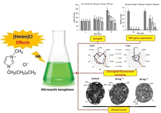
Inhibition Effect of Ionic Liquid [Hmim]Cl on Microcystis Growth and Toxin Production

1
College of Life Sciences, Henan Normal University, Xinxiang 453007, China
2
Department of Food Toxicology and Contaminants, National Research Centre, Dokki, Cairo 12622, Egypt
3
College of Life and Environmental Science, Wenzhou University, Wenzhou 325035, China
4
Wuhan Imagination Science and Technology Development Co., Ltd., Wuhan 443000, China
*
Authors to whom correspondence should be addressed.
Int. J. Environ. Res. Public Health 2022, 19(14), 8719; https://doi.org/10.3390/ijerph19148719
Submission received: 15 May 2022 / Revised: 15 July 2022 / Accepted: 17 July 2022 / Published: 18 July 2022
(This article belongs to the Special Issue Environmental Pollution and Ecological Health)

Abstract

:
Ionic liquids (ILs) are known as “green solvents” and widely used in industrial applications. However, little research has been conducted on cyanobacteria. This study was conducted to investigate the toxicity of ionic liquids ([Hmim]Cl) on Microcystis aeruginosa PCC7806. The EC50 (72 h) of [Hmim]Cl on the growth of Microcystis aeruginosa PCC 7806 was 10.624 ± 0.221 mg L−1. The possible mechanism of toxicity of [Hmim]Cl against M. aeruginosa PCC 7806 was evaluated by measuring cell growth, photosynthetic pigment contents, chlorophyll fluorescence transients, cell ultrastructure, and transcription of the microcystin-producing gene (mcyB). The concentrations of chlorophyll a and carotenoids were significantly reduced in treated M. aeruginosa cultures. The results of chlorophyll fluorescence transients showed that [Hmim]Cl could destruct the electron-accepting side of the photosystem II of M. aeruginosa PCC 7806. Transmission electron microscopy demonstrated cell damage including changes in the structure of the cell wall and cell membrane, thylakoid destruction, and nucleoid disassembly. The transcription of the mcyB gene was also inhibited under [Hmim]Cl stress. In summary, this study provides new insights into the toxicity of [Hmim]Cl on cyanobactreia.

Graphical Abstract

1. Introduction

Demand for green technologies has increased rapidly in recent years. Ionic liquids (ILs) have attracted great attention as promising organic solvents. ILs are low-temperature molten salts, which possess many unique physiochemical characteristics and excellent solvent properties (negligible vapor pressure, high thermal, chemical, and electrochemical stability) [1]. Therefore, ILs have been considered to be “green” substitutes for conventional volatile organic compounds (VOCs) used in synthetic and analytical processes [2]. Although ILs can reduce the potential risk of air pollution due to their negligible vapor pressures, they may pose ecological risks to aquatic ecosystems, due to their high solubility in water. In addition, many studies have found that ILs show poor biodegradability [3].
Algae are crucial primary producers for the food chain in aquatic ecosystems and play an important role in nutrient cycling. Algae have been used for hazard assessment of aquatic contaminants [4,5], because of their rapid response to environmental changes. Several studies have been done on the toxicity of ILs on aquatic organisms, such as green algae [6], diatoms [7], cladocerans [8], mussels [9], and fish [10]. However, little research has been conducted on cyanobacteria [11].
Global warming and eutrophication have enhanced the proliferation of cyanobacteria in freshwater bodies [12,13]. Microcystis aeruginosa (Kützing) Kützing 1846 is one of the most widespread and dominant cyanobacterial species [14,15]. M. aeruginosa also has the capability of producing secondary metabolites such as microcystin, the synthesis of which is sensitive to the surrounding environment [16,17]. Thus, the response of M. aeruginosa to IL stress can be used for assessing the impact of ILs on aquatic ecosystems. Oxygenic photosynthesis of cyanobacteria is easily affected by hazardous substances such as ILs [18]. The chlorophyll fluorescence measurement is an important index to evaluate the performance of photosynthesis processes and is usually measured via the redox states of photosystem II (PSII) and the electron-donating and -accepting sides of the active reaction centers. Krause et al. [19] and Strasser and Strasser [20] established the JIP-test, which conducts a kinetic analysis of the light-induced chlorophyll fluorescence rise curve based on the theory of biofilm energy flow. The JIP-test reflects the physiological state of cyanobacterial PSII by detecting the effects of different factors on its structure and function [18,21,22].
The compound 1-hexyl-3-methylimidazolium chloride, which is abbreviated as [Hmim]Cl, is one of the widely used ILs [23]. Until now, there has been no available study concerning the effects of [Hmim]Cl on cyanobacteria. The current study aims to illustrate the possible toxicological mechanism of [Hmim]Cl on cyanobacteria from the view of cell growth, photosynthesis, and gene expression.

2. Materials and Methods

The M. aeruginosa PCC 7806 was provided by the Institute of Hydrobiology, Chinese Academy of Sciences, and was cultured in a BG11 liquid medium [24]. The culture was maintained at a light–dark cycle of 12 h:12 h with a light intensity of 3000 lx under a controlled temperature (28 °C). The [Hmim]Cl (99% purity) was purchased from the Chengjie Chemical Company (Shanghai, China).

2.1. Measurement of Cell Growth and Photosynthetic Pigments Content

The [Hmim]Cl was prepared and added into the M. aeruginosa PCC 7806 culture with a cell density of 104 cells mL−1. The final concentrations of [Hmim]Cl were set at 5, 10, 20, and 30 mg L−1. All experiments were performed in triplicate. The cell density and pigment contents in the control and treated cultures were measured after 24 and 72 h of incubation. To measure the pigment contents, the cultures were centrifuged for 10 min at 8000 rpm, and the supernatants were discarded. The chlorophyll a(Chl a) and carotenoids were extracted from cell pellets using 80% acetone for 24 h in darkness at 4 °C. Absorbance of the extracts was measured using a spectrophotometer at 663, 645, and 450 nm. The following equations set by Richards et al. (1952) [25] were used for the calculation of the pigment contents.

2.2. Measurement of Chlorophyll Fluorescence Transients

The polyphasic rise of chlorophyll fluorescence transients was detected by PEA (AquaPen-C AP-C 100, Photon Systems Instruments, The Czech Republic). The JIP transients were measured after exposure to [Hmim]Cl stress for 24 h and 72 h, respectively. Cells from the control and treated cultures were placed on special plastic clips. The parameters of absorption (ABS/RC), trapping (TR0/RC), electron transport (ET0/RC), dissipation (DI0/RC), the probability that a trapped exciton moves an electron into the electron transport chain beyond QA (ψ0), the maximum quantum yield of primary photochemistry (φP0), the quantum yield of electron transport (φE0), the probability that an absorbed photon is dissipated (φD0), relative fluorescence values [(1 − VK/VJ)r], and the performance indexes of photosystem II were calculated from the chlorophyll fluorescence transients [26,27]. The intensity of actinic light was set at 30 μmol photons/(m2·s). The equations used for calculating the above parameters were as follows [27]:
ABS/RC = (M0/VJ)(1 − F0/FM)
TR0/RC = M0/VJ
ET0/RC = (TR0/RC)ψ0
DI0/RC = (ABS/RC) − (TR0/RC)
φP0 = 1 − (F0/FM)
ψ0 = 1 − VJ
φE0 = (1 − F0/FM)(1 − VJ)
φD0 = F0/FM
PIABS = [RC/ABS][φP0/(1 − φP0)][ψ0/(1 − ψ0)]
(1 − VK/VJ)r = [(1 − VK/VJ)treatment]/[(1 − VK/VJ)Control].

2.3. Observation of Cell Ultrastructure

Changes in the ultrastructure of the treated M. aeruginosa cells at 10 and 30 mg L−1 of [Hmim]Cl were detected using transmission electron microscopy. After 48 h of exposure, culture cells were centrifuged for 15 min at 4000 rpm under 4 °C, and the supernatant was discarded. The cell pellets were fixed with 2.5% glutaraldehyde solution. The samples were sent to the Institute of Hydrobiology, Chinese Academy of Sciences, for electron microscopic analysis. Changes in the ultrastructure of the treated cells were identified by comparison with the control group.

2.4. Transcription of mcyB Gene

2.4.1. RNA Extraction and Reverse Transcription

The cells of M. aeruginosa PCC 7806 were harvested after 24 h and 72 h of exposure to each concentration of [Hmim]Cl. The cultures were centrifuged at 6000 rpm for 10 min, and the supernatant was discarded. Cell pellets were re-suspended in a Trizol reagent (Invitrogen, Waltham, MA, USA) and then frozen and thawed for three times. Total RNAs were extracted following the manufacturer’s manual of Trizol reagent and dissolved with diethyl pyrocarbonate-treated water. Reverse transcription was conducted using a Transcriptor First Strand cDNA Synthesis Kit (Roche, Germany).

2.4.2. Determination of mcyB Gene Transcription

The primers 30F and 108R were used for the detection of the mcyB gene [16], whereas 5′-GGACGGGTGAGTAACGCGTA-3′ and 5′-CCCATTGCGGAAAATTCCCC-3′ were used to amplify 16S rRNA gene as the internal control [28]. The transcription of mcyB in M. aeruginosa was determined by qPCR after 24 h and 72 h of exposure to each concentration of [Hmim]Cl. The qPCR reaction was prepared with final volumes of 10 μL containing 5 μL of Master Mix (SYBR Green, CWBIO, China), 0.2 μL of each primer (10 μmol L−1), and 0.4 μL cDNA and 4 μL of sterile water. The qPCR conditions were set as follows: 95 °C for 10 min; 40 cycles at 95 °C for 15 s, 59 °C for 30 s, and 72 °C for 30 s. The relative transcription level of the mcyB gene was calculated using 2−ΔΔCt where ΔΔCt = (Ct target gene − Ct 16S rrn)stress − (Ct target gene − Ct 16Srrn)control [29].

2.5. Statistical Analysis

The experimental data were analyzed by SPSS 20.0 (ANOVA, LSD). Differences were considered to be significant at p < 0.05.

3. Results

3.1. Inhibition of [Hmim]Cl on M. aeruginosa PCC 7806 Growth

As displayed in Figure 1a, cell growth was not affected by [Hmim]Cl at the concentrations of 5 and 10 mg L−1 at 24 h. However, cell growth was significantly inhibited by higher concentrations of [Hmim]Cl with 25% decrease at 30 mg L−1. After 72 h, the cell density of the control culture and the culture exposed to 5 mg L−1 of [Hmim]Cl increased by 118% and 61%, respectively. For cultures exposed to higher concentrations of [Hmim]Cl, cell density decreased after 72 h of exposure. No growth was observed at 10 mg L−1 of [Hmim]Cl, and cell density decreased by 23% in culture exposed to 30 mg L−1 of [Hmim]Cl at 72 h compared with 0 time. [Hmim]Cl at the concentrations of 10, 20, and 30 mg L−1 inhibited cell growth by >50% when compared with the control culture after 72 h of incubation. Regression curves of the logarithmic values of [Hmim]Cl concentrations against the inhibitory rate after 72 h of exposure is shown in Figure 1b. The calculated EC50 of [Hmim]Cl on the growth of M. aeruginosa was 10.624 ± 0.221 mg L−1.

3.2. Effects of [Hmim]Cl on the Pigment Contents

The effects of [Hmim]Cl on the pigment contents were similar to those on the cell growth (Figure 2). The Chl a concentration in the control culture increased by 34.5% and 174.2% after 24 h and 72 h of incubation, respectively. However, little changes were observed in the treated cultures at 24 h when compared with 0 time. Although the Chl a concentrations in the treated cultures increased after 72 h of exposure, they remained significantly lower than in the control culture. The inhibition of [Hmim]Cl on carotenoids production was more apparent than that on Chl a production. The increase of the carotenoids’ concentrations in the treated cultures was lower than 50% in comparison to three times in the control culture after 72 h of incubation.

3.3. Characteristics of Chlorophyll Fluorescence Transients

The parameters of the JIP test are illustrated as a radar plot (Figure 3). At 24 h, TR0/RC, φP0, φD0, and (1 − VK/VJ)r were not affected by [Hmim]Cl, whereas PIABS was significantly inhibited at all concentrations of [Hmim]Cl. The parameters ABS/RC and DI0/RC at 5 mg L−1 were significantly larger than those of the control culture and ET0/RC and ψ0 were significantly lower at 30 mg L−1.
The effects of [Hmim]Cl on chlorophyll fluorescence transients were more apparent at 72 h compared with those at 24 h. The φP0 value was significantly lower than that of the control culture after 72 h of exposure. DI0/RC was doubled at 5 and 20 mg L−1 and increased 3 times at 10 mg L−1 when compared with the values at 24 h. The ABS/RC was significantly increased at all concentrations at 72 h, whereas PIABS was significantly inhibited.

3.4. Effect of [Hmim]Cl on the Cell Ultrastructure

Figure 4 shows the cell ultrastructure of M. aeruginosa PCC 7806 at 10 and 30 mg L−1 of [Hmim]Cl after 48 h of incubation. Exposure to 10 mg L−1 of [Hmim]Cl resulted in cell damage including thylakoid disorganization and nucleoid diffusion. More severe cell damage was observed at 30 mg L−1 of [Hmim]Cl, including extensive disassembly of intracellular structures and disorganization of the cell wall and cell membrane.

3.5. Transcription of mcyB Gene

The relative expression of mycB gene at different concentrations of [Hmim]Cl was significantly lower than that of the control culture (Figure 5). With the exception of 5 mg L−1, the gene expressions in treated cultures were increased significantly after 72 h of incubation.

4. Discussion

Recently, several studies have been published regarding the structural characteristics, toxicity, environmental fate, and application of ILs [30,31]. Although ILs have been validated as green solvents instead of the hazardous organic solvents, they can negatively affect the aquatic environment [3].
A toxicological assessment of ILs in aquatic organisms involves bacteria (Vibrio fischeri), green algae (Selenastrum capricornutum, Oocystis submarina), diatoms (Cyclotella meneghiniana), mollusks (Physa acuta), vertebrates (Danio rerio, Rana nigromaculata, Cyprinus carpio), and plants (Lemna minor) [7]. A few kinds of aquatic organisms are inferior and simple, and their cell membrane is the first target that can be affected by ILs [32,33]. ILs could cause the destruction of a membrane lipid structure and the change of composition due to their properties as ionic surfactants. The long alkyl chain of [Hmim]Cl in the cationic part increases the lipophilicity and consequently interacts with the phospholipid bilayer and hydrophobic part of membrane proteins. This leads to the leakage of cellular contents and then cell death [34]. Such interpretation can explain the decrease of the M. aeruginosa cell density in the current study. The experimental results revealed that the EC50 (72 h) of [Hmim]Cl for the growth of M. aeruginosa PCC 7806 was 10.624 ± 0.221 mg L−1, referring to the low biodegradability as mentioned in the previous studies on imidazole or pyridine ILs [35,36,37].
Previous study revealed that ILs can inhibit algal photosynthesis by causing a decrease in the content of chlorophyll, thereby affecting the growth of algae [38,39]. Similarly, the contents of both Chlorophyll a and carotenoids were significantly decreased after 72 h of exposure to [Hmim]Cl. These results may be attributed to the long alkyl chain of [Hmim]Cl, which can lead to a leakage of cellular contents [30,37,40].
Chlorophyll fluorescence transients could be used to indicate the structure and function of PSII [27] and have become an important method to examine the response of plants and cyanobacteria to environmental stress [41]. According to the energy-flow model diagram of Strasser and Strasser (1995) [20], φP0 can represent the electron-transfer performance of the electron donor side of the PSII reaction center. After 72 h of exposure, φP0 was significantly inhibited, indicating that the electron donor side of the PSII reaction center of M. aeruginosa is the target site of [Hmim]Cl. ψ0 and φE0 can reflect the performance of the electron acceptor side of the PSII reaction center. Inhibition of ψ0 and φE0 by [Hmim]Cl indicated that the electron acceptor side of the PSII reaction center is also the target site of [Hmim]Cl. ABS/RC, TR0/RC, DI0/RC, and φD0 values increased after extending the exposure time. These results indicate that although exposure to [Hmim]Cl leads to partial reaction center inactivation or cleavage, the efficiency of the active reaction center is improved. The values of (1 − VK/VJ)r can represent the state of the oxygen-evolving complex of PS II [26]. However, the relative value of (1 − VK/VJ)r showed no significant changes when compared with the control culture (Figure 3). Therefore, the oxygen-evolving complex was not the target of [Hmim]Cl. On the other hand, PIABS, which is the performance index of photosynthesis, was markedly decreased in all treatments. The results from this study show that [Hmim]Cl would cause serious damage to the photosynthetic system of M. aeruginosa. It also proves that PIABS is more sensitive to reflect the changes of the photosynthetic system.
Previous investigation reported that ionic liquids can impair cells via lipid peroxidation [42] and DNA damage [43]. The results of ultrastructural observation by TEM indicate that both the thylakoid membrane and the cellular membrane were all damaged by [Hmim]Cl at 30 mg L−1. These results suggest that the mechanism of the toxicity of [Hmim]Cl against M. aeruginosa PCC 7806 may also be involved in the damage of membrane. The experimental data show that [Hmim]Cl had a short-time strong inhibition on the transcription of mcyB, which recovered slowly after 72 h. Given that there is a positive relationship between the growth rate and microcystin production [44], the rapid inhibition of mcyB transcription provided further evidence for the toxic effects of [Hmim]Cl on M. aeruginosa at transcription level. It also reflects that the potential threat indicated by [Hmim]Cl caused the cells to allocate energy toward cell division instead of toxin production.
The results of this study found that an adverse effect was showed on M. aeruginosa under different concentrations of [Hmim]Cl, but this is just the indoor research. If many ILs flowed into the aquatic ecosystem, threats may be superimposed on the algae with other substances, such as heavy metals and pesticides. The combined toxicity of ILs requires further study.

5. Conclusions

The main conclusions may be summarized as follows.
i.
The EC50 of [Hmim]Cl on M. aeruginosa PCC 7806 was 10.624 ± 0.221 mg L−1 after 72 h of exposure.
ii.
[Hmim]Cl could destruct the electron-accepting side of the photosystem II of M. aeruginosa PCC 7806.
iii.
Cellular ultrastructure examination indicated that the distortion of the thylakoid membrane and the loss of the integrity of the cell membrane were associated with [Hmim]Cl treatment and concentration.
iv.
The transcriptional profiles of mcyB were depressed.

Author Contributions

Conceptualization, Y.L. and B.Z.; methodology, Y.L.; software, Y.Z. and Y.S.; validation, Y.Z., Y.S. and L.Y.; formal analysis, Y.Z., P.X. and Y.S.; investigation, L.Y. and H.L.; data curation, P.X. and H.L.; writing—original draft preparation, Y.L.; writing—review and editing, Y.L. and Y.S.; supervision, Y.L. and B.Z.; project administration, Y.L. and B.Z.; funding acquisition, Y.L. All authors have read and agreed to the published version of the manuscript.

Funding

This study was supported by the Key Scientific Research Projects of Henan Province college youth backbone teacher (2021118), the National Key Research and Development Program of China (2021YFE0112000).

Institutional Review Board Statement

Not applicable.

Informed Consent Statement

Not applicable.

Data Availability Statement

The authors declare that all data and materials are available to be shared on a formal request.

Acknowledgments

We thank Renhui Li for providing the strains of Microcystis aeruginosa and Gaofei Song for finishing the electron microscopy photos.

Conflicts of Interest

The authors declare no conflict of interest. The company (Wuhan Imagination Science and Technology Development Co. Ltd.) had no role in the design of the study; in the collection, analyses, or interpretation of data; in the writing of the manuscript, or in the decision to publish the results.

References

  1. Mei, X.; Yue, Z.; Ma, Q.; Dunya, H.; Mandal, B.K. Synthesis and electrochemical properties of new dicationic ionic liquids. J. Mol. Liq. 2018, 272, 1001–1018. [Google Scholar] [CrossRef]
  2. Gathergood, N.; Garcia, M.T.; Scammells, P.J. Biodegradable ionic liquids: Part I. Concept, preliminary targets and evaluation. Green Chem. 2004, 6, 166–175. [Google Scholar] [CrossRef] [Green Version]
  3. Cvjetko Bubalo, M.; Radošević, K.; Radojčić Redovniković, I.; Halambek, J.; Gaurina Srček, V. A brief overview of the potential environ-mental hazards of ionic liquids. Ecotoxicol. Environ. Saf. 2014, 99, 1–12. [Google Scholar] [CrossRef]
  4. Blaise, C.R. Practical Laboratory Applications with Microalgae for Hazard Assessment of Aquatic Contaminants; Richardson, M.L., Ed.; Ecotoxicology Monitoring VCH: Weinheim, Germany, 1993; pp. 83–108. [Google Scholar]
  5. Lewis, M.A. Algae and Vascular Plant Tests. In Fundamentals of Aquatic Toxicology: Effects, Environment Fate, and Risk Assessment; Rand, G.M., Ed.; Taylor and Francis: Washington, DC, USA, 1995; pp. 135–170. [Google Scholar]
  6. Liu, H.; Zhang, X.; Chen, C.; Du, S.; Dong, Y. Effects of imidazolium chloride ionic liquids and their toxicity to Scenedesmus obliquus. Ecotoxicol. Environ. Saf. 2015, 122, 83–90. [Google Scholar] [CrossRef]
  7. Flieger, J.; Flieger, M. Ionic Liquids Toxicity—Benefits and Threats. Int. J. Mol. Sci. 2020, 21, 6267. [Google Scholar] [CrossRef]
  8. Pretti, C.; Chiappe, C.; Baldetti, I.; Brunini, S.; Monni, G.; Intorre, L. Acute toxicity of ionic liquids for three freshwater organisms: Pseudokirchneriella subcapitata, Daphnia magna and Danio rerio. Ecotoxicol. Environ. Saf. 2009, 72, 1170–1176. [Google Scholar] [CrossRef]
  9. Tsarpali, V.; Belavgeni, A.; Dailianis, S. Investigation of toxic effects of imidazolium ionic liquids, [bmim][BF4] and [omim][BF4], on marine mussel Mytilus galloprovincialis with or without the presence of conventional solvents, such as acetone. Aquat. Toxicol. 2015, 164, 72–80. [Google Scholar] [CrossRef]
  10. Costa, S.P.F.; Azevedo, A.M.O.; Pinto, P.C.A.G.; Saraiva, M.L.M.F.S. Environmental Impact of Ionic Liquids: Recent Advances in (Eco)toxicology and (Bio)degradability. ChemSusChem 2017, 10, 2321–2347. [Google Scholar] [CrossRef]
  11. Pham, T.P.T.; Cho, C.-W.; Yun, Y.-S. Environmental fate and toxicity of ionic liquids: A review. Water Res. 2010, 44, 352–372. [Google Scholar] [CrossRef]
  12. Salmaso, N.; Boscaini, A.; Capelli, C.; Cerasino, L. Ongoing ecological shifts in a large lake are driven by climate change and eutrophication: Evidences from a three-decade study in Lake Garda. Hydrobiologia 2017, 824, 177–195. [Google Scholar] [CrossRef]
  13. Li, R.H.; Cheng, Y.; Geng, R.Z.; Cheng, X.L.; Xiao, P.; Zhang, H.; Liu, Y.; Ma, Z.L. Taxonomic separation and combination of cyanobacterial genera Cylindrospermopsis and Raphidiopsis. J. Henan Norm. Univ. 2022, 50, 39–46. [Google Scholar]
  14. Abdel-Rahman, G.N.; Sultan, Y.Y.; Hassoub, M.A.; Marrez, D. Cytotoxicity and antibacterial activity of the blue green alga Microcystis aeruginosa extracts against human cancer cell lines and foodborne bacteria. Egypt. J. Chem. 2020, 63, 4095–4105. [Google Scholar] [CrossRef]
  15. Liu, Y.; Chen, Y.; Fang, H.; Lu, H.; Wu, X.; Yu, G.; Nakano, S.-I.; Li, R. Relationship between morphospecies and microcystin-producing genotypes of Microcystis species in Chinese freshwaters. J. Oceanol. Limnol. 2021, 39, 1926–1937. [Google Scholar] [CrossRef]
  16. Kurmayer, R.; Kutzenberger, T. Application of Real-Time PCR for Quantification of Microcystin Genotypes in a Population of the Toxic Cyanobacterium Microcystis sp. Appl. Environ. Microbiol. 2003, 69, 6723–6730. [Google Scholar] [CrossRef] [Green Version]
  17. Ding, W.; Shangguan, Y.; Zhu, Y.; Sultan, Y.; Feng, Y.; Zhang, B.; Liu, Y.; Ma, J.; Li, X. Negative impacts of microcystin-LR and glyphosate on zebrafish intestine: Linked with gut microbiota and microRNAs? Environ. Pollut. 2021, 286, 117685. [Google Scholar] [CrossRef]
  18. Zhao, B.; Wang, J.; Gong, H.; Wen, X.; Ren, H.; Lu, C. Effects of heat stress on PSII photochemistry in a cyanobacterium Spirulina platensis. Plant Sci. 2008, 175, 556–564. [Google Scholar] [CrossRef]
  19. Krause, G.H.; Weis, E. Chlorophyll Fluorescence and Photosynthesis: The Basics. Annu. Rev. Plant Biol. 1991, 42, 313–349. [Google Scholar] [CrossRef]
  20. Strasser, B.J.; Strasser, R.J. Measuring Fast Fluorescence Transients to Address Environmental Questions: The JIP-Test. In Photosynthesis: From Light to Biosphere; Mathis, P., Ed.; KAP Press: Dordrecht, The Netherlands, 1995; pp. 977–980. [Google Scholar]
  21. Lu, C.; Vonshak, A. Characterization of PSII photochemistry in salt-adapted cells of cyanobacterium Spirulina platensis. New Phytol. 1999, 141, 231–239. [Google Scholar] [CrossRef] [Green Version]
  22. Bueno, M.; Fillat, M.F.; Strasser, R.J.; Maldonado-Rodriguez, R.; Marina, N.; Smienk, H.; Gómez-Moreno, C.; Barja, F. Effects of lindane on the photosynthetic apparatus of the cyanobacterium Anabaena. Environ. Sci. Pollut. Res. 2004, 11, 98–106. [Google Scholar] [CrossRef] [Green Version]
  23. Bilgili, S.; Atac, A.; Bardak, F. Theoretical and experimental investigation of the spectroscopic features of and interionic interactions in 1-hexyl-3-methylimidazolium chloride, 1-hexyl-3-methylimidazolium tetrafluoroborate and 1-hexyl-3-methylimidazolium hexafluorophosphate ionic liquids. J. Mol. Liq. 2020, 301, 112468. [Google Scholar] [CrossRef]
  24. Rippka, R.; Deruelles, J.; Waterbury, J.B.; Herdman, M.; Stanier, R.Y. Generic Assignments, Strain Histories and Properties of Pure Cultures of Cyanobacteria. Microbiology 1979, 111, 1–61. [Google Scholar] [CrossRef] [Green Version]
  25. Richards, F.A.; Thompson, T.G. The estimation and characterization of plankton populations by pigment analyses. II. A spectrophotometric method for the estimation of plankton pigments. J. Mar. Res. 1952, 11, 156–172. [Google Scholar]
  26. Appenroth, K.-J.; Stöckel, J.; Srivastava, A.; Strasser, R. Multiple effects of chromate on the photosynthetic apparatus of Spi-rodela polyrhiza as probed by OJIP chlorophyll a fluorescence measurements. Environ. Pollut. 2001, 115, 49–64. [Google Scholar] [CrossRef]
  27. Christen, D.; Schönmann, S.; Jermini, M.; Strasser, R.J.; Défago, G. Characterization and early detection of grapevine (Vitis vinifera) stress responses to esca disease by in situ chlorophyll fluorescence and comparison with drought stress. Environ. Exp. Bot. 2007, 60, 504–514. [Google Scholar] [CrossRef]
  28. Shao, J.; Wu, Z.; Yu, G.; Peng, X.; Li, R. Allelopathic mechanism of pyrogallol to Microcystis aeruginosa PCC7806 (Cyanobacteria): From views of gene expression and antioxidant system. Chemosphere 2009, 75, 924–928. [Google Scholar] [CrossRef]
  29. Shao, J.; He, Y.; Li, F.; Zhang, H.; Chen, A.; Luo, S.; Gu, J.-D. Growth inhibition and possible mechanism of oleamide against the toxin-producing cyanobacterium Microcystis aeruginosa NIES-843. Ecotoxicology 2015, 25, 225–233. [Google Scholar] [CrossRef]
  30. Pham, T.P.T.; Cho, C.-W.; Min, J.; Yun, Y.-S. Alkyl-chain length effects of imidazolium and pyridinium ionic liquids on photosynthetic response of Pseudokirchneriella subcapitata. J. Biosci. Bioeng. 2008, 105, 425–428. [Google Scholar] [CrossRef]
  31. Amde, M.; Liu, J.-F.; Pang, L. Environmental Application, Fate, Effects, and Concerns of Ionic Liquids: A Review. Environ. Sci. Technol. 2015, 49, 12611–12627. [Google Scholar] [CrossRef]
  32. Kumari, P.; Pillai, V.V.; Benedetto, A. Mechanisms of action of ionic liquids on living cells: The state of the art. Biophys. Rev. 2020, 12, 1187–1215. [Google Scholar] [CrossRef]
  33. Vilar, M.; Rodrigues, T.; Silva, L.; Pacheco, A.; Ferrão-Filho, A.; Azevedo, S. Ecophysiological Aspects and sxt Genes Expression Underlying Induced Chemical Defense in STX-Producing Raphidiopsis raciborskii (Cyanobacteria) against the Zooplankter Daphnia gessneri. Toxins 2021, 13, 406. [Google Scholar] [CrossRef]
  34. Kulacki, K.J.; Lamberti, G.A. Toxicity of imidazolium ionic liquids to freshwater algae. Green Chem. 2007, 10, 104–110. [Google Scholar] [CrossRef]
  35. Docherty, K.M.; Joyce, M.V.; Kulacki, K.J.; Kulpa, C.F. Microbial biodegradation and metabolite toxicity of three pyridinium-based cation ionic liquids. Green Chem. 2010, 12, 701–712. [Google Scholar] [CrossRef]
  36. Zhang, C.; Wang, H.; Malhotra, S.V.; Dodge, C.J.; Francis, A.J. Biodegradation of pyridinium-based ionic liquids by an axenic culture of soil Corynebacteria. Green Chem. 2010, 12, 851–858. [Google Scholar] [CrossRef]
  37. Shao, Y.; Du, Z.; Zhang, C.; Zhu, L.; Wang, J.; Wang, J. Acute Toxicity of Imidazole Nitrate Ionic Liquids with Varying Chain Lengths to Earthworms (Eisenia foetida). Bull. Environ. Contam. Toxicol. 2017, 99, 213–217. [Google Scholar] [CrossRef]
  38. Hui, M.; Peng, X.J.; Ning, D.; Chen, J. Toxicity of [C8mim]PF6 to aquatic organisms. China Environ. Sci. 2009, 29, 1196–1201, (In Chinese, English Summary). [Google Scholar]
  39. Sena, D.W.; Kulacki, K.J.; Chaloner, D.T.; Lamberti, G.A. The role of the cell wall in the toxicity of ionic liquids to the alga Chlamydomonas reinhardtii. Green Chem. 2010, 12, 1066–1071. [Google Scholar] [CrossRef]
  40. Cho, C.-W.; Pham, T.P.T.; Jeon, Y.-C.; Vijayaraghavan, K.; Choe, W.-S.; Yun, Y.-S. Toxicity of imidazolium salt with anion bromide to a phytoplankton Selenastrum capricornutum: Effect of alkyl-chain length. Chemosphere 2007, 69, 1003–1007. [Google Scholar] [CrossRef]
  41. Nakai, S.; Yamada, S.; Hosomi, M. Anti-cyanobacterial fatty acids released from Myriophyllum spicatum. Hydrobiologia 2005, 543, 71–78. [Google Scholar] [CrossRef]
  42. Zhang, B.; Li, X.; Chen, D.; Wang, J. Effects of 1-octyl-3-methylimidazolium bromide on the antioxidant system of Lemna minor. Protoplasma 2012, 250, 103–110. [Google Scholar] [CrossRef]
  43. Li, X.-Y.; Jing, C.-Q.; Zang, X.-Y.; Yang, S.; Wang, J.-J. Toxic cytological alteration and mitochondrial dysfunction in PC12 cells induced by 1-octyl-3-methylimidazolium chloride. Toxicol. Vitr. 2012, 26, 1087–1092. [Google Scholar] [CrossRef]
  44. Oh, H.-M.; Lee, S.J.; Jang, M.-H.; Yoon, B.-D. Microcystin Production by Microcystis aeruginosa in a Phosphorus-Limited Chemostat. Appl. Environ. Microbiol. 2000, 66, 176–179. [Google Scholar] [CrossRef] [PubMed] [Green Version]
Figure 1. Effect of [Hmim]Cl on M. aeruginosa PCC 7806 growth. (a) Cell density after 24 h and 72 h of incubation. (b) Regression analysis between [Hmim]Cl concentration and inhibitory rate after 72 h. Average values ± standard deviation (n = 3). Different uppercase letters represent significant differences (p < 0.05) between exposure times within the same treatment. Different lowercase letters represent significant differences (p < 0.05) between treatments within the same exposure time.
Ijerph 19 08719 g001
Figure 2. Effects of [Hmim]Cl on the photosynthetic pigment content of M. aeruginosa PCC 7806. (a) Chl a concentration; (b) carotenoids concentration. Different uppercase letters represent significant differences (p < 0.05) between exposure times within the same treatment. Different lowercase letters represent significant differences (p < 0.05) between treatments within the same exposure time.
Ijerph 19 08719 g002
Figure 3. Radar plots of the parameters originated from chlorophyll fluorescence transients after 24 h and 72 h of [Hmim]Cl treatment.
Ijerph 19 08719 g003
Figure 4. Effects of [Hmim]Cl on the ultrastructure of M. aeruginosa PCC 7806 after 48 h of exposure at 10 and 30 mg L−1, CW: cell wall; CM: cell membrane; TM: thylakoid membrane; N: nucleoid.
Ijerph 19 08719 g004
Figure 5. Effects of increasing [Hmim]Cl concentrations on the transcription of mcyB gene. Different uppercase letters represent significant differences (p < 0.05) between exposure times within the same treatment. Different lowercase letters represent significant differences (p < 0.05) between treatments within the same exposure time.
Ijerph 19 08719 g005
Publisher’s Note: MDPI stays neutral with regard to jurisdictional claims in published maps and institutional affiliations.

