Article

The L1 Retrotranspositional Stimulation by Particulate and Soluble Cadmium Exposure is Independent of the Generation of DNA Breaks

by
Shubha P. Kale
1,
Mary C. Carmichael
1,
Kelley Harris
1 and
Astrid M. Roy-Engel
2,*
1
Dept. of Biology, Xavier University of Louisiana, 1 Drexel Dr., New Orleans, LA 70125, USA
2
Tulane Cancer Center SL-66, Dept. of Epidemiology, Tulane University Health Sciences Center, 1430 Tulane Ave., New Orleans, LA 70112, USA
*
Author to whom correspondence should be addressed.
Int. J. Environ. Res. Public Health 2006, 3(2), 121-128; https://doi.org/10.3390/ijerph2006030015
Submission received: 5 October 2005 / Accepted: 2 June 2006 / Published: 30 June 2006

Abstract

:
Human exposure to toxic metals is a concern of the highest priority, due to their vast array of biological effects, including carcinogenicity. The particulate (water insoluble) form of several heavy metals presents a higher carcinogenic potential than its soluble counterparts. Our previous work demonstrates that the particulate forms of different heavy metals, such as nickel oxide, cadmium sulfide and mercury sulfide, stimulate human L1 mobile element activity leading to genomic instability. We present data demonstrating that the soluble form of CdCl2 also stimulates L1 retrotransposition in a dose-dependent manner comparable to the insoluble carcinogenic form of this compound. Reproducible results demonstrated a 2 to 3 fold dose-dependent increase in L1 retrotransposition compared to control cells. Heavy metals may cause DNA breaks through the generation of reactive oxygen species. However, evaluation of DNA damage by comet assay revealed no differences between the negative controls and the CdS-treated cells. In addition, active L1 elements express a protein with endonuclease activity that can generate toxicity through the creation of double strand breaks. To determine the contribution of the L1 endonuclease to the toxicity observed in our metal treatment assays, we compared the wild-type L1 vector with an L1 endonuclease-mutant vector. The presence of an active L1 endonuclease did not contribute significantly to the toxicity observed in any of the CdCl2 or CdS doses evaluated. No correlation between the creation of DNA breaks and L1 activity was observed. Alternatively, heavy metals inhibit enzymatic reactions by displacement of cofactors such as Zn and Mg from enzymes. Concomitant treatment with Mg(Ac)2 and Zn(Ac)2 ppb suppresses the stimulatory effect on L1 activity induced by the 3.8 ppb CdS treatment. Overall, these results are consistent with our previous observations, suggesting that the mechanism of L1 stimulation by heavy metals is most likely due to an overall inhibition of DNA repair proteins or other enzymes caused by the displacement of Mg and Zn from cellular proteins.

Introduction

Cadmium is a naturally occurring heavy metal found widespread throughout the environment. Human exposure is frequent both through occupational contact and cigarette smoke. Cadmium is of high concern due to its persistence, toxicity and carcinogenicity [1]. Cadmium exposure is associated with cancer in humans [1, 2] and the cause of pulmonary adenocarcinomas in rats exposed to inhaled cadmium [3]. The particulate (insoluble compound) form of the metal is known to be highly carcinogenic. Exposure to the metal particles is thought to be a more efficient mechanism of delivery to the cell, as phagocytosis allows for the localized delivery of a high dose of the metal directly into the cell [4]. However, the exposure to the soluble form of the metals also affects cell homeostasis. Interestingly, the adverse effects of cadmium, including its carcinogenic potential, are prevented or reduced by Zn or Mg treatments [5, 6].
Most heavy metals are capable of causing DNA damage (reviewed in [7, 8]). The production of reactive oxygen species (ROS) by these metals can alter DNA and other molecules, such as proteins and lipids. In addition, heavy metals induce aberrant gene expression, altering normal cell signaling and homeostasis [9]. Exposure to cadmium compounds is known to upregulate intracellular signaling pathways, which could lead to alterations favoring carcinogenesis [2, 10]. Cadmium stimulates a variety of protective molecules like metallothioneins, glutathione and heat shock proteins, as well as cellular proto-oncogenes like c-jun, c-fos, c-myc and cytokines in a dose-dependent manner [10]. An alternative mechanism proposes that carcinogenic activity of heavy metals results from their ability to inhibit DNA repair [11]. Although inhibition of DNA repair processes appears to be a common mechanism shared by several metals, different metals [2, 12]. One mechanism underlying the repair inhibition by heavy metals is due to their ability to displace essential metal ions required by some proteins [13, 14]. Cadmium inhibits both the nucleotide and base excision repair systems, which can be competed with Mg(II) and Zn(II). Clearer evidence demonstrated the specific inhibition of the DNA repair zinc finger proteins Fpg (bacterial) and XPA (mammalian) by cadmium [15]. In addition, cadmium can also inhibit the antimutagenic enzymes, 8-oxo-dGTPases [16].
We have recently demonstrated that heavy metals are capable of stimulating the activity of the mobile element, LINE-1, and introduced it as an additional mechanism of heavy metal-induced damage. LINE-1 elements (L1, Long, INterspersed Elements) are the only active member of the autonomous, non-LTR (long terminal repeat) retrotransposon family in humans. L1 elements are present at greater than half a million copies and represent 17% of the human genome [17]. Although the majority are 5′ truncated and thus incapable of retrotransposition, there are several thousand full length elements [18]. Many of these elements present other inactivating alterations, and only about 100 are estimated to be active in every human genome [19]. However, they still have a major impact, as L1 activity results in an estimated 1 new insert in 20–100 human births, causing 0.1% of human germ-line disease [20]. Therefore, any factor(s) that increases amplification of L1 elements, such as heavy metal exposure, could have a highly detrimental outcome on genomic stability.
L1 expression is detected in germ cells and in different somatic cell types of steroidogenic tissues, vascular endothelial cells, and differentiating neuronal cells [2123]. In addition, L1 expression is elevated in some tumors [2428]. It is thought that L1 expression is controlled by the methylation state of the L1 promoter [29,30], by the SRY family of transcription factors [31] and by post-transcriptional processing [32]. Increased expression of L1 can potentially contribute to malignant transformation. Examples include the insertions of L1 into the APC tumor suppressor gene in a human colon cancer [33] and into the c-myc proto-oncogene in a human breast carcinoma [34, 35], implicating these insertions as primary events in cancer initiation or progression.
L1 appears to preferentially integrate at DNA sequences cleaved by the L1 endonuclease through a target primed reverse transcription (TPRT) mechanism [36]. The L1 element encodes its own apurinic/apyrimidinic-like endonuclease [37, 38] and reverse transcriptase activity in its ORF2 protein [39, 40]. However, in some situations, L1 may utilize pre-existing nicks in chromosomal DNA in an endonuclease-independent pathway [41]. Furthermore, yeast retrotransposons have been reported to be “captured” in the sites of double strand breaks [42]. We hypothesized that the heavy metal stimulation of L1 elements could be due to the increased availability of DNA breaks generated by reactive oxygen species caused by heavy metal exposure. In this study, we demonstrated that under tissue culture conditions there is no correlation between generation of DNA breaks and increased L1 activity caused by CdS exposure. However, concomitant treatments of CdS and magnesium or zinc prevent the L1 stimulation observed, suggesting a potential role of Zn/Mg dependent enzymes in the L1 retrotransposition process.

Materials and Methods

Plasmids

The plasmids JM101/L1.3ΔCMV [43] utilized in the L1 retrotransposition assay (Figure 1A), TAM102/L1.3 (wildtype L1), and TAMD205A/L1.3 (L1.3 endomutant) [41] utilized in the toxicity assay, were a kind gift of Dr. John Moran. The TAM plasmids contain an L1-blasticidin gene in the reporter cassette instead of the neomycin gene. All plasmids contain a full-length L1 element with its two open reading frames either driven by the endogenous L1 pol II promoter or the CMV promoter. pIRES2-EGFP (Clontech) contains a neomycin resistance cassette and was used in parallel in the retrotransposition assays as a combined control for transfection and cytotoxicity. All plasmid DNA was purified by alkaline lysis and twice purified by cesium chloride buoyant density centrifugation. DNA quality was also evaluated by the visual assessment of ethidium bromide stained agarose gel electrophoresed aliquots.
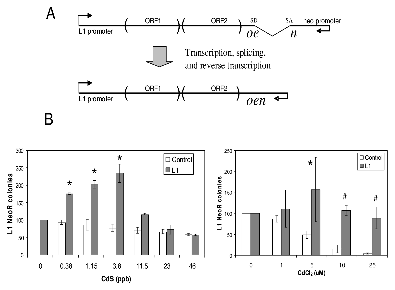
  • Schematic of the L1 retrotransposition plasmid (top). L1 RNA is transcribed by the L1 internal promoter. A neomycin resistance gene (neo) or a blasticidin resistance gene is located at the 3′ end in the opposite strand that contains a disrupting intron. The intron interrupting the neo can only be removed by splicing from RNA generated from the L1 promoter. During the L1 retrotransposition process the spliced RNA is reverse transcribed, and the cDNA inserted into the genome (bottom). The new L1 insert now contains a functional neo gene. Only newly integrated copies that retrotransposed from the spliced L1 RNA will present neomycin resistance. Promoters and transcription orientations are indicated by arrows. SD: splice donor, SA: splice acceptor.
  • Both CdS and CdCl2stimulate L1 retrotransposition in a dose dependent manner: NeoR colonies from separate L1 transfections (gray bars) treated with different doses of CdS, or CdCl2 (X axis) are shown. The no treatment (0 doses) for each experiment was defined as 100%. For toxicity control (white bars), cells were transfected in parallel with an unrelated plasmid with neomycin resistance and no L1 plasmid. Error bars indicate standard deviation. Statistically significant differences are indicated relative to the no treatment [t-test p<0.01(*)], or relative to no treatment after correcting for the cell death observed in the control [t-test p<0.01(*)].

L1 Retrotransposition Assay

The basic transient L1 retroposition assay was performed as previously described [44]. Briefly, HeLa cells (ATCC CCL2) were seeded in T-75 flasks at a density of 1.5 × 10⁵ cells/flask. Transient transfections were performed the next day using the Lipofectamine Plus (InVitrogen) following the manufacturer’s protocol, using 1 μg of either the L1 expressing vector (JM101/L1.3ΔCMV), or of the appropriate control plasmid, 0.3 μg pIRES2-EGFP (neomycin). Following removal of transfection cocktail, the cells were treated with varying doses of compounds for 48 hr. Treatment was removed and the cells were subsequently grown in media containing the appropriate selection, 400 μg/ml G418 (Fisher). After 14 days, cells were fixed and stained for 30 minutes with crystal violet (0.2% crystal violet in 5% acetic acid and 2.5% isopropanol). The rate of retrotransposition efficiency was then determined as the number of visible neoR-resistant colonies.

Toxicity Assay

HeLa cells were seeded in 6 well plates at a density of 1 × 10⁵ cells/well. Transient transfections were performed as described above using 50 ng/well of the L1 expressing vectors (TAM102/L1.3 and TAMD205A/L1.3). Cells were incubated with media containing varying doses of the metals for 48 hr. After treatment the cells were selected for 14 days either with 10 μg/ml blasticidin S (Sigma-Aldrich) to evaluate L1 retrotransposition capability or 125 μg/ml hygromycin B (InVitrogen) to evaluate the effect of L1 endonuclease toxicity on the integration of the L1 vector. Colonies were stained and evaluated as mentioned above.

Chemical Compounds

CdCl2 (Sigma-Aldrich) was used as a source of soluble cadmium, and CdS (Sigma-Aldrich) as a source of cadmium particles. The CdS concentration of the main stock (1150 ppb) was quantified / certified by AccuLab Inc. of Louisiana using method numbers 213.1 for CdS as described in USEPA Methods for Chemical Analysis of Water and Wastewater. Zinc acetate (C4H6O4Zn) and magnesium acetate (C4H6MgO4) were obtained from Sigma-Aldrich.

Comet Assay

The alkaline comet assay was performed using the slides and protocol provided by Trevigen. Briefly, suspensions of HeLa cells previously exposed to ± 3.8 ppb CdS for 24 hours were embedded in 1% low-melt agarose on glass slides. After exposure to alkaline conditions for 60 minutes to denature the DNA molecules, the slides were subjected to electrophoresis to allow the separation of the fragmented DNA molecules. The cells were stained with a DNA-intercalating fluorescent dye (SYBR green) and evaluated individually under a fluorescent microscope. For each condition (± CdS), 75 cells were evaluated with CometScore software (TriTek Corp.) for various characteristics associated with DNA damage, such as increases in comet tail length and comet tail moment (comet tail length × % DNA in the comet tail).

Results

Both Soluble and Particulate Cadmium Stimulate L1 Retrotransposition

The particulate forms of some heavy metals are known to be highly carcinogenic [45, 46]. We have previously demonstrated that several particulate forms of heavy metals, including CdS, stimulate L1 retrotransposition [44]. To determine if the L1 stimulation is only limited to particulate forms, we evaluated the capability of CdCl2 to stimulate L1 activity. HeLa cells were transiently transfected with the JM101/L1.3ΔCMV plasmid and treated with different doses of CdCl2 (0–25 μM) or CdS (0–46.5 ppb). A toxicity control using a plasmid expressing neomycin resistance was performed in parallel to evaluate the influence of the heavy metal toxicity on both transfection efficiency and colony formation. Our results demonstrate that CdCl2 stimulates L1 retrotransposition in a dose-dependent manner comparable to CdS (Figure 1B). The toxic effect of cadmium on the cells is observed as a decrease in the number of neoR colonies in the control. The stimulatory effect of 10 and 25 μM CdCl2 on L1 inserts is clear after correcting for cell death observed (data not shown).

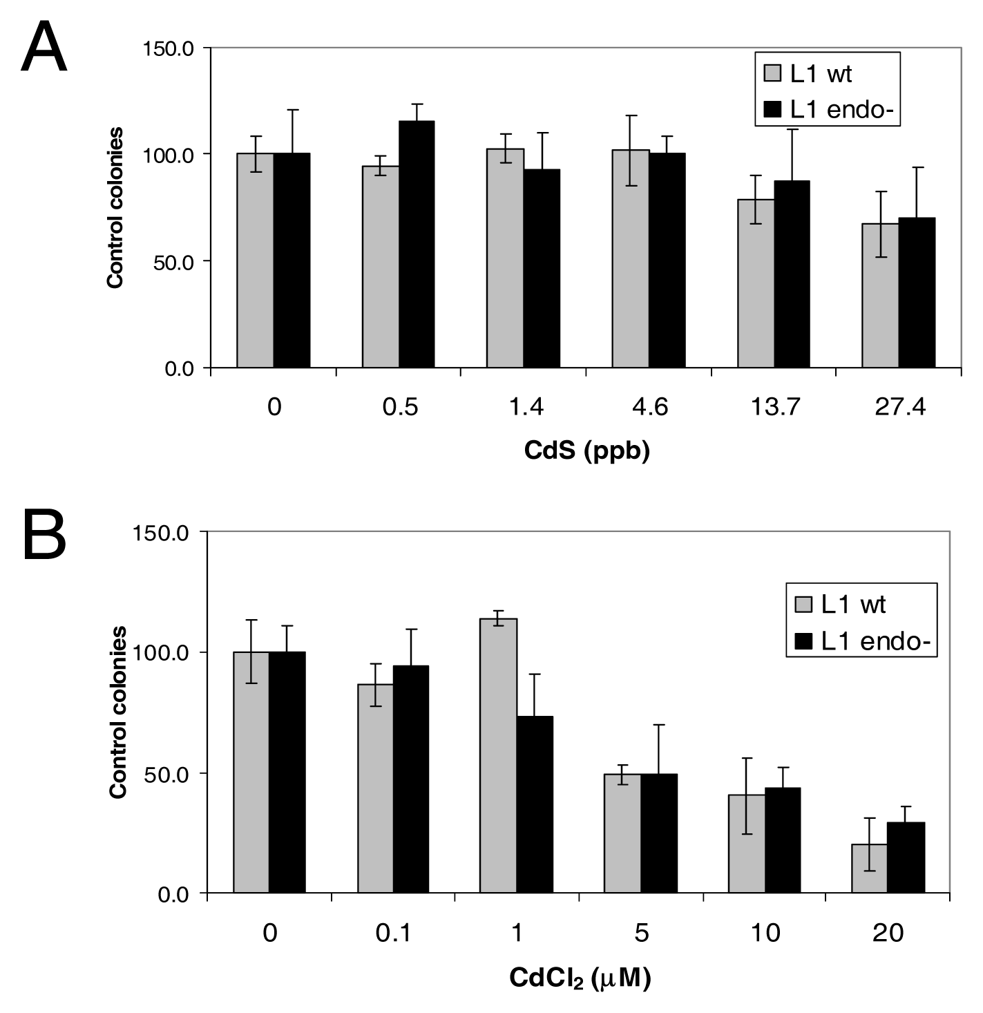
L1 Endonuclease Does Not Contribute to the Toxicity Observed

The L1 ORF2 encodes a multifunctional protein that contains an amino terminal endonuclease, an internal reverse transcriptase and a C-terminal cysteine-rich motif of unknown function [37, 39]. The endonuclease creates a nick at an AT-rich consensus site (TTTT′AA), thought to be used to prime the L1 mRNA polyA tail in the reverse transcription step [36]. The number of DNA nicks generated by the L1 endonuclease is higher by several orders of magnitude than the number of L1 inserts observed [64]. Therefore, active L1 elements can be toxic to cells, due to the endonuclease-generated DNA damage leading to cell death. In addition, cadmium inhibits DNA repair, potentially compounding the effect of the L1 endonuclease.
To evaluate if the endonuclease activity contributes to the toxicity observed in the cadmium treated cells, we compared cell survival by using plasmids containing a wildtype L1 and an endonuclease mutant L1 in our assay. In addition to the L1 reporter cassette, these plasmids contain a separate hygromycin resistance gene. Cell survival was assessed by determining the number of hygromycin-resistant colonies obtained after transfection. Because the only difference between the plasmids is the endonuclease inactivating point mutation, the number of hygromycin resistant colonies observed should only reflect toxic effects due to the endonuclease activity. In this assay, blasticidin selection was used to determine L1 retrotransposition because the plasmids contain a blasticidin resistance gene instead of neomycin in the L1 reporter cassette shown in Fig 1A.
HeLa cells were transfected with the L1 wildtype (TAM102/L1.3) or an L1 endo(-) vector (TAMD205A/L1.3), exposed to different doses of CdS or CdCl2, and grown under hygromycin selection to evaluate cell survival (Figure 2). The no treatment was designated as 100%. In this assay, the CdCl2 doses evaluated had a large effect on cell survival with a LD50 = ~5 μM (Figure 2B). However, no significant difference is observed in survival between the cells transfected with the L1 wildtype and L1 endo(-) vector [p≥0.344] for any of the treatments. As expected, the L1 retrotransposition activity was observed with wildtype L1 vector and no colonies for the L1 endo(-) after the two weeks of blasticidin selection, (data not shown).
HeLa cells were transfected with TAM102/L1.3, wildtype L1(wt), or TAMD205A/L1.3 (end Figure 2): L1 endonuclease does not significantly contribute to the toxicity observed. HeLa cells were transfected with TAM102/L1.3, wildtype L1(wt), or TAMD205A/L1.3 endo(-), which contains a point mutation in the endonuclease domain rendering the enzyme inactive. The transfected cells were exposed to different doses of CdS or CdCl2 and grown under hygromycin selection. The no treatment (0 dose) was designated as 100%. Error bars indicate standard deviation. No significant difference is observed in survival between the cells transfected with the L1 wildtype and L1 endo (-) vector [t-test p≥0.344]. Endo (-), which contains a point mutation in the endonuclease domain rendering the enzyme inactive. The transfected cells were exposed to different doses of CdS or CdCl2 and grown under hygromycin selection. The no treatment (0 dose) was designated as 100%. Error bars indicate standard deviation. No significant difference is observed in survival between the cells transfected with the L1 wildtype and L1 endo (-) vector [t-test p≥0.344].

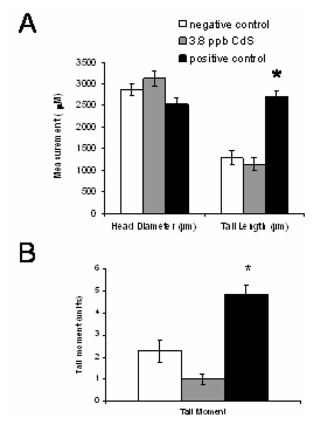
The L1 Stimulating Doses of Cds Do Not Contribute to DNA Breaks

Although L1 appears to preferentially insert at the sites generated by its own endonuclease, under certain circumstances it can utilize pre-existing nicks in chromosomal DNA [41]. Because heavy metals generate reactive oxygen species (ROS) that create DNA breaks, it may increase the availability of “potential” L1 insertion sites. We evaluated the amount of DNA damage caused by the CdS treatment with the highest effect on L1 activity. Analysis of DNA damage was performed using single cell alkaline gel electrophoresis (denaturing comet assay) on HeLa cells exposed to 3.8 ppb CdS, 5% H2O2 (positive control) or (untreated). Our data on both comet tail length and comet tail moment demonstrate that the CdS dose evaluated did not increase DNA breaks (Figure 3) and is significantly different from the positive control [unpaired t-test (p<0.001)]. These data support that the stimulation of L1 by CdS is independent of the generation of DNA breaks.
HeLa cells were treated with the cadmium dose (3.8 ppb CdS) shown to induce the highest L1 activity, H2O2 treatment (positive control) or untreated (negative control). Both double strand and single strand DNA breaks were evaluated using the alkaline comet assay. A total of 75 cells per treatment were analyzed for (A) head diameter and comet tail length and (B) comet tail moment. Error bars represent standard error of the mean. There is no significant difference in head diameter, tail length and tail moment between the cadmium treatment and negative control [unpaired t-test p=0.233, p=0.476 and p=0.024, respectively]. Statistically significant differences are indicated relative to the negative control [unpaired t-test p<0.001(*)].

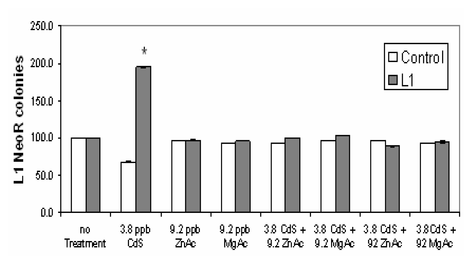
Magnesium and Zinc Reverse the L1 Stimulation Induced by Cds Exposure

Cadmium can inhibit enzymes through the displacement of magnesium and zinc cofactors. Zinc or magnesium treatments are reported to reverse or reduce the carcinogenic and genotoxic effects of cadmium [5, 6]. To determine if Mg and Zn affect the CdS stimulation of L1 retrotransposition, we transfected cells with the L1 reporter vector and treated with the highest L1 stimulating CdS dose (3.8 ppb) and 9.2 ppb or 92 ppb of magnesium acetate or zinc acetate. The toxicity control using a neoR vector was performed in parallel. As expected, L1 retrotransposition was significantly stimulated by CdS alone. However, no stimulation is observed when CdS exposed cells are concomitantly treated with any of the tested Mg and Zn doses (Figure 4). Our data demonstrate that both magnesium and zinc prevent the increase in L1 activity induced by cadmium. None of the magnesium or zinc treatments affected the toxicity control. Only the CdS dose alone presented some toxicity as observed by the reduction of neoR colonies when compared to the control. No difference was observed in the toxicity between the cells transfected with the wild type L1 (L1wt) or the endonuclease deficient (L1 endo-) in any of the CdS ± magnesium/zinc acetate doses evaluated (data not shown).
Cells were transfected with the L1 retrotransposition vector and treated with an L1 stimulating CdS dose (3.8 ppb) and 9.2 ppb or 92 ppb of magnesium acetate or zinc acetate. Error bars represent standard error of the mean (n=6). For toxicity control, cells were transfected with a neoR vector and treated in parallel. L1 retrotransposition was significantly stimulated by CdS alone (*p <0.001), but no stimulation is observed when cells are concomitantly treated with CdS plus Mg or Zn. Only the CdS treatment alone presented some toxicity as observed by the slight reduction of colonies observed in the control.

Discussion

The particulate forms of heavy metals are thought to be highly carcinogenic due to the internalization of the insoluble particles into cells by phagocytosis, where they are slowly dissolved providing a continuous source of the metal [4750]. We previously reported that exposure to several particulate forms of different heavy metals stimulate L1 retrotransposition in a tissue culture system [44]. In this study, we demonstrate that both soluble and particulate forms of cadmium increase L1 activity in a dose dependent manner. This has also been observed in our ongoing work with the soluble form of nickel, NiCl2[54]. In general, it is likely that both forms of a heavy metal will induce the same effect on L1 behavior, being stimulatory or not. However, some variances may occur in dose concentrations, due to the differences in the uptake of the two forms of the metal.
Because the carcinogenic mechanism of heavy metals is poorly defined, several pathways have been proposed: oxidative damage of DNA, enhanced cell proliferation (cell cycle alterations), and altered DNA repair [2, 8]. Evidence that indicate the DNA damage capability of metals includes effects such as DNA strand breaks, base modifications, DNA crosslinking, rearrangements and depurinations [7, 8, 51]. Cadmium is reported to induce genomic instability in cultured cells as determined by the detection of delayed cytogenetic aberrations and delayed cell death [52]. The cellular mechanism is thought to be the production of active oxygen and other radical species that alter not only the DNA, but also other molecules like proteins and lipids. However, these effects usually require high doses of the metals to occur [1, 53], possibly indicating that oxidative DNA damage is unlikely the primary carcinogenic mechanism of cadmium. This is in good agreement with our data, where the CdS doses capable of L1 stimulation do not generate detectable DNA breaks. Thus, the increase in L1 inserts in CdS treated cells seems to be independent of the availability of DNA breaks. This is also supported by our data on the effect of nickel treatments on L1 retrotransposition [54]. Most likely, other indirect genotoxic mechanism(s) are probably involved in L1 stimulation.
An alternate mechanism proposes that many of the adverse effects of heavy metals are due to their inhibition of cellular proteins such as DNA repair enzymes or chromatin proteins through the displacement of Mg and Zn. Our data demonstrate that the CdS stimulation of L1 is inhibited in the presence of additional Mg or Zn, suggesting that the inhibition of cellular proteins may allow L1 retrotransposition to occur more efficiently. Although many cellular processes depend on Mg and Zn, there is clear evidence that different heavy metals affect DNA repair in distinct manners and inhibit different enzymes, yielding different outcomes on L1 activity (Table 1). It is tempting to speculate that the DNA repair machinery recognizes some of the L1 intermediate forms during the insertion process as “damage” and proceeds to remove them before retrotransposition is completed. Although the complete molecular details of the L1 insertion mechanism are not understood, it is probable that host DNA repair enzymes contribute to L1 retrotransposition by either participating in or inhibiting the L1 insertion process. However, at this time more data are needed to properly address this hypothesis. Overall, CdS stimulation of L1 is independent of DNA break generation and is inhibited by the exogenous addition of magnesium or zinc source.
Figure 1. Stimulation of L1 retrotransposition by particulate and soluble cadmium.
Ijerph 03 00121f1
Figure 2. L1 endonuclease does not significantly contribute to the toxicity observed.
Ijerph 03 00121f2
Figure 3. Comet assay evaluation of CdS treated cells.
Ijerph 03 00121f3
Figure 4. Magnesium and zinc reverse the L1 stimulation induced by CdS exposure.
Ijerph 03 00121f4
Table 1. Effects of different metals on DNA repair pathways and L1 retrotransposition.
Heavy MetalDNA Repair Pathway AffectedL1 Stimulation
CadmiumNER-affect recognition step of DNA damage [55]; Strong inhibition of 8-oxo-dGTPase activity [16]; inhibition of APE-1 nuclease [56], inhibition of mismatch repair [57].Yes [44] (CdS, CdCl2§)
NickelNER-affect recognition step of DNA damage [55]; reduce the repair of DNA adducts [47]; weak inhibition of 8-oxo-dGTPase activity [16].Yes [44,54] (NiO, NiCl2)
CobaltNER-affect both incision and polymerization of repair patches [58]; weak inhibition of 8-oxo-dGTPase activity [16]No [54] (CoCl2)
MercuryInhibition of single strand DNA repair [59], inhibition of dUTPase and DNA polymerase alpha [60].Yes [44] (HgS)
Copper (Cu II)Strong inhibition of 8-oxo-dGTPase activity [16]; inhibition of single strand DNA repair [61].N.D.
Iron (Fe III)Inhibition of APE-1 nuclease [56].Yes
Arsenite (As III)Low doses inhibit PARP [62], and impair incision step [63].Yes
NER - Nucleotide excision repair.
N.D. - No data available.
Personal communication by Dr. El-Sawy.
§This study.

Acknowledgements

This research was supported by National Institutes of Environmental and Health Sciences ARCH grant, 1S11ES09996 and Office of Naval Research/9ONR: Tulane/Xavier Center for Bioenvironmental Research Funding Grant, N00014-05-1-0783 (SPK) and NIH P20 RR020152 (AMR-E).

References

  1. Meeting of the IARC working group on beryllium, cadmium, mercury and exposures in the glass manufacturing industry. Scand. J. Work Environ. Health 1993, 19(5), 360–363.
  2. Waalkes, M. P. Cadmium carcinogenesis in review. J. Inorg. Biochem 2000, 79(1–4), 241–244. [Google Scholar]
  3. Takenaka, S.; Oldiges, H.; Konig, H.; Hochrainer, D.; Oberdorster, G. Carcinogenicity of cadmium chloride aerosols in W rats. J. Natl. Cancer Inst 1983, 70(2), 367–373. [Google Scholar]
  4. Costa, M.; Sutherland, J. E.; Peng, W.; Salnikow, K.; Broday, L.; Kluz, T. Molecular biology of nickel carcinogenesis. Mol. Cell Biochem 2001, 222(1–2), 205–211. [Google Scholar]
  5. Szuster-Ciesielska, A.; et al. The inhibitory effect of zinc on cadmium-induced cell apoptosis and reactive oxygen species (ROS) production in cell cultures. Toxicology 2000, 145(2–3), 159–171. [Google Scholar]
  6. Littlefield, N. A.; Hass, B. S.; James, S. J.; Poirier, L. A. Protective effect of magnesium on DNA strand breaks induced by nickel or cadmium. Cell Biol. Toxicol 1994, 10(2), 127–135. [Google Scholar]
  7. Sunderman, F. W., Jr. Search for molecular mechanisms in the genotoxicity of nickel. Scand. J. Work Environ. Health 1993, 19 Suppl 1, 75–80. [Google Scholar]
  8. Kasprzak, K. S. Possible role of oxidative damage in metal-induced carcinogenesis. Cancer Invest 1995, 13(4), 411–430. [Google Scholar]
  9. Tully, D. Effects of arsenic, cadmium, chromium, and lead on gene expression regulated by a battery of 13 different promoters in recombinant HepG2 cells. Toxicol. Appl. Pharmacol 2000, 168(2), 79–90. [Google Scholar]
  10. Beyersmann, D.; Hechtenberg, S. Cadmium, gene regulation, and cellular signalling in mammalian cells. Toxicol. Appl. Pharmacol 1997, 144(2), 247–261. [Google Scholar]
  11. Asmuss, M.; Mullenders, L. H.; Hartwig, A. Interference by toxic metal compounds with isolated zinc finger DNA repair proteins. Toxicol. Lett 2000, 112–113, 227–231. [Google Scholar]
  12. Hartwig, A. Carcinogenicity of metal compounds: possible role of DNA repair inhibition. Toxicol. Lett 1998, 102–103, 235–239. [Google Scholar]
  13. Kopera, E.; Schwerdtle, T.; Hartwig, A.; Bal, W. Co(II) and Cd(II) substitute for Zn(II) in the zinc finger derived from the DNA repair protein XPA, demonstrating a variety of potential mechanisms of toxicity. Chem. Res. Toxicol 2004, 17(11), 1452–1458. [Google Scholar]
  14. Chen, X.; Chu, M.; Giedroc, D. P. Spectroscopic characterization of Co(II)-, Ni(II)-, and Cd(II)-substituted wild-type and non-native retroviral-type zinc finger peptides. J. Biol. Inorg. Chem 2000, 5(1), 93–101. [Google Scholar]
  15. Asmuss, M.; Mullenders, L. H.; Eker, A.; Hartwig, A. Differential effects of toxic metal compounds on the activities of Fpg and XPA, two zinc finger proteins involved in DNA repair. Carcinogenesis 2000, 21(11), 2097–2104. [Google Scholar]
  16. Kasprzak, K. S.; Bialkowski, K. Inhibition of antimutagenic enzymes, 8-oxo-dGTPases, by carcinogenic metals. Recent developments. J. Inorg. Biochem 2000, 79(1–4), 231–236. [Google Scholar]
  17. Lander, E. S.; et al. Initial sequencing and analysis of the human genome. Nature 2001, 409(6822), 860–921. [Google Scholar]
  18. Grimaldi, G.; Skowronski, J.; Singer, M. F. Defining the beginning and end of KpnI family segments. Embo J 1984, 3(8), 1753–1759. [Google Scholar]
  19. Brouha, B.; Schustak, J.; Badge, R. M.; Lutz-Prigge, S.; Farley, A. H.; Moran, J. V.; Kazazian, H. H., Jr. Hot L1s account for the bulk of retrotransposition in the human population. Proc. Natl. Acad. Sci. U. S. A 2003, 100(9), 5280–5285. [Google Scholar]
  20. Ostertag, E. M.; Kazazian, H. H., Jr. Biology of Mammalian l1 retrotransposons. Annu. Rev. Genet 2001, 355, 01–538. [Google Scholar]
  21. Trelogan, S. A.; Martin, S. L. Tightly regulated, developmentally specific expression of the first open reading frame from LINE-1 during mouse embryogenesis. Proc Natl Acad Sci U S A 1995, 92(5), 1520–1524. [Google Scholar]
  22. Ergun, S.; et al. Cell type-specific expression of LINE-1 open reading frames 1 and 2 in fetal and adult human tissues. J. Biol. Chem 2004, 279(26), 27753–27763. [Google Scholar]
  23. Muotri, A. R.; Chu, V. T.; Marchetto, M. C.; Deng, W.; Moran, J. V.; Gage, F. H. Somatic mosaicism in neuronal precursor cells mediated by L1 retrotransposition. Nature 2005, 435(7044), 903–910. [Google Scholar]
  24. Singer, M. F.; Krek, V.; McMillan, J. P.; Swergold, G. D.; Thayer, R. E. LINE-1: a human transposable element. Gene 1993, 135(1–2), 183–188. [Google Scholar]
  25. Skowronski, J.; Fanning, T. G.; Singer, M. F. Unit-length line-1 transcripts in human teratocarcinoma cells. Mol. Cell Biol 1988, 8(4), 1385–1397. [Google Scholar]
  26. Asch, H. L.; Eliacin, E.; Fanning, T. G.; Connolly, J. L.; Bratthauer, G.; Asch, B. B. Comparative expression of the LINE-1 p40 protein in human breast carcinomas and normal breast tissues. Oncol. Res 1996, 8(6), 239–247. [Google Scholar]
  27. Bratthauer, G. L.; Cardiff, R. D.; Fanning, T. G. Expression of LINE-1 retrotransposons in human breast cancer. Cancer 1994, 73(9), 2333–2336. [Google Scholar]
  28. Bratthauer, G. L.; Fanning, T. G. Active LINE-1 retrotransposons in human testicular cancer. Oncogene 1992, 7(3), 507–510. [Google Scholar]
  29. Chalitchagorn, K.; et al. Distinctive pattern of LINE-1 methylation level in normal tissues and the association with carcinogenesis. Oncogene 2004, 23(54), 8841–8846. [Google Scholar]
  30. Roman-Gomez, J.; et al. Promoter hypomethylation of the LINE-1 retrotransposable elements activates sense/antisense transcription and marks the progression of chronic myeloid leukemia. Oncogene 2005. epub. [Google Scholar]
  31. Tchenio, T.; Casella, J. F.; Heidmann, T. Members of the SRY family regulate the human LINE retrotransposons. Nucleic Acids Res 2000, 28(2), 411–415. [Google Scholar]
  32. Perepelitsa-Belancio, V.; Deininger, P. L. RNA truncation by premature polyadenylation attenuates human mobile element activity. Nat Genet 2003, 35(4), 363–366. [Google Scholar]
  33. Miki, Y.; Nishisho, I.; Horii, A.; Miyoshi, Y.; Utsunomiya, J.; Kinzler, K. W.; Vogelstein, B.; Nakamura, Y. Disruption of the APC gene by a retrotransposal insertion of L1 sequence in a colon cancer. Cancer Res 1992, 52(3), 643–645. [Google Scholar]
  34. Morse, B.; Rotherg, P. G.; South, V. J.; Spandorfer, J. M.; Astrin, S. M. Insertional mutagenesis of the myc locus by a LINE-1 sequence in a human breast carcinoma. Nature 1988, 333(6168), 87–90. [Google Scholar]
  35. Werle-Schneider, G.; von Brevern, M. C.; Sylla, B. S.; Hollstein, M. C. De novo retrotransposition of unbiased sequences in a human breast cancer cell clone. Genes Chromosomes. Cancer 1999, 26(1), 84–91. [Google Scholar]
  36. Luan, D. D.; Korman, M. H.; Jakubczak, J. L.; Eickbush, T. H. Reverse transcription of R2Bm RNA is primed by a nick at the chromosomal target site: a mechanism for non-LTR retrotransposition. Cell 1993, 72, 595–605. [Google Scholar]
  37. Martin, F.; Maranon, C.; Olivares, M.; Alonso, C.; Lopez, M. C. Characterization of a non-long terminal repeat retrotransposon cDNA (L1Tc) from Trypanosoma cruzi: homology of the first ORF with the ape family of DNA repairs enzymes. J. Mol. Biol 1995, 247(1), 49–59. [Google Scholar]
  38. Feng, Q.; Moran, J. V.; Kazazian, H. H., Jr; Boeke, J. D. Human L1 retrotransposon encodes a conserved endonuclease required for retrotransposition. Cell 1996, 87(5), 905–916. [Google Scholar]
  39. Scott, A. F.; et al. Origin of the human L1 elements: proposed progenitor genes deduced from a consensus DNA sequence. Genomics 1987, 1(2), 113–125. [Google Scholar]
  40. Mathias, S. L.; Scott, A. F.; Kazazian, H. H., Jr; Boeke, J. D.; Gabriel, A. Reverse transcriptase encoded by a human transposable element. Science 1991, 254, 1808–1810. [Google Scholar]
  41. Morrish, T. A.; Gilbert, N.; Myers, J. S.; Vincent, B. J.; Stamato, T. D.; Taccioli, G. E.; Batzer, M. A.; Moran, J. V. DNA repair mediated by endonuclease-independent LINE-1 retrotransposition. Nat. Genet 2002, 31(2), 159–165. [Google Scholar]
  42. Moore, J. K.; Haber, J. E. Capture of retrotransposon DNA at the sites of chromosomal double-strand breaks. Nature 1996, 383(6601), 644–646. [Google Scholar]
  43. Moran, J. V.; DeBerardinis, R. J.; Kazazian, H. H., Jr. Exon shuffling by L1 retrotransposition. Science 1999, 283(5407), 1530–1534. [Google Scholar]
  44. Kale, S. P.; Moore, L.; Deininger, P. L.; Roy-Engel, A. M. Heavy metals stimulate human LINE-1 retrotransposition. Int. J. Env. Res. Public Health 2005, 2, 84–90. [Google Scholar]
  45. Costa, M. Perspectives on the mechanism of nickel carcinogenesis gained from models of in vitro carcinogenesis. Environ. Health Perspect 1989, 81, 73–76. [Google Scholar]
  46. Singh, J.; Carlisle, D. L.; Pritchard, D. E.; Patierno, S. R. Chromium-induced genotoxicity and apoptosis: relationship to chromium carcinogenesis (review). Oncol. Rep 1998, 5(6), 1307–1318. [Google Scholar]
  47. Schwerdtle, T.; Seidel, A.; Hartwig, A. Effect of soluble and particulate nickel compounds on the formation and repair of stable benzo[a]pyrene DNA adducts in human lung cells. Carcinogenesis 2002, 23(1), 47–53. [Google Scholar]
  48. Harrison, P. T.; Heath, J. C. Apparent synergy in lung carcinogenesis: interactions between N-nitrosoheptamethyleneimine, particulate cadmium and crocidolite asbestos fibres in rats. Carcinogenesis 1986, 7(11), 1903–1908. [Google Scholar]
  49. Hartwig, A.; Schwerdtle, T. Interactions by carcinogenic metal compounds with DNA repair processes: toxicological implications. Toxicol. Lett 2002, 127(1–3), 47–54. [Google Scholar]
  50. Kasprzak, K. S.; Sunderman, F. W., Jr. Mechanisms of dissolution of nickel subsulfide in rat serum. Res. Commun. Chem. Pathol. Pharmacol 1977, 16(1), 95–108. [Google Scholar]
  51. Klein, C. B.; Frenkel, K.; Costa, M. The role of oxidative processes in metal carcinogenesis. Chem. Res. Toxicol 1991, 4(6), 592–604. [Google Scholar]
  52. Coen, N.; Mothersill, C.; Kadhim, M.; Wright, E. G. Heavy metals of relevance to human health induce genomic instability. J. Pathol 2001, 195(3), 293–299. [Google Scholar]
  53. Misra, R. R.; Smith, G. T.; Waalkes, M. P. Evaluation of the direct genotoxic potential of cadmium in four different rodent cell lines. Toxicology 1998, 126(2), 103–114. [Google Scholar]
  54. El Sawy, M.; Kale, S. P.; Dugan, C.; Nguyen, T. Q.; Belancio, V.; Bruch, H.; Roy-Engel, A.; Deininger, P. L. Nickel stimulates L1 retrotransposition by a post-transcriptional mechanism. J Mol. Biol 2005, 354(2), 246–257. [Google Scholar]
  55. Hartmann, M.; Hartwig, A. Disturbance of DNA damage recognition after UV-irradiation by nickel(II) and cadmium(II) in mammalian cells. Carcinogenesis 1998, 19(4), 617–621. [Google Scholar]
  56. McNeill, D. R.; Narayana, A.; Wong, H. K.; Wilson, D. M., III. Inhibition of Ape1 nuclease activity by lead; iron; and cadmium. Environ. Health Perspect 2004, 112(7), 799–804. [Google Scholar]
  57. Banerjee, S.; Flores-Rozas, H. Cadmium inhibits mismatch repair by blocking the ATPase activity of the MSH2-MSH6 complex. Nucleic Acids Res 2005, 33(4), 1410–1419. [Google Scholar]
  58. Kasten, U.; Mullenders, L. H.; Hartwig, A. Cobalt(II) inhibits the incision and the polymerization step of nucleotide excision repair in human fibroblasts. Mutat. Res 1997, 383(1), 81–89. [Google Scholar]
  59. Cantoni, O.; Costa, M. Correlations of DNA strand breaks and their repair with cell survival following acute exposure to mercury(II) and X-rays. Mol. Pharmacol 1983, 24(1), 84–89. [Google Scholar]
  60. Williams, M. V.; Winters, T.; Waddell, K. S. In vivo effects of mercury (II) on deoxyuridine triphosphate nucleotidohydrolase, DNA polymerase (alpha; beta), and uracil-DNA glycosylase activities in cultured human cells: relationship to DNA damage, DNA repair, and cytotoxicity. Mol. Pharmacol 1987, 31(2), 200–207. [Google Scholar]
  61. Snyder, R. D.; Lachmann, P. J. Thiol involvement in the inhibition of DNA repairs by metals in mammalian cells. Mol. Toxicol 1989, 2(2), 117–128. [Google Scholar]
  62. Yager, J. W.; Wiencke, J. K. Inhibition of poly (ADP-ribose) polymerase by arsenite. Mutat. Res 1997, 386(3), 345–351. [Google Scholar]
  63. Hartwig, A.; Groblinghoff, U. D.; Beyersmann, D.; Natarajan, A. T.; Filon, R.; Mullenders, L. H. Interaction of arsenic(III) with nucleotide excision repair in UV-irradiated human fibroblasts. Carcinogenesis 1997, 18(2), 399–405. [Google Scholar]
  64. Gasior, S. L.; Wakeman, T. P.; Xu, B.; Deininger, P. L. The human LINE-1 retrotransposon creates DNA double-strand breaks. In J. Mol. Biol; 2006; Volume 357, 5, pp. 1383–1393, * Personal communication was published after submission of manuscript. [Google Scholar]

