Detection of Human Plasma Glucose Using a Self-Powered Glucose Biosensor

1
Frank Reidy Research Center for Bioelectrics, Department of Electrical and Computer Engineering, Old Dominion University, Norfolk, VA 23508, USA
2
Department of Biochemistrry and Molecular Biology, Mayo Clinic, Jacksonville, FL 32224, USA
*
Author to whom correspondence should be addressed.
Energies 2019, 12(5), 825; https://doi.org/10.3390/en12050825
Submission received: 2 February 2019
/
Revised: 26 February 2019
/
Accepted: 26 February 2019
/
Published: 1 March 2019
(This article belongs to the Special Issue Biological Fuel Cells and Their Applications)
Abstract
:This work presents the characterization of a self-powered glucose biosensor using individual sequential assays of human plasma glucose obtained from diabetic patients. The self-powered glucose biosensor is exploited to optimize the assay parameters for sensing plasma glucose levels. In particular, the biofuel cell component of the system at pH 7.4, 37 °C generates a power density directly proportional to plasma glucose and exhibited a maximum power density of 0.462 mW·cm−2 at a cell voltage of 0.213 V in 5 mM plasma glucose. Plasma glucose is further sensed by monitoring the charge/discharge frequency (Hz) of the integrated capacitor functioning as the transducer. With this method, the plasma glucose is quantitatively detected in 100 microliters of human plasma with unprecedented sensitivity, as high as 104.51 ± 0.7 Hz·mM−1·cm−2 and a detection limit of 2.31 ± 0.3 mM. The results suggest the possibility to sense human plasma glucose at clinically relevant concentrations without the use of an external power source.
1. Introduction
The high demand for glucose biosensors is largely driven by the escalating mortality rate and life-threatening complications associated with the disease Type 1 diabetes mellitus, which accounts for about 387 million sufferers worldwide; this number is expected to rise to 592 million by 2035 [1]. Type 1 diabetes mellitus is a debilitating disease in which insulin-secreting pancreatic islets cease to function properly. This condition requires periodic insulin injections to regulate the patient’s metabolism. Studies have suggested that some diabetics choose not to strive for close blood glucose control due to the intrusiveness of current blood sampling and assay methods [2,3]. This situation is exacerbated because blood sampling is relatively infrequent, compared to the rate of blood glucose fluctuations. Moreover, blood sampling provides a discrete rather than the desired continuous blood glucose record, and is too infrequent and irregular to serve as an acceptable warning of rapid blood glucose imbalances.
Modern diabetes management systems rely heavily on invasive in vitro diagnostic tools, such as glucometers and continuous glucose monitors (CGMs) that provide an intermittent glucose record. The ability to continuously detect glucose biomarkers in bodily fluids at the point of care is of great importance for disease prevention, early diagnostics, disease management and individualized patient care [4,5,6,7]. Clinically relevant plasma glucose concentration below 5 mM is considered to be hypoglycemia and above 7.5 mM is considered to be hyperglycemia [7]. Moreover, a major challenge in diabetes management still remains, which is to maintain tight control over blood glucose. Therefore, considerable attention has been devoted recently to the development of enzymatic glucose biofuel cells as energy conversion technologies, especially for in vivo power sources for bioelectronics devices, such as pacemakers and even a glucometer [8,9,10,11]. To date, much effort has been devoted to addressing the micro-power output and stability of the biofuel cell [12,13,14,15,16] via a three-dimensional conducting matrix for biocatalysts/electrocatalysts. Currently, there are only a few reports on the performance of enzymatic glucose biofuel cells and self-powered glucose biosensors in complex biological fluids, i.e., human serum [14,17,18,19] and human blood [20,21]. Additionally, biofuel cell performance has been examined in vivo, i.e., crustaceans and mammals [10,14,17,18,19,22,23]. Based on the similarities in the power and current produced, it is widely held that a single enzymatic glucose biofuel cell cannot generate practical electrical power to either sense glucose nor power implantable devices [24].
It has been demonstrated that the micropower generated by a single enzymatic glucose biofuel cell can be amplified by stacking multiple biofuel cells in series [10] at the expense of device complexity and bulkiness. Du Toit et al. have demonstrated an approach to scale-up the electrical power produced from a glucose biofuel cell with a microfluidic design that embeds a cascade of three enzymatic fuel cells operating on iontophoresis extracts obtained from pig skin [25]. Toward early diagnostic applications, researchers have been able to successfully increase the power generated from a single enzymatic glucose biofuel cell, using a specially designed digital electronic circuit such as charge pumps to amplify the electrical power produced by the biofuel cells [26,27]. The amplified electrical power can further be conditioned using a step-up DC converter to deliver stable electrical power for implantable devices.
Our previous work exploits the direct electron transfer (DET) reactions of pyrolloquinoline quinone glucose dehydrogenase (PQQ-GDH), laccase, and bilirubin oxidase (BOD) that enabled us to fabricate and characterize the very first DET-based self-powered glucose biosensor optimally operating at pH 7 and 7.4 for laccase [24,28] and BOD [11] systems, respectively. The lower pH optima for the laccase-based system is the result of its bioelectrocatalytic activity optima at acidic pH of 5.5–6.0. In addition, we overcome the selectivity limitation because of the high selectivity of the self-powered biosensing system [11,29], wherein the device’s characteristics in glucose detection in the presence of unspecific mono- and disaccharides, and interfering species (e.g., ascorbic acid, etc) dissolved in buffer were characterized. No significant change in the charge/discharge frequency was observed upon introducing 5 mM glucose plus the respective 0.2 mM interfering analyte. In the presence of the respective interfering analyte independent of glucose, no electrical power generation and hence no charge/discharge frequency was observed [29]. Therefore, the evaluation of the self-powered glucose biosensor performance in biological samples obtained from diabetic patients is necessary in order to assess its potential as an early diagnostic biosensor and power source. In this paper, we characterized our novel self-powered glucose biosensor in human plasma obtained from diabetic patients. Stable electrical power density up to 0.917 mA/cm2 at 0.288 V was produced in the presence of 11.11 mM plasma glucose.
2. Materials and Methods
2.1. Chemicals
Buckypaper was purchased from Nanotech Labs, Yadkinville, NC, USA. 1-Pyrenebutanoic succinimidyl ester (PBSE) was purchased from AnaSpec. Inc (Fremont, CA, USA). PQQ-GDH was purchased from Toyobo. Co. Ltd. (Osaka, Japan). Bilirubin oxidase derived from Myrothecium was purchased from Sigma Aldrich (St. Louis, MO, USA). Potassium phosphate, D-(+)-Glucose, Calcium chloride and Nafion® were purchased from Sigma Aldrich (St. Louis, MO, USA) and all supplementary chemicals were of analytical grades and used without further purification. All solutions were prepared with 18.2 MΩ cm Milli-Q water (Millipore, St. Louis, MO, USA).
2.2. Electrochemical Instrumentation and Procedures
Electrochemical characterization was conducted on BASi potentiostat/galvanostats EC Epsilon (Bioanalytical Systems, Inc., West Lafayette, IN, USA) at 37 ± 1 °C. The electrochemical experiments were performed in a conventional three-electrode cell with the exception of the fuel cell characterizations. The Ag/AgCl (3 M KCl) and Pt wire were used as the reference and counter electrode, respectively. All potentials referenced in the present work are measured against the Ag/AgCl reference electrode.
The collection and processing of blood samples have been previously reported [30]. Blood samples were drawn from diabetic subjects with lithium-heparin monovettes (Sarstedt, Nümbrecht, Germany), and the plasma fraction was obtained by double centrifugation at 3000 × g for 10 min in K2 ethylenediaminetetraacetic acid (EDTA) tubes (BD Biosciences, San Jose, USA). The extracted plasma was placed in individual tubes and flash frozen in −70 °C freezer until assayed. Plasma glucose concentrations were measured in triplicate with a glucose analyzer (Beckman, Fullerton, CA, USA) using the glucose oxidase method. The plasma obtained naturally contained varying concentrations of glucose from 2.78–11.11 mM.
The self-powered biosensing system characterizations were carried out in the acquired 2.78–11.11 mM human plasma drawn from diabetic patients. The glucose biofuel cell component of the self-powered biosensing system consists of the PQQ-GDH bioanode and BOD biocathode in the single compartment cell. The polarization curves for the cell were obtained from the use of a series of external loads (1 MΩ to 1 kΩ) connected directly in parallel with the human plasma glucose biofuel cell. A Fluke 87V True RMS multimeter was used to capture the biofuel cell voltage (voltage across the bioanode and biocathode) and current readings. The power density and current density were obtained for the cell and have been normalized with the geometric surface of the electrodes. The charge/discharge frequency was acquired via an Agilent Oscilloscope (DS0X3054A, Santa Clara, CA, USA).
2.3. Bioelectrodes Preparation
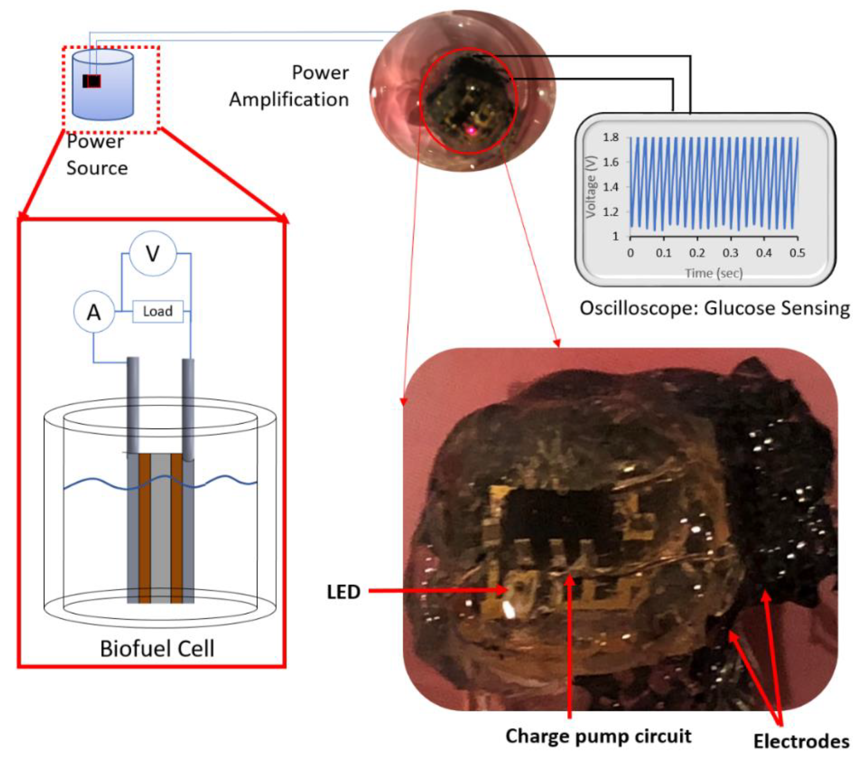
Buckypaper was employed as the three-dimensional electrode matrix because they comprise a dense mesh network of multi-walled carbon nabotubes that exhibit high surface area and high electrical conductivity. The electrodes were constructed using two 2 mm × 2 mm × 1 mm (l × w × h) buckypaper and a tungsten wire was affixed to the topmost edge using conductive wire glue to enable the delicate samples to be handled. Subsequently, the edges of the buckypaper were coated with Polyimide 2611 (HD Microsystems Inc, Parlin, NJ, USA) and cured at 150 °C to increase its rigidity. The cured electrodes were functionalized with 1 mM 1-Pyrenebutanoic succinimidyl ester (PBSE) in 10 mM dimethyl sulfoxide (DMSO) to serve as a crosslinker between the electrode and the enzyme over a geometric area of 1 mm × 1 mm. One of these electrodes was then chemically modified with 1 mM PQQ-GDH in 10 mM PBS (pH 7.0) + 1 mM CaCl2 (pH 7.0) and the other electrode was chemical modified with bilirubin oxidase (BOD). The PQQ-GDH functionalized electrode served as the bioanode, whereas the BOD functionalized electrode served as the biocathode. These bioelectrodes were further coated with 2 µL Nafion® to prevent any possible leeching of enzymes. The geometric area of the electrodes was designed to be 0.01 cm2. The resulting bioelectrodes were arranged in a coplanar orientation to form a single glucose biofuel cell as shown in Figure 1. The power amplification module comprises a charge pump circuit that can take an input voltage from the biofuel cell and generate a burst output power as described in our previous work [24]. This new system coupled with a capacitor is able to monitor the glucose concentrations.
3. Results and Discussion
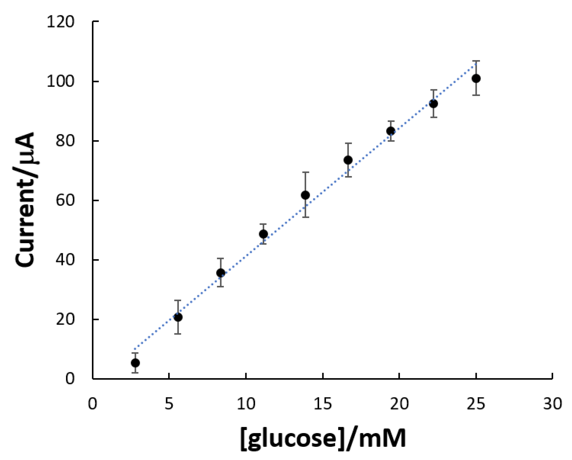
The self-powered glucose biosensor is completely packaged and encapsulated in medical grade silicone (Figure 1). The device comprised a single enzymatic glucose biofuel cell interfaced to a charge pump and a capacitor circuit. We have previously reported the development of an enzymatic glucose biofuel cell integrated with a charge pump circuit that takes the nominal input voltage (≥0.25 V) from the glucose biofuel cell and amplify it to generate a burst of power via a capacitor circuit. Additionally, the capacitor functions as a glucose transducer and is used to sense glucose by monitoring the capacitor’s charge/discharge frequency in hertz (Hz). The biocatalytic activity of the biofuel cell bioanode prior to packaging was characterized using amperometry in plasma glucose from diabetic patients to validate the presence of the biocatalyst functionalized on the dense mesh network of multiwalled carbon nanotubes (MWCNTs). The characterization was performed at increasing concentration of plasma glucose at the potential of 0.300 V. The oxidation reaction occurring at the PQQ-GDH electrode involves a one-step reaction, wherein glucose is oxidized to produce gluconolactone and release electrons and protons. Figure 2 displays the amperometric response to the successive addition of 2.78 mM human plasma glucose. It is reported in terms of steady state current versus glucose concentration. As expected, the PQQ-GDH/MWCNTs bioanode exhibited a linear response to the changes of glucose concentration. The PQQ-GDH/MWCNTs bioanode gives a linear dependence (R = 0.989) in the glucose concentration range of 2.78 mM to 25.02 mM with a sensitivity of 0.4 mA·mM−1.
The BOD biocathode was evaluated using chronoamperometry and after the addition of glucose, a small dip in the biocathdic current was observed. The chronoamperometric profiles for the PQQ-GDH bioanode and the BOD biocathode exhibited similar characteristics in the presence of glucose and oxygen and it is concluded that the reaction at the anode does not significantly affect the reaction at the biocathode [27] (pH 5–8, 25–37 °C), thereby making them attractive alternative to traditional fuel cells.
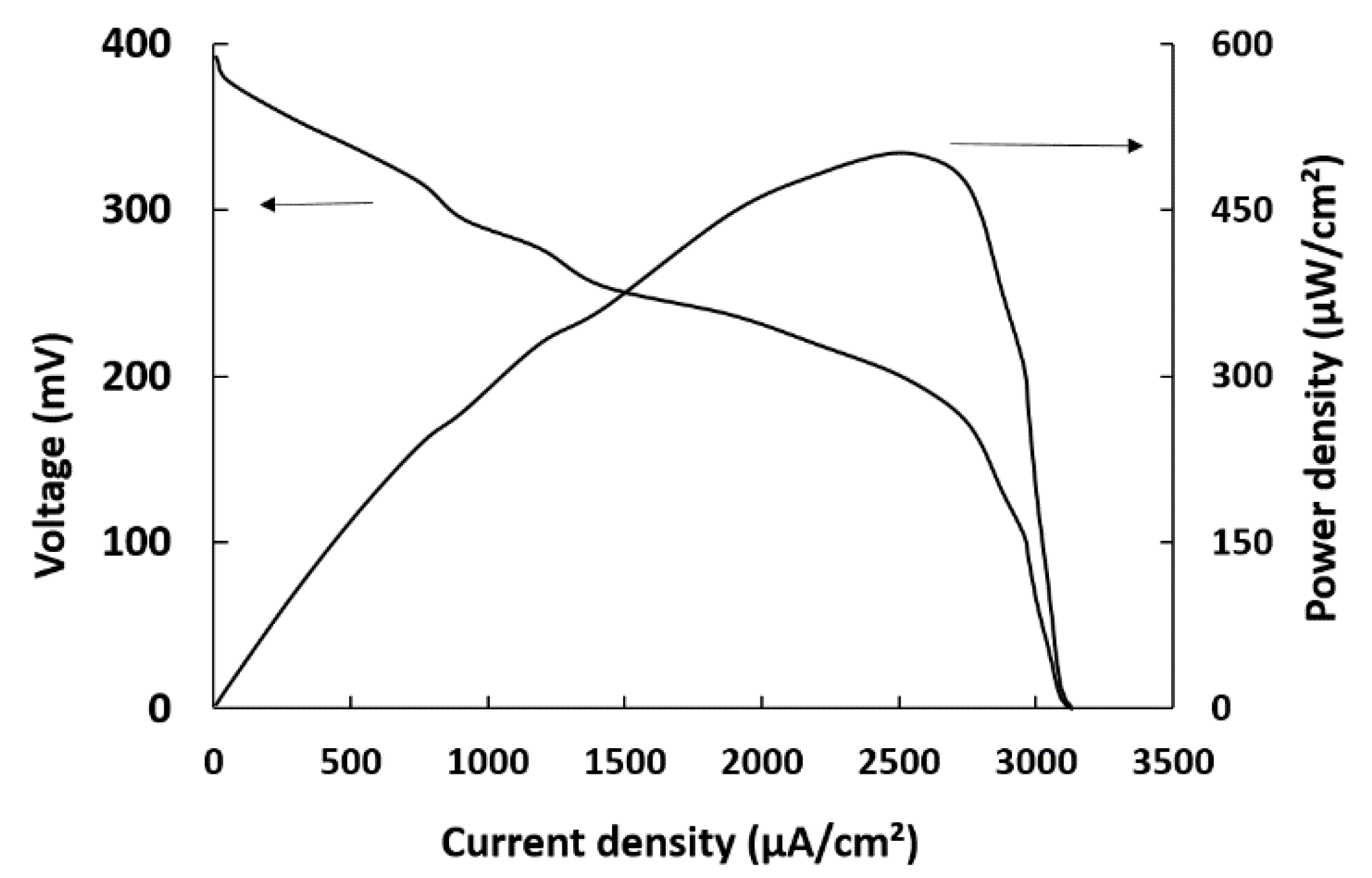
To evaluate the power generation from human plasma, the current density-voltage (I-V) polarization and power density-current curves were obtained with the packaged self-powered biosensing system. The performance of the biofuel cell was evaluated in a single compartment cell configuration at different external loads (1 kΩ to 1 MΩ) employing bare (abiotic) electrodes and the enzyme-modified electrodes. The observed open circuit potential and short circuit current for the abiotic cell were 27.6 mV and 4 μA, respectively. This confirms that the abiotic electrodes do not exhibit significant activity necessary to generate the electrical characteristics to serve as a self-powered glucose biosensor. In Figure 3, the polarization curve for the modified PQQ-GDH and BOD electrodes exhibited an open circuit voltage, short-circuit current and peak power density in a standard 5 mM glucose solution of 392 mV, 3.13 mA·cm−2 and 0.501 mW·cm−2 at a cell voltage of 203 mV under air, respectively. The electrical power produced by the biofuel cell originated from the redox reaction occurring simultaneously on the bioanode (oxidation of glucose) and biocathode (reduction of molecular oxygen).
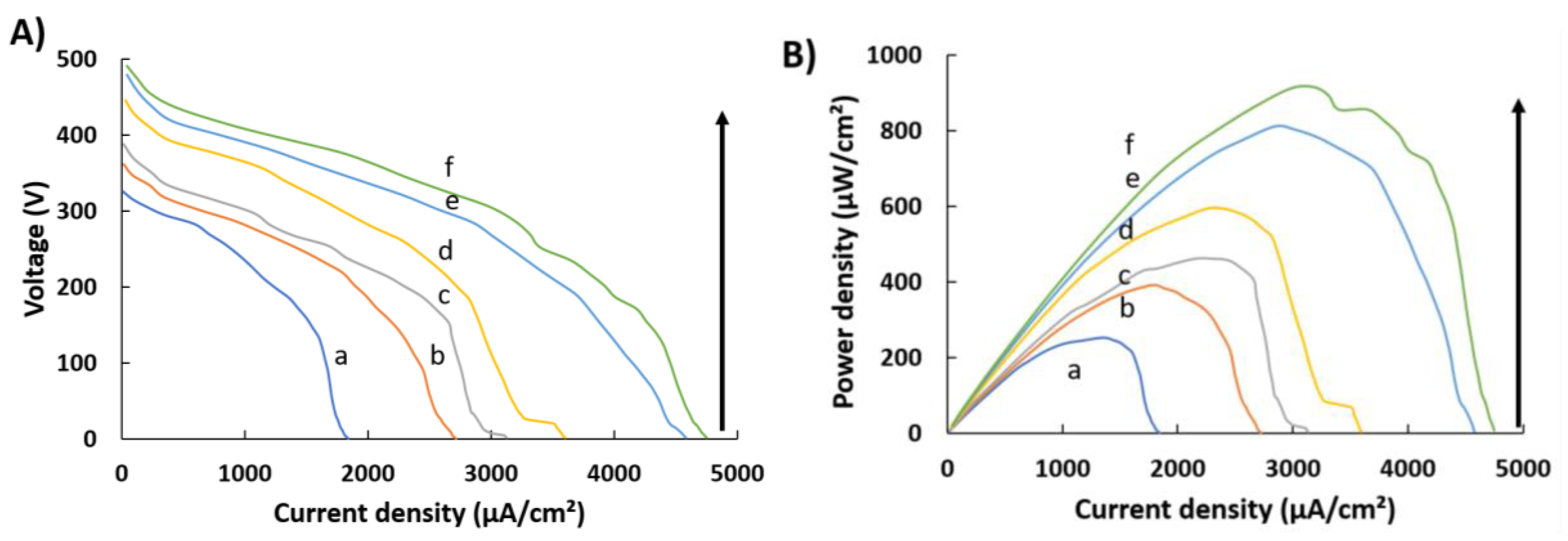
Figure 4 displays the performance of the biofuel cell in the presence of various human plasma glucose concentrations obtained from diabetic patients (2.78–11.11 mM). As shown in Figure 4A,B, the current and power output increased with increases in the plasma glucose concentration levels, since glucose oxidation is catalysed by PQQ-GDH at the bioanode, which in turn increases in a glucose concentration-dependent manner. The open circuit voltage, short circuit current and peak power density of the plasma glucose/oxygen biofuel cell operating in 5 mM plasma glucose were 392 mV, 3.13 mA·cm−2 and 0.462 mW·cm−2 at a cell voltage of 213 mV. Compared with those of the biofuel cell in standard 5 mM glucose solution, both the open circuit voltage and power density decreased in plasma glucose. This is consistent with those reported by Kwon et al. and Villarubia et al. [18,19]. The variation in the open circuit potential and peak power density between glucose solution and plasma glucose were approximately 1% and 7.8%, respectively. These small variations are expected because the biofuel cell has been demonstrated to selectively screen against interfering species in complex environments [29].
Despite the observed small variations in the peak power density produced in the presence of plasma glucose, the biofuel cell produced sufficient power to drive the charge pump circuit comprising a 0.1 µF capacitor functioning. Briefly, the charge pump circuit amplifies the nominal input voltage of 0.25 V from the biofuel cell via the capacitor to a start discharge voltage of 1.8 V until the capacitor voltage reaches the stop discharge voltage of approximately 1.2 V at which point, the power is charged up again via the capacitor. The charge/discharge frequency of the capacitor was used to sense glucose by monitoring the output of the capacitor using an oscilloscope. It was observed that the charge/discharge frequency of the capacitor increases in a glucose concentration-dependent manner. When the power produced by the glucose biofuel cell increases, this results in a rapid charge/discharge of the capacitor, thereby enabling the capacitor to function as a glucose transducer without the need for potentiostatic interrogation.
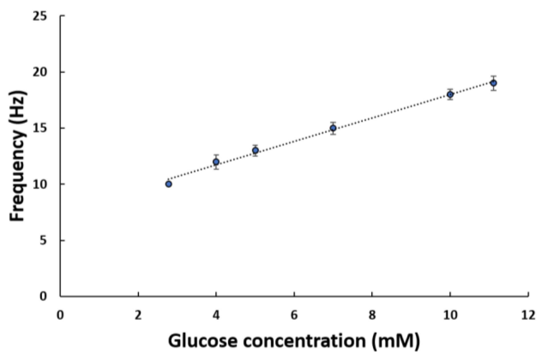
The plasma glucose concentrations were measured in triplicate using the self-powered glucose biosensing system by monitoring the charging/discharging of the capacitor. The glucose calibration curve obtained for the resulting self-powered glucose biosensor exhibited a linear correlation in the frequency (hertz) of charge/discharge across the 0.1 µF capacitor with glucose concentration and a dynamic linear range of 2.78–11.11 mM plasma glucose was realized. This range covers both the hypoglycemic as well as hyperglycemic including the normal glucose concentration range. As shown in Figure 5, it is evident that with an increase in glucose concentration, more electrical power is produced, thereby resulting in an increase in the charge/discharge frequency of the capacitor. Moreover, the self-powered glucose biosensor exhibited a sensitivity of 104.51 ± 0.7 Hz/mM·cm2 in the presence of human plasma and a limit of detection (LOD) of 2.31 ± 0.3 mM. The LOD is below the dynamic linear range, thereby corresponding to the ability of the self-powered glucose biosensor to sense the various clinical plasma glucose levels observed in patients suffering from Type 1 diabetes mellitus [30].
Moreover, this study demonstrates that self-powered glucose biosensing system among other enzymatic glucose biofuel cells can produce electrical power from biological fluids (Table 1).
The self-powered glucose biosensors mentioned by Katz et al., Valdés-Ramírez et al., and Cheng et al. are fundamentally different as it is a self-powered biosensor based on the correlation of changes in peak power density or voltage drop across a load with glucose concentration whereas here we demonstrated a self-powered biosensor employing an enzymatic glucose biofuel cell with a charge pump circuit and a capacitor to transduce the biochemical reaction occurring at the bioelectrodes [31,32,33]. To the extent of our knowledge, the system mentioned in Pellitero et al [34] describes a self-powered electrochromic biosensor that employs a potentiostatic system for interrogating the bioanode under constant polarization at 0.2 V vs. Ag/AgCl to achieve a dynamic range up 20 mM. The system we demonstrated is an improvement on these existing systems because it eliminates the use of variable resistive load, while enabling fast detection of glucose by monitoring the charge/discharge frequency of the transducer.
This self-powered glucose biosensor powered by a single enzymatic glucose biofuel cell overcomes the current limitations of glucose monitors that employ battery-operated potentiostats, by eliminating the need for a battery as the power source, thereby enabling the realization of miniaturized power sources and biosensors. Miniature glucose biofuel cells have the potential to serve as the only bio-friendly in vitro and in vivo power sources. They are of great importance for powering biosensors and closed-loop drug delivery systems. Here, the performance of our system in human plasma from diabetic pateints is assumed to be close to clinical applications, since these samples are clinically drawn from diabetic patients and the biosensor demonstrated good sensitivity and selectivity. This in vitro study serves as the preliminary studies for transitioning to the in vivo environment in future work to monitor blood glucose.
4. Conclusion
Using a self-powered glucose biosensor encapsulated in medical grade silicone, we demonstrated its operation in human plasma glucose at pH 7.4, 37 °C and at clinically relevant human plasma glucose concentrations (2.78–11.11 mM) with high sensitivity. The glucose biofuel cell component achieved an open-circuit voltage of 392 mV and delivered a maximum power density of 0.462 mW·cm−2 at a cell voltage of 213 mV when operating 5 mM plasma glucose. The highest peak current density of 4.75 mA·cm−2 was obtained in 11.11 mM. This work further demonstrates that human plasma can be used as an energy source for biofuel cells and concurrently sense plasma glucose levels. These results lead to the conclusion that the self-powered glucose biosensor is a very attractive biosensor platform for monitoring glucose and are of particular significance for point-of-care diagnostics, where high sensitivities and detection limits below basal clinical levels are desired to enable early diagnostics. A special feature of the self-powered glucose biosensor is that it generates high power output and provides clean electric energy production in human plasma. Additionally, the system can be easily miniaturized, thereby offering tremendous opportunities to overcome the scaling limitations of commercially available glucose monitoring technologies by providing a scalable self-powered glucose sensor device.
Author Contributions
Conceptualization, G.S.; Investigation, T.K.
Funding
This work was supported by National Science Foundation (Award ECCS# 1349603).
Acknowledgments
The authors thank Stephen N. Davis, Donna Tate and Lisa Younk at the University of Maryland School of Medicine, Department of Medicine, Diabetes Research Laboratory for the collection and processing of blood samples.
Conflicts of Interest
The authors declare no conflict of interest.
References
- International Diabetes Federation. IDF Diabetes Atlas. International Diabetes Federation: Brussels, Belgium, 2013, 6th ed. Available online: https://www.idf.org/e-library/epidemiology-research/diabetes-atlas/19-atlas-6th-edition.html (accessed on 12 October 2018).
- Gough, D.A.; Armour, J.C.; Baker, D.A. Advances and prospects in glucose assay technology. Diabetologia 1997, 40, s102. [Google Scholar] [CrossRef] [PubMed]
- Thome-Duret, V.; Reach, G.; Gangnerau, M.; Klein, J.C.; Zhang, Y.; Hu, Y.; Wilson, G. Use of a subcutaneous glucose sensor to detect decreases in glucose concentration prior to observation in blood. Anal. Chem. 1996, 68, 3822. [Google Scholar] [CrossRef] [PubMed]
- U.P. Group. Tight blood pressure control and risk of macrovascular and microvascular complications in type 2 diabetes: UKPDS 38. Br. Med. J. 1998, 12, 703. [Google Scholar]
- Inzucchi, S.E.; Bergenstal, R.M.; Buse, J.B.; Diamant, M.; Ferrannini, E.; Nauck, M.; Peters, A.L.; Tsapas, A.; Wender, R.; Matthews, D.R. Management of hyperglycemia in type 2 diabetes: a patient-centered approach: position statement of the American Diabetes Association (ADA) and the European Association for the Study of Diabetes (EASD). Diabetes Care 2012, 35, 1364. [Google Scholar] [CrossRef] [PubMed]
- Saleh, F.; Mumu, S.J.; Ara, F.; Hafez, M.A.; Ali, L. Non-adherence to self-care practices & medication and health related quality of life among patients with type 2 diabetes: a cross-sectional study. BMC Public Health 2014, 14, 431. [Google Scholar]
- König, M.; Bulik, S.; Holzhütter, H.-G. Quantifying the Contribution of the Liver to Glucose Homeostasis: A Detailed Kinetic Model of Human Hepatic Glucose Metabolism. PLoS Comput. Biol. 2012, 8, e1002577. [Google Scholar] [CrossRef] [PubMed]
- Kulkarni, T.; Slaughter, G. Application of semipermeable membranes in glucose biosensing. Membranes 2016, 6, 1. [Google Scholar] [CrossRef] [PubMed]
- Parrilla, M.; Cánovas, R.; Andrade, F.J. Paper-based enzymatic electrode with enhanced potentiometric response for monitoring glucose in biological fluids. Biosens. Bioelectron. 2017, 90, 110. [Google Scholar] [CrossRef] [PubMed]
- MacVittie, K.; Halámek, J.; Halámková, L.; Southcott, M.; Jemison, W.D.; Lobel, R.; Katz, E. From “cyborg” lobsters to a pacemaker powered by implantable biofuel cells. Energy Environ. Sci. 2013, 6, 81. [Google Scholar] [CrossRef]
- Slaughter, G.; Kulkarni, T. Highly selective and sensitive self-powered glucose sensor based on capacitor circuit. Nat. Sci. Rep. 2017, 7, 1471. [Google Scholar] [CrossRef] [PubMed]
- Slaughter, G.; Kulkarni, T. Fabrication of palladium nanowire array electrode for biofuel cell application. Microelec. Eng. 2016, 149, 92. [Google Scholar] [CrossRef]
- Slaughter, G.; Kulkarni, T. Enzymatic glucose biofuel cell and its application. J. Biochips Tissue Chips 2015, 5, 1. [Google Scholar] [CrossRef]
- Milton, R.D.; Lim, K.; Hickey, D.P.; Minteer, S.D. Employing FAD-dependent glucose dehydrogenase within a glucose/oxygen enzymatic fuel cell operating in human serum. Bioelectrochemistry 2015, 106, 56. [Google Scholar] [CrossRef] [PubMed]
- Siepenkoetter, T.; Salaj-Kosla, U.; Xiao, X.; Conghaile, P.Ó.; Pita, M.; Ludwig, R.; Magner, E. Immobilization of redox enzymes on nanoporous gold electrodes: applications in biofuel cells. ChemPlusChem 2017, 82, 553. [Google Scholar] [CrossRef]
- Kulkarni, T.; Slaughter, G. Self-powered glucose biosensor operating under physiological conditions. Proceedings of the IEEE Sens. 2016, 1–3. [Google Scholar]
- Szczupak, A.; Halámek, J.; Halámková, L.; Bocharova, V.; Alfonta, L.; Katz, E. Living battery—Biofuel cells operating in vivo in clams. Energy Environ. Sci. 2012, 5, 8891. [Google Scholar] [CrossRef]
- Kwon, C.; Lee, S.; Choi, Y.; Lee, J.; Kim, S.; Kim, H.; Spinks, G.; Wallace, G.; Lima, M.; Kozlov, M.; Baughman, R. High-power biofuel cell textiles from woven biscrolled carbon nanotube yarns. Nat. Commun. 2014, 5, 3928. [Google Scholar] [CrossRef] [PubMed]
- Villarrubia, C.; Soavi, F.; Santoro, C.; Arbizzani, C.; Serov, A.; Rojas-Carbonell, S.; Gupta, G.; Atanassov, P. Self-feeding paper based biofuel cell/self-powered hybrid μ-supercapacitor integrated system. Biosens. Bioelectron. 2016, 86, 459. [Google Scholar] [CrossRef] [PubMed]
- Cadet, M.; Gounel, S.; Stines-Chaumeil, C.; Brilland, X.; Rouhana, J.; Louerat, F.; Mano, N. An enzymatic glucose/O2 biofuel cell operating in human blood. Biosens. Bioelectron. 2016, 83, 60. [Google Scholar] [CrossRef] [PubMed]
- Conghaile, P.; Falk, M.; MacAodha, D.; Yakovleva, M.; Gonaus, C.; Peterbauer, C.; Gorton, L.; Shleev, S.; Leech, D. Fully Enzymatic Membraneless Glucose|Oxygen Fuel Cell That Provides 0.275 mA cm–2 in 5 mM Glucose, Operates in Human Physiological Solutions, and Powers Transmission of Sensing Data. Anal. Chem. 2016, 88, 2156. [Google Scholar] [CrossRef] [PubMed]
- Shoji, K.; Akiyama, Y.; Suzuki, M.; Hoshino, T.; Nakamura, N.; Ohno, H.; Morishima, K. Insect biofuel cells using trehalose included in insect hemolymph leading to an insect-mountable biofuel cell. Biomed. Microdevices 2012, 14, 1063. [Google Scholar] [CrossRef] [PubMed]
- Mano, N.; Mao, F.; Heller, A. Characteristics of a Miniature Compartment-less Glucose−O2 Biofuel Cell and Its Operation in a Living Plant. Journal of the American Chemical Society 2003, 125, 6588. [Google Scholar] [CrossRef] [PubMed]
- Slaughter, G.; Kulkarni, T. A self-powered glucose biosensing system. Biosens. Bioelectron. 2016, 78, 45. [Google Scholar] [CrossRef] [PubMed]
- Du Toit, H.; Rashidi, R.; Ferdani, D.W.; Delgado-Charro, M.B.; Sangan, C.M.; Di Lorenzo, M. Generating power from transdermal extracts using a multi-electrode miniature enzymatic fuel cell. Biosens. Bioelectron. 2016, 15, 411. [Google Scholar] [CrossRef] [PubMed]
- Southcott, M.; MacVittie, K.; Halámek, J.; Halámková, L.; Jemison, W.D.; Lobel, R.; Katz, E. A pacemaker powered by an implantable biofuel cell operating under conditions mimicking the human blood circulatory system—Battery not included. Phys. Chem. Chem. Phys. 2013, 15, 6278. [Google Scholar] [CrossRef] [PubMed]
- Hasan, M.Q.; Kuis, R.; Narayanan, J.S.; Slaughter, G. Fabrication of highly effective hybrid biofuel cell based on integral colloidal platinum and bilirubin oxidase on gold support. Nat. Sci. Rep. 2018, 8, 1. [Google Scholar] [CrossRef] [PubMed]
- Baingane, A.; Mburu, N.; Slaughter, G. Simultaneous Monitoring of Glucose and Lactate by Self-powered Biosensor. Sens. Transducers 2017, 214, 34. [Google Scholar]
- Kulkarni, T.; Slaughter, G. Characteristics of two self-powered glucose biosensors. IEEE Sens. J. 2017, 17, 3607. [Google Scholar] [CrossRef]
- Briscoe, V.J.; Ertl, A.C.; Tate, D.B.; Dawling, S.; Davis, S.N. Effects of the Selective Serotonin Reuptake Inhibitor Fluoxetine on Counterregulatory Responses to Hypoglycemia in Individuals with Type 1 Diabetes. Diabetes 2008, 57, 2453. [Google Scholar] [CrossRef] [PubMed]
- Katz, E.; Bückmann, A.F.; Willner, I. Self-Powered Enzyme-Based Biosensors. J. Am. Chem. Soc. 2001, 123, 10752. [Google Scholar] [CrossRef] [PubMed]
- Cheng, H.; Yu, P.; Lu, X.; Lin, Y.; Ohsaka, T.; Mao, L. Biofuel cell-based self-powered biogenerators for online continuous monitoring of neurochemicals in rat brain. Analyst 2013, 138, 179. [Google Scholar] [CrossRef] [PubMed]
- Valdés-Ramírez, G.; Li, Y.-C.; Kim, J.; Jia, W.; Bandodkar, A.J.; Nuñez-Flores, R.; Miller, P.R.; Wu, S.-Y.; Narayan, R.; Windmiller, J.R.; Polsky, R.; Wang, J. Microneedle-based self-powered glucose sensor. J. Electrochem. Commun. 2014, 47, 58. [Google Scholar] [CrossRef]
- Pellitero, M.A.; Guimera, A.; Kitsara, M.; Villa, R.; Rubio, C.; Lakard, B.; Doche, M.-L.; Hihn, J.-Y.; Javier del Campo, F. Quantitative self-powered electrochromic biosensors. Chem. Sci. 2017, 8, 1995. [Google Scholar] [CrossRef] [PubMed]
Figure 1.
Experimental set-up of glucose biofuel cell and glucose sensing. Charge pump circuit consisting of a 0.1 µF capacitor functioning as a transducer, where the charge pump circuit is powered by the electrical power generated by the glucose biofuel cell.
Figure 1.
Experimental set-up of glucose biofuel cell and glucose sensing. Charge pump circuit consisting of a 0.1 µF capacitor functioning as a transducer, where the charge pump circuit is powered by the electrical power generated by the glucose biofuel cell.

Figure 2.
The dependence of the current response versus the concentration of plasma glucose at PQQ-GDH/MWCNTs bioanode. the error bars represent standard errors of the mean triplicate values.
Figure 2.
The dependence of the current response versus the concentration of plasma glucose at PQQ-GDH/MWCNTs bioanode. the error bars represent standard errors of the mean triplicate values.

Figure 3.
Current density-voltage and power density-current characteristics for standard 5 mM glucose solution in air-saturated environment. pH = 7.4.
Figure 3.
Current density-voltage and power density-current characteristics for standard 5 mM glucose solution in air-saturated environment. pH = 7.4.

Figure 4.
(A) Current density-voltage and (B) power density-current characteristics for human plasma glucose (a) 2.78, (b) 4, (c) 5, (d) 7, (e) 10, and (f) 11.11 mM) in air, pH = 7.4.
Figure 4.
(A) Current density-voltage and (B) power density-current characteristics for human plasma glucose (a) 2.78, (b) 4, (c) 5, (d) 7, (e) 10, and (f) 11.11 mM) in air, pH = 7.4.

Figure 5.
Dependence of the average charging/discharging frequency of the capacitor on glucose concentration, as measured with the self-powered glucose biosensing system operating in human plasma glucose (2.78–11.11 mM). Error bars represent the standard deviation of the measurements from triplicate testing.


Table 1.
Performance of enzymatic glucose biofuel cell under in vitro and in vivo environments.
| Medium | Sample | Voc (mV) | Isc (µA·cm−2) | Pmax (µW·cm−2) | Ref. |
|---|---|---|---|---|---|
| in vivo | Orange | 600 | 330 | 335 | [10] |
| in vitro | Serum | 560 | 285.7 | 57.5 | [14] |
| in vivo | Clams | 800 | 100 | 20.8 | [17] |
| in vivo | Cockroach | 467 | 30 | 7.27 | [18] |
| in vivo | Grapes | 520 | 1037.5 | 537.5 | [19] |
| in vitro | Blood | 480 | 2.5 | 73 | [20] |
| in vitro | Human plasma | 392 | 3130 | 462 | this work |
© 2019 by the authors. Licensee MDPI, Basel, Switzerland. This article is an open access article distributed under the terms and conditions of the Creative Commons Attribution (CC BY) license (http://creativecommons.org/licenses/by/4.0/).
Share and Cite
MDPI and ACS Style
Slaughter, G.; Kulkarni, T. Detection of Human Plasma Glucose Using a Self-Powered Glucose Biosensor. Energies 2019, 12, 825. https://doi.org/10.3390/en12050825
AMA Style
Slaughter G, Kulkarni T. Detection of Human Plasma Glucose Using a Self-Powered Glucose Biosensor. Energies. 2019; 12(5):825. https://doi.org/10.3390/en12050825
Chicago/Turabian StyleSlaughter, Gymama, and Tanmay Kulkarni. 2019. "Detection of Human Plasma Glucose Using a Self-Powered Glucose Biosensor" Energies 12, no. 5: 825. https://doi.org/10.3390/en12050825
Note that from the first issue of 2016, this journal uses article numbers instead of page numbers. See further details here.