Article

Screen-Printed Electrodes: New Tools for Developing Microbial Electrochemistry at Microscale Level

by
Marta Estevez-Canales
1,
Antonio Berná
2,
Zulema Borjas
2 and
Abraham Esteve-Núñez
1,2,*
1
Department of Chemical Engineering, University of Alcalá, Alcalá de Henares, Madrid 28871, Spain
2
IMDEA WATER, Technological Park of Alcalá, Alcalá de Henares, Madrid 28805, Spain
*
Author to whom correspondence should be addressed.
Energies 2015, 8(11), 13211-13221; https://doi.org/10.3390/en81112366
Submission received: 1 October 2015 / Revised: 10 November 2015 / Accepted: 16 November 2015 / Published: 19 November 2015

Abstract

:
Microbial electrochemical technologies (METs) have a number of potential technological applications. In this work, we report the use of screen-printed electrodes (SPEs) as a tool to analyze the microbial electroactivity by using Geobacter sulfurreducens as a model microorganism. We took advantage of the small volume required for the assays (75 μL) and the disposable nature of the manufactured strips to explore short-term responses of microbial extracellular electron transfer to conductive materials under different scenarios. The system proved to be robust for identifying the bioelectrochemical response, while avoiding complex electrochemical setups, not available in standard biotechnology laboratories. We successfully validated the system for characterizing the response of Geobacter sulfurreducens in different physiological states (exponential phase, stationary phase, and steady state under continuous culture conditions) revealing different electron transfer responses. Moreover, a combination of SPE and G. sulfurreducens resulted to be a promising biosensor for quantifying the levels of acetate, as well as for performing studies in real wastewater. In addition, the potential of the technology for identifying electroactive consortia was tested, as an example, with a mixed population with nitrate-reducing capacity. We therefore present SPEs as a novel low-cost platform for assessing microbial electrochemical activity at the microscale level.

Graphical Abstract

1. Introduction

Since the discovery in the last decade of electrode-respiring microbes, such as Geobacter sulfurreducens and some species of Shewanella [1,2], there has been an increasing interest in their potential applications for designing several types of microbial electrochemical technologies (METs), like electricity production from wastewater in microbial fuel cells (MFC), bioremediation in microbial electroremediating cells (MERCs) [3], electrosynthesis, or biosensing [4]. Many attempts to optimize METs have been focused on reactor design and operational aspects [5,6]. Different electrochemical approaches, such as chronoamperometric assays, cyclic voltammetry (CV), sometimes coupled to spectroscopic techniques as infrared (IR) or Raman [7,8,9], have been applied for the study of this fascinating phenomenon. All of these have helped to investigate and analyze electroactive microorganisms and learn valuable information about how microbial physiology at different hierarchical levels affects the capability of electron transfer to solid electrodes at the whole biofilm, single cell and sub-cell (molecular) level [10,11,12].
Nevertheless, much remains unknown regarding the electrochemical properties of electroactive bacteria and their interaction with electrodes. This is partially due to difficulties in monitoring METs or the requirement of rather complex experimental setups for growing bacteria in electrochemical environments with limited access to microbial tools [13,14].
Although considerable efforts have been made to scale up METs for industrial applications, there is also a great interest in miniaturizing bioelectrochemical devices for research purposes where large current production is not the main goal. Those devices are typically designed to work under fixed potential conditions with a three-electrode setup. Some examples include flow cells, microfluidic devices or micro-sized microbial fuel cell [15,16,17].
Screen-printing is an established technique to fabricate electrochemical devices. A standard screen-printed electrode (SPE) comprises a variable number of electrodes and all the compounds necessary for completed the electric circuit placed over a supporting material [18]. Moreover, for carbon SPEs, the inks employed are based on graphite and have very similar properties to conventional electrodes, such as low background signal and a wide range of working potentials. With inherent advantages including miniaturization, versatility, low cost and the possibility of mass production. SPEs have been proved to be disposable, yet highly accurate and rapid devices [19], with a promising future as a tool for bioelectrochemical systems. SPE can be also combined with functionalization (through electrochemical techniques) of biomolecules [20] or bacteria [21]. Moreover, they require small volumes of the analyte (microlitres), they are suitable for off-the-bench assays and they are marketed by several companies. All these benefits make SPEs a natural choice that can serve as biosensors, as well as for a fast screening of the physiology and/or metabolism of electroactive microorganisms, among other potential applications, in kinds of assays that performed with conventional electrochemical devices involve more time and complexity [22,23,24].
Considering that G. sulfurreducens is a well-accepted model system in METs, it could be the ideal candidate for exploring its activity and its potential applications on SPEs, for which, to our knowledge, there is no precedent using Geobacter species. There is indeed one interesting study where SPEs are tested with Shewanella sp. [25], but Shewanella species are reported to release electron shuttles as the primary mechanism to make extracellular electrons [26,27], in contrast to Geobacter species that perform direct extracellular electron transfer (DEET) [28].
Herein, we report the use of SPEs as a tool to analyze the electrochemical behavior of Geobacter species and mixed cultures, under several physiological conditions. Our analysis was performed with planktonic cells in 75 µL drop-assays, at short term, using working electrodes made from SPEs either as electron acceptor and electron donor. SPEs provide us with the possibility to develop quick drop-assays and rapidly assess the electrochemical properties of a bacterial culture, avoiding complicated experimental setups. We, therefore, present SPEs as a novel, low-cost platform for studying microbial electrochemical activity at the microscale level.

2. Results and Discussion

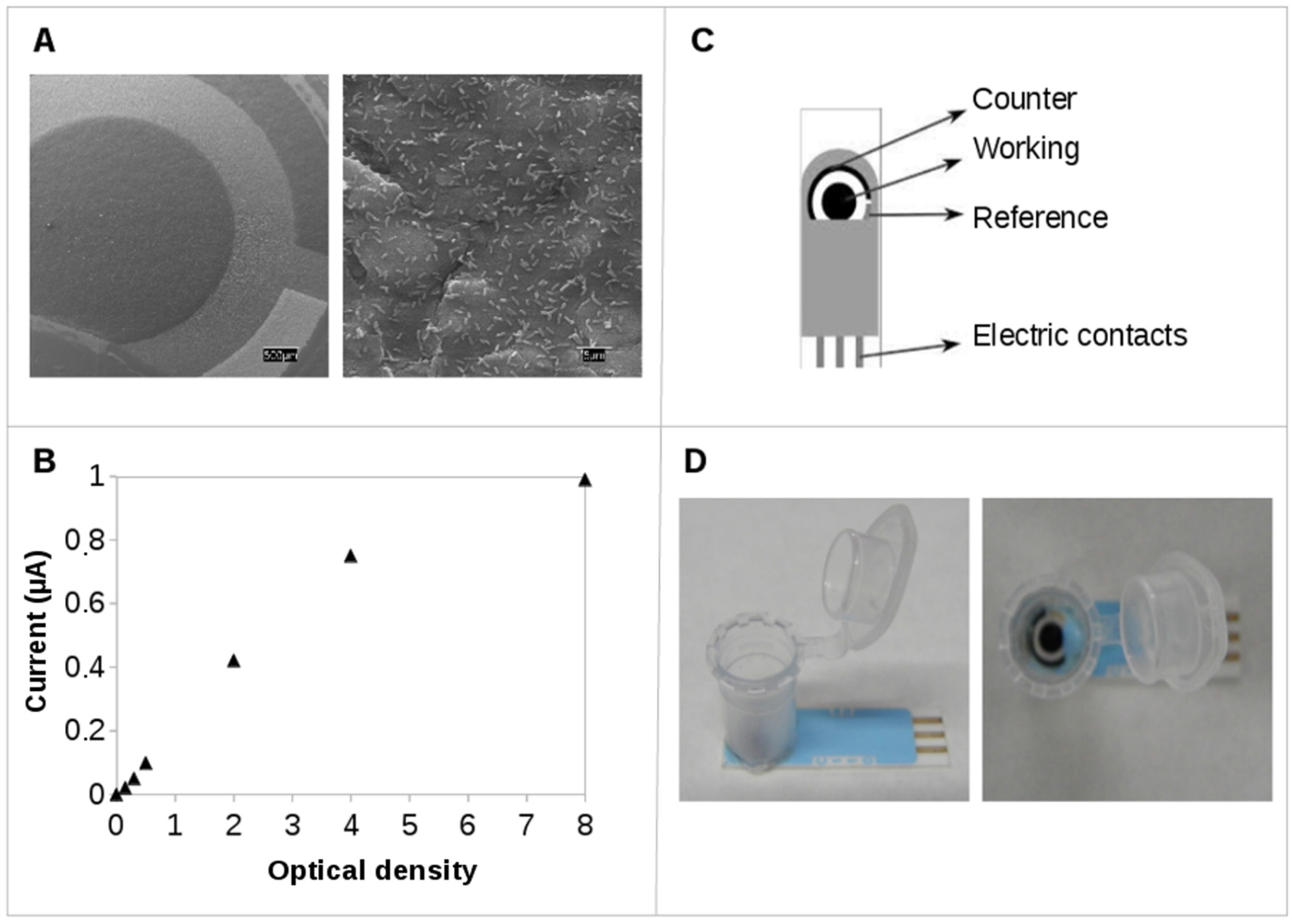
2.1. Screen-Printed Electrodes (SPEs): Testing Electroactivity in Geobacter

It is well known that the central metabolism of Geobacter can fully oxidized acetate to CO2 by coupling it with the extracellular electron transfer to electrodes of different nature [1,9,29]. The purpose of this work was to test if SPEs could be suitable tools for assaying such a reaction. With the aim of standardize our methodology, a preliminary study was performed by increasing thecell concentration of G. sulfurreducens and measuring the current production using carbon SPEs. Due to electrode sensitivity, we were able to detect changes in the current values by varying the amount of cells in contact with the electrode. We concluded that a cell suspension of bacteria with a value of 2 units of optical density (1.6 × 10⁹ cells/mL) can generate a current value in the mid-linear phase of the response (Figure 1B).
The electrogenic response of Geobacter sulfurreducens under electron acceptor limitation has been previously studied by electrochemical and spectroelectrochemical techniques [7,12]. Bacteria were grown in continuous culture with acetate as electron donor and fumarate as sole electron acceptor. Attenuated Total Reflection-Surface Enhanced Infrared Reflection Absorption Spectroscopy (ATR-SEIRAS) and CV, in a classical three-electrode chamber setup, showed that terminal electron acceptor (TEA) limited cells, exhibited a constitutive capacity for extracellular electron transfer (EET) [24]. The capability of cells in fumarate-limiting conditions to respire electrodes was analyzed on disposable miniaturized carbon SPEs, in order to compare the results to those previously obtained with a classical electrochemical cell configuration.
Figure 1. (A) Scanning electron microscopy (SEM) image of G. sulfurreducens attached to a carbon screen-printed electrode (SPE); (B) steady current production of G. sufurreducens at different cell density; (C) scheme of a SPE; and (D) detail of a lid-tube sealed to the SPE ceramic surface.
Energies 08 12366 g001
The fumarate-limited cells response showed that current production from metabolic acetate oxidation is predominant from the very beginning, displaying an electron transfer rate of 32.7 pmol electrons/s per cm2, which indicates the constitutive capacity for electron transfer to the electrode. In contrast, mid-log cells exhibit a lag phase, lower current production and ca. 2-fold lower electron transfer rate (15.6 pmol electrons/s per cm2), when they were tested with a SPE polarized at 0.2 V (Figure 2A). The electrochemical properties of both physiologies were also then tested by CV in SPEs. Once again, fumarate-limited cells presented a well-defined redox process showing the two characteristic redox peaks (Figure 2B), while mid-log cells exhibit a remarkably lower peak current under identical analysis conditions. This result is consistent with previous data that suggest a different bacterial electroactive response for each physiological condition [24].
Moreover, it is possible to use these SPEs for testing the electrochemical response corresponding to the growth phases of G. sulfurreducens, since it will be a key factor in the electrochemical performance. The availability of TEAs in microorganisms entails physiological changes in the cell, specially in those adapted to oligotrophic environments, such as G. sulfurreducens. As previous studies showed, the electron acceptor limitation triggers a metabolic adaptation that includes the overexpression of redox-active proteins, such c-type cytochromes [24]. Following this strategy, electrons generated from acetate metabolism can be stored in a c-type cytochrome network which act as capacitor-like element [30], that would discharge them as soon as a TEA is available. Interestingly, the current generated and the electron transfer rate (30.2 pmol electrons/s per cm2) by cells in stationary phase, was similar to the one found on fumarate-limited cells, suggesting that cells in stationary phase are also well adapted to EET (Figure 2). This behavior could be justified with the proteomic profile, which reveals the increase of several proteins at stationary phase, including cytochromes an oxidoreductase involved in electron transport [31,32]. In contrast, the significant lower electron transfer rate in mid-log phase cells, suggests that they still have not synthesized all the redox players for performing an efficient electron transfer to the electrode, so analyzing the electroactivity of a bacterial culture may help to predict the growth phase of the cells.
Figure 2. (A) Current production by G. sulfurreducens cells under different growth conditions: chemostat cells under fumarate-limiting conditions (red), early stationary cells (green), midlog cells (blue) and buffer control (black), polarized a 0.2 V; (B) Cyclic voltammetry (CV) of G. sulfurreducens cells under different growth conditions: chemostat cells under fumarate-limiting conditions (red), early stationary cells (green), mid-log cells (blue) and buffer control (black).
Energies 08 12366 g002
It is interesting to mention that cells are still able to produce current when adsorbed onto a polarized electrode, even if acetate is not present in the electrolyte solution (Figure 3A).
Figure 3. (A) Current production (at 0.2 V vs. Ag/AgCl) by G. sulfurreducens under acetate-excess conditions (red), and under acetate-limiting conditions just using intracellular acetate content (black); (B) CV of G. sulfurreducens on a graphene SPE (green) and a carbon SPE (black).
Energies 08 12366 g003
This can be explained by the presence of some intracellular acetate that remains as a source of electrons. Using this internal organic fuel, cells are able to produce current for ca. 3 min, after then intracellular acetate is not enough to produce a stable current value, and the current decreased to zero, presumably because the electron donor was fully consumed. From the gathered data, we calculated using the Faraday equation that the intracellular acetate content is about 1.51 × 10⁻⁶ pmol per cell. Regarding the acetate oxidation rate, it is worth noting that the remaining stored acetate is consumed ca. 4-fold slower (1 × 10⁻⁹ pmol/s per cell) than when it is at high concentration as 20 mM in the extracellular medium (4 × 10⁻⁹ pmols/s per cell).
During the last years, G. sulfurreducens’s electroactivity was tested on several materials serving as TEAs, such as graphite, gold, silver and platinum [9,29]. Among all the conductive materials that can be tested nowadays, graphene is for sure one of the most attractive ones. Its single atomic layer of graphite allows it to have higher electric conductivity than conventional carbon materials [33]. Despite the excellent features of graphene, there are few examples of its use in METs [34,35]. The electroactivity in stationary cells of G. sulfurreducens using graphene SPEs was also tested in this work, which resulted in a considerably increase of the signal intensity in contrast to carbon SPEs (Figure 3B). Moreover the redox signal appeared now at more negative values of potential (ca. 0 V) in contrast with the typical value of ca. 0.2 V detected on standard graphite electrodes [7,36]. The shift in the oxidation potential might be related with graphene’s better electric conductivity, which is several orders of magnitude higher than that of carbon [33]. In addition, it is well known that the working electrode material can influence the microbial response at METs. Even using the same material, a mere change in the active surface could lead to different electroactive microorganism behavior [29], so the improved response could be a combination of both electric conductivity and a different bacteria-electrode interaction. Graphene is by far the best up-and-coming material which is being implemented in several fields, so further studies will be required to take advantage of its unique conductive properties.

2.2. Practical Applications

2.2.1. Screening of Metabolic Features from a Microbial Consortium: Screen-Printed Electrodes (SPEs) Acting as Electron Donor

Another desirable property for a microorganism employed in METs, is the ability of using an electrode as electron donor source, conforming a biocathode. In comparison with an abiotic cathode, promoting the redox reaction at the cathode by microorganisms, increases the operational sustainability as well as reduces the cost of construction and operation of METs [37]. This trait could be exploited with the aim of screening predominant metabolic pathways from a mixed culture, by monitoring the electroactive response in the presence of several oxidizing substrates.
As a proof of concept, a consortium well adapted to nitrate reduction obtained from an anaerobic reactor was tested. In contrast, a culture of G. sulfurreducens was used as a negative control for electroactive bacteria unable to reduce nitrate. The current consumption of both types of cells was evaluated in the presence of the selected electron acceptor nitrate (2 mM) after polarizing the SPE-working electrode at −0.5 V.
After a brief conditioning period of 5 min, we observed that the current consumption was 20-fold higher in the microbial consortium assay in comparison with the pure culture of G. sulfurreducens (Figure 4). The results are not unexpected, considering that the consortium was fully adapted to nitrate-reducing conditions, while G. sulfurreducens is very well known for its ability to interchange electrons with electrodes, but not with nitrate as sole electron acceptor [38].
Aside from this example, the use of SPEs appears as an easy and quick method to identify potential microbial cultures showing activity in biocathodes. The microbial metabolism in biocathodes, indeed provides an excellent opportunity to find candidates for produce useful products performing bioelectrosynthesis [39] or for removing unwanted compounds from water like sulfate [40], nitrate [41], uranium [42], chloroaromatic compounds [43], which could be exploited in METs.
Figure 4. Electricity consumption by a microbial consortium (red) and G. sulfurreducens (blue) and buffer control (black) using nitrate (2 mM) as TEA, polarized at −0.5 V.
Energies 08 12366 g004

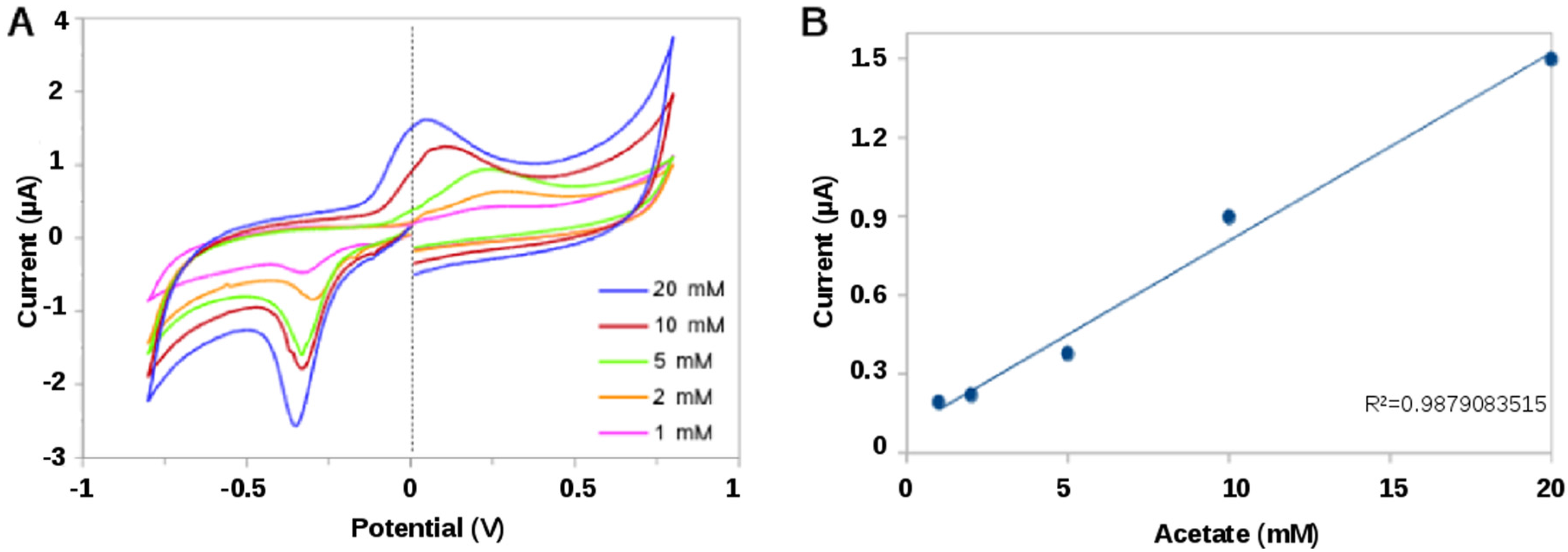
2.2.2. Bioelectrochemical Sensing of Acetate

Among the METs, there are a few examples of whole-cell biosensors as the electric signal transducer for generating and quantifying soluble organic matter [16,44], or for detecting the presence of a toxic compound like formaldehyde [15]. In contrast to those studies, we combine the use of SPEs with G. sulfurreducens to follow a CV-based strategy and construct a prototype microbial electrochemical sensor for quantifying acetate. Our assays revealed that the microbial current displayed by the CV at a potential of 0 V seems to be proportional to the concentration of acetate (Figure 5). The fact of using voltammograms for extrapolating current produced at 0 V, allows us to take advantage of both electrochemical methods. Amperometry makes it possible to detect down to picoamperes, while performing CV offers a low noise signal on top of verifying the proper electrode-bacteria interaction [45]. Indeed, the current response to acetate concentration at this conditions, exhibited a coefficient of determination (R2) of 0.98 with a detection limit between 20 mM and 1 mM. Similar results were previously obtained by other authors, although they used more sophisticated microfluidic devices [16]. Although further investigation for optimization of the sensor will be required, nevertheless, to our knowledge, this is the first approach to an amperometric acetate biosensor using a pure culture of G. sulfurreducens in combination with a SPE.
Figure 5. (A) Voltammograms corresponding to SPE-monolayer of G. sulfurreducens exposed to solutions with different acetate concentration; (B) Calibration curve for acetate concentration between 1 mM and 20 mM, calculated from current display at 0 V according to the CV.
Energies 08 12366 g005

2.2.3. Effects of a Background of Real Urban Wastewater

In contrast with the fascinating profile of electron acceptors (Fe-oxides, uranium, humic acids, graphite or gold electrode) that Geobacter sulfurreducens can use [46], its ability for oxidizing organic electron donors is very limited. Actually just acetate [1] and lactate [47] can be converted into electrical current by this strain. This apparent limitation can nevertheless be a positive feature because just acetate will be converted into electrical current regardless of the complexity of mixture. This is specially relevant if we consider that acetate is the end-product of the acetogenic phase in the anaerobic wastewater treatments, and its presence is directly related with the formation of biogas in anaerobic digestion [48].
Therefore, as a first approach for future applications, we tested the current production of our microbial-SPE-based in a background sample of real urban wastewater, using acetate as electron donor. According to the electrical current production, there was no substance in the sample of non-buffered wastewater able to significantly compete with the electrode as TEA. Moreover, current production was doubled when the acetate concentration was artificially increased from 5 mM to 10 mM demonstrating that the system was robust enough for performing assays in any kind of medium (Figure 6).
Figure 6. Current production of G. sulfurreducens from a real urban wastewater supplemented with 0 (black), 5 (green) and 10 mM acetate (blue). Working electrode was polarized a 0 V (vs. Ag/AgCl).
Energies 08 12366 g006

3. Experimental Section

3.1. Bacterial Culture

Geobacter sulfurreducens (strain DSM 12127; ATCC 51573) was grown at 30 °C in freshwater medium containing the following mineral salts (per liter): 2.5 g of NaHCO3, 0.25 of NH4Cl, 0.06 g of NaH2PO4·H2O and 0.1 g of KCl, 0.024 g of C6H5FeO7 (ferric citrate), 10 mL of a vitamins mix and 10 mL of a trace mineral mix [49]. Anaerobic conditions were achieved by flushing the culture media with N2:CO2 (80:20) to remove oxygen and to keep the pH of the bicarbonate buffer at pH = 7. For batch cultures, acetate (20 mM) was supplied as the sole carbon source and electron donor, and fumarate (40 mM) as electron acceptor. Mid-log cells and stationary cells were harvested 40 h and 72 h, respectively, after the inoculation procedure.
Geobacter sulfurreducens was also cultured in continuous mode using a chemostat. This growth method allowed to achieve fumarate-limiting conditions, a situation that led to a better EET, as previously described [24,50].
The nitrate reducing microbial consortium was directly harvested from an anaerobic reactor operating under nitrate reducing conditions.

3.2. Electrochemical Analysis

Bacterial cultures were harvested by centrifugation at 6000 rpm during 10 min. Subsequently, bacterial pellet were resuspended (OD600 = 2) in filtered wastewater or 30 mM phosphate buffer solution (pH = 6.8) containing 30 mM KCl. Then, 75 µL of a Geobacter sulfurreducens cell suspension was added to the SPE and, immediately, the electrochemical assays were performed without any preconditioning period. All electrochemical assays were performed in a screen-printed three electrode configuration using electrodes made of carbon (DRP-110, Dropsens, Asturias, Spain) or graphene (DRP-110GPH, Dropsens). These electrodes consist of a working electrode of carbon ink (surface = 0.12 cm2), a carbon counter electrode and a silver reference electrode; assembled on a ceramic platform (3.4 cm × 1.0 cm × 0.05 cm), connected to a potentiostat and controlled by specific software (Nanoelectra, Madrid, Spain). All potentials were reported versus a Ag/AgCl electrode. For chronoamperommetric assays the current was registered every 0.5 s and two electrode potentials were explored: current producing assays by supplying acetate as electron donor to the cell suspension and fixing the potential at 0.2 V; and current consuming assays, by supplying nitrate as electron acceptor and fixing the potential at −0.5 V. For CV, the potential window was scanned between −0.8 V and 0.8 V at 0.005 V s−1. All experiments were performed under anaerobic conditions by using an anoxic chamber (Coy, Grass Lake, MI, USA) or, alternatively, a sealed cap-tube. The sealed cap-tube was assembled by gluing the upper section of a cut-tube (1.5 mL, Eppendorf, Hamburg, Germany) to the ceramic surface of the SPE. Anoxic conditions inside the tube were achieved by flushing the headspace with N2 (Figure 1).

3.3. Perfomance of the Acetate Biosensor Assay

Early stationary bacterial cultures were harvested by centrifugation at 6000 rpm during 10 min. Then, samples were resuspended (OD600 = 5) in 30 mM phosphate buffer solution (pH = 6.8) containing 30 mM KCl. Subsequently, a drop of 75 µL of the cell suspension was placed on the electrode and cells were fixed by cycling the working electrode between 0.1 V and −0.1 V at 100 mV·s−1 during 100 scans [20]. Then, the electrode was washed with phosphate buffer in order to remove the unattached cells. Finally, a 75 µL drop of acetate solutions (1–20 mM) in phosphate buffer were added, and the electrode was polarized at 0.2 V during 15 min. For each acetate concentration a new SPE was used.

3.4. Scanning Electron Microscopy (SEM)

The microbial attachment of the electrode surface was observed by SEM. Microbial SPEs were fixed by immersion for one hour at room temperature in cacodylate buffer (0.2 M, pH = 7.2) containing 5% glutaraldehyde. The samples were rinsed two times in 0.2 M cacodylate buffer, pH = 7.2 for 10 min, and subsequently dehydrated by a graded ethanol series (25%, 50%, 70%, 90%, 100% and 100%; 10 min each stage). Then, the samples were rinsed two times in acetone for 10 min and immersed in anhydrous acetone at 4 °C overnight. Finally, dehydrated cells were dried in CO2 at the critical point and processed using a scanning electron microscope DSM-950 (Zeiss, Oberkochen, Alemania).

4. Conclusions

The results presented in this work demonstrate that screen-printed disposable electrodes can be used as a novel platform to assess within minutes the electron transfer capacities of electroactive microorganisms in quick drop assays that only require microlitres of culture samples. SPEs have shown to be sensitive to different physiological conditions in Geobacter sulfurreducens and excellent working electrode materials. Furthermore, SPEs could be exploited for fast screening methods to select tailor made biocathodes. SPE were designed for electroanalytical applications, and we have demonstrated that they are ready to accept whole-living cells and explored biosensor development for uses such as acetate detection in wastewater. Finally, we understand this work as a proof of concept for exploring new scenarios to investigate microbial electrochemistry without setting-up large electrochemical devices.

Acknowledgments

This work was supported by the European Union through the FP7 BacWire Project (Contract-No.: MNP4-SL-2009-2293337). The PhD fellowship for Marta Estevez is funded by the Formación de Profesorado Universitario (FPU) program of the University of Alcalá.

Author Contributions

Marta Estevez-Canales contributed to and practical implementation, as well as in writing and editing the manuscript. Antonio Berná contributed to the analysis and review of theoretical and practical subjects. , Zulema Borjas contributed to the practical implementation. Abraham Esteve-Núñez contributed to the analysis and review of theoretical and practical subjects, as well as in writing and editing the manuscript.

Conflicts of Interest

The authors declare no conflict of interest.

References

  1. Bond, D.R.; Lovley, D.R. Electricity production by Geobacter sulfurreducens attached to electrodes. Appl. Environ. Microbiol. 2003, 69, 1548–1555. [Google Scholar] [CrossRef] [PubMed]
  2. Kim, H.J.; Park, H.S.; Hyun, M.S.; Chang, I.S.; Kim, M.; Kim, B.H. A mediator-less microbial fuel cell using a metal reducing bacterium, Shewanella putrefaciens. Enzym. Microb. Technol. 2002, 30, 145–152. [Google Scholar] [CrossRef]
  3. Rodrigo, J.; Boltes, K.; Esteve-Nuñez, A. Microbial-electrochemical bioremediation and detoxification of dibenzothiophene-polluted soil. Chemosphere 2014, 101, 61–65. [Google Scholar] [CrossRef] [PubMed]
  4. Schröder, U.; Harnisch, F.; Angenent, L.T. Microbial electrochemistry and technology: Terminology and classification. Energy Environ. Sci. 2015, 8, 513–519. [Google Scholar] [CrossRef]
  5. Rozendal, R.A.; Hamelers, H.V.M.; Rabaey, K.; Keller, J.; Buisman, C.J.N. Towards practical implementation of bioelectrochemical wastewater treatment. Trends Biotechnol. 2008, 26, 450–459. [Google Scholar] [CrossRef] [PubMed]
  6. Clauwaert, P.; Aelterman, P.; Pham, T.H.; de Schamphelaire, L.; Carballa, M.; Rabaey, K.; Verstraete, W. Minimizing losses in bio-electrochemical systems: The road to applications. Appl. Microbiol. Biotechnol. 2008, 79, 901–913. [Google Scholar] [CrossRef] [PubMed]
  7. Busalmen, J.P.; Esteve-Nuñez, A.; Feliu, J.M. Whole cell electrochemistry of electricity-producing microorganisms evidence an adaptation for optimal exocellular electron transport. Environ. Sci. Technol. 2008, 42, 2445–2450. [Google Scholar] [CrossRef] [PubMed]
  8. Kuzume, A.; Zhumaev, U.; Li, J.; Fu, Y.; Fueeg, M.; Esteve-Nunez, A.; Wandlowski, T. An in-situ surface electrochemistry approach toward whole-cell studies: Charge transfer between Geobacter sulfurreducens and electrified metal/electrolyte interfaces through linker molecules. Electrochim. Acta 2013, 112, 933–942. [Google Scholar] [CrossRef]
  9. Kuzume, A.; Zhumaev, U.; Li, J.; Fu, Y.; Füeg, M.; Estévez, M.; Borjas, Z.; Wandlowski, T.; Esteve-Nuñez, A. An in situ surface electrochemistry approach towards whole-cell studies: The structure and reactivity of a Geobacter sulfurreducens submonolayer on electrified metal/electrolyte interfaces. Phys. Chem. Chem. Phys. 2014, 16, 22229–22236. [Google Scholar] [CrossRef] [PubMed]
  10. Schrott, G.D.; Bonanni, P.S.; Robuschi, L.; Esteve-Nuñez, A.; Busalmen, J.P. Electrochemical insight into the mechanism of electron transport in biofilms of Geobacter sulfurreducens. Electrochim. Acta 2011, 56, 10791–10795. [Google Scholar] [CrossRef]
  11. Carmona-Martínez, A.A.; Harnisch, F.; Kuhlicke, U.; Neu, T.R.; Schröder, U. Electron transfer and biofilm formation of Shewanella putrefaciens as function of anode potential. Bioelectrochemistry 2013, 93, 23–29. [Google Scholar] [CrossRef] [PubMed]
  12. Busalmen, J.P.; Esteve-Núñez, A.; Berná, A.; Feliu, J.M. C-type cytochromes wire electricity-producing bacteria to electrodes. Angew. Chem. Int. Ed. 2008, 47, 4874–4877. [Google Scholar] [CrossRef] [PubMed]
  13. Harnisch, F.; Rabaey, K. The diversity of techniques to study electrochemically active biofilms highlights the need for standardization. ChemSusChem 2012, 5, 1027–1038. [Google Scholar] [CrossRef] [PubMed]
  14. Schröder, U. Editorial: Microbial Fuel Cells and Microbial Electrochemistry: Into the Next Century! ChemSusChem 2012, 5, 959. [Google Scholar] [CrossRef] [PubMed]
  15. Davila, D.; Esquivel, J.P.; Sabate, N.; Mas, J. Silicon-based microfabricated microbial fuel cell toxicity sensor. Biosens. Bioelectron. 2011, 26, 2426–2430. [Google Scholar] [CrossRef] [PubMed]
  16. Li, Z.; Zhang, Y.; LeDuc, P.R.; Gregory, K.B. Microbial electricity generation via microfluidic flow control. Biotechnol. Bioeng. 2011, 108, 2061–2069. [Google Scholar] [CrossRef] [PubMed]
  17. Fraiwan, A.; Dai, C.; Sidhu, N.K.; Rastogi, A.; Choi, S. A micro-sized microbial fuel cell with electrochemical sensing functionality. In Proceedings of the 2014 9th IEEE International Conference on Nano/Micro Engineered and Molecular Systems (NEMS), Waikiki Beach, HI, USA, 13–16 April 2014.
  18. Hayat, A.; Marty, J.L. Disposable screen printed electrochemical sensors: Tools for environmental monitoring. Sensors 2014, 14, 10432–10453. [Google Scholar] [CrossRef] [PubMed]
  19. Taleat, Z.; Khoshroo, A.; Mazloum-Ardakani, M. Screen-printed electrodes for biosensing: A review (2008–2013). Microchim. Acta 2014, 181, 865–891. [Google Scholar] [CrossRef]
  20. Gómez-Mingot, M.; Iniesta, J.; Montiel, V.; Kadara, R.O.; Banks, C.E. Screen printed graphite macroelectrodes for the direct electron transfer of cytochrome c. Analyst 2011, 136, 2146–2150. [Google Scholar] [CrossRef] [PubMed]
  21. Chang, J.-L.; Lien, C.-H.; Vijayakumar, P.S.; Hsieh, P.-H.; Zen, J.-M. Electrochemical regulation of microbial growth on disposable screen printed carbon electrodes. Electrochim. Acta 2012, 82, 103–108. [Google Scholar] [CrossRef]
  22. Tran, H.V.; Piro, B.; Reisberg, S.; Huy Nguyen, L.; Dung Nguyen, T.; Duc, H.T.; Pham, M.C. An electrochemical ELISA-like immunosensor for miRNAs detection based on screen-printed gold electrodes modified with reduced graphene oxide and carbon nanotubes. Biosens. Bioelectron. 2014, 62, 25–30. [Google Scholar] [CrossRef] [PubMed]
  23. Asturias-Arribas, L.; Asuncion Alonso-Lomillo, M.; Dominguez-Renedo, O.; Julia Arcos-Martinez, M. Cytochrome P450 2D6 based electrochemical sensor for the determination of codeine. Talanta 2014, 129, 315–319. [Google Scholar] [CrossRef] [PubMed]
  24. Esteve-Núñez, A.; Busalmen, J.P.; Berná, A.; Gutiérrez-Garrán, C.; Feliu, J.M. Opportunities behind the unusual ability of Geobacter sulfurreducens for exocellular respiration and electricity production. Energy Environ. Sci. 2011, 4, 2066–2069. [Google Scholar] [CrossRef]
  25. Sudhakara Prasad, K.; Arun, A.B.; Rekha, P.D.; Young, C.-C.; Chang, J.-L.; Zen, J.-M. A microbial sensor based on direct electron transfer at Shewanella sp. drop-coated screen-printed carbon electrodes. Electroanalysis 2009, 21, 1646–1650. [Google Scholar] [CrossRef]
  26. Marsili, E.; Baron, D.B.; Shikhare, I.D.; Coursolle, D.; Gralnick, J.A.; Bond, D.R. Shewanella secretes flavins that mediate extracellular electron transfer. Proc. Natl. Acad. Sci. USA 2008, 105, 3968–3973. [Google Scholar] [CrossRef] [PubMed] [Green Version]
  27. Kotloski, N.J.; Gralnick, J.A. Flavin electron shuttles dominate extracellular electron transfer by Shewanella oneidensis. MBio 2013, 4. [Google Scholar] [CrossRef] [PubMed]
  28. Lovley, D.R. Live wires: Direct extracellular electron exchange for bioenergy and the bioremediation of energy-related contamination. Energy Environ. Sci. 2011, 4, 4896–4906. [Google Scholar] [CrossRef]
  29. Maestro, B.; Ortiz, J.M.; Schrott, G.; Busalmen, J.P.; Climent, V.; Feliu, J.M. Crystallographic orientation and electrode nature are key factors for electric current generation by Geobacter sulfurreducens. Bioelectrochemistry 2014, 98, 11–19. [Google Scholar] [CrossRef] [PubMed] [Green Version]
  30. Esteve-Núñez, A.; Sosnik, J.; Visconti, P.; Lovley, D.R. Fluorescent properties of c-type cytochromes reveal their potential role as an extracytoplasmic electron sink in Geobacter sulfurreducens. Environ. Microbiol. 2008, 10, 497–505. [Google Scholar] [CrossRef] [PubMed]
  31. Bansal, R.; Helmus, R.A.; Stanley, B.A.; Zhu, J.; Liermann, L.J.; Brantley, S.L.; Tien, M. Survival during long-term starvation: Global proteomics analysis of Geobacter sulfurreducens under prolonged electron-acceptor limitation. J. Proteome Res. 2013, 12, 4316–4326. [Google Scholar] [CrossRef] [PubMed]
  32. Ding, Y.-H.R.; Hixson, K.K.; Giometti, C.S.; Stanley, A.; Esteve-Núñez, A.; Khare, T.; Tollaksen, S.L.; Zhu, W.; Adkins, J.N.; Lipton, M.S.; et al. The proteome of dissimilatory metal-reducing microorganism Geobacter sulfurreducens under various growth conditions. Biochim. Biophys. Acta 2006, 1764, 1198–1206. [Google Scholar] [CrossRef] [PubMed]
  33. Castro Neto, A.H.; Guinea, F.; Peres, N.M.R.; Novoselov, K.S.; Geim, A.K. The electronic properties of graphene. Rev. Mod. Phys. 2009, 81, 109–162. [Google Scholar] [CrossRef]
  34. Guo, W.; Cui, Y.; Song, H.; Sun, J. Layer-by-layer construction of graphene-based microbial fuel cell for improved power generation and methyl orange removal. Bioprocess Biosyst. Eng. 2014, 37, 1749–1758. [Google Scholar] [CrossRef] [PubMed]
  35. Kumar, G.G.; Hashmi, S.; Karthikeyan, C.; GhavamiNejad, A.; Vatankhah-Varnoosfaderani, M.; Stadler, F.J. Graphene oxide/carbon nanotube composite hydrogels-versatile materials for microbial fuel cell applications. Macromol. Rapid Commun. 2014, 35, 1861–1865. [Google Scholar] [CrossRef] [PubMed]
  36. Estevez-Canales, M.; Kuzume, A.; Borjas, Z.; Füeg, M.; Lovley, D.; Wandlowski, T.; Esteve-Núñez, A. A severe reduction in the cytochrome C content of Geobacter sulfurreducens eliminates its capacity for extracellular electron transfer. Environ. Microbiol. Rep. 2015, 7, 219–226. [Google Scholar] [CrossRef] [PubMed]
  37. Rabaey, K.; Keller, J. Microbial fuel cell cathodes: From bottleneck to prime opportunity? Water Sci. Technol. 2008, 57, 655–659. [Google Scholar] [CrossRef] [PubMed]
  38. Caccavo, F.; Lonergan, D.J.; Lovley, D.R.; Davis, M.; Stolz, J.F.; McInerney, M.J. Geobacter sulfurreducens sp. nov., a hydrogen- and acetate-oxidizing dissimilatory metal-reducing microorganism. Appl. Environ. Microbiol. 1994, 60, 3752–3759. [Google Scholar] [PubMed]
  39. Nevin, K.P.; Woodard, T.L.; Franks, A.E.; Summers, Z.M.; Lovley, D.R. Microbial electrosynthesis: Feeding microbes electricity to convert carbon dioxide and water to multicarbon extracellular organic compounds. MBio 2010, 1. [Google Scholar] [CrossRef] [PubMed]
  40. Coma, M.; Puig, S.; Pous, N.; Balaguer, M.D.; Colprim, J. Biocatalysed sulphate removal in a BES cathode. Bioresour. Technol. 2013, 130, 218–223. [Google Scholar] [CrossRef] [PubMed]
  41. Clauwaert, P.; Rabaey, K.; Aelterman, P.; de Schamphelaire, L.; Pham, T.H.; Boeckx, P.; Boon, N.; Verstraete, W. Biological denitrification in microbial fuel cells. Environ. Sci. Technol. 2007, 41, 3354–3360. [Google Scholar] [CrossRef] [PubMed]
  42. Gregory, K.B.; Lovley, D.R. Remediation and recovery of uranium from contaminated subsurface environments with electrodes. Environ. Sci. Technol. 2005, 39, 8943–8947. [Google Scholar] [CrossRef] [PubMed]
  43. Aulenta, F.; Fina, A.; Potalivo, M.; Petrangeli Papini, M.; Rossetti, S.; Majone, M. Anaerobic transformation of tetrachloroethane, perchloroethylene, and their mixtures by mixed-cultures enriched from contaminated soils and sediments. Water Sci. Technol. 2005, 52, 357–362. [Google Scholar] [PubMed]
  44. Tront, J.M.; Fortner, J.D.; Plötze, M.; Hughes, J.B.; Puzrin, A.M. Microbial fuel cell biosensor for in situ assessment of microbial activity. Biosens. Bioelectron. 2008, 24, 586–590. [Google Scholar] [CrossRef] [PubMed]
  45. Su, L.; Jia, W.; Hou, C.; Lei, Y. Microbial biosensors: A review. Biosens. Bioelectron. 2011, 26, 1788–1799. [Google Scholar] [CrossRef] [PubMed]
  46. Lovley, D.R.; Ueki, T.; Zhang, T.; Malvankar, N.S.; Shrestha, P.M.; Flanagan, K.A.; Aklujkar, M.; Butler, J.E.; Giloteaux, L.; Rotaru, A.-E.; et al. Geobacter: The Microbe’s Electric Physiology, Ecology, and Practical Applications; Academic Press: London, UK, 2011. [Google Scholar]
  47. Speers, A.M.; Reguera, G. Electron donors supporting growth and electroactivity of Geobacter sulfurreducens anode biofilms. Appl. Environ. Microbiol. 2012, 78, 437–444. [Google Scholar] [CrossRef] [PubMed]
  48. Henze, M. Biological Wastewater Treatment: Principles, Modelling and Design; IWA Publishing: London, UK, 2008. [Google Scholar]
  49. Lovley, D.R.; Phillips, E.J.P. Novel mode of microbial energy metabolism: Organic carbon oxidation coupled to dissimilatory reduction of iron or manganese. Appl. Environ. Microbiol. 1988, 54, 1472–1480. [Google Scholar] [PubMed]
  50. Esteve-Núñez, A.; Rothermich, M.; Sharma, M.; Lovley, D. Growth of Geobacter sulfurreducens under nutrient-limiting conditions in continuous culture. Environ. Microbiol. 2005, 7, 641–648. [Google Scholar] [CrossRef] [PubMed]

