
Potential Applications of Nanocellulose-Containing Materials in the Biomedical Field

 ,
,  , ,
, ,
Abstract
:1. Introduction
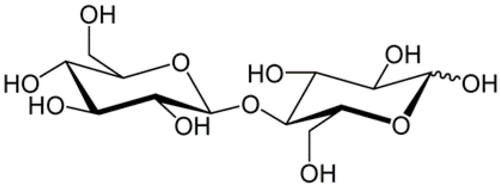
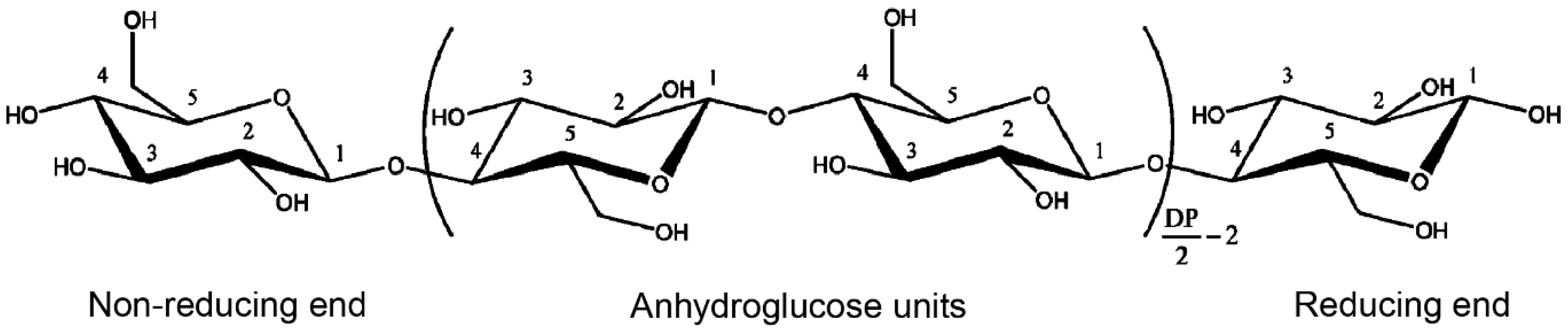
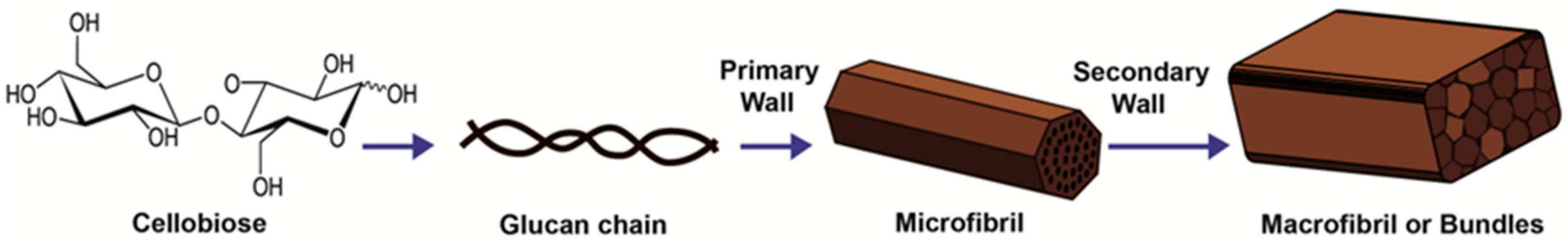
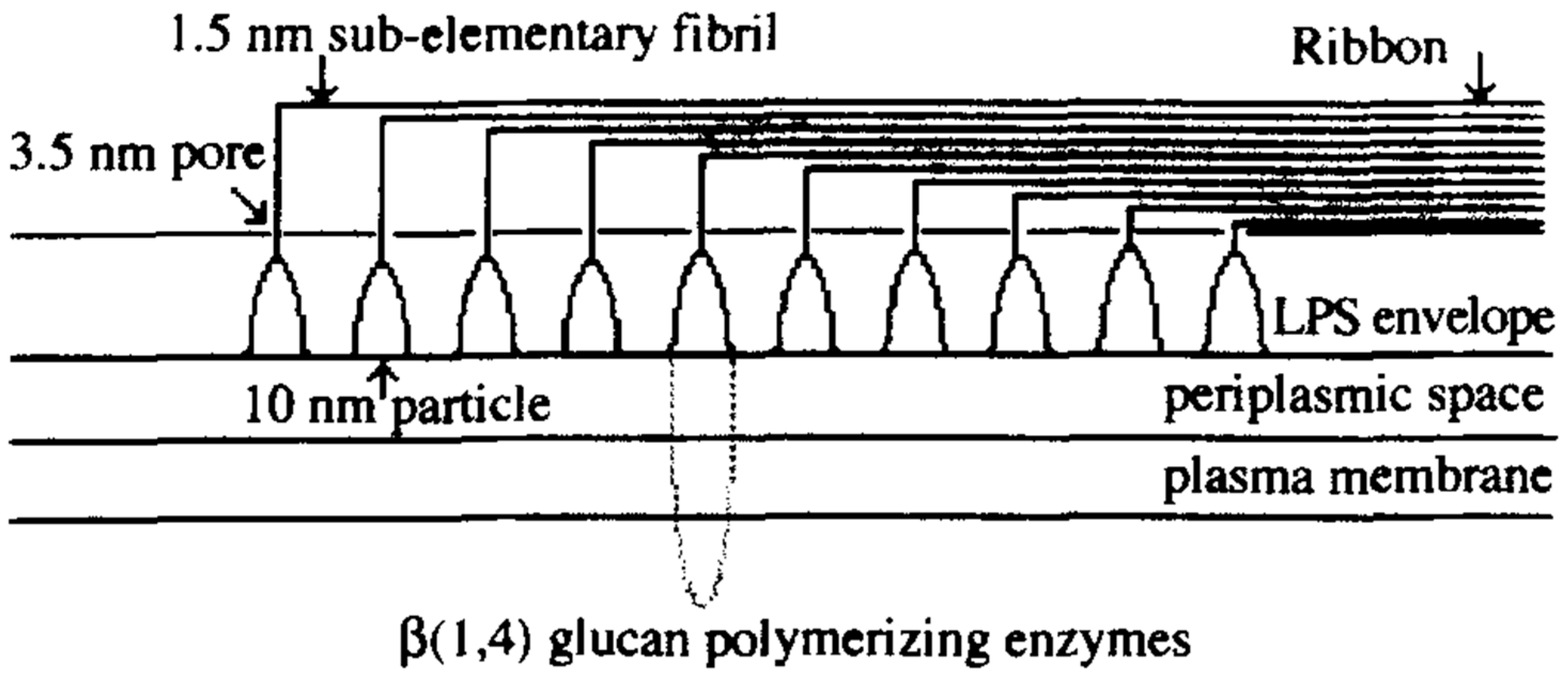
2. Chemical and Structural Composition of Cellulose Fibers
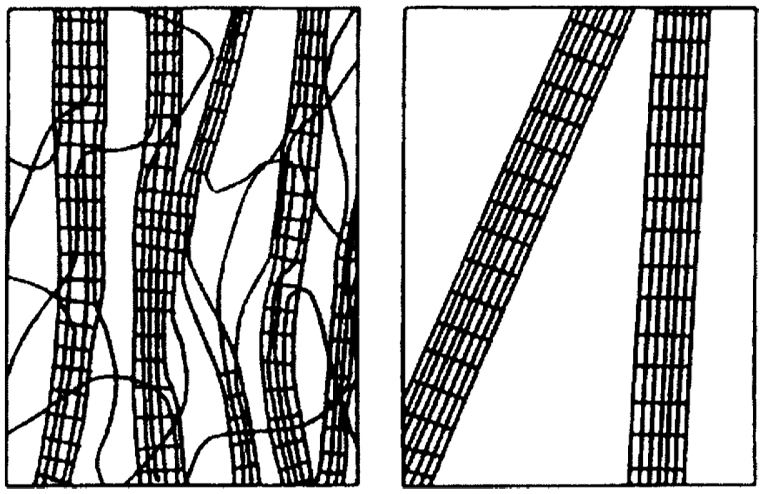
3. Sources and Properties of Cellulose
4. Isolation and Chemical Modifications of Cellulose
4.1. Nanofibrillar Cellulose (NFC)
4.2. Cellulose Nanocrystal (CNC)
4.3. Microcrystalline Cellulose (MCC)
4.4. Chemical Modification of Nano Cellulose
5. Biomedical Applications
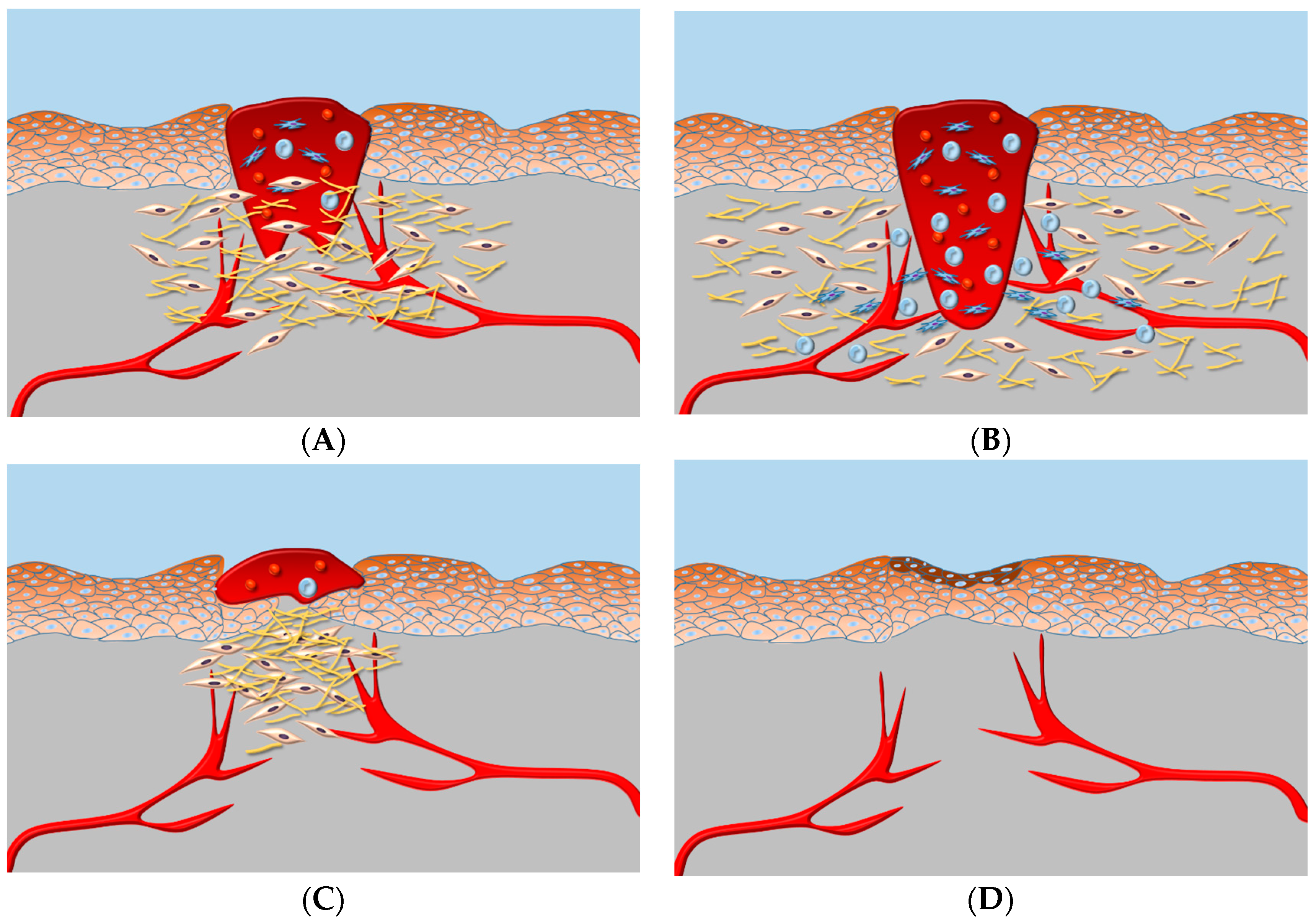
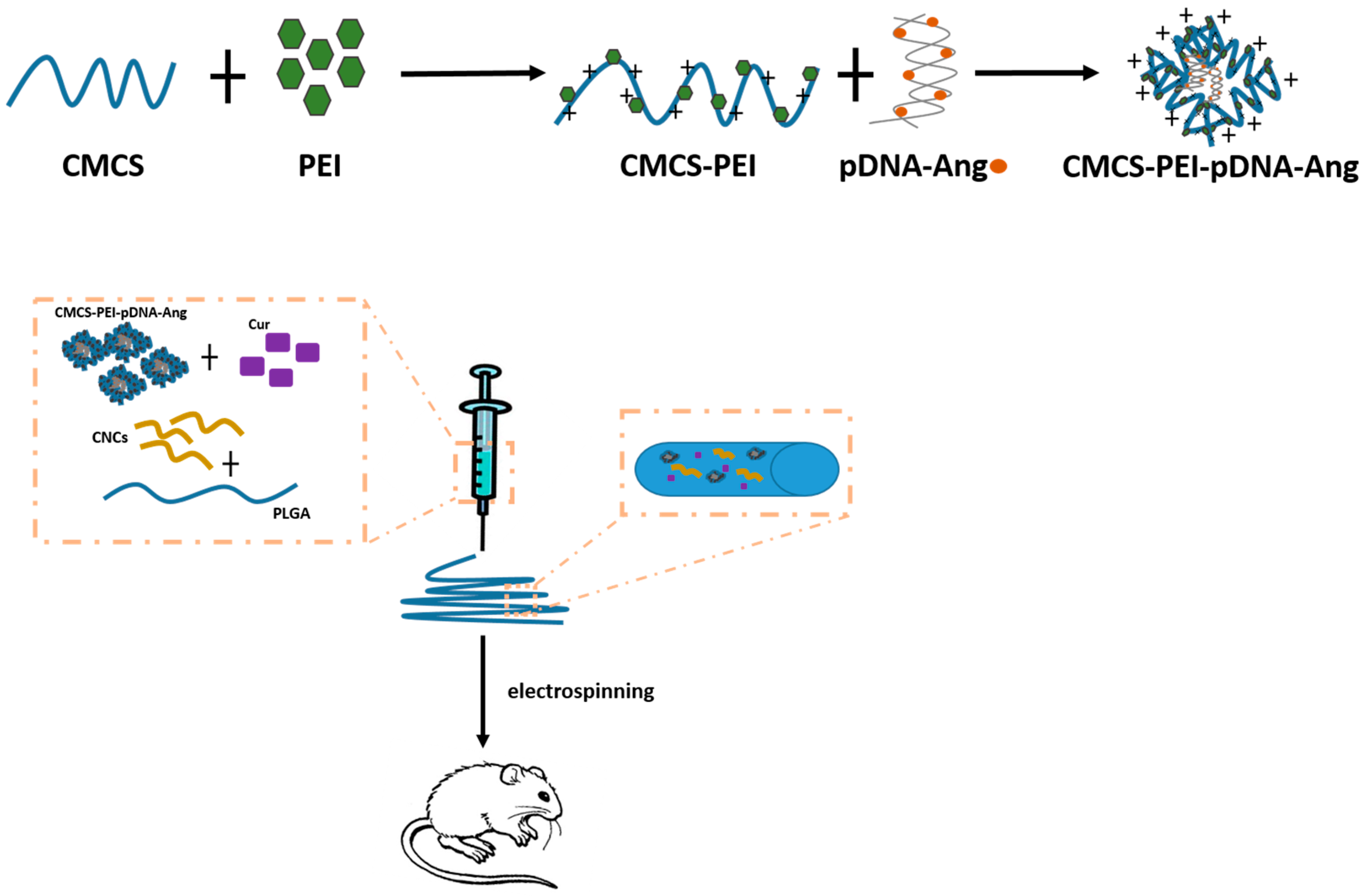
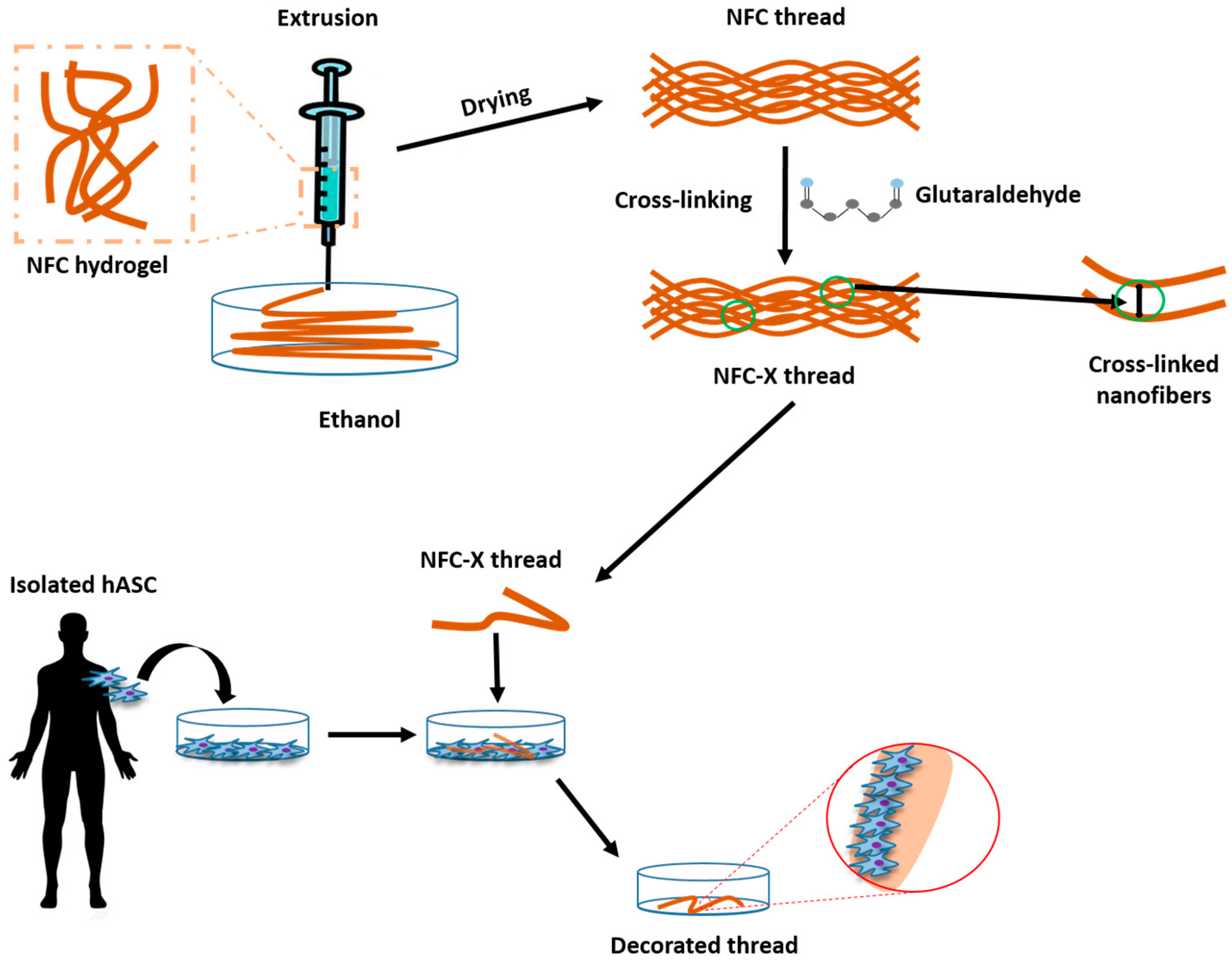
5.1. Wound Healing
Nano-Sized Cellulose-Based Material in Wound Healing
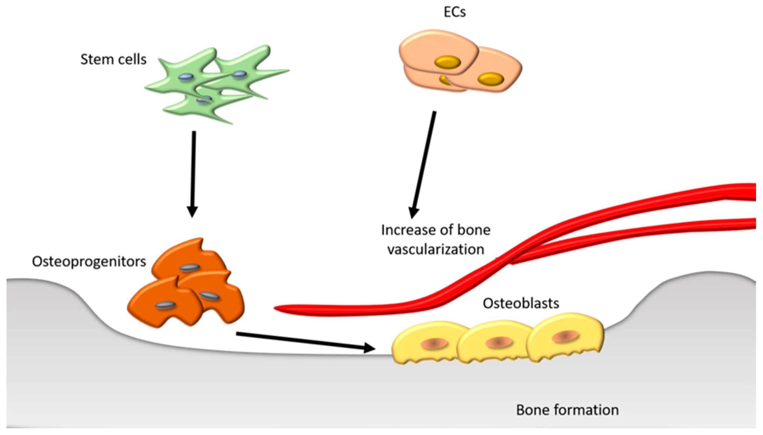
5.2. Bone Regeneration
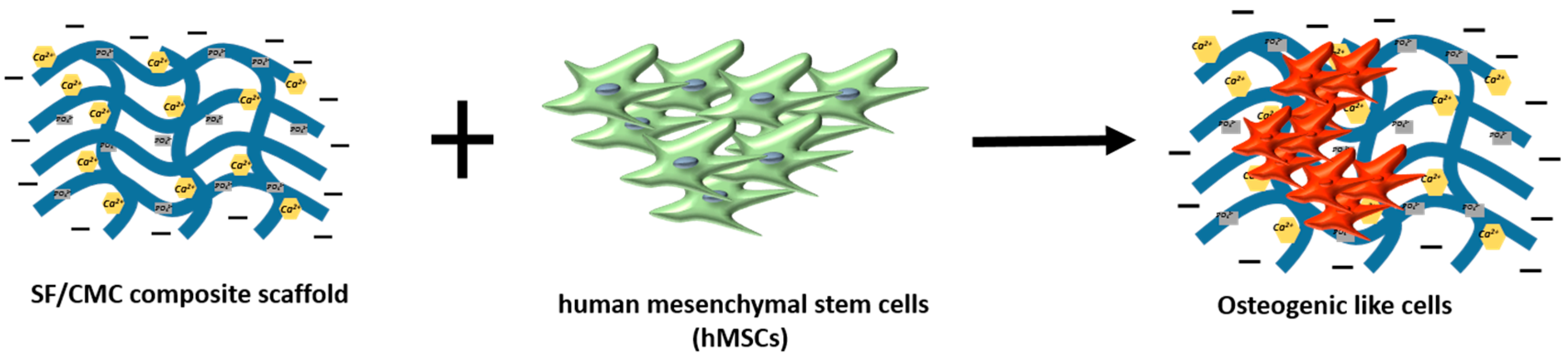
Nano-Sized Cellulose-Based Material in Bone Regeneration
5.3. Cartilage Regeneration
Nano-Sized Cellulose-Based Material in Cartilage Regeneration
5.4. Dental Applications
5.5. Other Applications
5.5.1. Cancer Diseases
5.5.2. Other Diseases
5.6. Clinical Trials
6. Conclusions
Acknowledgments
Author Contributions
Conflicts of Interest
References
- Klemm, D.; Schumann, D.; Kramer, F.; Hessler, N.; Hornung, M.; Schmauder, H.; Marsch, S. Nanocelluloses as innovative polymers in research and application. Polysaccharides 2006, 205, 49–96. [Google Scholar]
- Henriksson, M.; Berglund, L. Structure and properties of cellulose nanocomposite films containing melamine formaldehyde. J. Appl. Polym. Sci. 2007, 106, 2817–2824. [Google Scholar] [CrossRef]
- Iwamoto, S.; Nakagaito, A.; Yano, H. Nano-fibrillation of pulp fibers for the processing of transparent nanocomposites. Appl. Phys. A Mater. Sci. Process. 2007, 89, 461–466. [Google Scholar] [CrossRef]
- Jonas, R.; Farah, L. Production and application of microbial cellulose. Polym. Degrad. Stab. 1998, 59, 101–106. [Google Scholar] [CrossRef]
- Qin, C.; Soykeabkaew, N.; Xiuyuan, N.; Peijs, T. The effect of fibre volume fraction and mercerization on the properties of all-cellulose composites. Carbohydr. Polym. 2008, 71, 458–467. [Google Scholar] [CrossRef]
- Yamanaka, S.; Watanabe, K.; Kitamura, N.; Iguchi, M.; Mitsuhashi, S.; Nishi, Y.; Uryu, M. The structure and mechanical properties of sheets prepared from bacterial cellulose. J. Mater. Sci. 1989, 24, 3141–3145. [Google Scholar] [CrossRef]
- Endes, C.; Camarero-Espinosa, S.; Mueller, S.; Foster, E.J.; Petri-Fink, A.; Rothen-Rutishauser, B.; Weder, C.; Clift, M.J. A critical review of the current knowledge regarding the biological impact of nanocellulose. J. Nanobiotechnol. 2016, 14, 78. [Google Scholar] [CrossRef] [PubMed]
- Mondal, S. Preparation, properties and applications of nanocellulosic materials. Carbohydr. Polym. 2017, 163, 301–316. [Google Scholar] [CrossRef] [PubMed]
- Delmer, D.; Amor, Y. Cellulose Biosynthesis. Plant Cell 1995, 7, 987–1000. [Google Scholar] [CrossRef] [PubMed]
- Kalia, S.; Dufresne, A.; Cherian, B.; Kaith, B.; Avérous, L.; Njuguna, J.; Nassiopoulos, E. Cellulose-Based Bio- and Nanocomposites: A Review. Int. J. Polym. Sci. 2011, 1–35. [Google Scholar] [CrossRef]
- Habibi, Y.; Lucia, L.; Rojas, O. Cellulose Nanocrystals: Chemistry, Self-Assembly, and Applications. Chem. Rev. 2010, 110, 3479–3500. [Google Scholar] [CrossRef] [PubMed]
- Brown, R.M.J. The Biosynthesis Of Cellulose. Pure Appl. Chem. 1996, 33, 1345–1373. [Google Scholar] [CrossRef]
- Rowland, S.; Roberts, E.J. The nature of accessible surfaces in the microstructure of cotton cellulose. J. Polym. Sci. Part A Polym. Chem. 1972, 10, 2447–2461. [Google Scholar] [CrossRef]
- Mood, S.; Golfeshan, A.; Tabatabaei, M.; Jouzani, G.; Najafi, G.; Gholami, M.; Ardjmand, M. Lignocellulosic biomass to bioethanol, a comprehensive review with a focus on pretreatment. Renew. Sustain. Energy Rev. 2013, 27, 77–93. [Google Scholar] [CrossRef]
- El-Saied, H.; Basta, A.; Gobran, R. Research progress in friendly environmental technology for the production of cellulose products (bacterial cellulose and its application). Polym.-Plast. Technol. Eng. 2004, 43, 797–820. [Google Scholar] [CrossRef]
- Dammström, S.; Salmén, L.; Gatenholm, P. The effect of moisture on the dynamic properties of bacterial cellulose/glucuronoxylan nanocomposites. Polymer 2005, 46, 10364–10371. [Google Scholar] [CrossRef]
- Kalia, S.; Kaith, B.; Kaur, I. Pre-treatments of natural fibers and their application as reinforcing material in polymer composites-a review. Polym. Eng. Sci. 2009, 49, 1253–1272. [Google Scholar] [CrossRef]
- Iguchi, M.; Yamanaka, S.; Budhiono, A. Review Bacterial Cellulose—A masterpiece of nature’s arts. J. Mater. Sci. 2000, 35, 261–270. [Google Scholar] [CrossRef]
- Food and Drug Administration. Pyrogens and endotoxins testing: Questions and answers. In Guidance for Industry; FDA: Silver Spring, MD, USA, 2012. [Google Scholar]
- Zaar, K. The biogenesis of cellulose by Acetobacter xylinum. Cytobiologie 1977, 16, 1–15. [Google Scholar]
- Brown, R.M.J.; Willison, J.; Richardson, C. Cellulose biosynthesis in Acetobacter xylinum: Visualization of the site of synthesis and direct measurement of the in vivo process. Proc. Natl. Acad. Sci. USA 1976, 73, 4565–4569. [Google Scholar] [CrossRef] [PubMed]
- Yamanaka, S.; Ishihara, M.; Sugiyama, J. Structural modification of bacterial cellulose. Cellulose 2000, 7, 213–225. [Google Scholar] [CrossRef]
- Ben-Hayyim, G.; Ohad, I. Synthesis of cellulose by Acetobacter xylinum. VIII. On the Formation and Orientation of Bacterial Cellulose Fibrils in the Presence of Acidic Polysaccharides. J. Cell Biol. 1965, 25, 191–207. [Google Scholar] [CrossRef] [PubMed]
- Czaja, W.; Krystynowicz, A.; Bielecki, S.; Brown, R.M., Jr. Microbial cellulose-the natural power to heal wounds. Biomaterials 2006, 27, 145–151. [Google Scholar] [CrossRef] [PubMed]
- Hult, E.; Yamanaka, S.; Ishihara, M.; Sugiyama, J. Aggregation of ribbons in bacterial cellulose induced by high pressure incubation. Carbohydr. Polym. 2003, 53, 9–14. [Google Scholar] [CrossRef]
- Wan, W.; Hutter, J.; Millon, L.; Guhados, G. Bacterial cellulose and its nanocomposites for biomedical applications. In Cellulose Nanocomposites. Processing Characterization, and Properties; Oksman, K., Sain, M., Eds.; American Chemical Society: Washington, DC, USA, 2006. [Google Scholar]
- Barud, H.; Barrios, C.; Regiani, T.; Marques, R.; Verelst, M.; Dexpert-Ghys, J.; Messaddeq, Y.; Ribeiro, S. Selfsupported silver nanoparticles containing bacterial cellulose membranes. Mater. Sci. Eng. C 2008, 28, 515–518. [Google Scholar] [CrossRef]
- De Wulf, P.; Joris, K.; Vandamme, E.J. Improved cellulose fromation by an Acetobacer xylinum mutant limited in (keto)gluconate synthesis. J. Chem. Technol. Biotechnol. 1996, 67, 376–380. [Google Scholar] [CrossRef]
- Oikawa, T.; Morino, T.; Ameyama, M. Production of Cellulose from D Arabitol by Acetobacter xylinum KU-1. Biosci. Biotechnol. Biochem. 1995, 59, 1564–1565. [Google Scholar] [CrossRef]
- Oikawa, T.; Ohtori, T.; Ameyama, M. Production of Cellulose from D Mannitol by Acetobacter xylinum KU-1. Biosci. Biotechnol. Biochem. 1995, 59, 331–332. [Google Scholar] [CrossRef]
- Masaoka, S.; Ohe, T.; Sakota, N. Production of cellulose from glucose by Acetobacter xylinum. J. Ferment. Bioeng. 1993, 75, 18–22. [Google Scholar] [CrossRef]
- Toyosaki, H.; Naritomi, T.; Seto, A.; Matsuoka, M.; Tsuchida, T.; Yoshinaga, F. Screening of Bacterial Cellulose-producing Acetobacter Strains Suitable for Agitated Culture. Biosci. Biotechnol. Biochem. 1995, 59, 1498–1502. [Google Scholar] [CrossRef]
- Surip, S.; Wan Jaafar, W.; Azmi, N.; Anwar, U. Microscopy Observation on Nanocellulose from Kenaf Fibre. Adv. Mater. Res. 2012, 488, 72–75. [Google Scholar] [CrossRef]
- Siró, I.; Plackett, D. Microfibrillated cellulose and new nanocomposite materials: A review. Cellulose 2010, 17, 459–494. [Google Scholar] [CrossRef]
- Shin, E.J.; Choi, S.M.; Singh, D.; Zo, S.M.; Lee, Y.H.; Kim, J.H.; Han, S.S. Fabrication of cellulose-based scaffold with microarchitecture using a leaching technique for biomedical applications. Cellulose 2014, 21, 3515–3525. [Google Scholar] [CrossRef]
- Randy, B. The cellulose micelles. Tappi 1952, 35, 53–58. [Google Scholar]
- Hubbe, M.; Rojas, O.; Lucia, L.; Sain, M. Cellulosic nanocomposites: A review. BioResources 2008, 3, 929–980. [Google Scholar]
- Šturcova, A.; Davies, G.; Eichhorn, S. Elastic modulus and stress-transfer properties of tunicate cellulose whiskers. Biomacromolecules 2005, 6, 1055–1061. [Google Scholar] [CrossRef] [PubMed]
- Samir, M.; Alloin, F.; Dufresne, A. Review of recent research into cellulosic whiskers, their properties and their application in nanocomposite field. Biomacromolecules 2005, 6, 612–626. [Google Scholar] [CrossRef] [PubMed]
- Dufresne, A. Comparing the mechanical properties of high performances polymer nanocomposites from biological sources. J. Nanosci. Nanotechnol. 2006, 6, 322–330. [Google Scholar] [CrossRef] [PubMed]
- Dufresne, A. Polysaccharide nanocrystals reinforced nanocomposites. J. Chem. 2008, 86, 484–494. [Google Scholar] [CrossRef]
- Araki, J.; Wada, M.; Kuga, S.; Okano, T. Flow properties of microcrystalline cellulose suspension prepared by acid treatment of native cellulose. Colloids Surf. A 1998, 142, 75–82. [Google Scholar] [CrossRef]
- Roman, M.; Winter, W. Effect of Sulfate Groups from Sulfuric Acid Hydrolysis on the Thermal Degradation Behavior of Bacterial Cellulose. Biomacromolecules 2004, 5, 1671–1677. [Google Scholar] [CrossRef] [PubMed]
- Johari, N.; Ahmad, I.; Halib, N. Comparison Study of Hydrogels Properties Synthesized with Micro- and Nano- Size Bacterial Cellulose Particles Extracted from Nata de coco. Chem. Biochem. Eng. Q. 2012, 26, 399–404. [Google Scholar]
- Yin, O.; Ahmad, I.; Amin, M. Effect of Cellulose Nanocrystals Content and pH on Swelling Behaviour of Gelatin Based Hydrogel. Sains Malays. 2015, 44, 793–799. [Google Scholar]
- Ludueña, L.; Fasce, D.; Alvarez, V.; Stefani, P. Nanocellulose from rice husk following alkaline treatment to remove silica. BioResource 2011, 6, 1440–1453. [Google Scholar]
- Sheltami, R.; Abdullah, I.; Ahmad, I.; Dufresne, A.; Kargarzadeh, H. Extraction of cellulose nanocrystals from mengkuang leaves (Pandanus tectorius). Carbohydr. Polym. 2012, 88, 772–779. [Google Scholar] [CrossRef]
- Ahmad, N.; Ahmad, I. Extraction and Characterization of Nanocellulose from Coconut Fiber. Malays. J. Anal. Sci. 2013, 17, 109–118. [Google Scholar]
- Kargarzadeh, H.; Ahmad, I.; Abdullah, I.; Dufresne, A.; Zainudin, S.; Sheltami, R. Effects of hydrolysis conditions on the morphology, crystallinity, and thermal stability of cellulose nanocrystals extracted from kenaf bast fibers. Cellulose 2012, 19, 855–866. [Google Scholar] [CrossRef]
- Mandal, A.; Chakrabarty, D. Isolation of nanocellulose from waste sugarcane bagasse (SCB) and its Characterization. Carbohydr. Polym. 2011, 86, 1291–1299. [Google Scholar] [CrossRef]
- Morán, J.; Alvarez, V.; Cyras, V.; Vázquez, A. Extraction of cellulose and preparation of nanocellulose from sisal fibers. Cellulose 2008, 15, 149–159. [Google Scholar]
- Reddy, J.; Rhim, J.W. Characterization of bionanocomposite films prepared with agar and paper-mulberry pulp nanocellulose. Carbohydr. Polym. 2014, 110, 480–488. [Google Scholar] [CrossRef] [PubMed]
- Lani, N.; Ngadi, N.; Johari, A.; Jusoh, M. Isolation, Characterization, and Application of Nanocellulose from Oil Palm Empty Fruit Bunch Fiber as Nanocomposites. J. Nanomater. 2014. [Google Scholar] [CrossRef]
- Pereira, A.; Do Nascimento, D.; Morais, J.; Vasconcelos, N.; Feitosa, J.; Brígida, A.; Rosa, M. Improvement of polyvinyl alcohol properties by adding nanocrystalline cellulose isolated from banana pseudostems. Carbohydr. Polym. 2014, 112, 165–172. [Google Scholar] [CrossRef] [PubMed]
- Morais, J.; Rosa, M.; Souza Filho, M. Extraction and characterization of nanocellulose structures from raw cotton linter. Carbohydr. Polym. 2013, 91, 229–235. [Google Scholar] [CrossRef] [PubMed]
- Oun, A.; Rhim, J.W. Characterization of nanocelluloses isolated from Ushar (Calotropis procera) seed fiber: Effect of isolation method. Mater. Lett. 2016, 168, 146–150. [Google Scholar] [CrossRef]
- Satyamurthy, P.; Vigneshwaran, N. A novel process for synthesis of spherical nanocellulose by controlled hydrolysis of microcrystalline cellulose using anaerobic microbial consortium. Enzym. Microb. Technol. 2013, 52, 20–25. [Google Scholar] [CrossRef] [PubMed]
- Levis, S.; Deasy, P. Production and evaluation of size reduced grades of microcrystalline cellulose. Int. J. Pharm. 2001, 213, 13–24. [Google Scholar] [CrossRef]
- Janardhnan, S.; Sain, M. Isolation of cellulose microfibrils—An enzymathic approach. Bioresources 2006, 1, 176–188. [Google Scholar]
- Sassi, J.; Chanzy, H. Ultrastructural aspects of the acetylation of cellulose. Cellulose 1995, 2, 111–127. [Google Scholar] [CrossRef]
- Li, J.; Zhang, L.; Peng, F.; Bian, J.; Yuan, T.; Xu, F.; Sun, R. Microwave-Assisted Solvent-Free Acetylation of Cellulose with Acetic Anhydride in the Presence of Iodine as a Catalyst. Molecules 2009, 14, 3551–3566. [Google Scholar] [CrossRef] [PubMed]
- Wu, J.; Zhang, J.; Zhang, H.; He, J.; Ren, Q.; Guo, M. Homogeneous Acetylation of Cellulose in a New Ionic Liquid. Biomacromolecules 2004, 5, 266–268. [Google Scholar] [CrossRef] [PubMed]
- Charpentier, D.; Mocanu, G.; Carpov, A.; Chapelle, S.; Merle, L.; Muller, G. New hydrophobically modified carboxymethyl cellulose derivatives. Carbohydr. Polym. 1997, 33, 177–186. [Google Scholar] [CrossRef]
- Toðrul, H.; Arslan, N. Production of carboxymethyl cellulose from sugar beet pulp cellulose and rheological behaviour of carboxymethyl cellulose. Carbohydr. Polym. 2003, 54, 73–82. [Google Scholar]
- Harper, B.J.; Clendaniel, A.; Sinche, F.; Way, D.; Hughes, M.; Schardt, J.; Simonsen, J.; Stefaniak, A.B.; Harper, S.L. Impacts of chemical modification on the toxicity of diverse nanocellulose materials to developing zebrafish. Cellulose 2016, 23, 1763–1775. [Google Scholar] [CrossRef] [PubMed]
- Liu, Y.; Ye, H.; Satkunendrarajah, K.; Yao, G.S.; Bayon, Y.; Fehlings, M.G. A self-assembling peptide reduces glial scarring, attenuates post-traumatic inflammation and promotes neurological recovery following spinal cord injury. Acta Biomater. 2013, 9, 8075–8088. [Google Scholar] [CrossRef] [PubMed]
- Liekens, S.; De, C.E.; Neyts, J. Angiogenesis: Regulators and clinical applications. Biochem. Pharmacol. 2001, 61, 253–270. [Google Scholar] [CrossRef]
- Sharpe, J.R.; Martin, Y. Strategies Demonstrating Efficacy in Reducing Wound Contraction In Vivo. Adv. Wound Care 2013, 2, 167–175. [Google Scholar] [CrossRef] [PubMed]
- Heo, D.N.; Yang, D.H.; Lee, J.B.; Bae, M.S.; Kim, J.H.; Moon, S.H.; Chun, H.J.; Kim, C.H.; Lim, H.N.; Kwon, I.K. Burn-wound healing effect of gelatin/polyurethane nanofiber scaffold containing silver-sulfadiazine. J. Biomed. Nanotechnol. 2013, 9, 511–515. [Google Scholar] [CrossRef] [PubMed]
- Mo, Y.; Guo, R.; Zhang, Y.; Xue, W.; Cheng, B.; Zhang, Y. Controlled Dual Delivery of Angiogenin and Curcumin by Electrospun Nanofibers for Skin Regeneration. Tissue Eng. Part A 2017, 23, 597–608. [Google Scholar] [CrossRef] [PubMed]
- Grassi, M.; Cavallaro, G.; Scirè, S.; Scaggiante, B.; Daps, B.; Farra, R.; Baiz, D.; Giansante, C.; Guarnieri, G.; Perin, D.; et al. Current Strategies to Improve the Efficacy and the Delivery of Nucleic Acid Based Drugs. Curr. Signal Transduct. Ther. 2010, 5, 92–120. [Google Scholar] [CrossRef]
- Posocco, B.; Dreussi, E.; De Santa, J.; Toffoli, G.; Abrami, M.; Musiani, F.; Grassi, M.; Farra, R.; Tonon, F.; Grassi, G.; et al. Polysaccharides for the Delivery of Antitumor Drugs. Materials 2015, 8, 2569–2615. [Google Scholar] [CrossRef] [Green Version]
- Guo, R.; Lan, Y.; Xue, W.; Cheng, B.; Zhang, Y.; Wang, C.; Ramakrishna, S. Collagen-cellulose nanocrystal scaffolds containing curcumin-loaded microspheres on infected full-thickness burns repair. J. Tissue Eng. Regen. Med. 2017. [Google Scholar] [CrossRef] [PubMed]
- Alkhatib, Y.; Dewaldt, M.; Moritz, S.; Nitzsche, R.; Kralisch, D.; Fischer, D. Controlled extended octenidine release from a bacterial nanocellulose/Poloxamer hybrid system. Eur. J. Pharm. Biopharm. 2017, 112, 164–176. [Google Scholar] [CrossRef] [PubMed]
- Dumortier, G.; Grossiord, J.L.; Agnely, F.; Chaumeil, J.C. A review of poloxamer 407 pharmaceutical and pharmacological characteristics. Pharm. Res. 2006, 23, 2709–2728. [Google Scholar] [CrossRef] [PubMed]
- Khalid, A.; Khan, R.; Ul-Islam, M.; Khan, T.; Wahid, F. Bacterial cellulose-zinc oxide nanocomposites as a novel dressingsystem for burn woundsAyesha. Carbohydr. Polym. 2017, 164, 214–221. [Google Scholar] [CrossRef] [PubMed]
- Reckhenrich, A.K.; Kirsch, B.M.; Wahl, E.A.; Schenck, T.L.; Rezaeian, F.; Harder, Y.; Foehr, P.; Machens, H.G.; Egana, J.T. Surgical sutures filled with adipose-derived stem cells promote wound healing. PLoS ONE 2014, 9, e91169. [Google Scholar] [CrossRef] [PubMed]
- Barbash, I.M.; Chouraqui, P.; Baron, J.; Feinberg, M.S.; Etzion, S.; Tessone, A.; Miller, L.; Guetta, E.; Zipori, D.; Kedes, L.H.; et al. Systemic delivery of bone marrow-derived mesenchymal stem cells to the infarcted myocardium: Feasibility, cell migration, and body distribution. Circulation 2003, 108, 863–868. [Google Scholar] [CrossRef] [PubMed]
- Mertaniemi, H.; Escobedo-Lucea, C.; Sanz-Garcia, A.; Gandia, C.; Makitie, A.; Partanen, J.; Ikkala, O.; Yliperttula, M. Human stem cell decorated nanocellulose threads for biomedical applications. Biomaterials 2016, 82, 208–220. [Google Scholar] [CrossRef] [PubMed]
- Fang, B.; Wan, Y.Z.; Tang, T.T.; Gao, C.; Dai, K.R. Proliferation and osteoblastic differentiation of human bone marrow stromal cells on hydroxyapatite/bacterial cellulose nanocomposite scaffolds. Tissue Eng. Part A 2009, 15, 1091–1098. [Google Scholar] [CrossRef] [PubMed]
- Singh, B.N.; Panda, N.N.; Mund, R.; Pramanik, K. Carboxymethyl cellulose enables silk fibroin nanofibrous scaffold with enhanced biomimetic potential for bone tissue engineering application. Carbohydr. Polym. 2016, 151, 335–347. [Google Scholar] [CrossRef] [PubMed]
- Gaihre, B.; Jayasuriya, A.C. Fabrication and characterization of carboxymethyl cellulose novel microparticles for bone tissue engineering. Mater. Sci. Eng. C 2016, 69, 733–743. [Google Scholar] [CrossRef] [PubMed]
- Chen, Y.; Roohani-Esfahani, S.I.; Lu, Z.; Zreiqat, H.; Dunstan, C.R. Zirconium ions up-regulate the BMP/SMAD signaling pathway and promote the proliferation and differentiation of human osteoblasts. PLoS ONE 2015, 10, e0113426. [Google Scholar] [CrossRef] [PubMed]
- Ao, C.; Niu, Y.; Zhang, X.; He, X.; Zhang, W.; Lu, C. Fabrication and characterization of electrospun cellulose/nano-hydroxyapatite nanofibers for bone tissue engineering. Int. J. Biol. Macromol. 2017, 97, 568–573. [Google Scholar] [CrossRef] [PubMed]
- Saugspier, M.; Felthaus, O.; Viale-Bouroncle, S.; Driemel, O.; Reichert, T.E.; Schmalz, G.; Morsczeck, C. The differentiation and gene expression profile of human dental follicle cells. Stem Cells Dev. 2010, 19, 707–717. [Google Scholar] [CrossRef] [PubMed]
- Atila, D.; Keskin, D.; Tezcaner, A. Crosslinked pullulan/cellulose acetate fibrous scaffolds for bone tissue engineering. Mater. Sci. Eng. C 2016, 69, 1103–1115. [Google Scholar] [CrossRef] [PubMed]
- Fricain, J.C.; Schlaubitz, S.; Le, V.C.; Arnault, I.; Derkaoui, S.M.; Siadous, R.; Catros, S.; Lalande, C.; Bareille, R.; Renard, M.; et al. A nano-hydroxyapatite: Pullulan/dextran polysaccharide composite macroporous material for bone tissue engineering. Biomaterials 2013, 34, 2947–2959. [Google Scholar] [CrossRef] [PubMed]
- Liu, L.; He, D.; Wang, G.S.; Yu, S.H. Bioinspired crystallization of CaCO3 coatings on electrospun cellulose acetate fiber scaffolds and corresponding CaCO3 microtube networks. Langmuir 2011, 27, 7199–7206. [Google Scholar] [CrossRef] [PubMed]
- Oliveira, H.; Catros, S.; Castano, O.; Rey, S.; Siadous, R.; Clift, D.; Marti-Munoz, J.; Batista, M.; Bareille, R.; Planell, J.; et al. The proangiogenic potential of a novel calcium releasing composite biomaterial: Orthotopic in vivo evaluation. Acta Biomater. 2017, 54, 377–385. [Google Scholar] [CrossRef] [PubMed]
- Weiss, P.; Layrolle, P.; Clergeau, L.P.; Enckel, B.; Pilet, P.; Amouriq, Y.; Daculsi, G.; Giumelli, B. The safety and efficacy of an injectable bone substitute in dental sockets demonstrated in a human clinical trial. Biomaterials 2007, 28, 3295–3305. [Google Scholar] [CrossRef] [PubMed] [Green Version]
- Oliveira, H.; Catros, S.; Boiziau, C.; Siadous, R.; Marti-Munoz, J.; Bareille, R.; Rey, S.; Castano, O.; Planell, J.; Amedee, J.; et al. The proangiogenic potential of a novel calcium releasing biomaterial: Impact on cell recruitment. Acta Biomater. 2016, 29, 435–445. [Google Scholar] [CrossRef] [PubMed]
- Aguirre, A.; Gonzalez, A.; Planell, J.A.; Engel, E. Extracellular calcium modulates in vitro bone marrow-derived Flk-1+ CD34+ progenitor cell chemotaxis and differentiation through a calcium-sensing receptor. Biochem. Biophys. Res. Commun. 2010, 393, 156–161. [Google Scholar] [CrossRef] [PubMed]
- Laschke, M.W.; Harder, Y.; Amon, M.; Martin, I.; Farhadi, J.; Ring, A.; Torio-Padron, N.; Schramm, R.; Rucker, M.; Junker, D.; et al. Angiogenesis in tissue engineering: Breathing life into constructed tissue substitutes. Tissue Eng. 2006, 12, 2093–2104. [Google Scholar] [CrossRef] [PubMed]
- Markstedt, K.; Mantas, A.; Tournier, I.; Martinez, A.H.; Hagg, D.; Gatenholm, P. 3D Bioprinting Human Chondrocytes with Nanocellulose-Alginate Bioink for Cartilage Tissue Engineering Applications. Biomacromolecules 2015, 16, 1489–1496. [Google Scholar] [CrossRef] [PubMed]
- Turco, G.; Donati, I.; Grassi, M.; Marchioli, G.; Lapasin, R.; Paoletti, S. Mechanical spectroscopy and relaxometry on alginate hydrogels: A comparative analysis for structural characterization and network mesh size determination. Biomacromolecules 2011, 12, 1272–1282. [Google Scholar] [CrossRef] [PubMed]
- Möller, T.; Amoroso, M.; Hägg, D.; Brantsing, C.; Rotter, N.; Apelgren, P.; Lindahl, A.; Kölby, L.; Gatenholm, P. In Vivo Chondrogenesis in 3D Bioprinted Human Cell-laden Hydrogel Constructs. Plast. Reconstr. Surg. Glob. Open 2017, 1–7. [Google Scholar] [CrossRef]
- De Windt, T.S.; Hendriks, J.A.; Zhao, X.; Vonk, L.A.; Creemers, L.B.; Dhert, W.J.; Randolph, M.A.; Saris, D.B. Concise review: Unraveling stem cell cocultures in regenerative medicine: Which cell interactions steer cartilage regeneration and how? Stem Cells Transl. Med. 2014, 3, 723–733. [Google Scholar] [CrossRef] [PubMed]
- Martinez, A.H.; Feldmann, E.M.; Pleumeekers, M.M.; Nimeskern, L.; Kuo, W.; De Jong, W.C.; Schwarz, S.; Muller, R.; Hendriks, J.; Rotter, N.; et al. Novel bilayer bacterial nanocellulose scaffold supports neocartilage formation in vitro and in vivo. Biomaterials 2015, 44, 122–133. [Google Scholar] [CrossRef] [PubMed]
- Silva, R.; Pereira, F.; Mota, F.; Watanabe, E.; Soares, S.; Santos, M. Dental glass ionomer cement reinforced by cellulose microfibers and cellulose nanocrystals. Mater. Sci. Eng. 2016, 58, 389–395. [Google Scholar] [CrossRef] [PubMed]
- Nitanda, J.; Matsui, H.; Matsui, A.; Kasahara, Y.; Wakasa, K.; Yamaki, M. Denture base materials reinforced with glass fibres. Part1. The application of industrial glass fibres distributed at random. J. Mater. Sci. 1987, 22, 1875–1878. [Google Scholar] [CrossRef]
- Nakamura, M.; Takahashi, H.; Hayakawa, I. Reinforcement of denture base resin with short-rod glass fiber. Dent. Mater. J. 2007, 26, 733–738. [Google Scholar] [CrossRef] [PubMed]
- Xu, D.; Cheng, X.; Zhang, Y.; Wang, J.; Cheng, H. The flexural behaviour of denture base reinforced by different contents of ultrahigh-modulus polyethylene fiber. J. Wuhan Univ. Technol. (Mater. Sci. Ed.) 2003, 18, 69–71. [Google Scholar]
- Xua, J.; Lia, Y.; Yua, T.; Cong, L. Reinforcement of denture base resin with short vegetable fiber. Dent. Mater. 2013, 29, 1273–1279. [Google Scholar] [CrossRef] [PubMed]
- Sukumar, S.; Drizhal, I. Bone Grafts in Periodontal Therapy. Acta Med. (Hradec Kralove) 2008, 51, 203–207. [Google Scholar] [CrossRef] [PubMed]
- Nasr, H.; Aichelmann-Reidy, M.; Yukna, R. Bone and bone substitutes. Periodontology Guided Tissue Regeneration. Br. Dent. J. 1991, 171, 125–127. [Google Scholar]
- Fassman, A. Øízená Tkáòová a Kostní Regenerace ve Stomatologii; Grada Publishing Inc.: Praha, Czech Republic, 2002; Volume 13, p. 14. [Google Scholar]
- Takata, T.; Wang, H.L.; Miyauchi, M. Migration of osteoblastic cells on various guided bone regeneration membranes. Clin. Oral. Implant Res. 2001, 12, 332–338. [Google Scholar] [CrossRef]
- Olyveira, G.; Acasigua, G.; Costa, L.; Scher, C.; Filho, L.; Pranke, P.; Basmaji, P. Human Dental Pulp Stem Cell Behavior Using Natural Nanotolith/Bacterial Cellulose Scaffolds for Regenerative Medicine. J. Biomed. Nanotechnol. 2013, 9, 1–8. [Google Scholar] [CrossRef]
- Bhandari, J.; Mishra, H.; Mishra, P.K.; Wimmer, R.; Ahmad, F.J.; Talegaonkar, S. Cellulose nanofiber aerogel as a promising biomaterial for customized oral drug delivery. Int. J. Nanomed. 2017, 12, 2021–2031. [Google Scholar] [CrossRef] [PubMed]
- Meneguin, A.B.; Ferreira Cury, B.S.; Dos Santos, A.M.; Franco, D.F.; Barud, H.S.; Da Silva Filho, E.C. Resistant starch/pectin free-standing films reinforced with nanocellulose intended for colonic methotrexate release. Carbohydr. Polym. 2017, 157, 1013–1023. [Google Scholar] [CrossRef] [PubMed]
- Kumar, S.U.; Gopinath, P. Controlled delivery of bPEI-niclosamide complexes by PEO nanofibers and evaluation of its anti-neoplastic potentials. Colloids Surf. B Biointerfaces 2015, 131, 170–181. [Google Scholar] [CrossRef] [PubMed]
- Nurani, M.; Akbari, V.; Taheri, A. Preparation and characterization of metformin surface modified cellulose nanofiber gel and evaluation of its anti-metastatic potentials. Carbohydr. Polym. 2017, 165, 322–333. [Google Scholar] [CrossRef] [PubMed]
- Fischer, U.M.; Harting, M.T.; Jimenez, F.; Monzon-Posadas, W.O.; Xue, H.; Savitz, S.I.; Laine, G.A.; Cox, C.S., Jr. Pulmonary passage is a major obstacle for intravenous stem cell delivery: The pulmonary first-pass effect. Stem Cells Dev. 2009, 18, 683–692. [Google Scholar] [CrossRef] [PubMed]
- Moussa, L.; Pattappa, G.; Doix, B.; Benselama, S.L.; Demarquay, C.; Benderitter, M.; Semont, A.; Tamarat, R.; Guicheux, J.; Weiss, P.; et al. A biomaterial-assisted mesenchymal stromal cell therapy alleviates colonic radiation-induced damage. Biomaterials 2017, 115, 40–52. [Google Scholar] [CrossRef] [PubMed]
- Grassi, G.; Marini, J.C. Ribozymes: Structure, function, and potential therapy for dominant genetic disorders. Ann. Med. 1996, 28, 499–510. [Google Scholar] [CrossRef] [PubMed]
- Grassi, G.; Dawson, P.; Guarnieri, G.; Kandolf, R.; Grassi, M. Therapeutic potential of hammerhead ribozymes in the treatment of hyper-proliferative diseases. Curr. Pharm. Biotechnol. 2004, 5, 369–386. [Google Scholar] [CrossRef] [PubMed]
- Dapas, B.; Farra, R.; Grassi, M.; Giansante, C.; Fiotti, N.; Uxa, L.; Rainaldi, G.; Mercatanti, A.; Colombatti, A.; Spessotto, P.; et al. Role of E2F1-cyclin E1-cyclin E2 circuit in human coronary smooth muscle cell proliferation and therapeutic potential of its downregulation by siRNAs. Mol. Med. 2009, 15, 297–306. [Google Scholar] [CrossRef] [PubMed]
- Grassi, G.; Schneider, A.; Engel, S.; Racchi, G.; Kandolf, R.; Kuhn, A. Hammerhead ribozymes targeted against cyclin E and E2F1 cooperate to down-regulate coronary smooth muscle cell proliferation. J. Gene Med. 2005, 7, 1223–1234. [Google Scholar] [CrossRef] [PubMed]
- Farra, R.; Dapas, B.; Pozzato, G.; Giansante, C.; Heidenreich, O.; Uxa, L.; Zennaro, C.; Guarnieri, G.; Grassi, G. Serum response factor depletion affects the proliferation of the hepatocellular carcinoma cells HepG2 and JHH6. Biochimie 2010, 92, 455–463. [Google Scholar] [CrossRef] [PubMed]
- Farra, R.; Dapas, B.; Pozzato, G.; Scaggiante, B.; Agostini, F.; Zennaro, C.; Grassi, M.; Rosso, N.; Giansante, C.; Fiotti, N.; et al. Effects of E2F1-cyclin E1–E2 circuit down regulation in hepatocellular carcinoma cells. Dig. Liver Dis. 2011, 43, 1006–1014. [Google Scholar] [CrossRef] [PubMed]
- Scaggiante, B.; Dapas, B.; Bonin, S.; Grassi, M.; Zennaro, C.; Farra, R.; Cristiano, L.; Siracusano, S.; Zanconati, F.; Giansante, C.; et al. Dissecting the expression of EEF1A1/2 genes in human prostate cancer cells: The potential of EEF1A2 as a hallmark for prostate transformation and progression. Br. J. Cancer 2012, 106, 166–173. [Google Scholar] [CrossRef] [PubMed]
- Grassi, G.; Scaggiante, B.; Dapas, B.; Farra, R.; Tonon, F.; Lamberti, G.; Barba, A.; Fiorentino, S.; Fiotti, N.; Zanconati, F.; et al. Therapeutic potential of nucleic acid-based drugs in coronary hyper- proliferative vascular diseases. Curr. Med. Chem. 2013, 20, 3515–3538. [Google Scholar] [CrossRef] [PubMed]
- Scaggiante, B.; Dapas, B.; Farra, R.; Grassi, M.; Pozzato, G.; Giansante, C.; Fiotti, N.; Grassi, G. Improving siRNA bio-distribution and minimizing side effects. Curr. Drug Metab. 2011, 12, 11–23. [Google Scholar] [CrossRef] [PubMed]
- Scarabel, L.; Perrone, F.; Garziera, M.; Farra, R.; Grassi, M.; Musiani, F.; Russo, S.C.; Salis, B.; De, S.L.; Toffoli, G.; et al. Strategies to optimize siRNA delivery to hepatocellular carcinoma cells. Expert Opin. Drug Deliv. 2017, 14, 797–810. [Google Scholar] [CrossRef] [PubMed]
- Grassi, G.; Farra, R.; Noro, E.; Voinovivh, D.; Lapasin, R.; Dapas, B.; Alpar, O.; Zennaro, C.; Carraro, M.; Giansante, C.; et al. Charactherization of nucleid acid molecule/liposome complexes and rheological effects on pluronic/alginate matrices. J. Drug Deliv. Sci. Technol. 2007, 17, 325–331. [Google Scholar] [CrossRef]
- Ntoutoume, G.M.A.; Grassot, V.; Bregier, F.; Chabanais, J.; Petit, J.M.; Granet, R.; Sol, V. PEI-cellulose nanocrystal hybrids as efficient siRNA delivery agents-Synthesis, physicochemical characterization and in vitro evaluation. Carbohydr. Polym. 2017, 164, 258–267. [Google Scholar] [CrossRef] [PubMed]
- Dong, S.; Cho, H.J.; Lee, Y.W.; Roman, M. Synthesis and cellular uptake of folic acid-conjugated cellulose nanocrystals for cancer targeting. Biomacromolecules 2014, 15, 1560–1567. [Google Scholar] [CrossRef] [PubMed]
- Furst, T.; Dakwar, G.R.; Zagato, E.; Lechanteur, A.; Remaut, K.; Evrard, B.; Braeckmans, K.; Piel, G. Freeze-dried mucoadhesive polymeric system containing pegylated lipoplexes: Towards a vaginal sustained released system for siRNA. J. Control. Release 2016, 236, 68–78. [Google Scholar] [CrossRef] [PubMed] [Green Version]
- Barba, A.A.; Lamberti, G.; Sardo, C.; Dapas, B.; Abrami, M.; Grassi, M.; Farra, R.; Tonon, F.; Forte, G.; Musiani, F.; et al. Novel Lipid and Polymeric Materials as Delivery Systems for Nucleic Acid Based Drugs. Curr. Drug Metab. 2015, 16, 427–452. [Google Scholar] [CrossRef] [PubMed]
- Gupta, P.N.; Pattani, A.; Curran, R.M.; Kett, V.L.; Andrews, G.P.; Morrow, R.J.; Woolfson, A.D.; Malcolm, R.K. Development of liposome gel based formulations for intravaginal delivery of the recombinant HIV-1 envelope protein CN54gp140. Eur. J. Pharm. Sci. 2012, 46, 315–322. [Google Scholar] [CrossRef] [PubMed]
- Malik, N.S.; Ahmad, M.; Minhas, M.U. Cross-linked beta-cyclodextrin and carboxymethyl cellulose hydrogels for controlled drug delivery of acyclovir. PLoS ONE 2017, 12, e0172727. [Google Scholar] [CrossRef] [PubMed]
- Lademann, J.; Richter, H.; Schanzer, S.; Knorr, F.; Meinke, M.; Sterry, W.; Patzelt, A. Penetration and storage of particles in human skin: Perspectives and safety aspects. Eur. J. Pharm. Biopharm. 2011, 77, 465–468. [Google Scholar] [CrossRef] [PubMed]
- Pan-In, P.; Wongsomboon, A.; Kokpol, C.; Chaichanawongsaroj, N.; Wanichwecharungruang, S. Depositing alpha-mangostin nanoparticles to sebaceous gland area for acne treatment. J. Pharmacol. Sci. 2015, 129, 226–232. [Google Scholar] [CrossRef] [PubMed]
- Suwannateep, N.; Banlunara, W.; Wanichwecharungruang, S.P.; Chiablaem, K.; Lirdprapamongkol, K.; Svasti, J. Mucoadhesive curcumin nanospheres: Biological activity, adhesion to stomach mucosa and release of curcumin into the circulation. J. Control. Release 2011, 151, 176–182. [Google Scholar] [CrossRef] [PubMed]
- Ahn, J.H.; Lim, H.W.; Hong, H.R. The clinical application and efficacy of sodium hyaluronate-carboxymethylcellulose during tympanomastoid surgery. Laryngoscope 2012, 122, 912–915. [Google Scholar] [CrossRef] [PubMed]
- Mencucci, R.; Boccalini, C.; Caputo, R.; Favuzza, E. Effect of a hyaluronic acid and carboxymethylcellulose ophthalmic solution on ocular comfort and tear-film instability after cataract surgery. J. Cataract Refract. Surg. 2015, 41, 1699–1704. [Google Scholar] [CrossRef] [PubMed]
- Valerieva, A.; Popov, T.A.; Staevska, M.; Kralimarkova, T.; Petkova, E.; Valerieva, E.; Mustakov, T.; Lazarova, T.; Dimitrov, V.; Church, M.K. Effect of micronized cellulose powder on the efficacy of topical oxymetazoline in allergic rhinitis. Allergy Asthma Proc. 2015, 36, 134–139. [Google Scholar] [CrossRef] [PubMed]
- Peppas, N.; Buri, P. Surface, interfacial and molecular aspects of polymer bioadhesion on soft tissues. J. Control. Release 1985, 2, 257–275. [Google Scholar] [CrossRef]
- Lee, J.H.; Ahn, H.S.; Kim, E.K.; Kim, T.I. Efficacy of sodium hyaluronate and carboxymethylcellulose in treating mild to moderate dry eye disease. Cornea 2011, 30, 175–179. [Google Scholar] [CrossRef] [PubMed]
- Abeer, M.M.; Mohd Amin, M.C.; Martin, C. A review of bacterial cellulose-based drug delivery systems: Their biochemistry, current approaches and future prospects. J. Pharm. Pharmacol. 2014, 66, 1047–1061. [Google Scholar] [CrossRef] [PubMed]


| Sources of Cellulose | Hydrolysis Procedure | Nanocellulose Dimension | References |
|---|---|---|---|
| Rice husk fibers | 10.0 mol L−1 of sulphuric acid (pre-heated) at 50 °C for 40 min under continuous stirring. 60 wt % H2SO4 solution at 45 °C for 45 min. | TEM micrograph Diameter and aspect ratio in the range of 15–20 nm and 10–15 respectively AFM micrograpgh Average diameter 12.4 ± 4.6 nm. | Johari et al. [44] Ludueña et al. [46] |
| Pandanus tectorius | 60 wt % H2SO4 solution at 45 °C for 45 min. | TEM micrograph Length from 50 to 400 nm, with an average value around 200 nm. Diameter was in the range 5–25 nm. | Sheltami et al. [47] |
| Coconut husk fiber | 64%brt H2SO4 solution at 45 °C for 30 min under continuous stirring. | TEM micrograph Average length 172.34 ± 8.4 nm. Average diameter 13.7 ± 6.2. Aspect ratio 14.23 ± 8 nm. | Ahmad and Ahmad [48] |
| kenaf bast fibers | 65 wt % H2SO4 120 min | TEM micrograph Average length 124.3 ± 45.3 Average diameter 11.3 ± 2.6 (nm) | Kargarzade et al. [49] |
| waste sugarcane bagasse | 60% (w/v) sulfuric acid (fiber to liquor ratio of 1:20) for 5 h at 50 °C under strong agitation. | TEM micrograph Length and diameter: 170 nm × 35 nm. | Mandal and Chakrabart [50] |
| Sisal fibers | 60 wt % sulphuric acid solution at 45 °C, 30 min under continuous agitation. | AFM micrographs Diameter, average size of 30.9 ± 12.5 nm | Mora´n et al. [51] |
| 47 wt % sulfuric acid with fiber to solution ratio of 1:20 by refluxing for 3 h at 60 °C under strong agitation. | TEM micrograph Length of 200–350 nm. Diameter of 40–50 nm | Reddy and Rhim [52] | |
| Oil Palm Empty Fruit Bunch Fiber | 64 wt % sulfuric acid solution under strong agitation at 45 °C for 45 min | TEM micrograph Diameter range of 4–15 nm | Lani et al. [53] |
| banana pseudostems fibers (Musa sp.) | 50.0 mL of 64% (w/w) sulfuric acid at 45 °C and stirred for 70 min | TEM micrograph Mean length (L) of the nanostructures was 135.0 ± 12 nm, the mean 269 diameter (D) was 7.2 ± 1.9 nm, and the mean aspect ratio (L/D) was 21.2 ± 2.8 | Pereira et al. [54] |
| Raw cotton linter | Sulfuric acid (60%, w/w) with a Teflon© bar dispersing element, at 45 °C, for 60 min. | TEM micrograph 177 nm long (ranging from 161 to 193), 12 nm wide (ranging from 10 to 13), and have an aspect ratio (L/D) of 19 (ranging from 20 to 24). | Morais et al. [55] |
| Ushar (Calotropis procera) seed fiber | 64 wt % sulfuric acid solution with fiber to acid ratio of 1:20 at 50 °C for 75 min with strong agitation | Diameter of 14–24 nm and length of 140–260 nm. | Oun and Rhim [56] |
| Micro crystalline cellulose | Enzymatic degradation (Clostridium and Coccobacillus–cellulase producing organisms) | AFM micrograph smaller particles 43 ± 13 nm bigger particles 119 ± 9 nm | Satyamurth and Vigneshwaran [57] |
| Type of Cellulose | Material Realesed | In Vitro Tests | In Vivo Tests | Molecular Effects | References |
|---|---|---|---|---|---|
| PLGA/CNC/CMCS | Cur and ANG | HUVEC cells | Skin full-thickness burn Rat model | Increase of ANG expression, improving wound healing | Mo et al. [70] |
| Cur/GMs/Col-CNC scaffold | Curcumin | Escherichia coli, Staphylococcus aureus and Pseudomonas aeruginosa | Skin full-thickness burn rat model in which the dorsal skin was artificially burned and infected with Pseudomonas aeruginosa | in vitro: antimicrobial activity; in vivo: exhibited higher epithelializing rates | Guo et al. [73] |
| NFC- PEOx-PPOy-PEOx | Octenidine | Vertical diffusion cells and antimicrobial tests | Down regulation of bacterial infection | Alkhatib et al. [74] | |
| NFC | Zinc oxide (ZnO) | Skin burn mice model | Enhanced wound healing and tissue regeneration activity | Khalid et al. [76] | |
| NFC-X | hASC cells | The cells adhere, migrate and proliferate properly | Mertaniemi et al [79] |
| Type of Cellulose | Material Realesed | In Vitro Tests | In Vivo Tests | Molecular Effects | References |
|---|---|---|---|---|---|
| SF/CMC | hMSCs | Uniform deposition and nucleation of Ca/P over the surface of the scaffold. The SF/CMC improved osteoblastic differentiation of hMSCs | Singh et al. [81] | ||
| Zr-CS-CMC | Cefazolin | Murine OB-6 cell line | Promotion of osteogenic cell proliferation | Gaire et al. [82] | |
| Cotton cellulose | Nano-Ha | HDFCs | Improved HDFCs adherence and proliferation on the scaffold | Ao et al. [84] | |
| CA-P-STMP | Saos-2 | Formation of new bone and capillaries | Atila et al. [86] | ||
| HPMC-CaP | Calcium | PDECs | Rat bone defect model | In vitro: promotion of the VEGF expression (vasculogenesis); in vivo: increased bone regeneration (osteogenesis) and vessels density | Oliveira et al. [89] |
| Type of Cellulose | Material Realesed | In Vitro Tests | In Vivo Tests | Molecular Effects | References |
|---|---|---|---|---|---|
| NFC/A | hBMSCs and hNC cells | 6-weeks-old nude female mice | Cartilage reconstruction and chondrocytes proliferation | Moller et al. [96] | |
| Bacterial NFC bilayer | hNC (in vitro) hNC and MNC (in vivo) | NC cell culture with BCN-bilayer | Nude mice | Chondrogenesis promotion | Martinez et al. [98] |
| Type of Cellulose | Characteristics | Applications | References |
|---|---|---|---|
| GIC with NFC from eucalyptus pulp | length (L) and diameter (D) of the isolated nanocellulose were determined to be 145 ± 25 nm and 6 ± 1.5 nm, respectively, with an aspect ratio (L/D) of 24. | reinforcing agent in dental restorative material | Silva et al. [99] |
| Ramie fiber | extracted from the phloem tissue of the plant | reinforcing agent for denture base resin with higher flexural modulus, but weak interfacial adhesion | Xua et al. [103] |
| Collagen and polylactic acid | Resorbable membrane barriers | disallowing of fast-growing gum tissue from getting into the regenerative site | Fassman, A. et al. [106] |
| Ethyl cellulose | Non resorbable membrane barriers | disallowing of fast-growing gum tissue from getting into the regenerative site | Fassman, A. et al. [106] |
| MP containing cellulose | co-polymer of cellulose acetate and nitrocellulose | uniform and enhanced cell migration in bone regeneration | Takata, T. et al. [107] |
| Polylactic-acid membrane | Less uniform and enhanced cell migration in bone regeneration than MP | Takata, T. et al. [107] | |
| otolith/collagen/bacterial cellulose nanocomposites | Cellulose from bacteria Gluconacetobacter xylinus; ultra-fine gel network of cellulose nanofibres (3–8 nm), with nanotolith gels. | Good scaffold for bone and tissue regeneration | Olyveira, G. et al. [108] |
| Type of Cellulose | Material Realesed | In Vitro Tests | In Vivo Tests | Molecular Effects | References |
|---|---|---|---|---|---|
| NFC | Bendamustine | Swelling tests at different pH | Rat model | Good drug loading and release | Bhandari et al. [109] |
| NFC/RS/P | Methotrexate | In vitro: good delivery and permanence in the colon mucosa | Meneguin et al. [110] | ||
| NFC | Metformin | B16F10 cells | Reduction of the migration, invasion and ashesion of B16F10 cells | Nurani et al. [112] | |
| Si-HPMC | hASCs | Rat model | Colonic biological conditions improving in rats with colon damage | Moussa et al. [114] | |
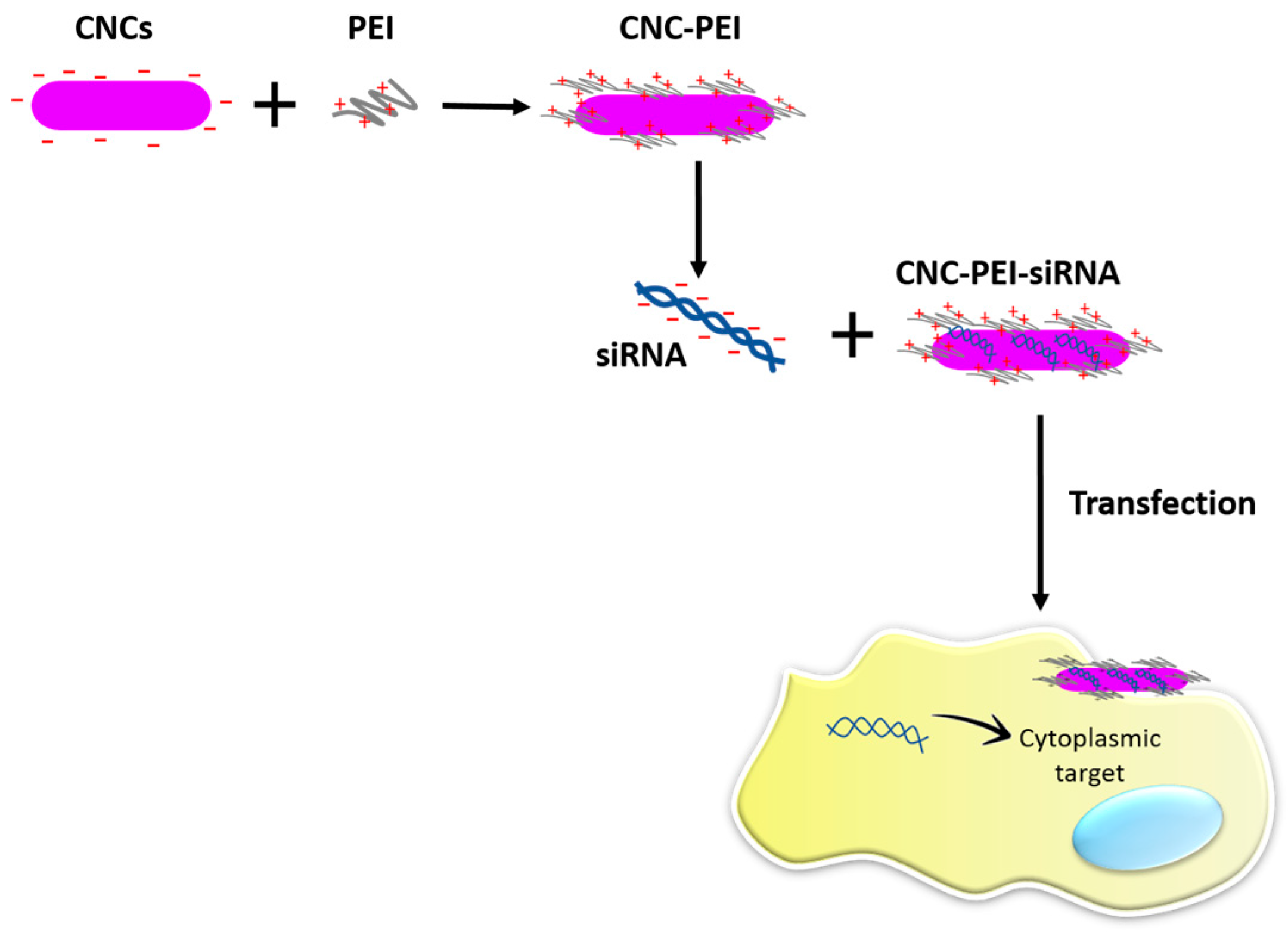
| CNC-PEI | siRNA | C2C12 murine myoblastic cells | Decrease of the cells growth (anti-proliferative) | Ndong Ntoutoume et al. [126] | |
| HEC-PEG | siRNA | Muco-adhesive properties | Furst et al. [128] | ||
| CMC/β-C/AA | Acyclovir | Swelling tests at different pH | Basic pH favors the maximal swelling of the CMC/β-C/AA releasing acyclovir in the small intestine | Malik et al. [131] |
| Type of Cellulose | Strategy | Effect | References |
|---|---|---|---|
| EC-MC | Delivery α-mangostin compound | Anti-acne effect | Pan-In et al. [133] |
| HA-CMC | HA-CMC soaked with Gelfoam | Hearing improvement without significant collateral effects | Ahn et al. [135] |
| HA-CMC | Artificial tears composed of hyaluronate 0.1% and CMC 0.5% | Improvement of the tear-film in eyes after cataract surgery | Mencucci et al. [136] |
| HPMC | Delivery oxymetazoline compound | Improvement of mucosal barrier in allergic rhinitis | Valerieva et al. [137] |
© 2017 by the authors. Licensee MDPI, Basel, Switzerland. This article is an open access article distributed under the terms and conditions of the Creative Commons Attribution (CC BY) license (http://creativecommons.org/licenses/by/4.0/).
Share and Cite
Halib, N.; Perrone, F.; Cemazar, M.; Dapas, B.; Farra, R.; Abrami, M.; Chiarappa, G.; Forte, G.; Zanconati, F.; Pozzato, G.; et al. Potential Applications of Nanocellulose-Containing Materials in the Biomedical Field. Materials 2017, 10, 977. https://doi.org/10.3390/ma10080977
Halib N, Perrone F, Cemazar M, Dapas B, Farra R, Abrami M, Chiarappa G, Forte G, Zanconati F, Pozzato G, et al. Potential Applications of Nanocellulose-Containing Materials in the Biomedical Field. Materials. 2017; 10(8):977. https://doi.org/10.3390/ma10080977
Chicago/Turabian StyleHalib, Nadia, Francesca Perrone, Maja Cemazar, Barbara Dapas, Rossella Farra, Michela Abrami, Gianluca Chiarappa, Giancarlo Forte, Fabrizio Zanconati, Gabriele Pozzato, and et al. 2017. "Potential Applications of Nanocellulose-Containing Materials in the Biomedical Field" Materials 10, no. 8: 977. https://doi.org/10.3390/ma10080977