Article

Disruption of Autographa Californica Multiple Nucleopolyhedrovirus ac111 Results in Reduced per os Infectivity in a Host-Dependent Manner

1
Department of Biology, Zhaoqing University, Zhaoqing 526061, China
2
State Key Laboratory of Biocontrol, Sun Yat-sen University, Guangzhou 510275, China
*
Author to whom correspondence should be addressed.
Viruses 2018, 10(10), 527; https://doi.org/10.3390/v10100527
Submission received: 12 August 2018 / Revised: 18 September 2018 / Accepted: 20 September 2018 / Published: 27 September 2018
(This article belongs to the Special Issue Baculovirus Advances and Applications)

Abstract

:
The Autographa californica multiple nucleopolyhedrovirus (AcMNPV) ac111 gene is highly conserved in lepidopteran-specific baculoviruses, and its function in the AcMNPV life cycle is still unknown. To investigate the function of ac111, an ac111-knockout AcMNPV (vAc111KO) was constructed through homologous recombination in Escherichia coli. Viral growth curve analysis and plaque assays showed that the deletion of ac111 had no effect on infectious budded virion production. Quantitative real-time polymerase chain reaction analysis confirmed that viral DNA replication was unaffected in the absence of ac111. Electron microscopy revealed that the ac111 deletion did not affect nucleocapsid assembly, occlusion-derived virion formation, or the embedding of occlusion-derived virions into the occlusion bodies. However, in vivo bioassays showed that although the deletion of ac111 did not affect the per os infectivity of AcMNPV in Spodoptera exigua larvae, it led to an approximately five-fold reduction in infectivity of AcMNPV in Trichoplusia ni larvae, and vAc111KO took approximately 21 h longer to kill Trichoplusia ni larvae than the wild-type viruses. Taken together, our results demonstrated that although ac111 is not essential for virus replication in vitro, it plays an important role in the per os infectivity of AcMNPV in a host-dependent manner.

1. Introduction

Baculoviruses are a family of rod-shaped, enveloped, double-stranded DNA viruses which are specifically pathogenic to invertebrate species, mainly belonging to the orders Lepidoptera, Hymenoptera, and Diptera [1]. Baculoviridae consists of Alphabaculovirus, Betabaculovirus, Gammabaculovirus, and Deltabaculovirus [2]. Based on phylogenetic analysis of the polyhedrin (polh) gene, Alphabaculovirus can be further subdivided into Group I and Group II nucleopolyhedroviruses (NPVs) [3]. Autographa californica multiple NPV (AcMNPV) is the archetype of the Baculoviridae family and the first baculovirus to be completely genome-sequenced [4]. The AcMNPV genome is approximately 134 kbp and encodes 154 potential open reading frames (ORFs) [4]. To date, more than 80 baculovirus genomes have been completely sequenced, and there are 38 genes conserved in all these genomes. These 38 common genes are considered to be the baculovirus core genes [1,5,6,7].
During the baculovirus infection cycle, two morphologically distinct but genetically identical progeny virion phenotypes, budded virions (BVs), and occlusion-derived virions (ODVs), are produced. Although BVs and ODVs are similar in their nucleocapsid structure, they differ in their origin and the composition of their envelopes, which reflects their distinct roles in the baculovirus life cycle [8,9]. In the early infection phase, newly assembled nucleocapsids egress from the nucleus, migrate across the cytoplasm, and bud from the plasma membrane to form BVs. BVs are responsible for spreading infections among susceptible insect tissues [10]. During the late phase of infection, ODVs are formed and are subsequently embedded in the paracrystalline protein matrix to form occlusion bodies (OBs). ODVs can initiate primary infection in the insect hosts [9,11].
The site of baculovirus primary infection is the midgut of host insects. Upon oral ingestion, the OBs are dissolved under alkaline conditions in the insect midgut, and they release ODVs, which then penetrate the peritrophic matrix and infect epithelial cells. A number of ODV envelope-associated proteins that play important roles in oral infectivity are designated as per os infectivity factors (PIFs), including P74, and PIF-1 to 8 [6,12,13,14,15,16,17,18,19,20,21,22,23]. In addition, the Ac108 homolog in Spodoptera frugiperda NPV, Sf58, which is only conserved in lepidopteran baculoviruses, has been shown to be a PIF of lepidopteran-infecting baculoviruses [24]. Moreover, some other less-conserved proteins, such as Ac18, Ac32, ODV-E66, Ac114, Ac124, Ac145, and Ac150 have been shown to be involved in the oral infection of AcMNPV [25,26,27,28,29,30,31]. ac18 is a highly conserved gene in lepidopteran NPVs, though ac18 deletion did not affect viral propagation in vitro or AcMNPV infectivity for Trichoplusia ni larvae; however, the LT50 was 24 h longer for the ac18 knockout virus in T. ni larvae compared to the wild-type virus [28]. Previous studies have also demonstrated that although ac114 and ac124 are nonessential for AcMNPV replication in vitro, ac114 and ac124 selectively affect viral infectivity and viral killing speed, respectively, of AcMNPV in S. exigua larvae [27,29]. Therefore, baculovirus per os infection is a complex process involving numerous virus proteins coordinating the establishment of the primary infection.
The AcMNPV orf111 (ac111) is located between AcMNPV nucleotides (nt) 101,624 and 101,827 [4], and its homologs are present in all sequenced Group I NPVs, certain Group II NPVs, and granuloviruses (GVs). Transcriptional analysis identified that ac111 is transcribed as an early gene [32]. Recently, the Ac111 homolog in Bombyx mori NPV, Bm93, has been found not to be essential for virus replication [33]. However, currently the role of ac111 in the AcMNPV life cycle is still unknown.
In this study, an ac111 knockout AcMNPV (vAc111KO) was constructed to investigate the role of ac111 in the AcMNPV life cycle. We found that the ac111 deletion had no significant effect on infectious BV production, viral DNA replication, or viral morphogenesis. However, the oral infectivity bioassays showed that, in T. ni larvae, the infectivity of vAc111KO was reduced by about five times relative to the wild-type virus, and the mutant virus took approximately 21 h longer to kill these larvae. Interestingly, the deletion of ac111 did not affect the infectivity of AcMNPV in another lepidopteran host, S. exigua larvae. Therefore, our results suggest that Ac111 plays an important role in virus per os infectivity in a host-dependent manner.

2. Materials and Methods

2.1. Computer-Assisted Analysis

HHpred analysis [34], NCBI’s Conserved Domain Database [35], Phrye [36], and SMART [37] were used to predict the conserved domains of Ac111. The Position-Specific Iterated BLAST algorithm was used to search the homologues of Ac111 in the NCBI database. ClustalX [38] and GeneDoc [39] were used to perform and edit multiple sequence alignments, respectively.

2.2. Cell Lines, Viruses, and Insect Larvae

The Sf9 cell line and Se301 cell line [40], which are derived from the fall armyworm Spodoptera frugiperd and S. exigua, respectively, were cultured at 27 °C in TNM-FH medium (Invitrogen, Carlsbad, CA, USA) supplemented with 10% fetal bovine serum, penicillin (100 μg/mL), and streptomycin (30 μg/mL). The bMON14272 bacmid (Invitrogen, Carlsbad, CA, USA), which contains the AcMNPV genome, was propagated in DH10B cells as previously described [41]. T. ni and S. exigua larvae were reared on an artificial diet at 28 °C [42]. BV titers were determined using a 50% tissue culture infective dose (TCID50) endpoint dilution assay in Sf9 cells or Se301 cells, as previously described [43].

2.3. Construction of an ac111-Knockout Virus

Employing the bacmid bMON14272, an ac111-knockout AcMNPV bacmid (bAc111KO) was generated through ET homologous recombination in Escherichia coli, as previously described [44]. Briefly, a 400-bp fragment that was homologous to the 5′ region of the ac111 ORF (AcMNPV nt 101,822 to 102,221) was PCR-amplified from bMON14272 using the primers ac111PF1/ac111PR1 (the PCR primers used in this study are listed in Table 1). A 340-bp fragment homologous to the 3′ region of the ac111 ORF (AcMNPV nt 101,402 to 101,741) was PCR-amplified from bMON14272 using the primers ac111PF2/ac111PR2. The 5′- and 3′-PCR products were digested with PstI/HindIII and SacI/BamHI, respectively, and ligated into pUC18-Cm [45], which contains a chloramphenicol resistant (Cm) gene cassette to generate pUC18-111US-Cm-111DS. The plasmid was then digested with SacI and HindIII to obtain a linear 1778-bp fragment in which the Cm gene cassette was flanked by ac111 5′- and 3′-regions. Homologous recombination between the deletion region of ac111 (AcMNPV nt 101,742 to 101,821) and the Cm cassette in the linear fragment was performed to obtain an ac111-knockout bacmid, bAc111KO. The replacement in bAc111KO was confirmed by PCR and sequencing.
To facilitate the detection of viral infection, the pFB1-PH-GFP donor plasmid, containing the AcMNPV polh gene and the enhanced green fluorescence protein (egfp; referred to as gfp in the present study) gene, was transformed into electrocompetent DH10B cells harboring the bAc111KO bacmid and the pMON7124 helper plasmid as previously described [45], to generate the ac111-knockout virus, vAc111KO. To generate the ac111-repaired virus, vAc111:HA, the donor plasmid pFB1-Ac111:HA-PH-GFP was constructed using the following steps. The 531-bp DNA fragment containing the promoter and the ORF of ac111 tagged with an HA epitope prior to the stop codon was PCR amplified from bMON14272 using the primers ac111PF3/ac111PR3, and it was cloned into the SacI/BamHI-digested pUC18-SV40 plasmid [46] to construct the pUC18-Ac111:HA plasmid. The Ac111:HA fragment was digested from pUC18-Ac111:HA with SacI/XbaI, and subcloned into SacI/XbaI-digested pFB1-PH-GFP to generate the donor plasmid pFB1-Ac111:HA-PH-GFP. The pFB1-Ac111:HA-PH-GFP was then transformed into electrocompetent DH10B cells harboring the bAc111KO bacmid and the helper plasmid pMON7124 to generate the ac111-repaired virus, vAc111:HA. Similarly, vAcWT, a wild type virus, was also constructed by inserting polh and gfp genes into bMON14272.

2.4. Viral Propagation Analysis

Sf9 cells (1.0 × 10⁶) were transfected with 1.0 μg bacmid DNA using the Cellfectin liposome reagent (Invitrogen Life Technologies, Carlsbad, CA, USA). For infection, Sf9 cells (1.0 × 10⁶) or Se301 cells (1.0 × 10⁶) were infected with BVs at a multiplicity of infection (MOI) of 5 TCID50/cell. At the designated time points post transfection (p.t.) or post infection (p.i.), the supernatants containing BVs were harvested and titered. To further evaluate the effect of the ac111 deletion on BV production, a plaque assay was performed as described previously [43,47].

2.5. Real-Time PCR (qPCR) Analysis

qPCR analysis was performed to assess the effect of the ac111 deletion on viral DNA synthesis as described previously [7,46].

2.6. Transmission Electron Microscopy (TEM)

1.0 × 10⁶ Sf9 cells were transfected with 1.0 μg bacmid DNA of vAc111KO, vAc111:HA, or vAcWT. At 72 h p.t., the cells were dislodged with a cell scraper, pelleted at 1000 × g for 10 min, and prepared for TEM, as previously described [7]. The samples were observed with a JEOL JEM-1400 transmission electron microscope (Tokyo, Japan) at an accelerating voltage of 120 kV.

2.7. Bioassays in T. ni and S. exigua Larvae

In order to determine whether the ac111 deletion had any effect on the infectivity of AcMNPV in insect larvae, bioassays were performed using T. ni or S. exigua larvae, as described previously [28]. Briefly, OBs and BVs were obtained from vAc111KO-, vAc111:HA-, or vAcWT-infected Sf9 cells, as previously described [48]. Different doses of BVs were hemocoelically injected into fourth-instar T. ni or S. exigua larvae to examine the infectivity of BVs in vivo. H2O was used as a blank control. For the oral infectivity bioassays, the OBs of vAc111KO, vAc111:HA, or vAcWT purified from infected Sf9 cells, were resuspended in double-distilled water and administered to newly molted T. ni or S. exigua larvae via oral inoculation. To determine the median lethal dose (LD50), 1 μL water (control) or 1 μL OB suspensions of each virus was administered to a small piece of diet of newly molted third-instar T. ni or S. exigua larvae, respectively. For each virus in T. ni, the doses were 9, 30, 90, 3 × 10², and 9 × 10² OBs per larva. For each virus in S. exigua, the doses were 9 × 10², 3 × 10³, 9 × 10³, 3 × 10⁴, and 9 × 10⁴ OBs per larva. Larvae were reared individually in 24-well plates until all of the diet was consumed, then fresh diet was added. Only the larvae that completely ingested the diet with OBs within 12 h were further reared and monitored daily, until all larvae had either pupated or died. The median lethal time (LT50) was determined using the droplet feeding method [49]. The newly molted third-instar T. ni or fourth-instar S. exigua larvae were starved for 6 h, and then they were fed with 1 μL OB suspensions of each virus with concentrations of 1 × 10⁷ or 1 × 10⁸ OBs/mL, respectively. For each virus, the final mortalities were about 99% or 86% in T. ni or S. exigua, respectively. The larvae that have ingested virus were reared individually and the mortality was recorded every 12 h until all larvae had either pupated or died. The experiments with each dose in both oral and injection treatments were performed three times (30 larvae per dose). The LD50 value and the LT50 value were determined by probit analysis and the Kaplan-Meier estimator, respectively.

3. Results

3.1. Sequence Analysis of Ac111 and Its Homologues

Homologues of ac111 are present in lepidopteran-specific baculoviruses. Sequence-based searches have shown that Ac111 belongs to the Baculo-8kDa superfamily with no homology to other identified functional domains. The alignment of Ac111 orthologs from several selected lepidopteran-specific baculovirus groups, including AcMNPV, Hyphantria cunea NPV (HcNPV), Orgyia pseudotsugata MNPV (OpMNPV), Rachiplusia ou MNPV (RoMNPV), Euproctis pseudoconspersa NPV (EupsNPV), Chrysodeixis chalcites NPV (ChchNPV), Helicoverpa armigera NPV (HearNPV), Lymantria dispar MNPV (LdMNPV), Xestia c-nigrum GV (XecnGV), and Spodoptera litura GV (SpliGV), revealed that a T-T/S-L-H/Y-X2-N motif (where X was any other amino acid) was conserved in all Ac111 homologues (Figure 1). Currently, no information about this motif in the function of ac111 is available and further studies need to be undertaken to determine its importance.

3.2. Construction of an ac111-Knockout AcMNPV

Based on AcMNPV transcriptomics [32], the 5′ transcription start site of ac110 is located 38 nt downstream of the 3′ end of ac111, and the 5′ transcription start site of ac112 is located 59 nt upstream of the initiation codon ATG of ac111. To investigate the role of Ac111 in the baculovirus life cycle, the ac111-knockout bacmid, bAc111KO, was constructed via the λ Red recombination system, as described previously [7]. In bAc111KO bacmid, 6 nt of the 5′-end and 118 nt of the 3′-end of ac111 ORF were retained to avoid affecting the transcription of ac110 and ac112; an 80-bp region of ac111 was deleted and replaced with a 1038-bp Cm cassette (Figure 2A).
The absence of ac111 and the replacement with the Cm gene in AcMNPV bacmid were confirmed by PCR analysis. As shown in Figure 2B, the primers ac111PF4/ac111PR2 produced no PCR product in bAc111KO, while a 420-bp fragment was amplified in AcMNPV bacmid bMON14272. Similarly, the primers ac111PF1/ac111PR4 produced no PCR product in bAc111KO but produced a 480-bp fragment in AcMNPV bacmid bMON14272. The primers CmF/CmR produced no PCR product in AcMNPV bacmid bMON14272, but a 1038-bp fragment was amplified in bAc111KO. Finally, the primers ac111PF1/ac111PR2 produced a 1778-bp fragment in bAc111KO but an 820-bp fragment in AcMNPV bacmid bMON14272. These results demonstrated that the target deletion region of ac111 was successfully replaced by the Cm gene.
In order to facilitate the observation of the viral infection progress, the polh gene of AcMNPV and the gfp gene were inserted into the polh locus of bAc111KO via Tn7-mediated transposition to construct the ac111-knockout AcMNPV, vAc111KO, as described previously [45]. To confirm the phenotype observed in vAc111KO resulted from the deletion of ac111, a rescue virus, vAc111:HA, was constructed by inserting the ac111 gene under the control of its native promoter with an HA tag prior to the ac111 stop codon, along with the polh and gfp genes, into the polh locus of bAc111KO (Figure 2A). Similarly, a wild-type AcMNPV control, vAcWT, was generated by inserting the polh and gfp genes into bMON14272 (Figure 2A).

3.3. Ac111 is Not Required forBV Production

To study the effect of ac111 deletion on viral replication, Sf9 cells were transfected with vAc111KO, vAc111:HA, or vAcWT bacmid DNA, and virus infections were monitored using a fluorescence microscope. As shown in Figure 3A, similar amounts of GFP-positive cells were observed in the three groups at 24 h p.t., indicating comparable transfection efficiencies. By 72 h p.t., almost all vAc111KO-, vAc111:HA-, and vAcWT-transfected cells showed GFP fluorescence, indicating the infection had spread from the cells initially transfected. Light microscopy showed that OBs were produced in the three groups, and there were no obvious differences in the OB production (Figure 3A), suggesting similar infection progress among all three viruses.
To further evaluate the effect of the ac111 deletion on virus propagation, viral growth curve analysis was performed. Sf9 cells transfected with vAc111KO, vAc111:HA, or vAcWT or infected with BVs showed a normal and steady increase in virus production and similar virus growth kinetics (Figure 3B). In Se301 cells infected with vAc111KO, vAc111:HA, or vAcWT, the three virus growth curves were similar [50]. In addition, no obvious differences were observed in plaque sizes in Sf9 cells transfected with vAc111KO, vAc111:HA, or vAcWT [50].
Taken together, these results demonstrated that the deletion of ac111 had no apparent effect on viral replication in Sf9 cells or Se301 cells.

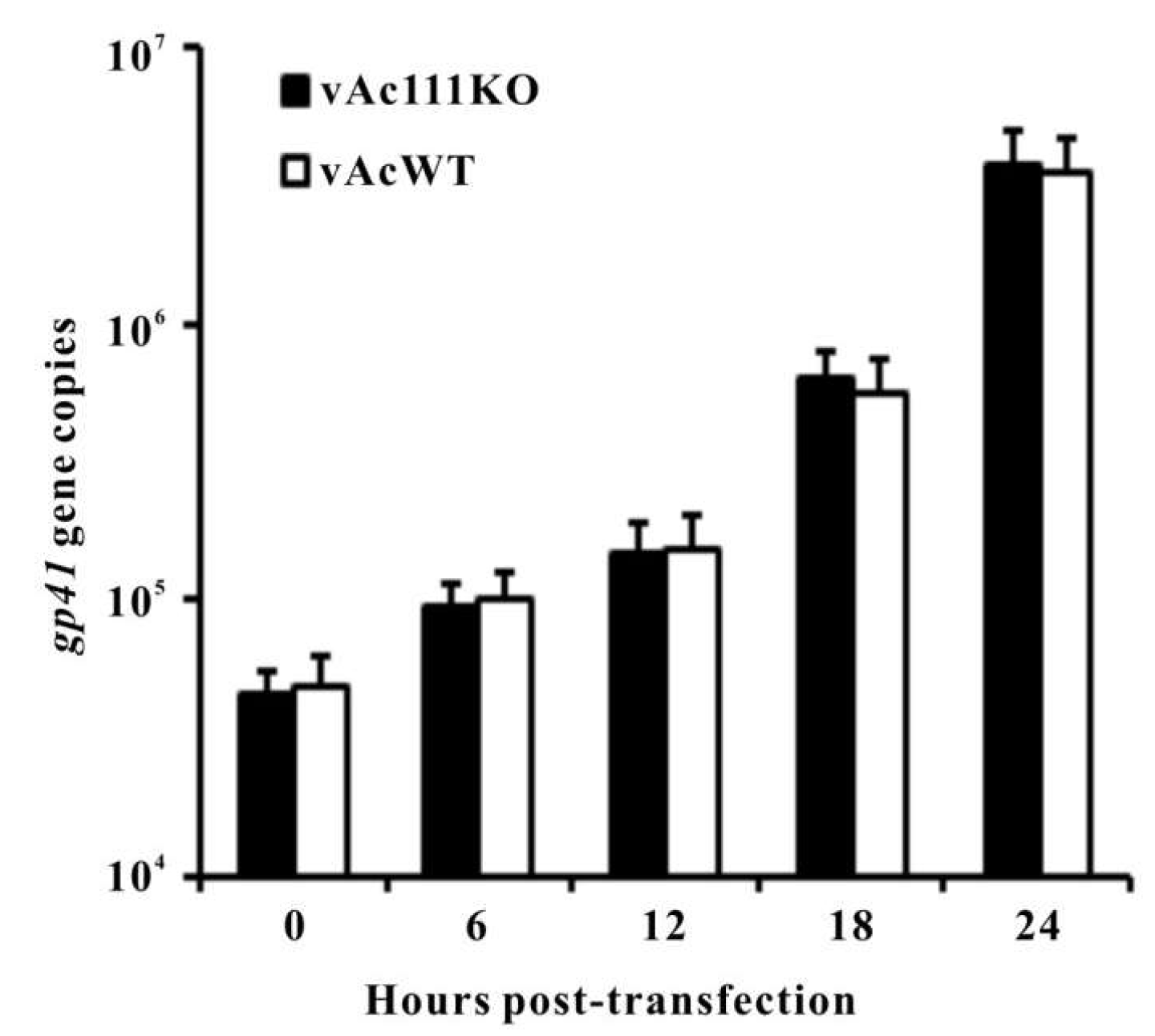
3.4. Ac111 is Not Required for Viral DNA Replication

To assess whether ac111 deletion leads to a defect in viral DNA replication, qPCR was performed to measure the levels of viral DNA synthesis in vAc111KO-transfected cells during a single replication cycle. vAcWT was used as a positive control. Total cellular DNA was isolated from vAc111KO- or vAcWT-transfected cells at the designated time points, and treated with DpnI to eliminate all input bacmid DNA. As shown in Figure 4, viral DNA replication levels of vAc111KO-transfected cells and vAcWT-transfected cells were comparable throughout a 24-h time course. The qPCR results demonstrated that the deletion of ac111 did not affect the onset or levels of viral DNA synthesis.

3.5. Ac111 is Not Essential for Virus Morphogenesis

Electron microscopy observations were performed using thin sections of vAc111KO-, vAcWT-, and vAc111:HA-transfected Sf9 cells at 72 h p.t., to further investigate whether the ac111 deletion had any effect on virus morphogenesis (Figure 5). In vAc111:HA-transfected Sf9 cells, the typical characteristics were similar to those of vAcWT-transfected cells, including a well-defined virogenic stroma (VS) enriched with electron-dense, rod-shaped nucleocapsids in the center of the nucleus (Figure 5A), and developing OBs embedding numerous mature ODVs in the ring zone (Figure 5B). The cells transfected with vAc111KO exhibited morphologically indistinguishable characteristics to those of vAc111:HA and vAcWT (Figure 5C,D). These results indicated that the deletion of ac111 did not affect virus morphogenesis.

3.6. Ac111 Plays a Role in Oral Infectivity in a Host-Dependent Manner

To determine the effect of ac111 deletion on the infectivity of AcMNPV in insect larvae, bioassays with BVs and OBs grown in Sf9 cells were performed in two host species, T. ni and S. exigua. Intrahemocoelic injection of vAc111KO BV supernatant into fourth-instar T. ni at 1 TCID50 units/larva produced 67% mortality, which was not significantly different from that of vAc111:HA (63%) or vAcWT (63%) (Student’s t-test, p> 0.05) (Table 2). Intrahemocoelic injection of vAc111KO, vAc111:HA or vAcWT BV supernatant into fourth-instar S. exigua at 2 TCID50 units/larva produced 70%, 67%, or 70% mortalities, respectively, and there was no significant difference among the mortalities (Student’s t-test, p> 0.05) (Table 2). However, bioassays with OBs revealed that ac111 affected the oral infectivity of AcMNPV in the two host species in a different way. Bioassays with OBs in T. ni larvae showed that, the LD50 values of vAc111:HA and vAcWT were 59 and 54 OBs/larva, respectively with overlapping 95% confidence interval indicating they were not significantly different, the LD50 of vAc111KO (300 OBs/larva) was approximately five times more than that of vAcWT (Table 2). The LT50 value of vAc111:HA (86 h) was comparable to that of vAcWT (85 h), but the LT50 value of vAc111KO (106 h) was 21 h longer than that of vAcWT (Table 2). Therefore, the deletion of ac111 reduced the infectivity and killing speed of AcMNPV in T. ni larvae. However, in S. exigua larvae, no significant differences in infectivity or killing speed were observed among vAc111KO, vAc111:HA and vAcWT, with the LD50 values being 5006, 4650, and 4535 OBs/larva, and the LT50 values being 148 h, 141 h and 140 h, respectively (as determined by overlapping 95% confidence interval) (Table 2). This suggests that the deletion of ac111 did not impact per os infectivity of AcMNPV in S. exigua larvae.
Taken together, the bioassays demonstrated that ac111 plays a role in primary oral infection of AcMNPV and the effect is host dependent.

4. Discussion

ac111 is one of the highly conserved lepidopteran-specific baculovirus genes, but its role in the AcMNPV replication remains unknown. In this study, we demonstrated that ac111 is important for AcMNPV to efficiently establish per os infection in T. ni larvae specifically, while is dispensable for per os infectivity of AcMNPV in S. exigua larvae, thus providing evidence for its important role as a conserved baculovirus gene.
To investigate the function of ac111 in the AcMNPV life cycle, an ac111-knockout virus was generated and characterized. We found that ac111 was dispensable for virus replication in vitro as evidenced by indistinguishable characteristics such as a normal viral growth curve, normal plaque, steady viral DNA replication and normal virus morphogenesis, compared to those of vAcWT. These results were consistent with the study on Bm93 [33]. Bioassays showed that there was no significant difference between the LD50 and LT50 of vAc111KO OBs and those of vAcWT in S. exigua larvae. However, the LD50 of vAc111KO OBs was approximate five times higher than that of vAcWT OBs in T. ni larvae, and the LT50 of vAc111KO was 21 h longer than that of vAcWT in T. ni larvae. Furthermore, the deletion of ac111 did not affect BV infectivity through intrahemocoelic injection in T. ni and S. exigua larvae. Taken together, these results indicated that ac111 affects the per os infectivity of AcMNPV in a host-dependent manner.
Of the 154 putative ORFs encoded in the AcMNPV genome, the function of more than 70% of these ORFs has been determined. While some genes were found to be nonessential and could be deleted without affecting virus replication, some genes are essential for virus replication in vitro and participate in critical steps in the virus life cycle, such as DNA replication, early/late gene transcription, or nucleocapsid assembly. There are some other genes that are dispensable for virus replication in vitro but function during oral infection of insect larvae. Genes belonging to this category include the nine pifs and several virus virulence-related genes, such as ac111, ac145, and ac150. Baculoviruses initiate infection orally when their insect hosts consume and digest OBs in the alkaline pH of the midgut, releasing ODVs to infect the midgut epithelial cells. A number of viral membrane proteins, including the PIF proteins, have been identified to be essential for this process. Considering the pivotal role of PIFs, these proteins are encoded by baculovirus core genes [6], indicating that they play pivotal roles in per os infection of all baculoviruses and thus have been conserved during baculovirus evolution. Other virus virulence-related genes are only conserved in certain baculoviruses, implying that they might function distinctly compared to PIFs in a host dependent manner and facilitate the efficient establishment of baculovirus infection in specific hosts.
Similar to ac111, previous studies have demonstrated that two AcMNPV genes, ac145 and ac150 affect oral infectivity in a host dependent manner [26]. ac145 and ac150 encode proteins related to the 11K proteins with a characteristic C6 motif of conserved cysteine residues in a well-defined spacing pattern. Previous studies have found that the deletion of ac145 led to a six-fold drop in infectivity of AcMNPV in T. ni larvae but not in Heliothis virescens larvae [26]. On the other hand, the deletion of ac150 had no effect on infectivity of AcMNPV in either host [26]. However, the deletion of both ac145 and ac150 significantly reduced the infectivity of AcMNPV (39-fold) in H. virescens larvae [26]. In another study, significantly more OBs were required per LD50 for ac150 deletion AcMNPVcompared with wild-type virus in H. virescens, S. exigua, and T. ni larvae (4.1-, 5.6-, and 18-fold increases, respectively), suggesting that Ac150 indeed had an enhancing effect on per os infection and that the effect was host-specific [31]. In this study, we showed that ac111 specifically affected viral infectivity and killing speed of AcMNPV in T. ni larvae but is dispensable in S. exigua larvae. Therefore, although AcMNPV could establish infection in T. ni, S. exigua, and H. virescens larvae, differences might exist that subtly affect the fitness of AcMNPV in these different insect hosts. It is intriguing to speculate that a group of proteins in AcMNPV, such as Ac111, Ac145, and Ac150, might specifically affect the infectivity of a virus in different hosts. We also cannot rule out the possibility that these viral proteins might interact with each other or with specific host proteins, and these interactions may eventually determine the host tropism of AcMNPV. Further studies are needed to verify this assumption.
Taken together, this study has shown that ac111 was not required for AcMNPV replication in vitro, but it played a pivotal role in the viral infectivity in vivo in T. ni but not in S. exigua larvae. Further experiments are needed to unravel the specific role of ac111 during AcMNPV infection in T. ni larvae. These studies will likely shed light on our understanding of the mechanism of baculovirus–insect interactions, to ascertain the fitness of baculoviruses in different hosts.

Author Contributions

S.L. conceived and designed the experiments; L.L. performed the viral growth curve analysis, S.L. and H.Z. performed all other experiments; S.L. wrote the manuscript; W.L. provided manuscript revision.

Funding

This work was supported by the National Natural Science Foundation of China (31501705) and the Provincial Natural Science Foundation of Guangdong (2014A030313664).

Acknowledgments

We are grateful to Professor Kai Yang of Sun Yat-sen University, China, for valuable suggestions.

Conflicts of Interest

The authors declare no conflict of interest.

References

  1. Herniou, E.A.; Olszewski, J.A.; Cory, J.S.; O’Reilly, D.R. The genome sequence and evolution of baculoviruses. Annu. Rev. Entomol. 2003, 48, 211–234. [Google Scholar] [CrossRef] [PubMed]
  2. Jehle, J.A.; Blissard, G.W.; Bonning, B.C.; Cory, J.S.; Herniou, E.A.; Rohrmann, G.F.; Theilmann, D.A.; Thiem, S.M.; Vlak, J.M. On the classification and nomenclature of baculoviruses: A proposal for revision. Arch. Virol. 2006, 151, 1257–1266. [Google Scholar] [CrossRef] [PubMed] [Green Version]
  3. De Zanotto, A.P.M.; Kessing, B.D.; Maruniak, J.E. Phylogenetic interrelationships among baculoviruses: Evolutionary rates and host associations. J. Invertebr. Pathol. 1993, 62, 147–164. [Google Scholar] [CrossRef]
  4. Ayres, M.D.; Howard, S.C.; Kuzio, J.; Lopez-Ferber, M.; Possee, R.D. The complete DNA sequence of Autographa californica nuclear polyhedrosis virus. Virology 1994, 202, 586–605. [Google Scholar] [CrossRef] [PubMed]
  5. Garavaglia, M.J.; Miele, S.A.; Iserte, J.A.; Belaich, M.N.; Ghiringhelli, P.D. The ac53, ac78, ac101, and ac103 genes are newly discovered core genes in the family Baculoviridae. J. Virol. 2012, 86, 12069–12079. [Google Scholar] [CrossRef] [PubMed]
  6. Javed, M.A.; Biswas, S.; Willis, L.G.; Harris, S.; Pritchard, C.; van Oers, M.M.; Donly, B.C.; Erlandson, M.A.; Hegedus, D.D.; Theilmann, D.A. Autographa californica multiple nucleopolyhedrovirus AC83 is a per os infectivity factor (PIF) protein required for occlusion-derived virus (ODV) and budded virus nucleocapsid assembly as well as assembly of the PIF complex in ODV envelopes. J. Virol. 2017, 91. [Google Scholar] [CrossRef] [PubMed]
  7. Yuan, M.; Huang, Z.; Wei, D.; Hu, Z.; Yang, K.; Pang, Y. Identification of Autographa californica nucleopolyhedrovirus ac93 as a core gene and its requirement for intranuclear microvesicle formation and nuclear egress of nucleocapsids. J. Virol. 2011, 85, 11664–11674. [Google Scholar] [CrossRef] [PubMed]
  8. Braunagel, S.C.; Summers, M.D. Autographa californica nuclear polyhedrosis virus, PDV, and ECV viral envelopes and nucleocapsids: Structural proteins, antigens, lipid and fatty acid profiles. Virology 1994, 202, 315–328. [Google Scholar] [CrossRef] [PubMed]
  9. Slack, J.; Arif, B.M. The baculoviruses occlusion-derived virus: Virion structure and function. Adv. Virus Res. 2007, 69, 99–165. [Google Scholar] [PubMed]
  10. Blissard, G.W.; Wenz, J.R. Baculovirus gp64 envelope glycoprotein is sufficient to mediate pH-dependent membrane fusion. J. Virol. 1992, 66, 6829–6835. [Google Scholar] [PubMed]
  11. Braunagel, S.C.; Summers, M.D. Molecular biology of the baculovirus occlusion-derived virus envelope. Curr. Drug Targets 2007, 8, 1084–1095. [Google Scholar] [CrossRef] [PubMed]
  12. Fang, M.; Nie, Y.; Harris, S.; Erlandson, M.A.; Theilmann, D.A. Autographa californica multiple nucleopolyhedrovirus core gene ac96 encodes a per os infectivity factor (pif-4). J. Virol. 2009, 83, 12569–12578. [Google Scholar] [CrossRef] [PubMed]
  13. Fang, M.; Nie, Y.; Wang, Q.; Deng, F.; Wang, R.; Wang, H.; Vlak, J.M.; Chen, X.; Hu, Z. Open reading frame 132 of Helicoverpa armigera nucleopolyhedrovirus encodes a functional per os infectivity factor (PIF-2). J. Gen. Virol. 2006, 87, 2563–2569. [Google Scholar] [CrossRef] [PubMed]
  14. Faulkner, P.; Kuzio, J.; Williams, G.V.; Wilson, J.A. Analysis of p74, a PDV envelope protein of Autographa californica nucleopolyhedrovirus required for occlusion body infectivity in vivo. J. Gen. Virol. 1997, 78, 3091–3100. [Google Scholar] [CrossRef] [PubMed]
  15. Harrison, R.L.; Sparks, W.O.; Bonning, B.C. Autographa californica multiple nucleopolyhedrovirus ODV-E56 envelope protein is required for oral infectivity and can be substituted functionally by Rachiplusia ou multiple nucleopolyhedrovirus ODV-E56. J. Gen. Virol. 2010, 91, 1173–1182. [Google Scholar] [CrossRef] [PubMed]
  16. Jiantao, L.; Zhu, L.; Zhang, S.; Deng, Z.; Huang, Z.; Yuan, M.; Wu, W.; Yang, K. The Autographa californica multiple nucleopolyhedrovirus ac110 gene encodes a new per os infectivity factor. Virus Res. 2016, 221, 30–37. [Google Scholar] [CrossRef] [PubMed]
  17. Kikhno, I.; Gutierrez, S.; Croizier, L.; Croizier, G.; Ferber, M.L. Characterization of pif, a gene required for the per os infectivity of Spodoptera littoralis nucleopolyhedrovirus. J. Gen. Virol. 2002, 83, 3013–3022. [Google Scholar] [CrossRef] [PubMed]
  18. Nie, Y.; Fang, M.; Erlandson, M.A.; Theilmann, D.A. Analysis of the Autographa californica multiple nucleopolyhedrovirus overlapping gene pair lef3 and ac68 reveals that ac68 is a per os infectivity factor and that lef3 is critical, but not essential, for virus replication. J. Virol. 2012, 86, 3985–3994. [Google Scholar] [CrossRef] [PubMed]
  19. Ohkawa, T.; Washburn, J.O.; Sitapara, R.; Sid, E.; Volkman, L.E. Specific binding of Autographa californica M nucleopolyhedrovirus occlusion-derived virus to midgut cells of Heliothis virescens larvae is mediated by products of pif genes ac119 and ac022 but not by ac115. J. Virol. 2005, 79, 15258–15264. [Google Scholar] [CrossRef] [PubMed]
  20. Pijlman, G.P.; Pruijssers, A.J.; Vlak, J.M. Identification of pif-2, a third conserved baculovirus gene required for per os infection of insects. J. Gen. Virol. 2003, 84, 2041–2049. [Google Scholar] [CrossRef] [PubMed]
  21. Sparks, W.O.; Harrison, R.L.; Bonning, B.C. Autographa californica multiple nucleopolyhedrovirus ODV-E56 is a per os infectivity factor, but is not essential for binding and fusion of occlusion-derived virus to the host midgut. Virology 2011, 409, 69–76. [Google Scholar] [CrossRef] [PubMed]
  22. Xiang, X.; Chen, L.; Guo, A.; Yu, S.; Yang, R.; Wu, X. The Bombyx mori nucleopolyhedrovirus (BmNPV) ODV-E56 envelope protein is also a per os infectivity factor. Virus Res. 2011, 155, 69–75. [Google Scholar] [CrossRef] [PubMed]
  23. Zhu, S.; Wang, W.; Wang, Y.; Yuan, M.; Yang, K. The baculovirus core gene ac83 is required for nucleocapsid assembly and per os infectivity of Autographa californica nucleopolyhedrovirus. J. Virol. 2013, 87, 10573–10586. [Google Scholar] [CrossRef] [PubMed]
  24. Simon, O.; Palma, L.; Williams, T.; Lopez-Ferber, M.; Caballero, P. Analysis of a naturally-occurring deletion mutant of Spodoptera frugiperda multiple nucleopolyhedrovirus reveals sf58 as a new per os infectivity factor of lepidopteran-infecting baculoviruses. J. Invertebr. Pathol. 2012, 109, 117–126. [Google Scholar] [CrossRef] [PubMed]
  25. Detvisitsakun, C.; Cain, E.L.; Passarelli, A.L. The Autographa californica M nucleopolyhedrovirus fibroblast growth factor accelerates host mortality. Virology 2007, 365, 70–78. [Google Scholar] [CrossRef] [PubMed]
  26. Lapointe, R.; Popham, H.J.; Straschil, U.; Goulding, D.; O’Reilly, D.R.; Olszewski, J.A. Characterization of two Autographa californica nucleopolyhedrovirus proteins, Ac145 and Ac150, which affect oral infectivity in a host-dependent manner. J. Virol. 2004, 78, 6439–6448. [Google Scholar] [CrossRef] [PubMed]
  27. Liang, C.; Lan, D.; Zhao, S.; Liu, L.; Xue, Y.; Zhang, Y.; Wang, Y.; Chen, X. The Ac124 protein is not essential for the propagation of Autographa californica multiple nucleopolyhedrovirus, but it is a viral pathogenicity factor. Arch. Virol. 2015, 160, 275–284. [Google Scholar] [CrossRef] [PubMed]
  28. Wang, Y.; Wu, W.; Li, Z.; Yuan, M.; Feng, G.; Yu, Q.; Yang, K.; Pang, Y. ac18 is not essential for the propagation of Autographa californica multiple nucleopolyhedrovirus. Virology 2007, 367, 71–81. [Google Scholar] [CrossRef] [PubMed]
  29. Wei, W.; Zhou, Y.; Lei, C.; Sun, X. Autographa californica multiple nucleopolyhedrovirus orf114 is not essential for virus replication in vitro, but its knockout reduces per os infectivity in vivo. Virus Genes 2012, 45, 360–369. [Google Scholar] [CrossRef] [PubMed]
  30. Xiang, X.; Chen, L.; Hu, X.; Yu, S.; Yang, R.; Wu, X. Autographa californica multiple nucleopolyhedrovirus odv-e66 is an essential gene required for oral infectivity. Virus Res. 2011, 158, 72–78. [Google Scholar] [CrossRef] [PubMed]
  31. Zhang, J.H.; Ohkawa, T.; Washburn, J.O.; Volkman, L.E. Effects of Ac150 on virulence and pathogenesis of Autographa californica multiple nucleopolyhedrovirus in noctuid hosts. J. Gen. Virol. 2005, 86, 1619–1627. [Google Scholar] [CrossRef] [PubMed] [Green Version]
  32. Chen, Y.R.; Zhong, S.; Fei, Z.; Hashimoto, Y.; Xiang, J.Z.; Zhang, S.; Blissard, G.W. The transcriptome of the baculovirus Autographa californica multiple nucleopolyhedrovirus in Trichoplusia ni cells. J. Virol. 2013, 87, 6391–6405. [Google Scholar] [CrossRef] [PubMed]
  33. Ono, C.; Kamagata, T.; Taka, H.; Sahara, K.; Asano, S.; Bando, H. Phenotypic grouping of 141 BmNPVs lacking viral gene sequences. Virus Res. 2012, 165, 197–206. [Google Scholar] [CrossRef] [PubMed] [Green Version]
  34. Soding, J.; Biegert, A.; Lupas, A.N. The HHpred interactive server for protein homology detection and structure prediction. Nucl. Acids Res. 2005, 33, W244–W248. [Google Scholar] [CrossRef] [PubMed]
  35. Marchler-Bauer, A.; Anderson, J.B.; Derbyshire, M.K.; DeWeese-Scott, C.; Gonzales, N.R.; Gwadz, M.; Hao, L.; He, S.; Hurwitz, D.I.; Jackson, J.D.; et al. CDD: A conserved domain database for interactive domain family analysis. Nucl. Acids Res. 2007, 35, D237–D240. [Google Scholar] [CrossRef] [PubMed]
  36. Kelley, L.A.; Sternberg, M.J. Protein structure prediction on the Web: A case study using the Phyre server. Nat. Protoc. 2009, 4, 363–371. [Google Scholar] [CrossRef] [PubMed]
  37. Schultz, J.; Milpetz, F.; Bork, P.; Ponting, C.P. SMART, a simple modular architecture research tool: Identification of signaling domains. Proc. Natl. Acad. Sci.USA 1998, 95, 5857–5864. [Google Scholar] [CrossRef] [PubMed] [Green Version]
  38. Larkin, M.A.; Blackshields, G.; Brown, N.P.; Chenna, R.; McGettigan, P.A.; McWilliam, H.; Valentin, F.; Wallace, I.M.; Wilm, A.; Lopez, R.; et al. Clustal W and clustal X version 2.0. Bioinformatics 2007, 23, 2947–2948. [Google Scholar] [CrossRef] [PubMed]
  39. Nicholas, K.B.; Nicholas, H.B.; Deerfield, D.W. GeneDoc: Analysis and visualization of genetic variation. Embnew News 1997, 4, 14. [Google Scholar]
  40. Hara, K.; Funakoshi, M.; Tsuda, K.; Kawarabata, T. New Spodoptera exigua cell lines susceptible to Spodoptera exigua nuclear polyhedrosis virus. Vitro Cell. Dev. Biol. Anim. 1993, 29A, 904–907. [Google Scholar] [CrossRef]
  41. Luckow, V.A.; Lee, S.C.; Barry, G.F.; Olins, P.O. Efficient generation of infectious recombinant baculoviruses by site-specific transposon-mediated insertion of foreign genes into a baculovirus genome propagated in Escherichia coli. J. Virol. 1993, 67, 4566–4579. [Google Scholar] [PubMed]
  42. Li, G.; Pang, Y.; Chen, Q.; Su, Z.; Wen, X. Studies on the artificial diet for beet armyworm, Spodoptera exigua. Chin. J. Biol. Control 2002, 18, 132–134. [Google Scholar]
  43. O’Reilly, D.R.; Miller, L.K.; Luckow, V.A. Baculovirus Expression Vectors: A Laboratory Manual; Oxford University Press: New York, NY, USA, 1992. [Google Scholar]
  44. Yuan, M.; Wu, W.; Liu, C.; Wang, Y.; Hu, Z.; Yang, K.; Pang, Y. A highly conserved baculovirus gene p48 (ac103) is essential for BV production and ODV envelopment. Virology 2008, 379, 87–96. [Google Scholar] [CrossRef] [PubMed]
  45. Wu, W.; Lin, T.; Pan, L.; Yu, M.; Li, Z.; Pang, Y.; Yang, K. Autographa californica multiple nucleopolyhedrovirus nucleocapsid assembly is interrupted upon deletion of the 38k gene. J. Virol. 2006, 80, 11475–11485. [Google Scholar] [CrossRef] [PubMed]
  46. Cai, Y.; Long, Z.; Qiu, J.; Yuan, M.; Li, G.; Yang, K. An ac34 deletion mutant of Autographa californica nucleopolyhedrovirus exhibits delayed late gene expression and a lack of virulence in vivo. J. Virol. 2012, 86, 10432–10443. [Google Scholar] [CrossRef] [PubMed]
  47. Li, S.N.; Wang, J.Y.; Yuan, M.J.; Yang, K. Disruption of the baculovirus core gene ac78 results in decreased production of multiple nucleocapsid-enveloped occlusion-derived virions and the failure of primary infection in vivo. Virus Res. 2014, 191, 70–82. [Google Scholar] [CrossRef] [PubMed]
  48. Wu, W.; Passarelli, A.L. Autographa californica multiple nucleopolyhedrovirus Ac92 (ORF92, P33) is required for budded virus production and multiply enveloped occlusion-derived virus formation. J. Virol. 2010, 84, 12351–12361. [Google Scholar] [CrossRef] [PubMed]
  49. Hughes, P.; vanBeek, N.; Wood, H. A modified droplet feeding method for rapid assay of Bacillus thuringiensis and baculoviruses innoctuid larvae. J. Invertebr. Pathol. 1986, 48, 187–192. [Google Scholar] [CrossRef]
  50. Li, S.; Department of Biology, Zhaoqing University, China. Viral growth curveanalyses in Se301 cells and plaque assays. 2018.
Figure 1. Multiple-sequence alignment of 10 selected Ac111 homologues. The alignment was performed and edited using Clustal X and GeneDoc, respectively. The Ac111 homologue sequences used are as follows: NP_054141.1 for AcMNPV ORF111, YP_473232.1 for HcNPV ORF44, NP_046268.1 for OpMNPV ORF112, AAN28146.1 for RoMNPV ORF106, YP_002854670.1 for EupsNPV ORF60, YP_249675.1 for ChchNPV ORF71, NP_203672.1 for HearNPV ORF116, NP_047713.1 for LdMNPV ORF76, NP_059308.1 for XecnGV ORF160, and YP_001257077.1 for SpliGV ORF126. Black, dark gray, or light gray shading denote 100%, 80%, or 60% conservation, respectively. The predicted T-T/S-L-H/Y-X2-N motif found in all of the homologues is indicated.
Viruses 10 00527 g001
Figure 2. Construction of recombinant viruses and their confirmation. (A) Construction of the recombinant viruses. The ac111 ORF has 204 nt. The first nucleotide of the initiation codon ATG of ac111 is indicated as +1. The 5′ transcription start site of ac110 is located 242 nt downstream of the 5′ end of ac111, and the 5′ transcription start site of ac112 is located 59 nt upstream of the initiation codon ATG of ac111. The ac111 deletion bacmid, bAc111KO, was generated by replacing an 80-bp fragment between the 6 and 87 nt of ac111 in the bMON14272 genome with a 1038-bp Cm cassette via ET homologous recombination. The polh and gfp genes were inserted into the polh locus of bAc111KO by Tn7-mediated transposition to construct the ac111 deletion virus, vAc111KO. The rescue virus, vAc111:HA, was constructed by inserting the ac111 gene under the control of its native promoter with an HA tag (indicated as a grey triangle) prior to the ac111 stop codon, together with the polh and gfp genes, into the polh locus of bAc111KO. The polh and gfp genes were inserted into bMON14272 to generate the wild-type control virus, vAcWT. The approximate locations of primers (arrows) used in the confirmation of the disruption of ac111 and the correct insertion of the Cm gene cassette are shown in the diagram; (B) Confirmation of bacmids by PCR. Bacmid DNA templates are shown above each lane, and the primer pairs used are shown below. The migration of DNA markers is shown to the left in basepairs (bp).
Viruses 10 00527 g002
Figure 3. Analysis of viral replication. (A) Microscopic analyses of Sf9 cells transfected with vAc111KO, vAc111:HA, or vAcWT bacmid DNA. Fluorescence microscopy revealed the progress of the viral infection for the three viruses at 24 and 72 h p.t. Light microscopy revealed the formation of occlusion bodies (OBs) for the three viruses at 96 h p.t.; scale bar, 50 um; (B) Viral growth curves. Sf9 cells were transfected with vAc111KO, vAc111:HA, or vAcWT bacmid DNA (upper graph), or infected with the three viruses (MOI = 5 TCID50/cell) (lower graph). The supernatants were harvested at the designated time points and the viral titers were determined using a TCID50 endpoint dilution assay. Each data point represents the average of three independent assays; the error bars represent the standard deviations.
Viruses 10 00527 g003
Figure 4. qPCR analysis of viral DNA synthesis in Sf9 cells. Sf9 cells were transfected with vAc111KO or vAcWT bacmid DNA. At the designated time points, total intracellular DNA was extracted, further digested with the restriction enzyme DpnI to eliminate input bacmid DNA, and quantified by qPCR. The values represent the average of three independent replication assays. The error bars indicate standard deviations.
Viruses 10 00527 g004
Figure 5. Electron micrographs of Sf9 cells transfected with vAc111:HA (A,B) or vAc111KO (C,D) at 72 h p.t. (A,C) Partial view of the virogenic stroma (VS) at a higher magnification. Normal nucleocapsids (NC) are found in the VS; (B,D) Image of OB showing embedded multiple-nucleocapsid ODVs. Scale bar, 500 nm.
Viruses 10 00527 g005
Table 1. Primer sequences used in this study.
ApplicationsPrimerSequence
Amplification of ac111 5′ flanking sequencesac111PF15′-GAGCTCGTTTCCATAATAATGAGGATACGCT-3′
ac111PR15′-GGATCCATCCATAGTGCAGCACAGGCAAT-3′
Amplification of ac111 3′ flanking sequencesac111PF25′-CTGCAGATCGGTGTACGAAGATGTCACATATA-3′
ac111PR25′-AAGCTTAGATTATTTTAATTTGTGAACTCGTACC-3′
Amplification of the 5′ end of the ac111 native promoter and ORFac111PF35′-GAGCTCTACAAAACGATGCATTTATAGCGC-3′
Amplification of the 3′ end of ac111 tagged with an HAac111PR35′-GGATCCTTAGGCGTAATCTGGGACGTCGTATGGGTA-
TTTATATTTGTTTTCTTTGTTATAACCG-3′
Amplification of the 5′ end of ac111 target deletion regionac111PF45′-AATTATTCGGTGCAAAATTTTTACAAC-3′
Amplification of the 3′ end of ac111 target deletion regionac111PR45′-TTCTTGATGTTACCATCGTGAAGCGTTG-3′
Amplification of Cm cassetteCmPF5′-CATGTCTGGATCCTTCGAATAAATACCTGTGACGG-3′
CmPR5′-GGATTCTAAACCAGCAATAGACATAAGCGGC-3’
Real-time PCR, specific for AcMNPV DNAgp41-F5′-CGTAGTGGTAGTAATCGCCGC-3′
gp41-R5′-AGTCGAGTCGCGTCGCTTT-3′
Table 2. Dose-mortality and time-mortality responses of insect larvae infected with different viruses.
Inoculation Methods and Viruses or ControlInsect Larvae aDoseMortality at 7 Days p.i. (%)Dose-Mortality RegressionLT50 (hr) (95% Confidence Interval)
LD50c (95% Confidence Interval)slope ± SEChi2d/dfe
Injection
vAcWTT. ni1 TCID5063
vAc111:HAT. ni1 TCID5063
vAc111KOT. ni1 TCID5067
vAcWTS. exigua2 TCID5070
vAc111:HAS. exigua2 TCID5067
vAc111KOS. exigua2 TCID5070
H2OT. ni
S. exigua
3.3b
Per os
vAcWTT. ni 54 (34–84)1.33 ± 0.224.80/385 (81–90)
vAc111:HAT. ni 59 (36–92)1.37 ± 0.224.76/386 (82–91)
vAc111KOT. ni 300 (187–482)1.39 ± 0.281.82/2106 (101–112)
vAcWTS. exigua 4535 (2823–7283)1.13 ± 0.187.72/3140 (126–155)
vAc111:HAS. exigua 4650 (2989–6877)1.42 ± 0.202.59/3141 (129–154)
vAc111KOS. exigua 5006 (2975–8423)1.51 ± 0.212.21/3148 (133–164)
a A total of 30 larvae were used for each treatment. b Larval death not due to virus infection (microscopy and PCR analysis). c Expressed as OBs/larva. d Heterogeneity of deviations from model. e Number of degrees of freedom of Chi2 values.

