


Abstract
The order Mononegavirales harbors numerous viruses of significant relevance to human health, including both established and emerging infections. Currently, vaccines are only available for a small subset of these viruses, and antiviral therapies remain limited. Being obligate cellular parasites, viruses must utilize the cellular machinery for their replication and spread. Therefore, targeting cellular pathways used by viruses can provide novel therapeutic approaches. One of the key challenges confronted by both hosts and viruses alike is the successful folding and maturation of proteins. In cells, this task is faced by cellular molecular chaperones, a group of conserved and abundant proteins that oversee protein folding and help maintain protein homeostasis. In this review, we summarize the current knowledge of how the Mononegavirales interact with cellular chaperones, highlight key gaps in our knowledge, and discuss the potential of chaperone inhibitors as antivirals.
2. Protein Folding, a Common Challenge for Both Viruses and Their Hosts
One of the most complex tasks faced by cells and viruses alike is the successful production of correctly folded, functional proteins and protein complexes. This is because reaching the correctly folded structure is essential for functionality, yet a vast number of possible conformations can be adopted by any given polypeptide chain. The challenge of protein folding is exacerbated by the fact that newly synthesized proteins emerge from ribosomes in a linear fashion, exposing hydrophobic, aggregation-prone regions that are normally buried inside the protein core during a relatively prolonged period of time (the translation rate in mammalian cells is ~20 s for a 100 amino acid protein [13]), promoting misfolding and aggregation. Furthermore, folding may be delayed until complete domains are synthesized or, for oligomeric proteins, until assembly into protein complexes. All of these events take place in a highly crowded environment [14] that favors misfolding and aggregation. Finally, cellular stress conditions, such as elevated temperatures or oxidation, can further promote incorrect folding [15].
Not only is protein folding complex, but errors in this process can have grave consequences for cells, resulting in a loss of function of the misfolded protein and the formation of toxic aggregates [15]. In addition, as ~10% of the energy in cells is consumed by translation [16], errors in protein folding can be energetically costly. To avoid potentially dangerous and costly errors in protein folding, cells are equipped with an extensive network of cellular proteins, the molecular chaperones, which oversee protein folding and maintain proteostasis. The network of chaperones and co-chaperones that comprise the chaperome is estimated to include >340 proteins in human cells [17], and constitutes ~10% of total cellular protein mass under normal conditions [18].
Several features of RNA viruses may render them particularly likely to depend on chaperones for successful replication. First, limited by their genetic coding capacity, RNA viruses encode few proteins that coordinate an astounding array of functions; this likely necessitates unique and complex structural features. Indeed, viral proteins have been shown to have distinct biochemical properties [19]. Second, RNA viruses hijack the cellular machinery to rapidly produce high levels of very few viral proteins, potentially saturating the particular folding pathways that are required for their maturation. Third, viruses rely heavily on the formation of multimeric complexes that are comprised of thousands of proteins, such as the NC or the virion matrix network. Finally, the replication of RNA viruses is extremely error-prone and thus, it generates an array of mutant proteins [20]; as most mutations are destabilizing for folding [21], a hyper-dependence on chaperones for maintaining the functionality of such proteins is likely. Hence, it is not surprising to find that viruses have evolved to universally employ chaperones for the folding of their proteins. However, despite the numerous studies that have been carried out to date on virus–chaperone interactions, the role of a relatively small fraction of the chaperome has been investigated. In this review, we provide a brief summary of the human chaperome and the current state of the knowledge of how members of the Mononegavirales interact with these key cellular factors, highlighting gaps in our knowledge. Finally, we discuss the potential of chaperone inhibitors as antiviral drugs.
3. The Human Chaperome
The task of the chaperome is to oversee the correct folding of newly synthesized proteins, attempt to refold misfolded or aggregated proteins, and to interface with cellular degradation pathways to maintain proteostasis, including the ubiquitin–proteasome system (UPS) and autophagy [15,22,23,24]. This is accomplished by a set of relatively few ATP-dependent core chaperones, together with a large network of co-chaperones that provide client specificity, regulate chaperone activity, and provide links to different proteostasis components. Many chaperones are upregulated under conditions of stress to help maintain proteostasis, and failure to do so can result in apoptosis, directly coupling chaperone function to cell viability [25,26,27,28]. Below is a brief summary of the human chaperome (Figure 1).
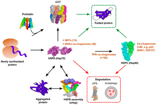
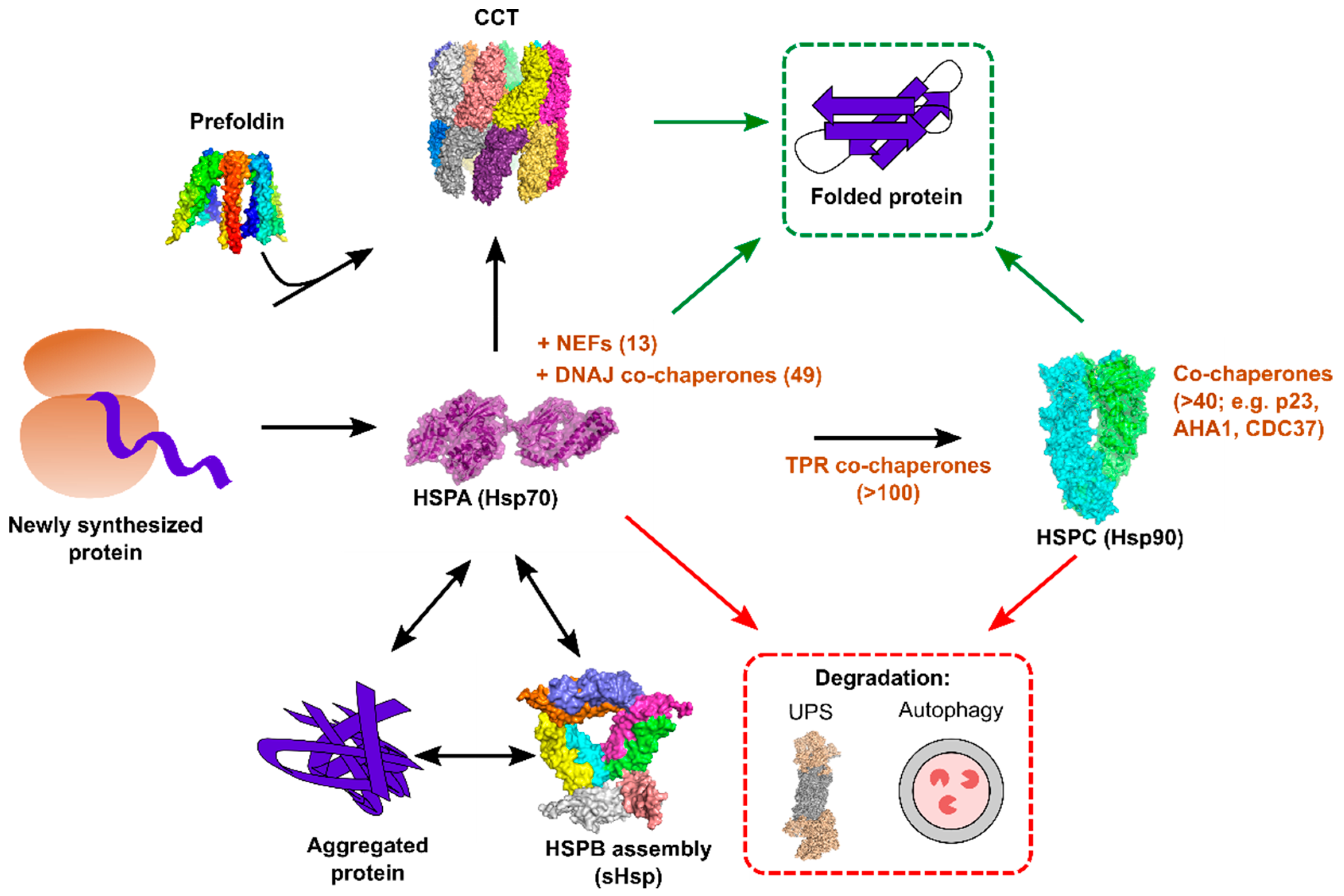
Figure 1.
Overview of the major cytoplasmic chaperone systems in human cells. Newly translated proteins interact cotranslationally with HSPAs/Hsp70s, which function together with 49 co-chaperones of the DNAJ family and 13 nucleotide exchange factors (NEFs). Client proteins can either complete their folding via interaction with Hsp70s (green arrows) or be transferred to downstream chaperone systems. Certain client proteins require further interaction with HSPC/Hsp90, and numerous tetratricopeptide repeat (TPR) domain co-chaperones help bridge these two chaperone systems. As for Hsp70, Hsp90 function is critically dependent on a large number of co-chaperones. The chaperonin containing tailless complex polypeptide 1 (CCT) can either act downstream of Hsp70 to fold proteins, or it can receive client proteins from its co-chaperone, prefoldin. Under conditions of aberrant folding or cellular stress, protein aggregates can form in the cell, and these are refolded by Hsp70, together with small heat shock proteins (HSPB/sHsps). Finally, both Hsp70 and Hsp90 can direct proteins towards cellular degradation pathways for their disposal (red arrows). Structures were produced using PyMol version 3.5.2, and they are: Hsp70 (PDB: 2KHO), Hsp90 (PDB: 2O1V), CCT (PDB: 3IYF), sHSP (PDB: 1SHS), Proteasome (PDB: 5GJR).
3.1. The HSPA Chaperone Family
The HSPAs, better known as Hsp70s and Hsc70, comprise some of the most conserved and ubiquitous chaperones. These exist in all cellular compartments where ATP is found, and mediate a staggering number of cellular processes, including assisting the folding of newly translated and stress-denatured proteins, protein translocation into organelles, protein complex formation, uncoating of clathrin vesicles, as well as supporting the degradation of misfolded or aggregated proteins [28,29,30,31]. Humans encode 13 HSPA gene products, including HSPA5 (BiP) and HSPA13 in the endoplasmic reticulum (ER), the mitochondrial HSPA9 (Grp75), and numerous cytoplasmic members that are either stress-induced (Hsp70s) or constitutively expressed (Hsc70). These share a similar domain organization, with an N-terminal ATP binding and hydrolyzing domain, and a C-terminal substrate binding domain, which are connected by a hydrophobic linker [28]. In addition, HSPAs can have N-terminal localization motifs, as well as variable C-terminal sequences that mediate co-chaperone binding. HSPAs recognize short protein stretches that are enriched in hydrophobic residues that are typically buried in protein cores [32]. The HSPA chaperone cycle is dependent upon interaction with co-chaperones, nucleotide exchange factors (NEFs), and ATP [28,29]. In the ATP-bound state, the interaction of HSPAs with clients is characterized by low affinity and fast exchange rates; in contrast, in the ADP bound state, HSPAs show high affinity and slow exchange rates. Client protein delivery is mediated by DNAJ domain containing co-chaperones (DNAJs or Hsp40s), which transfer the client to ATP-bound HSPA and stimulate ATP hydrolysis [33], yielding a stable complex of ADP-bound HSPA with the client protein. Completion of the cycle is facilitated by specific NEFs, which stimulate the exchange of ADP for ATP, resulting in client release [34].
The complexity of the HSPA chaperone system stems from the large number of co-chaperones that regulate its ATPase activity and client binding, and which provide a link to other cellular proteostasis systems. For example, there are 49 DNAJ co-chaperones in humans. These share a conserved J domain that binds HSPAs and that stimulates ATP hydrolysis [33], but are otherwise divergent, allowing for: the binding and delivery of a wide range of clients to the HSPAs, networking with distinct chaperone systems (e.g., HSPB/small heat shock proteins and HSPC/Hsp90; see below), ribosomes, clathrin-coated vesicles, the UPS, as well as autophagy, facilitating HSPA disaggregation activity, and participating in the process of protein import into organelles, and the export of misfolded proteins from the ER [23,24,30,33]. In addition to DNAJs, >100 co-chaperones with tetratricopeptide repeat (TPR) domains interact with both HSPAs and Hsp90, providing a link between these systems [17,35].
As with the DNAJ co-chaperones, NEFs also confer specialized functions to this chaperone system. Four divergent families of HSPA NEFs can be found in human cells: the bacterial homologues of GrpE (two in the mitochondria), the Hsp110 NEF family (three cytoplasmic and one in the ER), the armadillo repeats-containing NEFs (one cytoplasmic and one in the ER), and five BAG domain-containing NEFs in the cytoplasm and nucleus [34]. While all stimulate the exchange of ADP for ATP, specialized functions have been attributed to specific NEFs. For example, Hsp110 NEFs have been implicated in protein disaggregation [24], and BAG domain NEFs have been shown to play a role in protein degradation by both the UPS and autophagy [23,36].
3.2. The HSPB Chaperone Family
There are a total of 10 HSPBs (small heat shock proteins; sHsps) in humans [37], which are exclusively cytoplasmic and nuclear. These are characterized by their small size (12–42 kDa), and by the presence of a conserved alpha-crystallin domain [38]. sHsps form dynamic structures ranging from monomers to homo- and hetero-oligomeric complexes of up to 50 subunits. The assembly of sHsps is important for their function, and is regulated by post-translational modifications [37,38]. sHsps do not utilize ATP, and they are thought to act as “holdases”, binding and assembling around misfolded proteins in order to maintain them in a conformation that is competent for refolding, disaggregation, or degradation by other chaperone systems [37].
3.3. The HSPC Chaperone Family
The HSPCs (Hsp90s) comprise an abundant and conserved chaperone system, constituting >2.5% of the cellular protein mass under normal conditions [18]. In humans, there are a total of five HSPC genes: HSPC1 (Hsp90AA1) and HSPC3 (Hsp90AB1) exist in the cytosol along with HSPC2 (HspAA2), a shorter isoform of HSPC1 of unclear function, as well as HSPC4 (Grp94) in the ER, and HSPC5 (Trap-1) in the mitochondria [27]. These exist largely as homodimers, and share common structural features of an N-terminal ATP binding and hydrolyzing domain, a middle domain where client binding occurs, and a C-terminal domain that is responsible for dimerization, and where numerous co-chaperones bind [39].
As with the HSPAs, the chaperone cycle of HSPCs is regulated by ATP and a large array of co-chaperones [27,39]. In the nucleotide-free state, the chaperone adopts an open V conformation, with the C-terminal domains dimerized and the N-terminal domains open, allowing for interaction with client proteins. ATP binding induces structural rearrangements that result in the association of the N-terminal domains, adopting a closed structure that stimulates ATP hydrolysis. Subsequently, ADP release results in a return to the open conformation.
The cytoplasmic HSPCs (hereafter referred to as Hsp90) act post-translationally to facilitate the folding and stabilization of client-proteins. These include many key kinases, transcription factors, and steroid hormone receptors (for an updated list, see https://www.picard.ch/downloads/Hsp90interactors.pdf), and tend to be metastable, being rapidly degraded upon Hsp90 inhibition. Hsp90 binds client proteins downstream of the Hsp70 system, with co-chaperones harboring TPR domains providing a link between these two systems [35,39]. Interestingly, Hsp90 was recently shown to play a key role in supporting the protein folding of Hsp70 client proteins, breaking the Hsp70 folding cycle to allow for subsequent Hsp70 independent folding [40]. In addition to the TPR domain co-chaperones, numerous other co-chaperones regulate Hsp90 activity. These include proteins that stabilize the open conformation, favoring client loading, such as STIP1 and CDC37, AHA1, which stimulates ATP hydrolysis, and p23, which stabilizes the closed state [39]. Finally, the co-chaperone CHIP links Hsp90, Hsp70, and the UPS [39].
Of all the chaperones, the role of Hsp90 in viral replication is best studied [41,42]. This is largely due to the availability of highly specific inhibitors [26,43] which facilitate testing, whether this chaperone system is involved in the replication cycle (Table 1). In addition, the identification of viral proteins that interact with Hsp90 is aided by the fact that many client proteins are degraded upon Hsp90 inhibition, helping unmask the relevant viral protein, as well as the ability to isolate Hsp90 in complex with client proteins. To date, Hsp90 seems to be universally employed by viruses for their replication, with the exception of the picornavirus hepatitis A [44].
Table 1.
Antiviral activity of chaperone inhibitors against the Mononegavirales.
3.4. The Chaperonin CCT
Two different chaperonins exist in human cells. The type I chaperonin (Hsp60) is found in the mitochondria, together with its co-chaperone Hsp10. The type II chaperonin, the chaperonin containing tailless complex polypeptide 1 (CCT; also known as TCP-1 Ring Complex, TRiC), is found in the cytoplasm. CCT is a 1 MDa complex comprised of two back-to-back rings, each comprised of eight different subunits, which form a cavity in which folding can occur in isolation from the cytoplasmic environment [45]. Each subunit contains three domains, an equatorial domain that mediates inter-ring interactions, a middle domain that binds and hydrolyzes ATP together with the equatorial domain, and an apical domain that binds client proteins. The apical domain harbors helical protrusions that form an iris upon ATP hydrolysis, isolating client proteins or individual domains within the chaperonin cavity. CCT is essential for viability, and it is estimated to help the folding of ~5–10% of proteins [46], including the key structural proteins tubulin and actin. Client-protein delivery to CCT can be mediated by interaction with Hsp70s or the co-chaperone prefoldin [15]. In addition, CCT can also support the formation of protein complexes. Despite its essential nature and unique chaperoning mechanism, CCT has been shown to be involved in the life cycle of relatively few viruses to date.
3.5. Folding in the Endoplasmic Reticulum (ER)
The ER is the site of folding for all membrane and secretory proteins [47]. As all of the Mononegavirales encode glycoproteins that are essential for their infectious cycle, these viruses must interact with ER chaperone systems. Moreover, as ER stress resulting from the accumulation of misfolded proteins can lead to the suppression of cellular translation and cell death [25], the interaction of viruses with the ER folding machinery is likely to be carefully regulated.
Nascent proteins enter the ER cotranslationally via the translocon, where they encounter a unique folding environment that is characterized by increased oxidative conditions and high calcium concentrations, helping to mimic the extracellular environment [47]. As in the cytosol, proteostasis is maintained by a set of chaperones; however, the ER lacks degradation capabilities, and therefore, misfolded proteins must be retro-translocated into the cytoplasm in a process termed ER-associated degradation (ERAD) [47,48]. Analogous systems to the cytosolic HSPA/Hsp70 and HSPC/Hsp90 systems are present in the ER to facilitate both protein folding and quality control. The ER Hsp70 system is composed of the Hsp70 BiP, five DNAJ co-chaperones, and two NEFs. It is an essential aspect of folding of all ER proteins, from their translocation into the ER, until their final maturation or their exit from the ER for degradation. The ER Hsp90 system is composed of Grp94 (HSPC4), which represents the major glycoprotein of the ER. Unlike cytoplasmic Hsp90, co-chaperones of Grp94 are poorly defined, and relatively few client proteins have been identified to depend on this chaperone for folding [47]. In addition to folding, Grp94 also plays a key role in quality control and ERAD [49].
The ER includes an additional chaperone system that acts in concert with the Hsp70 and Hsp90 systems to fold glycoproteins, the lectin-binding chaperone system [47]. It consists of the chaperones calreticulin and calnexin, N-glycan processing enzymes that prevent aggregation and premature export from the ER, and a UDP-glucose:glycoprotein glucosyltransferase that is involved in quality control. Additional factors, such as peptidyl-propyl isomerases and protein disulfide isomerases help in the folding and maturation of ER proteins. As in the cytoplasm, the ER chaperome is composed of an interconnected network that cooperates to regulate proteostasis [50].
6. Conclusions
Despite the progress that has been made in deciphering the interactions between chaperones and the Mononegavirales, significant gaps in our knowledge remain. The chaperone dependence of numerous viral proteins from the Mononegavirales remains to be defined (e.g., SH, VP24, NS1, NS2, M2-1, M2-2, V, and C). Proteomics studies frequently identify chaperones as interacting partners [11,76,79,82,93,112,120], but many times they lack validation of the interaction and formal testing of the relevance of the identified chaperone in the context of infection. Even for well-studied interactions, such as that of the NC with Hsp70, or the L protein with Hsp90, the role of co-chaperones or upstream and downstream chaperone systems remains to be fully elucidated. For example, while a clear interaction between NC and Hsp70 has been established for MeV, the relevant DNAJ and NEF co-chaperones remain unknown. Moreover, the generality of this finding for other Mononegavirales is not clear. Similarly, Hsp90 has been shown to be involved in the folding of L from numerous viruses. However, Hsp90 works downstream of Hsp70/Hsc70, and thus far, an interaction between the latter and L has only been formally shown for MuV [80]. As for Hsp70, our knowledge of Hsp90 co-chaperones involved in the folding of L remains limited. Thus far, of the numerous Hsp90 co-chaperones, the only one identified to date to play a role in the replication of any of Mononegavirales is CDC37 for chaperoning the RABV P protein [89]. Due to the large number of co-chaperones for both Hsp70 and Hsp90, it is possible that functional redundancies may hinder the identification of individual co-chaperones involved in viral replication. In favor of this argument, a study using RSV found that knocking down two co-chaperones identified in their proteomic study to bind L, STIP1 and DNAJA2, had no effect on viral replication [79]. However, contrary to this argument, a recent study investigating the function of DNAJ proteins in the replication of dengue virus was able to identify a role for several of these co-chaperones in distinct stages of the viral replication cycle [121]. Finally, it is important to note that the role of key chaperone systems in the cells, such as the sHsps and CCT, remains undefined for nearly all the Mononegavirales.
While much can be learned about viral biology from studying chaperones, the opposite may be true as well. In particular, recent works have appreciated the fact that chaperones exist in dynamic multi-chaperone complexes that are likely to confer specific functions that are required to meet cellular proteostasis demands [122]. In this regard, viral infection may provide a valuable tool for the reproducible induction of alterations in the composition of chaperone complexes that can help to decipher both the basis and functional importance of such changes.
Funding
This research was funded by a grant from the Conselleria de Educación, Investigación, Cultura y Deporte (SEJI/2017/006) and by a 2017 Research Grant by the European Society of Clinical Microbiology and Infectious Diseases (ESCMID) to R.G., F.M. is supported by a predoctoral fellowship from the Spanish Ministerio de Ciencia, Innovación y Universidades (BES-2016-076677). R.G. is supported by the Ramon y Cajal fellowship from the Spanish Ministry of Economy and Competitiveness (RYC-2015-17517).
Acknowledgments
We thank Jose Manuel Cuevas for helpful comments and suggestions.
Conflicts of Interest
The authors declare no conflict of interest. The funders had no role in the design of the study; in the collection, analyses, or interpretation of data; in the writing of the manuscript, or in the decision to publish the results.
References
- Amarasinghe, G.K.; Aréchiga Ceballos, N.G.; Banyard, A.C.; Basler, C.F.; Bavari, S.; Bennett, A.J.; Blasdell, K.R.; Briese, T.; Bukreyev, A.; Caì, Y.; et al. Taxonomy of the order Mononegavirales: Update 2018. Arch. Virol. 2018, 163, 2283–2294. [Google Scholar] [CrossRef] [Scilit] [PubMed]
- Thibault, P.A.; Watkinson, R.E.; Moreira-Soto, A.; Drexler, J.F.; Lee, B. Zoonotic Potential of Emerging Paramyxoviruses: Knowns and Unknowns. Adv. Virus Res. 2017, 98, 1–55. [Google Scholar] [CrossRef] [Scilit] [PubMed]
- Coltart, C.E.M.; Lindsey, B.; Ghinai, I.; Johnson, A.M.; Heymann, D.L. The Ebola outbreak, 2013-2016: Old lessons for new epidemics. Phil. Trans. R. Soc. Lond. B Biol. Sci. 2017, 372. [Google Scholar] [CrossRef] [Scilit] [PubMed]
- Whelan, S.P.J.; Barr, J.N.; Wertz, G.W. Transcription and replication of nonsegmented negative-strand RNA viruses. Curr. Top. Microbiol. Immunol. 2004, 283, 61–119. [Google Scholar] [CrossRef] [Scilit] [PubMed]
- Lamb, R.A. Mononegavirales. In Fields Virology; Knipe, D.M., Howley, P.M., Eds.; Wolters Kluwer Health/Lippincott Williams & Wilkins: Philadelphia, PA, USA, 2013; pp. 880–884. ISBN 9781451105636. [Google Scholar]
- Longhi, S. Nucleocapsid structure and function. Curr. Top. Microbiol. Immunol. 2009, 329, 103–128. [Google Scholar] [PubMed]
- Morin, B.; Kranzusch, P.J.; Rahmeh, A.A.; Whelan, S.P.J. The polymerase of negative-stranded RNA viruses. Curr. Opin. Virol. 2013, 3, 103–110. [Google Scholar] [CrossRef] [Scilit] [PubMed]
- Longhi, S.; Bloyet, L.-M.; Gianni, S.; Gerlier, D. How order and disorder within paramyxoviral nucleoproteins and phosphoproteins orchestrate the molecular interplay of transcription and replication. Cell. Mol. Life Sci. 2017, 74, 3091–3118. [Google Scholar] [CrossRef] [Scilit] [PubMed]
- Liljeroos, L.; Butcher, S.J. Matrix proteins as centralized organizers of negative-sense RNA virions. Front. Biosci. 2013, 18, 696–715. [Google Scholar] [CrossRef] [Scilit]
- Cantoni, D.; Rossman, J.S. Ebolaviruses: New roles for old proteins. PLoS Negl. Trop. Dis. 2018, 12, e0006349. [Google Scholar] [CrossRef] [Scilit] [PubMed]
- Komarova, A.V.; Combredet, C.; Meyniel-Schicklin, L.; Chapelle, M.; Caignard, G.; Camadro, J.-M.; Lotteau, V.; Vidalain, P.-O.; Tangy, F. Proteomic analysis of virus-host interactions in an infectious context using recombinant viruses. Mol. Cell. Proteom. 2011, 10, M110–007443. [Google Scholar] [CrossRef] [Scilit] [PubMed]
- de Chassey, B.; Meyniel-Schicklin, L.; Vonderscher, J.; André, P.; Lotteau, V. Virus-host interactomics: New insights and opportunities for antiviral drug discovery. Genome Med. 2014, 6, 115. [Google Scholar] [CrossRef] [Scilit] [PubMed]
- Wu, B.; Eliscovich, C.; Yoon, Y.J.; Singer, R.H. Translation dynamics of single mRNAs in live cells and neurons. Science 2016, 352, 1430–1435. [Google Scholar] [CrossRef] [Scilit] [PubMed]
- Ellis, R.J. Macromolecular crowding: Obvious but underappreciated. Trends Biochem. Sci. 2001, 26, 597–604. [Google Scholar] [CrossRef] [Scilit]
- Balchin, D.; Hayer-Hartl, M.; Hartl, F.U. In vivo aspects of protein folding and quality control. Science 2016, 353, aac4354. [Google Scholar] [CrossRef] [Scilit] [PubMed]
- Schwanhüusser, B.; Busse, D.; Li, N.; Dittmar, G.; Schuchhardt, J.; Wolf, J.; Chen, W.; Selbach, M. Global quantification of mammalian gene expression control. Nature 2011, 473, 337–342. [Google Scholar] [CrossRef] [Scilit] [PubMed]
- Brehme, M.; Voisine, C.; Rolland, T.; Wachi, S.; Soper, J.H.; Zhu, Y.; Orton, K.; Villella, A.; Garza, D.; Vidal, M.; et al. A chaperome subnetwork safeguards proteostasis in aging and neurodegenerative disease. Cell Rep. 2014, 9, 1135–1150. [Google Scholar] [CrossRef] [Scilit] [PubMed]
- Finka, A.; Goloubinoff, P. Proteomic data from human cell cultures refine mechanisms of chaperone-mediated protein homeostasis. Cell Stress Chaperones 2013, 18, 591–605. [Google Scholar] [CrossRef] [Scilit] [PubMed]
- Tokuriki, N.; Oldfield, C.J.; Uversky, V.N.; Berezovsky, I.N.; Tawfik, D.S. Do viral proteins possess unique biophysical features? Trends Biochem. Sci. 2009, 34, 53–59. [Google Scholar] [CrossRef] [Scilit] [PubMed]
- Sanjuan, R.; Domingo-Calap, P. Mechanisms of viral mutation. Cell. Mol. Life Sci. 2016, 73, 4433–4448. [Google Scholar] [CrossRef] [Scilit] [PubMed]
- Tokuriki, N.; Tawfik, D.S. Stability effects of mutations and protein evolvability. Curr. Opin. Struct. Biol. 2009, 19, 596–604. [Google Scholar] [CrossRef] [Scilit] [PubMed]
- Kim, Y.E.; Hipp, M.S.; Bracher, A.; Hayer-Hartl, M.; Hartl, F.U. Molecular chaperone functions in protein folding and proteostasis. Annu. Rev. Biochem. 2013, 82, 325–355. [Google Scholar] [CrossRef] [Scilit] [PubMed]
- Ciechanover, A.; Kwon, Y.T. Protein quality control by molecular chaperones in neurodegeneration. Front. Neurosci. 2017, 11, 185. [Google Scholar] [CrossRef] [Scilit] [PubMed]
- Mogk, A.; Bukau, B.; Kampinga, H.H. Cellular Handling of Protein Aggregates by Disaggregation Machines. Mol. Cell 2018, 69, 214–226. [Google Scholar] [CrossRef] [Scilit] [PubMed]
- Frakes, A.E.; Dillin, A. The UPRER: Sensor and Coordinator of Organismal Homeostasis. Mol. Cell 2017, 66, 761–771. [Google Scholar] [CrossRef] [Scilit] [PubMed]
- Chatterjee, S.; Burns, T.F. Targeting heat shock proteins in cancer: A promising therapeutic approach. Int. J. Mol. Sci. 2017, 18, 1978. [Google Scholar] [CrossRef] [Scilit]
- Radons, J. The Hsp90 Chaperone Machinery: An Important Hub in Protein Interaction Networks. Br. J. Med. Med. Res. 2016, 14, 1–32. [Google Scholar] [CrossRef] [Scilit]
- Radons, J. The human HSP70 family of chaperones: Where do we stand? Cell Stress Chaperones 2016, 21, 379–404. [Google Scholar] [CrossRef] [Scilit]
- Mayer, M.P.; Bukau, B. Hsp70 chaperones: Cellular functions and molecular mechanism. Cell. Mol. Life Sci. 2005, 62, 670–684. [Google Scholar] [CrossRef] [Scilit]
- Sousa, R.; Lafer, E.M. The role of molecular chaperones in clathrin mediated vesicular trafficking. Front. Mol. Biosci. 2015, 2, 26. [Google Scholar] [CrossRef] [Scilit]
- Craig, E.A. Hsp70 at the membrane: Driving protein translocation. BMC Biol. 2018, 16, 11. [Google Scholar] [CrossRef] [Scilit]
- Clerico, E.M.; Tilitsky, J.M.; Meng, W.; Gierasch, L.M. How hsp70 molecular machines interact with their substrates to mediate diverse physiological functions. J. Mol. Biol. 2015, 427, 1575–1588. [Google Scholar] [CrossRef] [Scilit] [PubMed]
- Kampinga, H.H.; Craig, E.A. The HSP70 chaperone machinery: J proteins as drivers of functional specificity. Nat. Rev. Mol. Cell Biol. 2010, 11, 579–592. [Google Scholar] [CrossRef] [Scilit] [PubMed]
- Bracher, A.; Verghese, J. GrpE, Hsp110/Grp170, HspBP1/Sil1 and BAG domain proteins: Nucleotide exchange factors for Hsp70 molecular chaperones. Subcell. Biochem. 2015, 78, 1–33. [Google Scholar] [CrossRef] [Scilit] [PubMed]
- Allan, R.K.; Ratajczak, T. Versatile TPR domains accommodate different modes of target protein recognition and function. Cell Stress Chaperones 2011, 16, 353–367. [Google Scholar] [CrossRef] [Scilit] [PubMed]
- Stürner, E.; Behl, C. The Role of the Multifunctional BAG3 Protein in Cellular Protein Quality Control and in Disease. Front. Mol. Neurosci. 2017, 10, 177. [Google Scholar] [CrossRef] [Scilit] [PubMed]
- Kampinga, H.H.; de Boer, R.; Beerstra, N. The Multicolored World of the Human HSPB Family. In The Big Book on Small Heat Shock Proteins; Tanguay, R.M., Hightower, L.E., Eds.; Springer International Publishing: Cham, Switzerland, 2015; pp. 3–26. ISBN 978-3-319-16077-1. [Google Scholar]
- Haslbeck, M.; Vierling, E. A first line of stress defense: Small heat shock proteins and their function in protein homeostasis. J. Mol. Biol. 2015, 427, 1537–1548. [Google Scholar] [CrossRef] [Scilit] [PubMed]
- Schopf, F.H.; Biebl, M.M.; Buchner, J. The HSP90 chaperone machinery. Nat. Rev. Mol. Cell Biol. 2017, 18, 345–360. [Google Scholar] [CrossRef] [Scilit]
- Morán Luengo, T.; Kityk, R.; Mayer, M.P.; Rüdiger, S.G.D. Hsp90 Breaks the Deadlock of the Hsp70 Chaperone System. Mol. Cell 2018, 70, 545–552. [Google Scholar] [CrossRef] [Scilit]
- Wang, Y.; Jin, F.; Wang, R.; Li, F.; Wu, Y.; Kitazato, K.; Wang, Y. HSP90: A promising broad-spectrum antiviral drug target. Arch. Virol. 2017, 162, 3269–3282. [Google Scholar] [CrossRef] [Scilit]
- Geller, R.; Taguwa, S.; Frydman, J. Broad action of Hsp90 as a host chaperone required for viral replication. Biochim. Biophys. Acta 2012, 1823, 698–706. [Google Scholar] [CrossRef] [Scilit]
- Yuno, A.; Lee, M.-J.; Lee, S.; Tomita, Y.; Rekhtman, D.; Moore, B.; Trepel, J.B. Clinical Evaluation and Biomarker Profiling of Hsp90 Inhibitors. In Chaperones: Methods and Protocols; Calderwood, S.K., Prince, T.L., Eds.; Springer: New York, NY, USA, 2018; pp. 423–441. ISBN 978-1-4939-7477-1. [Google Scholar]
- Aragonès, L.; Guix, S.; Ribes, E.; Bosch, A.; Pintó, R.M. Fine-tuning translation kinetics selection as the driving force of codon usage bias in the hepatitis A virus capsid. PLoS Pathog. 2010, 6, e1000797. [Google Scholar] [CrossRef] [Scilit] [PubMed]
- Lopez, T.; Dalton, K.; Frydman, J. The Mechanism and Function of Group II Chaperonins. J. Mol. Biol. 2015, 427, 2919–2930. [Google Scholar] [CrossRef] [Scilit] [PubMed]
- Yam, A.Y.; Xia, Y.; Lin, H.-T.J.; Burlingame, A.; Gerstein, M.; Frydman, J. Defining the TRiC/CCT interactome links chaperonin function to stabilization of newly made proteins with complex topologies. Nat. Struct. Mol. Biol. 2008, 15, 1255–1262. [Google Scholar] [CrossRef] [Scilit] [PubMed]
- Hebert, D.N.; Molinari, M. In and out of the ER: Protein folding, quality control, degradation, and related human diseases. Physiol. Rev. 2007, 87, 1377–1408. [Google Scholar] [CrossRef] [Scilit] [PubMed]
- Hegde, R.S.; Ploegh, H.L. Quality and quantity control at the endoplasmic reticulum. Curr. Opin. Cell Biol. 2010, 22, 437–446. [Google Scholar] [CrossRef] [Scilit] [PubMed]
- Araki, K.; Nagata, K. SUP: Protein folding and quality control in the ER. Cold Spring Harb. Perspect. Biol. 2012, 4, a015438. [Google Scholar] [CrossRef] [Scilit] [PubMed]
- Jansen, G.; Maattanen, P.; Denisov, A.Y.; Scarffe, L.; Schade, B.; Balghi, H.; Dejgaard, K.; Chen, L.Y.; Muller, W.J.; Gehring, K.; et al. An Interaction Map of Endoplasmic Reticulum Chaperones and Foldases. Mol. Cell. Proteom. 2012, 11, 710–723. [Google Scholar] [CrossRef] [Scilit] [PubMed]
- Katoh, H.; Kubota, T.; Kita, S.; Nakatsu, Y.; Aoki, N.; Mori, Y.; Maenaka, K.; Takeda, M.; Kidokoro, M. Heat shock protein 70 regulates degradation of the mumps virus phosphoprotein via the ubiquitin-proteasome pathway. J. Virol. 2015, 89, 3188–3199. [Google Scholar] [CrossRef] [Scilit] [PubMed]
- Zhang, R.; Liu, C.; Cao, Y.; Jamal, M.; Chen, X.; Zheng, J.; Li, L.; You, J.; Zhu, Q.; Liu, S.; et al. Rabies viruses leader RNA interacts with host Hsc70 and inhibits virus replication. Oncotarget 2017, 8, 43822–43837. [Google Scholar] [CrossRef] [Scilit]
- Lahaye, X.; Vidy, A.; Fouquet, B.; Blondel, D. Hsp70 Protein Positively Regulates Rabies Virus Infection. J. Virol. 2012, 86, 4743–4751. [Google Scholar] [CrossRef] [Scilit]
- Wheeler, D.S.; Chase, M.A.; Senft, A.P.; Poynter, S.E.; Wong, H.R.; Page, K. Extracellular Hsp72, an endogenous DAMP, is released by virally infected airway epithelial cells and activates neutrophils via Toll-like receptor (TLR)-4. Respir. Res. 2009, 10, 31. [Google Scholar] [CrossRef] [Scilit] [PubMed]
- Baturcam, E.; Snape, N.; Yeo, T.H.; Schagen, J.; Thomas, E.; Logan, J.; Galbraith, S.; Collinson, N.; Phipps, S.; Fantino, E.; et al. Human Metapneumovirus Impairs Apoptosis of Nasal Epithelial Cells in Asthma via HSP70. J. Innate Immun. 2017, 9, 52–64. [Google Scholar] [CrossRef] [Scilit] [PubMed]
- Oliveira, A.P.; Simabuco, F.M.; Tamura, R.E.; Guerrero, M.C.; Ribeiro, P.G.G.; Libermann, T.A.; Zerbini, L.F.; Ventura, A.M. Human respiratory syncytial virus N, P and M protein interactions in HEK-293T cells. Virus Res. 2013, 177, 108–112. [Google Scholar] [CrossRef] [Scilit] [PubMed]
- Kim, M.Y.; Oglesbee, M. Virus-heat shock protein interaction and a novel axis for innate antiviral immunity. Cells 2012, 1, 646–666. [Google Scholar] [CrossRef] [Scilit] [PubMed]
- Mayer, M.P. Recruitment of Hsp70 chaperones: A crucial part of viral survival strategies. In Reviews of Physiology, Biochemistry and Pharmacology; Springer: Berlin/Heidelberg, Germany, 2005; Volume 153, pp. 1–46. ISBN 0303-4240. [Google Scholar]
- Collins, P.L.; Hightower, L.E. Newcastle disease virus stimulates the cellular accumulation of stress (heat shock) mRNAs and proteins. J. Virol. 1982, 44, 703–707. [Google Scholar]
- Oglesbee, M.; Krakowka, S. Cellular stress response induces selective intranuclear trafficking and accumulation of morbillivirus major core protein. Lab. Investig. 1993, 68, 109–117. [Google Scholar] [PubMed]
- Brown, G.; Rixon, H.W.M.; Steel, J.; McDonald, T.P.; Pitt, A.R.; Graham, S.; Sugrue, R.J. Evidence for an association between heat shock protein 70 and the respiratory syncytial virus polymerase complex within lipid-raft membranes during virus infection. Virology 2005, 338, 69–80. [Google Scholar] [CrossRef] [Scilit] [PubMed]
- Radhakrishnan, A.; Yeo, D.; Brown, G.; Myaing, M.Z.; Iyer, L.R.; Fleck, R.; Tan, B.-H.; Aitken, J.; Sanmun, D.; Tang, K.; et al. Protein analysis of purified respiratory syncytial virus particles reveals an important role for heat shock protein 90 in virus particle assembly. Mol. Cell. Proteom. 2010, 9, 1829–1848. [Google Scholar] [CrossRef] [Scilit]
- Lahaye, X.; Vidy, A.; Pomier, C.; Obiang, L.; Harper, F.; Gaudin, Y.; Blondel, D. Functional Characterization of Negri Bodies (NBs) in Rabies Virus-Infected Cells: Evidence that NBs Are Sites of Viral Transcription and Replication. J. Virol. 2009, 83, 7948–7958. [Google Scholar] [CrossRef] [Scilit]
- Oglesbee, M.J.; Kenney, H.; Kenney, T.; Krakowka, S. Enhanced production of morbillivirus gene-specific RNAs following induction of the cellular stress response in stable persistent infection. Virology 1993, 192, 556–567. [Google Scholar] [CrossRef] [Scilit]
- Oglesbee, M.J.; Liu, Z.; Kenney, H.; Brooks, C.L. The Highly Inducible Member of the 70 kDa Family of Heat Shock Proteins Increases Canine Distemper Virus Polymerase Activity. J. Gen. Virol. 1996, 77, 2125–2135. [Google Scholar] [CrossRef] [Scilit] [PubMed]
- Vasconcelos, D.; Norrby, E.; Oglesbee, M. The cellular stress response increases measles virus-induced cytopathic effect. J. Gen. Virol. 1998, 79 Pt 7, 1769–1773. [Google Scholar] [CrossRef] [Scilit]
- Parks, C.L.; Lerch, R.A.; Walpita, P.; Sidhu, M.S.; Udem, S.A. Enhanced measles virus cDNA rescue and gene expression after heat shock. J. Virol. 1999, 73, 3560–3566. [Google Scholar] [PubMed]
- Heller, M.; Vasconcelos, D.; Cummins, J.; Oglesbee, M. Interferon-alpha inhibits the emergence of cellular stress response-dependent morbillivirus large plaque variants. Antivir. Res. 1998, 38, 195–207. [Google Scholar] [CrossRef] [Scilit]
- Vasconcelos, D.Y.; Cai, X.H.; Oglesbee, M.J. Constitutive overexpression of the major inducible 70 kDa heat shock protein mediates large plaque formation by measles virus. J. Gen. Virol. 1998, 79 Pt 9, 2239–2247. [Google Scholar] [CrossRef] [Scilit]
- Zhang, X.; Glendening, C.; Linke, H.; Parks, C.L.; Brooks, C.; Udem, S.A.; Oglesbee, M. Identification and characterization of a regulatory domain on the carboxyl terminus of the measles virus nucleocapsid protein. J. Virol. 2002, 76, 8737–8746. [Google Scholar] [CrossRef] [Scilit]
- Carsillo, T.; Traylor, Z.; Choi, C.; Niewiesk, S.; Oglesbee, M. hsp72, a host determinant of measles virus neurovirulence. J. Virol. 2006, 80, 11031–11039. [Google Scholar] [CrossRef] [Scilit]
- Kim, M.Y.; Ma, Y.; Zhang, Y.; Li, J.; Shu, Y.; Oglesbee, M. hsp70-Dependent Antiviral Immunity against Cytopathic Neuronal Infection by Vesicular Stomatitis Virus. J. Virol. 2013, 87, 10668–10678. [Google Scholar] [CrossRef] [Scilit]
- Zhang, X.; Bourhis, J.M.; Longhi, S.; Carsillo, T.; Buccellato, M.; Morin, B.; Canard, B.; Oglesbee, M. Hsp72 recognizes a P binding motif in the measles virus N protein C-terminus. Virology 2005, 337, 162–174. [Google Scholar] [CrossRef] [Scilit]
- Couturier, M.; Buccellato, M.; Costanzo, S.; Bourhis, J.-M.; Shu, Y.; Nicaise, M.; Desmadril, M.; Flaudrops, C.; Longhi, S.; Oglesbee, M. High affinity binding between Hsp70 and the C-terminal domain of the measles virus nucleoprotein requires an Hsp40 co-chaperone. J. Mol. Recognit. 2010, 23, 301–315. [Google Scholar] [CrossRef] [Scilit]
- Carsillo, T.; Zhang, X.; Vasconcelos, D.; Niewiesk, S.; Oglesbee, M. A single codon in the nucleocapsid protein C terminus contributes to in vitro and in vivo fitness of Edmonston measles virus. J. Virol. 2006, 80, 2904–2912. [Google Scholar] [CrossRef] [Scilit] [PubMed]
- García-Dorival, I.; Wu, W.; Armstrong, S.D.; Barr, J.N.; Carroll, M.W.; Hewson, R.; Hiscox, J.A. Elucidation of the Cellular Interactome of Ebola Virus Nucleoprotein and Identification of Therapeutic Targets. J. Proteome Res. 2016, 15, 4290–4303. [Google Scholar] [CrossRef] [Scilit] [PubMed]
- Nelson, E.V.; Pacheco, J.R.; Hume, A.J.; Cressey, T.N.; Deflubé, L.R.; Ruedas, J.B.; Connor, J.H.; Ebihara, H.; Mühlberger, E. An RNA polymerase II-driven Ebola virus minigenome system as an advanced tool for antiviral drug screening. Antivir. Res. 2017, 146, 21–27. [Google Scholar] [CrossRef] [Scilit] [PubMed]
- Tchesnokov, E.P.; Raeisimakiani, P.; Ngure, M.; Marchant, D.; Götte, M. Recombinant RNA-Dependent RNA Polymerase Complex of Ebola Virus. Sci. Rep. 2018, 8, 3970. [Google Scholar] [CrossRef] [Scilit] [PubMed]
- Munday, D.C.; Wu, W.; Smith, N.; Fix, J.; Noton, S.L.; Galloux, M.; Touzelet, O.; Armstrong, S.D.; Dawson, J.M.; Aljabr, W.; et al. Interactome Analysis of the Human Respiratory Syncytial Virus RNA Polymerase Complex Identifies Protein Chaperones as Important Cofactors That Promote L-Protein Stability and RNA Synthesis. J. Virol. 2015, 89, 917–930. [Google Scholar] [CrossRef] [Scilit] [PubMed]
- Katoh, H.; Kubota, T.; Nakatsu, Y.; Tahara, M.; Kidokoro, M.; Takeda, M. Heat Shock Protein 90 Ensures Efficient Mumps Virus Replication by Assisting with Viral Polymerase Complex Formation. J. Virol. 2017, 91, e02220-16. [Google Scholar] [CrossRef] [Scilit]
- Sztuba-Solinska, J.; Diaz, L.; Kumar, M.R.; Kolb, G.; Wiley, M.R.; Jozwick, L.; Kuhn, J.H.; Palacios, G.; Radoshitzky, S.R.; Le Grice, S.F.J.; et al. A small stem-loop structure of the Ebola virus trailer is essential for replication and interacts with heat-shock protein A8. Nucleic Acids Res. 2016, 44, 9831–9846. [Google Scholar] [CrossRef] [Scilit]
- Wu, W.; Tran, K.C.; Teng, M.N.; Heesom, K.J.; Matthews, D.A.; Barr, J.N.; Hiscox, J.A. The interactome of the human respiratory syncytial virus NS1 protein highlights multiple effects on host cell biology. J. Virol. 2012, 86, 7777–7789. [Google Scholar] [CrossRef] [Scilit]
- Spurgers, K.B.; Alefantis, T.; Peyser, B.D.; Ruthel, G.T.; Bergeron, A.A.; Costantino, J.A.; Enterlein, S.; Kota, K.P.; Boltz, R.C.D.; Aman, M.J.; et al. Identification of essential filovirion-associated host factors by serial proteomic analysis and RNAi screen. Mol. Cell. Proteom. 2010, 9, 2690–2703. [Google Scholar] [CrossRef] [Scilit]
- Liang, J.; Sagum, C.A.; Bedford, M.T.; Sidhu, S.S.; Sudol, M.; Han, Z.; Harty, R.N. Chaperone-Mediated Autophagy Protein BAG3 Negatively Regulates Ebola and Marburg VP40-Mediated Egress. PLoS Pathog. 2017, 13, e1006132. [Google Scholar] [CrossRef] [Scilit]
- Liang, B.; Li, Z.; Jenni, S.; Rahmeh, A.A.; Morin, B.M.; Grant, T.; Grigorieff, N.; Harrison, S.C.; Whelan, S.P.J. Structure of the L Protein of Vesicular Stomatitis Virus from Electron Cryomicroscopy. Cell 2015, 162, 314–327. [Google Scholar] [CrossRef] [Scilit]
- Bloyet, L.-M.; Welsch, J.; Enchery, F.; Mathieu, C.; de Breyne, S.; Horvat, B.; Grigorov, B.; Gerlier, D. HSP90 Chaperoning in Addition to Phosphoprotein Required for Folding but Not for Supporting Enzymatic Activities of Measles and Nipah Virus L Polymerases. J. Virol. 2016, 90, 6642–6656. [Google Scholar] [CrossRef] [Scilit] [PubMed]
- Geller, R.; Andino, R.; Frydman, J. Hsp90 inhibitors exhibit resistance-free antiviral activity against respiratory syncytial virus. PLoS ONE 2013, 8, e56762. [Google Scholar] [CrossRef] [Scilit]
- Connor, J.H.; McKenzie, M.O.; Parks, G.D.; Lyles, D.S. Antiviral activity and RNA polymerase degradation following Hsp90 inhibition in a range of negative strand viruses. Virology 2007, 362, 109–119. [Google Scholar] [CrossRef] [Scilit] [PubMed]
- Xu, Y.; Liu, F.; Liu, J.; Wang, D.; Yan, Y.; Ji, S.; Zan, J.; Zhou, J. The co-chaperone Cdc37 regulates the rabies virus phosphoprotein stability by targeting to Hsp90AA1 machinery. Sci. Rep. 2016, 6, 27123. [Google Scholar] [CrossRef] [Scilit]
- Zhang, J.; Wu, X.; Zan, J.; Wu, Y.; Ye, C.; Ruan, X.; Zhou, J. Cellular Chaperonin CCT Contributes to Rabies Virus Replication during Infection. J. Virol. 2013, 87, 7608–7621. [Google Scholar] [CrossRef] [Scilit] [PubMed]
- Zhang, J.; Ye, C.; Ruan, X.; Zan, J.; Xu, Y.; Liao, M.; Zhou, J. The chaperonin CCTα is required for efficient transcription and replication of rabies virus. Microbiol. Immunol. 2014, 58, 590–599. [Google Scholar] [CrossRef] [Scilit] [PubMed]
- Roobol, A.; Sahyoun, Z.P.; Carden, M.J. Selected subunits of the cytosolic chaperonin associate with microtubules assembled in vitro. J. Biol. Chem. 1999, 274, 2408–2415. [Google Scholar] [CrossRef] [Scilit]
- García-Dorival, I.; Wu, W.; Dowall, S.; Armstrong, S.; Touzelet, O.; Wastling, J.; Barr, J.N.; Matthews, D.; Carroll, M.; Hewson, R.; et al. Elucidation of the Ebola virus VP24 cellular interactome and disruption of virus biology through targeted inhibition of host-cell protein function. J. Proteome Res. 2014, 13, 5120–5135. [Google Scholar] [CrossRef] [Scilit]
- Roux, L. Selective and transient association of Sendai virus HN glycoprotein with BiP. Virology 1990, 175, 161–166. [Google Scholar] [CrossRef] [Scilit]
- Watowich, S.S.; Morimoto, R.I.; Lamb, R. A Flux of the paramyxovirus hemagglutinin-neuraminidase glycoprotein through the endoplasmic reticulum activates transcription of the GRP78-BiP gene. J. Virol. 1991, 65, 3590–3597. [Google Scholar] [PubMed]
- Bitko, V.; Barik, S. An endoplasmic reticulum-specific stress-activated caspase (caspase-12) is implicated in the apoptosis of A549 epithelial cells by respiratory syncytial virus. J. Cell. Biochem. 2001, 80, 441–454. [Google Scholar] [CrossRef] [Scilit]
- Bolt, G. The measles virus (MV) glycoproteins interact with cellular chaperones in the endoplasmic reticulum and MV infection upregulates chaperone expression. Arch. Virol. 2001, 146, 2055–2068. [Google Scholar] [CrossRef] [Scilit] [PubMed]
- Patrick Reid, S.; Shurtleff, A.C.; Costantino, J.A.; Tritsch, S.R.; Retterer, C.; Spurgers, K.B.; Bavari, S. HSPA5 is an essential host factor for Ebola virus infection. Antivir. Res. 2014, 109, 171–174. [Google Scholar] [CrossRef] [Scilit] [PubMed]
- Ng, D.T.; Randall, R.E.; Lamb, R.A. Intracellular maturation and transport of the SV5 type II glycoprotein hemagglutinin-neuraminidase: Specific and transient association with GRP78-BiP in the endoplasmic reticulum and extensive internalization from the cell surface. J. Cell Biol. 1989, 109, 3273–3289. [Google Scholar] [CrossRef] [Scilit] [PubMed]
- Ng, D.T.; Hiebert, S.W.; Lamb, R.A. Different roles of individual N-linked oligosaccharide chains in folding, assembly, and transport of the simian virus 5 hemagglutinin-neuraminidase. Mol. Cell. Biol. 1990, 10, 1989–2001. [Google Scholar] [CrossRef] [Scilit] [PubMed]
- Anderson, K.; Stott, E.J.; Wertz, G.W. Intracellular processing of the human respiratory syncytial virus fusion glycoprotein: Amino acid substitutions affecting folding, transport and cleavage. J. Gen. Virol. 1992, 73 Pt 5, 1177–1188. [Google Scholar] [CrossRef] [Scilit]
- Gaudin, Y. Folding of rabies virus glycoprotein: Epitope acquisition and interaction with endoplasmic reticulum chaperones. J. Virol. 1997, 71, 3742–3750. [Google Scholar]
- Booth, L.; Roberts, J.L.; Ecroyd, H.; Tritsch, S.R.; Bavari, S.; Reid, S.P.; Proniuk, S.; Zukiwski, A.; Jacob, A.; Sepúlveda, C.S.; et al. AR-12 Inhibits Multiple Chaperones Concomitant With Stimulating Autophagosome Formation Collectively Preventing Virus Replication. J. Cell. Physiol. 2016, 231, 2286–2302. [Google Scholar] [CrossRef] [Scilit]
- Roberts, J.L.; Tavallai, M.; Nourbakhsh, A.; Fidanza, A.; Cruz-Luna, T.; Smith, E.; Siembida, P.; Plamondon, P.; Cycon, K.A.; Doern, C.D.; et al. GRP78/Dna K Is a Target for Nexavar/Stivarga/Votrient in the Treatment of Human Malignancies, Viral Infections and Bacterial Diseases. J. Cell. Physiol. 2015, 230, 2552–2578. [Google Scholar] [CrossRef] [Scilit]
- Booth, L.; Roberts, J.L.; Cash, D.R.; Tavallai, S.; Jean, S.; Fidanza, A.; Cruz-Luna, T.; Siembiba, P.; Cycon, K.A.; Cornelissen, C.N.; et al. GRP78/BiP/HSPA5/Dna K is a universal therapeutic target for human disease. J. Cell. Physiol. 2015, 230, 1661–1676. [Google Scholar] [CrossRef] [Scilit] [PubMed]
- Wang, B.; Wang, Y.; Frabutt, D.A.; Zhang, X.; Yao, X.; Hu, D.; Zhang, Z.; Liu, C.; Zheng, S.; Xiang, S.-H.; et al. Mechanistic understanding of N-glycosylation in Ebola virus glycoprotein maturation and function. J. Biol. Chem. 2017, 292, 5860–5870. [Google Scholar] [CrossRef] [Scilit]
- Hammond, C.; Helenius, A. Folding of VSV G protein: Sequential interaction with BiP and calnexin. Science 1994, 266, 456–458. [Google Scholar] [CrossRef] [Scilit] [PubMed]
- Peterson, J.R.; Ora, A.; Van, P.N.; Helenius, A. Transient, lectin-like association of calreticulin with folding intermediates of cellular and viral glycoproteins. Mol. Biol. Cell 1995, 6, 1173–1184. [Google Scholar] [CrossRef] [Scilit]
- Bloor, S.; Maelfait, J.; Krumbach, R.; Beyaert, R.; Randow, F. Endoplasmic reticulum chaperone gp96 is essential for infection with vesicular stomatitis virus. Proc. Natl. Acad. Sci. USA 2010, 107, 6970–6975. [Google Scholar] [CrossRef] [Scilit]
- Smith, D.R.; McCarthy, S.; Chrovian, A.; Olinger, G.; Stossel, A.; Geisbert, T.W.; Hensley, L.E.; Connor, J.H. Inhibition of heat-shock protein 90 reduces Ebola virus replication. Antivir. Res. 2010, 87, 187–194. [Google Scholar] [CrossRef] [Scilit] [PubMed]
- Liu, C.; Erlichman, C.; McDonald, C.J.; Ingle, J.N.; Zollman, P.; Iankov, I.; Russell, S.J.; Galanis, E. Heat shock protein inhibitors increase the efficacy of measles virotherapy. Gene Ther. 2008, 15, 1024–1034. [Google Scholar] [CrossRef] [Scilit]
- Munday, D.C.; Howell, G.; Barr, J.N.; Hiscox, J.A. Proteomic analysis of mitochondria in respiratory epithelial cells infected with human respiratory syncytial virus and functional implications for virus and cell biology. J. Pharm. Pharmacol. 2015, 67, 300–318. [Google Scholar] [CrossRef] [Scilit]
- Geller, R.; Vignuzzi, M.; Andino, R.; Frydman, J. Evolutionary constraints on chaperone-mediated folding provide an antiviral approach refractory to development of drug resistance. Genes Dev. 2007, 21, 195–205. [Google Scholar] [CrossRef] [Scilit]
- Okudera, M.; Gojoubori, T.; Tsujino, I.; Asano, M. Effect of ionomycin on interaction of calnexin with vesicular stomatitis virus glycoprotein is cell type-specific. J. Oral Sci. 2015, 57, 305–312. [Google Scholar] [CrossRef] [Scilit]
- Chang, J.; Warren, T.K.; Zhao, X.; Gill, T.; Guo, F.; Wang, L.; Comunale, M.A.; Du, Y.; Alonzi, D.S.; Yu, W.; et al. Small molecule inhibitors of ER α-glucosidases are active against multiple hemorrhagic fever viruses. Antivir. Res. 2013, 98, 432–440. [Google Scholar] [CrossRef] [Scilit] [PubMed]
- McLaughlin, M.; Vandenbroeck, K. The endoplasmic reticulum protein folding factory and its chaperones: New targets for drug discovery? Br. J. Pharmacol. 2011, 162, 328–345. [Google Scholar] [CrossRef] [Scilit] [PubMed]
- Kim, M.Y.; Shu, Y.; Carsillo, T.; Zhang, J.; Yu, L.; Peterson, C.; Longhi, S.; Girod, S.; Niewiesk, S.; Oglesbee, M. hsp70 and a novel axis of type I interferon-dependent antiviral immunity in the measles virus-infected brain. J. Virol. 2013, 87, 998–1009. [Google Scholar] [CrossRef] [Scilit] [PubMed]
- Carsillo, T.; Carsillo, M.; Niewiesk, S.; Vasconcelos, D.; Oglesbee, M. Hyperthermic pre-conditioning promotes measles virus clearance from brain in a mouse model of persistent infection. Brain Res. 2004, 1004, 73–82. [Google Scholar] [CrossRef] [Scilit]
- Pockley, A.G.; Henderson, B. Extracellular cell stress (heat shock) proteins-immune responses and disease: An overview. Philos. Trans. R. Soc. Lond. B Biol. Sci. 2018, 373. [Google Scholar] [CrossRef] [Scilit] [PubMed]
- Munday, D.C.; Hiscox, J.A.; Barr, J.N. Quantitative proteomic analysis of A549 cells infected with human respiratory syncytial virus subgroup B using SILAC coupled to LC-MS/MS. Proteom. 2010, 10, 4320–4334. [Google Scholar] [CrossRef] [Scilit] [PubMed]
- Taguwa, S.; Maringer, K.; Li, X.; Bernal-Rubio, D.; Rauch, J.N.; Gestwicki, J.E.; Andino, R.; Fernandez-Sesma, A.; Frydman, J. Defining Hsp70 Subnetworks in Dengue Virus Replication Reveals Key Vulnerability in Flavivirus Infection. Cell 2015, 163, 1108–1123. [Google Scholar] [CrossRef] [Scilit] [PubMed]
- Joshi, S.; Wang, T.; Araujo, T.L.S.; Sharma, S.; Brodsky, J.L.; Chiosis, G. Adapting to stress—Chaperome networks in cancer. Nat. Rev. Cancer 2018, 1–14. [Google Scholar] [CrossRef] [Scilit] [PubMed]

© 2018 by the authors. Licensee MDPI, Basel, Switzerland. This article is an open access article distributed under the terms and conditions of the Creative Commons Attribution (CC BY) license (http://creativecommons.org/licenses/by/4.0/).