Article

A Virus in American Blackcurrant (Ribes americanum) with Distinct Genome Features Reshapes Classification in the Tymovirales

by
Thanuja Thekke-Veetil
1,
Thien Ho
1,
Joseph D. Postman
2,
Robert R. Martin
3 and
Ioannis E. Tzanetakis
1,*
1
Department of Plant Pathology, Division of Agriculture, University of Arkansas System, Fayetteville, AR 72701, USA
2
National Clonal Germplasm Repository, United States Department of Agriculture, Corvallis, OR 97333, USA
3
Horticultural Crops Research Unit, United States Department of Agriculture, Corvallis, OR 97331, USA
*
Author to whom correspondence should be addressed.
Viruses 2018, 10(8), 406; https://doi.org/10.3390/v10080406
Submission received: 21 April 2018 / Revised: 16 July 2018 / Accepted: 26 July 2018 / Published: 3 August 2018
(This article belongs to the Special Issue Fruit Tree Viruses and Viroids)

Abstract

:
A novel virus with distinct genome features was discovered by high throughput sequencing in a symptomatic blackcurrant plant. The virus, tentatively named Ribes americanum virus A (RAVA), has distinct genome organization and molecular features bridging genera in the order Tymovirales. The genome consists of 7106 nucleotides excluding the poly(A) tail. Five open reading frames were identified, with the first encoding a putative viral replicase with methyl transferase (MTR), AlkB, helicase, and RNA dependent RNA polymerase (RdRp) domains. The genome organization downstream of the replicase resembles that of members of the order Tymovirales with an unconventional triple gene block (TGB) movement protein arrangement with none of the other four putative proteins exhibiting significant homology to viral proteins. Phylogenetic analysis using replicase conserved motifs loosely placed RAVA within the Betaflexiviridae. Data strongly suggest that RAVA is a novel virus that should be classified as a species in a new genus in the Betaflexiviridae or a new family within the order Tymovirales.

1. Introduction

Technological developments in high throughput sequencing (HTS) have resulted in the rapid discovery and characterization of several novel plant viruses [1,2,3,4]. The USDA National Clonal Germplasm Repository (NCGR) maintains and distributes various accessions of specialty fruit and nut crop species from around the world [5]. This includes currant (Ribes spp.), a berry known for its potential health effects [6]. This study was initiated when an American blackcurrant maintained in the USDA-NCGR, Corvallis, Oregon (Ribes americanum; PI 617879) showed virus-like symptoms (Figure 1). The plant was infected with two viruses [3]: A waikavirus [7] and an undescribed virus in the order Tymovirales.
The order Tymovirales includes five families, namely Alphaflexiviridae, Betaflexiviridae, Gammaflexiviridae, Deltaflexiviridae, and Tymoviridae [8]. Members of this group are non-enveloped, flexuous, filamentous viruses primarily infecting plants, and have genomes resembling eukaryotic mRNAs [9]. This manuscript describes the characterization of a novel virus tentatively named as Ribes americanum virus A (RAVA). The evolutionary relationship of RAVA with other known members of the order Tymovirales indicated that RAVA is a virus with unique genome features, possibly representing a new genus in the Betaflexiviridae family, or being the type representative of a yet to be described family. Discovery of this novel virus adds information to the current knowledge on this diverse group of viruses, and expands the number of virus species reported in the order.

2. Materials and Methods
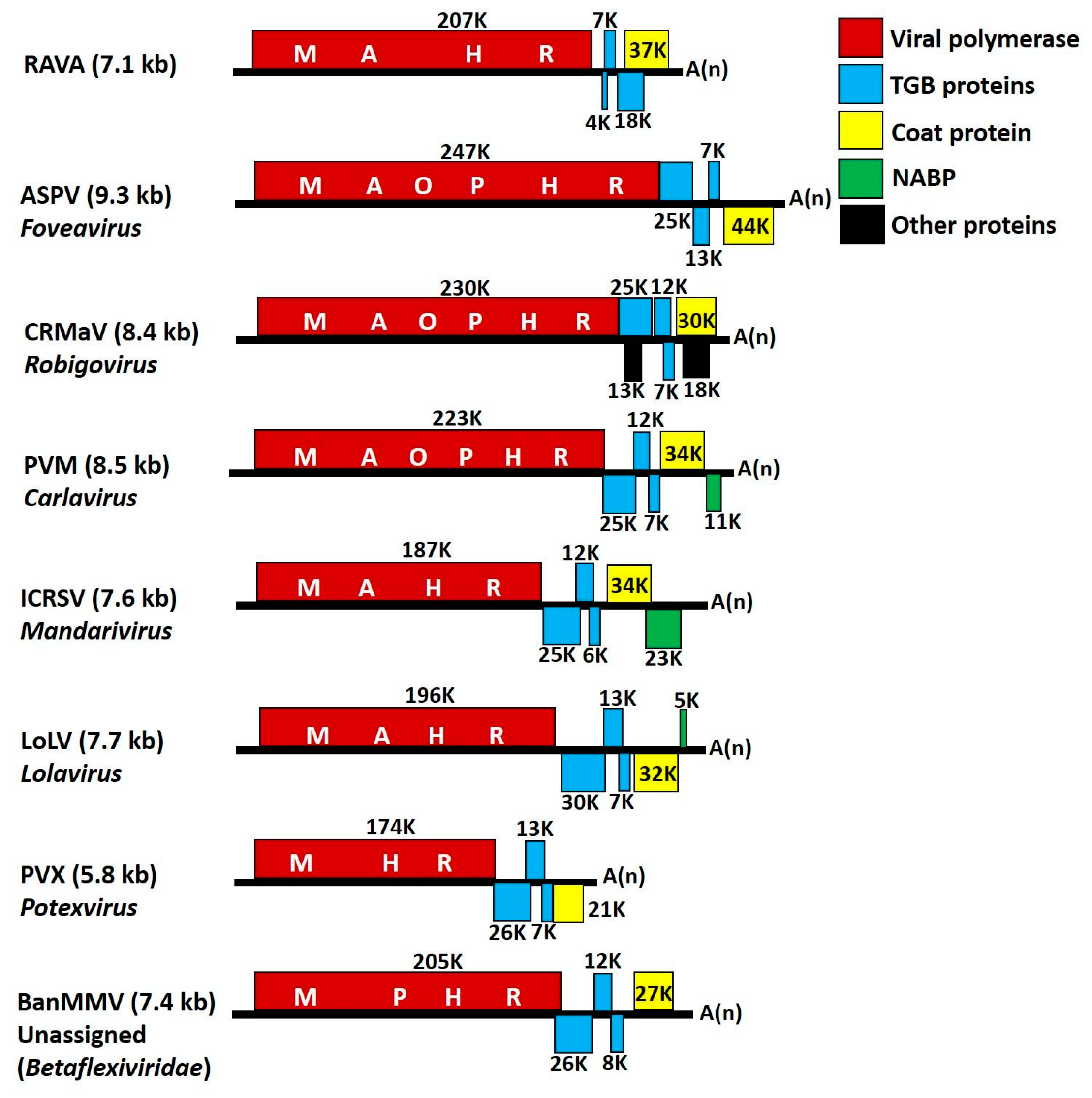
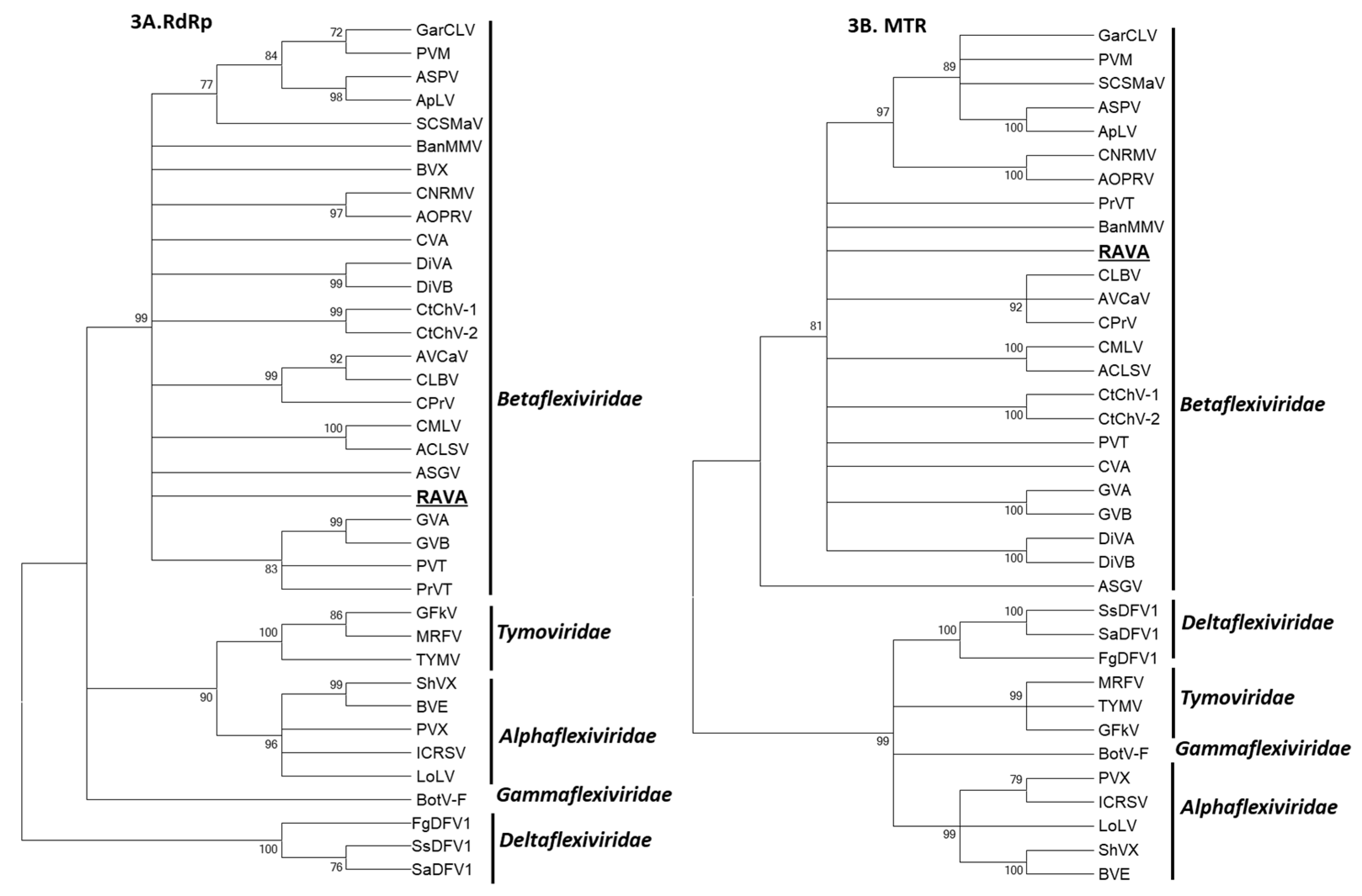
Double-stranded RNAs were extracted from 20 g of symptomatic leaves from currant accession PI 617879 (Figure 1) and subjected to a degenerate oligonucleotide-primed reverse transcription-PCR (DOP RT-PCR) followed by HTS and sequence assembly as described [10]. The FirstChoice® RLM-RACE Kit (ThermoFisher Scientific, Waltham, MA, USA) was used to obtain the 5′ terminus. The 5′ and 3′ RACE-RT-PCRs were performed using virus-specific primers and programs described in Table S1. For RACE and confirmation of genome regions downstream of ORF 1 total nucleic acids were extracted as described [3]. For all amplification reactions, Takara LA Taq polymerase (Takara Bio USA Inc, Mountain View, CA, USA) was used according to the manufacturer’s instructions. All PCR products were sequenced for at least three-fold coverage of the regions. The complete nucleotide sequence of RAVA was deposited in GenBank under accession number MF166685.
The genome organization, putative ORFs, and protein sequences were derived using the NCBI ORF Finder [11]. RNA binding amino acid (aa) residues present in RAVA proteins were identified using Pprint (Prediction of Protein RNA-Interaction; [12]) and transmembrane domains identified using TMHMM Server v. 2.0 [13]. The secondary structures of the proteins were predicted by PSI-Pred, I-TASSER [14,15], and nuclear localization signals identified with cNLS Mapper [16]. The protein sequences of members belonging to the families in the order Tymovirales were obtained from GenBank (Table 1) and aligned with the virus putative proteins using ClustalW on Bioedit [17]. Phylogenetic analyses were performed using the maximum likelihood method with 1000 bootstrap replicates on MEGA v. 7 [18] and phylogenetic trees displayed using Treeview in MEGA v. 7.
Tissue, tested positive for the virus by RT-PCR, was examined by transmission electron microscopy as follows: Leaf and root tissues were either thin sectioned using a microtome or macerated on a glass slide in water. Sap was applied to a Formvar coated copper EM grid and incubated for 1 min at room temperature. The excess liquid was drawn off with filter paper and samples stained by adding 4 μL of 2% stain for 1 min at room temp before the excess liquid was drawn off using filter paper. For thin sectioning, material was cut into 2 mm sections and fixed in modified Karnovsky fixative overnight at 4 °C. After fixation, the samples were rinsed in 0.1 M sodium cacodylate buffer for 10 min followed by post fixation in 1.5% potassium ferrocyanide and 2% osmium tetroxide in water. Staining was followed by incubations with uranyl acetate overnight and lead aspartate for 30 min at 60 °C. Samples were then dehydrated in acetone (10%, 30%, 50%, 70%, 90%, 95%, 100%) for 10 min each and infiltrated with Araldite resin. Ultrathin sections were done using an RMC PowerTome PC microtome. Grids were stained with either uranyl acetate or phosphotungstic acid and examined in an FEI Titan ChemiSTEM TEM.

3. Results

The genome of RAVA is 7106 nucleotides (nt), excluding the poly-A tail and begins with the pentanucleotide motif GAAAA1–5. The genome encodes five ORFs with untranslated regions (UTRs) of 140 and 172 nt for the 5′ and 3′ termini, respectively (Figure 2).
ORF 1 encodes a putative protein of 207 K (p1; 1826 aa), presumably involved in virus replication as it contains domains of methyl transferase (MTR; aa 44–334; Cdd: pfam01660), AlkB homologue of 2OG-Fe(II) oxygenase (aa 712–806; pfam13532), helicase (HEL; aa 1031–1266; pfam01443), and RNA dependent RNA polymerase (RdRp; aa 1526–1732; pfam00978). The polyprotein contains five nuclear localization signals; one monopartite signal, AVRKRLRFA1436-44 and four bipartite signals; FAKCRQLDPENLLLSEALLVNDLIKWLRE328-65, KIRLGSKDRVIGSCKDWTTKVIKISKG913-39, GCGKTKPLMDLILKSNDNILILVPRKRLGDS1034-64, and PRKRLGDSWTSKMGHKKNVRV1057-78. The entire protein shares low aa identities with replicases of other members of Tymovirales showing the highest identity (23%) to the orthologs in members of the genus Chordovirus (carrot Ch virus 1 and 2; CtChV-1 and CtChV-2). The sequence identity of the conserved domains of RdRp ranges from 21–57%; the highest being with members of Betaflexiviridae (Trichovirus and Prunevirus; 55–57%) and the lowest with members of the Tymoviridae, Alpha-, Gamma- and Deltaflexiviridae (21–32%) (data not shown). The MTR domain also showed higher identity to members of the Betaflexiviridae (24–33%) compared to the other families in Tymovirales.
Predicted products of ORFs 2–5 showed no significant similarity to viral proteins, and for this reason, HTS products were verified by Sanger sequencing performed on RT-PCR products of viral dsRNA as well as total nucleic acids extracted to eliminate the possibility of contamination (Table S1). ORF 2, which is separated from ORF 1 by an intergenic region of 49 nt encodes a putative peptide of 4 K (p2, 39 aa) with a transmembrane domain (aa13-35). ORF 3 partially overlaps ORF 2 and encodes a peptide of 60 aa (p3) with a calculated molecular mass of 7 K with two predicted transmembrane domains at the N and C termini (aa7-24,34-56). The secondary structures of the transmembrane regions contain one and two α-helixes respectively (Figure S1). In p2 the exposed hydrophilic regions were positively charged containing four Lys residues at the N terminal and two Arg residues at the C terminal. Both hydrophilic and hydrophobic regions contained nine aa residues predicted to interact with RNA (Table 2). P3 contains only two RNA interacting aa residues and their significance in RNA binding is to be determined (Table 2).
Separated by an intergenic region of five nt, the fourth ORF encodes a protein of 159 aa with molecular mass of 18K (p4) (Figure 2). This putative protein has marginal identity (24% identity, blossom 45) with a bacterial superfamily 1 (SF1) ATP-dependent DNA helicase domain. The predicted secondary structure (Figure S1) of the protein contains coils, α-helixes, and β-sheets with 60% of the aa residues predicted to interact with RNA (Table 2). These aa appeared as long stretches of primarily positively charged residues (Lys and Arg) which were located in the coils present in the N- and C-termini as well as in the α-helixes. The central region of the protein contained the longest stretch (50 RNA interacting aa) in two α-helixes, which was highly rich in Lys and Arg (30%) in a region mapped to the helicase domain (Table 2).
ORF 5 encodes a putative 37 K (p5, 321 aa) protein of unknown function speculated to be the capsid protein (CP) of the virus. This protein contains several α-helixes and coils and a single β-sheet (Figure S1). Among several RNA binding residues throughout the protein (Table 2), a stretch of 15 residues was predicted in the second and third α-helixes from the N-terminal containing seven Arg residues. The putative CP also has two aa residues (Glu280,283) predicted to be involved in ligand binding with calcium (Ca) (I-TASSER; [15]).
The phylogenetic analysis using the conserved domains of the RdRp and MTR (conserved in the alphavirus-like SF [19]) loosely placed RAVA with members of Betaflexiviridae (Figure 3A,B), with clustering only supported by low bootstrap values. The analysis based on p4 and CP with the putative orthologs yielded unreliable phylogeny due to extremely low bootstrap values, and is not presented here.
Tissue tested positive for the virus by RT-PCR was used in electron microscopy. No virus-like particles were identified, nor were there any structures that could belong to an endophyte, a potential host of the virus (data not shown).

4. Discussion

RAVA has features bridging genera in both the Alphaflexiviridae and Betaflexiviridae families. Because of the unique genome organization, we employed electron microscopy on tissue that was tested positive for the virus to determine whether a bacterial or fungal endophyte may be the cryptic host of the virus. In the absence of any structures other than those associated with the plant cell, we determined that RAVA is a plant virus. The genome initiated with the pentanucleotide sequence (GAAAA1-5) that is conserved in Alpha- and Betaflexiviridae [20,21,22,23] and thought to be essential for positive-strand RNA synthesis and protein translation [24,25]. The genome encodes five ORFs with resemblance to the genomes of members with TGB movement proteins, such as Potexvirus, Allexivirus, Lolavirus, Mandarivirus (all belonging to Alphaflexiviridae) and Foveavirus, Robigovirus, Carlavirus, and the unassigned members (in the Betaflexiviridae) (Figure 2). The replicase size is characteristic of a ‘carlavirus-like replicase’ (>195 K), when compared to the ‘potex-like’ (<195 K) [9,26]. This protein contains conserved domains of MTR, HEL, AlkB, and RdRp domains that are present in members of both families. The RAVA replicase showed the highest aa identity to its conserved orthologous domains (MTR and RdRp) of members of the Betaflexiviridae.
Despite the similarity in the 5′ part of the genome coding for the replicase, the RAVA genome differed significantly from genomes of members of the Alphaflexiviridae and Betaflexiviridae (Figure 2). First and foremost, the putative RAVA MP (p2–p4) and CP do not share any detectable similarity to any known viral proteins. In general, TGB movement proteins are of two types: ‘Hordei-like’ (rod shaped viruses of Hordeivirus, Benyvirus, Pomovirus, and Pecluvirus), and ‘potex-like’ (members of Tymovirales), and are usually overlapping [27], with the exception of TGBps reported in the genus Robigovirus and an unassigned member, sugarcane striate mosaic-associated virus, in which only two, TGBp 2 and TGBp 3, are overlapping.
The organization of TGB ORFs is highly conserved among plant viruses [27,28]. Potex-like TGB proteins occur in the order of a bigger sized protein, TGBp 1 (~25 K) followed by smaller proteins, TGBp 2 and TGBp 3, (~12 K and ~7 K respectively; Figure 2) in which TGBp 2 has two and TGBp 3 has single transmembrane domains bordered by charged residues. TGBp 2 and TGBp 3 are membrane proteins and localize on endomembranes and cell walls [26,27]. TGBp 1 of these viruses usually harbors an NTP binding helicase domain of SFI and has ATPase, RNA binding, and RNA helicase activity, and is believed to increase the size exclusion limit of plasmodesmata [26]. Based on protein size and characteristics, RAVA seems to have potex-like TGB proteins, however the arrangement of the putative TGB ORFs (ORFs 2–4) is reversed. The RNA binding site of potexviral TGBp1 conserved positively charged residue (Arg), which is essential for interaction with RNA [27], as with the case of RAVA p4 protein (Table 2). The size of these three proteins was significantly different from other members (Figure 2). Although there was no significant sequence identity with its putative orthologs, the presence of transmembrane domains in p2 and p3 proteins and the NTP binding helicase domain in p4 makes us hypothesize that these proteins constitute the TGB movement proteins of RAVA.
ORFs downstream of the viral replicase are thought to be translated from 3′-terminal subgenomic RNAs in which TGB proteins get translated through leaky scanning mechanisms in all families of the order other than Tymoviridae [27]. Sequence context around the initiation codon affects the translation efficiency of mRNAs. The consensus optimal context for translation initiation in mammals is GCC(A/G)CCAUGG (the initiation codon in bold; [29]). In some cases, viruses also follow this rule [30]. The Kozak context of translation initiation was observed in RAVA ORF 5, although its initiation codon is 15 nt downstream of the overlapping ORF 4. This is expected if the protein is expressed, given that the ribosomes first identify the start codon of p4, and the CP initiation context needs to be optimal if any ribosomes are to bypass that of p4.
The last ORF of most Alpha- and Betaflexiviridae members is the CP (Figure 2). However, the last ORF of RAVA lacked the domains conserved in the CP of flexuous viruses [31]. The predicted RNA binding region of the protein is rich in Arg. Arg-rich RNA binding motif in the CP is known to be involved in genome binding and subsequent packaging [32]. The protein also has aa residues thought to be involved in Ca binding. Calcium plays an important role in the assembly and disassembly and/or replication processes in viruses [33,34,35]. The size of the protein is similar to the range shown by members that have TGB MP (28–45 K; Figure 2). The position in the genome, predicted size, Arg-rich RNA binding region, and presence of Ca binding sites suggests that the protein is the RAVA CP.
The genomes of the members of Tymovirales are highly diverse with respect to the number and organization of genes, indicative of the major role of recombination in the evolution of this virus group. There is significant diversity in the 3′ region downstream of viral replicase (30 K/TGB MP, CP, and NABP) suggestive of possible recombination events in which the ancestral viruses had a common 5′ part while acquiring the 3′ genes from different origins, which further diversified over time [36]. Several studies have also described evidence of recombination in the members of this group [37,38,39,40,41]. Martelli et al. [26] suggested coevolution of MP and CP in this virus group. RAVA genome follows this theory in which the replicase, although with low bootstrap values, groups with the Betaflexiviridae, whereas the remaining proteins are phylogenetically distant. Changes in phylogenetic relationships for different regions of the genome of a virus is an indication of recombination [42]. RAVA polymerase is similar to the members of the Trivirinae subfamily, however the rest of the genome is Quinvirinae-like (Carlavirus, Foveavirus, Robigovirus; Figure 2) in which the TGBp-like proteins’ organization is reversed, suggesting the role of recombination between these two subfamilies in the evolution of this novel virus.

5. Conclusions

RAVA is a unique, previously undescribed virus of tymo-like lineage which cannot be assigned to any of the currently recognized virus taxa. With regard to its molecular features such as the number of genes encoded in the genome, organization, and the presence of conserved pentanucleotide at 5′ termini, RAVA resembles members of the order Tymovirales. Phylogenetic analysis based on conserved RdRp domain, the principal determinant in the evolutionary framework of positive-strand RNA viruses, and MTR domain, clearly qualify RAVA as a putative species in the Betaflexiviridae family. However, differences in the genome downstream of polymerase (organization of TGBp-like proteins and lack of conserved domains of ortholog proteins) and the distant phylogenetic relationship with other members distinguishes it from both existing genera and unassigned members of the family, suggesting a tentative classification. Therefore, we propose that RAVA represents a new, monotypic genus in the family Betaflexiviridae or a family (given the low replicase aa identities with existing viruses) for which the names Ravavirus/Ravaviridae are proposed.

Supplementary Materials

The following are available online at https://www.mdpi.com/1999-4915/10/8/406/s1, Figure S1: Predicted secondary structures (PSI-Pred; [14]) of the Ribes americanum virus A proteins. Table S1: Primers and PCR conditions used for the amplification of the Ribes americanum virus A genome.

Author Contributions

T.T.-V., T.H., and I.E.T. conceived and designed the experiments; T.T.-V. and T.H. performed the experiments; T.T.-V. and T.H. analyzed the data; T.T.-V. and I.E.T. wrote the paper; J.D.P. and R.R.M. did the electron microscopy work. All authors approved the final version of the manuscript.

Funding

This research was funded by USDA awards 14-8130-0392-CA and Hatch project 1002361.

Conflicts of Interest

The authors declare no conflict of interest. The funding sponsors had no role in the design of the study; in the collection, analyses, or interpretation of data; in the writing of the manuscript, and in the decision to publish the results.

References

  1. Barba, M.; Czosnek, H.; Hadidi, A. Historical perspective, development and applications of next-generation sequencing in plant virology. Viruses 2014, 6, 106–136. [Google Scholar] [CrossRef] [PubMed]
  2. Di Bello, P.L.; Laney, A.G.; Druciarek, T.; Ho, T.; Gergerich, R.C.; Keller, K.E.; Martin, R.R.; Tzanetakis, I.E. A novel emaravirus is associated with redbud yellow ringspot disease. Virus Res. 2016, 222, 41–47. [Google Scholar] [CrossRef] [PubMed] [Green Version]
  3. Ho, T.; Tzanetakis, I.E. Developing a virus detection and discovery pipeline using next generation sequencing. Virology 2014, 471–473, 54–60. [Google Scholar] [CrossRef] [PubMed]
  4. Massart, S.; Olmos, A.; Jijakli, H.; Candresse, T. Current impact and future directions of high throughput sequencing in plant virus diagnostics. Virus Res. 2014, 188, 90–96. [Google Scholar] [CrossRef] [PubMed]
  5. Postman, J.; Hummer, K.; Stover, E.; Krueger, R.; Forsline, P.; Grauke, L.J.; Zee, F.; Ayala-Silva, T.; Irish, B. Fruit and nut genebanks in the US national plant germplasm system. HortScience 2006, 41, 1188–1194. [Google Scholar]
  6. Terry, L. Health-Promoting Properties of Fruit and Vegetables; CABI: Wallingford, UK, 2014; p. 432. [Google Scholar]
  7. Thekke-Veetil, T.; Ho, T.; Postman, J.D.; Tzanetakis, I.E. Molecular characterization of a new member of the genus Waikavirus. Phytopathology 2017, 107, 102. [Google Scholar]
  8. ICTV. Virus Taxonomy 2017 Release, Order Tymovirales. Available online: https://talk.ictvonline.org/taxonomy/ (accessed on 31 July 2018).
  9. Adams, M.J.; Antoniw, J.F.; Bar-Joseph, M.; Brunt, A.A.; Candresse, T.; Foster, G.D.; Martelli, G.P.; Milne, R.G.; Fauquet, C.M. The new plant virus family Flexiviridae and assessment of molecular criteria for species demarcation. Arch. Virol. 2004, 149, 1045–1060. [Google Scholar] [CrossRef] [PubMed]
  10. Ho, T.; Martin, R.R.; Tzanetakis, I.E. Next-generation sequencing of elite berry germplasm and data analysis using a bioinformatics pipeline for virus detection and discovery. In Plant Pathology: Techniques and Protocols (Methods in Molecular Biology); Lacomme, C., Ed.; Springer: New York, NY, USA, 2015; pp. 301–313. [Google Scholar]
  11. Wheeler, D.L.; Church, D.M.; Federhen, S.; Lash, A.E.; Madden, T.L.; Pontius, J.U.; Schuler, G.D.; Schriml, L.M.; Sequeira, E.; Tatusova, T.A.; et al. Database resources of the National Center for Biotechnology. Nucleic Acids Res. 2003, 31, 28–33. [Google Scholar] [CrossRef] [PubMed] [Green Version]
  12. Kumar, M.; Gromiha, M.M.; Raghava, G.P.S. Prediction of RNA binding sites in a protein using SVM and PSSM profile. Proteins 2008, 71, 189–194. [Google Scholar] [CrossRef] [PubMed]
  13. Krogh, A.; Larsson, B.; von Heijne, G.; Sonnhammer, E.L.L. Predicting transmembrane protein topology with a hidden Markov model: Application to complete genomes. J. Mol. Biol. 2001, 305, 567–580. [Google Scholar] [CrossRef] [PubMed]
  14. Jones, D.T. Protein secondary structure prediction based on position-specific scoring matrices. J. Mol. Biol. 1999, 292, 195–202. [Google Scholar] [CrossRef] [PubMed]
  15. Yang, J.; Yan, R.; Roy, A.; Xu, D.; Poisson, J.; Zhang, Y. The I-TASSER Suite: Protein structure and function prediction. Nat. Methods 2015, 12, 7–8. [Google Scholar] [CrossRef] [PubMed]
  16. Kosugi, S.; Hasebe, M.; Tomita, M.; Yanagawa, H. Systematic identification of yeast cell cycle-dependent nucleocytoplasmic shuttling proteins by prediction of composite motifs. Proc. Natl. Acad. Sci. USA 2009, 106, 10171–10176. [Google Scholar] [CrossRef] [PubMed]
  17. Hall, T.A. BioEdit: A user-friendly biological sequence alignment editor and analysis program for Windows 95/98/NT. Nucleic Acids Symp. Ser. 1999, 41, 95–98. [Google Scholar]
  18. Kumar, S.; Stecher, G.; Tamura, K. MEGA7: Molecular evolutionary genetics analysis version 7.0 for bigger datasets. Mol. Biol. Evol. 2016, 33, 1870–1874. [Google Scholar] [CrossRef] [PubMed]
  19. Koonin, E.V.; Dolja, V.V. Evolution and taxonomy of positive-strand RNA viruses: Implications of comparative analysis of amino acid sequences. Crit. Rev. Biochem. Mol. Biol. 1993, 28, 375–430. [Google Scholar] [CrossRef] [PubMed]
  20. Li, R.; Mock, R. Characterization of a flowering cherry strain of cherry necrotic rusty mottle virus. Arch. Virol. 2008, 153, 973–978. [Google Scholar] [CrossRef] [PubMed]
  21. Sabanadzovic, S.; Abou Ghanem-Sabanadzovic, N.; Tzanetakis, I.E. Blackberry virus E: An unusual flexivirus. Arch. Virol. 2011, 156, 1665–1669. [Google Scholar] [CrossRef] [PubMed]
  22. Villamor, D.V.; Druffel, K.L.; Eastwell, K.C. Complete nucleotide sequence of a virus associated with rusty mottle disease of sweet cherry (Prunus avium). Arch. Virol. 2013, 158, 1805–1810. [Google Scholar] [CrossRef] [PubMed]
  23. Zhang, Y.P.; Kirkpatrick, B.C.; Smart, C.D.; Uyemoto, J.K. cDNA cloning and molecular characterization of cherry green ring mottle virus. J. Gen. Virol. 1998, 79, 2275–2281. [Google Scholar] [CrossRef] [PubMed]
  24. Verchot-Lubicz, J.; Ye, C.M.; Bamunusinghe, D. Molecular biology of potexviruses: Recent advances. J. Gen. Virol. 2007, 88, 1643–1655. [Google Scholar] [CrossRef] [PubMed]
  25. Wong, S.M.; Mahtani, P.H.; Lee, K.C.; Yu, H.H.; Tan, Y.; Neo, K.K.; Chan, Y.; Wu, M.; Chng, C.G. Cymbidium mosaic potexvirus RNA: Complete nucleotide sequence and phylogenetic analysis. Arch. Virol. 1997, 142, 383–391. [Google Scholar] [CrossRef] [PubMed]
  26. Martelli, G.P.; Adams, M.J.; Kreuze, J.F.; Dolja, V.V. Family Flexiviridae: A case study in virion and genome plasticity. Annu. Rev. Phytopathol. 2007, 45, 73–100. [Google Scholar] [CrossRef] [PubMed]
  27. Morozov, S.Y.; Solovyev, A.G. Triple gene block: Modular design of a multifunctional machine for plant virus movement. J. Gen. Virol. 2003, 84, 1351–1366. [Google Scholar] [CrossRef] [PubMed]
  28. Rupasov, V.V.; Morozov, S.Y.; Kanyuka, K.V.; Zavriev, S.K. Partial nucleotide sequence of potato virus M RNA shows similarities to potexviruses in gene arrangement and the encoded amino acid sequences. J. Gen. Virol. 1989, 70, 1861–1869. [Google Scholar] [CrossRef] [PubMed]
  29. Kozak, M. Point mutations define a sequence flanking the AUG initiator codon that modulates translation by eukaryotic ribosomes. Cell 1986, 44, 283–292. [Google Scholar] [CrossRef]
  30. Sánchez-Puig, J.M.; Blasco, R. AUG context and mRNA translation in vaccinia virus. Span. J. Agric. Res. 2008, 6, 73–80. [Google Scholar] [CrossRef]
  31. Dolja, V.V.; Boyko, V.P.; Agranovsky, A.A.; Koonin, E.V. Phylogeny of capsid proteins of rod-shaped and filamentous RNA plant viruses: Two families with distinct patterns of sequence and probably structure conservation. Virology 1991, 184, 79–86. [Google Scholar] [CrossRef]
  32. Choi, Y.G.; Rao, A.L.N. Molecular studies on bromovirus capsid protein: VII. Selective packaging of BMV RNA4 by specific N-terminal arginine residues. Virology 2000, 275, 207–217. [Google Scholar] [CrossRef] [PubMed]
  33. Abdel-Meguid, S.; Yamane, S.T.; Fukuyama, K.; Rossmann, M.G. The location of calcium ions in southern bean mosaic virus. Virology 1981, 114, 81–85. [Google Scholar] [CrossRef]
  34. Durham, A.C.; Hendry, D.A. Cation binding by tobacco mosaic virus. Virology 1977, 77, 510–519. [Google Scholar] [CrossRef]
  35. Gajardo, R.; Vende, P.; Poncet, D.; Cohen, J. Two proline residues are essential in the calcium-binding activity of rotavirus VP7 outer capsid protein. J. Virol. 1997, 71, 2211–2216. [Google Scholar] [PubMed]
  36. Marais, A.; Faure, C.; Mustafayev, E.; Candresse, T. Characterization of new isolates of apricot vein clearing-associated virus and of a new Prunus-Infecting virus: Evidence for recombination as a driving force in Betaflexiviridae evolution. PLoS ONE 2015, 10, e0129469. [Google Scholar] [CrossRef] [PubMed]
  37. Alabi, O.J.; Al Rwahnih, M.; Mekuria, T.A.; Naidu, R.A. Genetic diversity of grapevine virus A in Washington and California vineyards. Phytopathology 2014, 104, 548–560. [Google Scholar] [CrossRef] [PubMed]
  38. Singh, L.; Hallan, V.; Martin, D.P.; Ram, R.; Zaidi, A.A. Genomic sequence analysis of four new chrysanthemum virus B isolates: Evidence of RNA recombination. Arch. Virol. 2012, 157, 531–537. [Google Scholar] [CrossRef] [PubMed]
  39. Villamor, D.E.; Eastwell, K.C. Viruses associated with rusty mottle and twisted leaf diseases of sweet cherry are distinct species. Phytopathology 2013, 103, 1287–1295. [Google Scholar] [CrossRef] [PubMed]
  40. Yoon, J.Y.; Joa, J.H.; Choi, K.S.; Do, K.S.; Lim, H.C.; Chung, B.M. Genetic diversity of a natural population of apple stem pitting virus isolated from apple in Korea. Plant Pathol. J. 2014, 30, 195–199. [Google Scholar] [CrossRef] [PubMed]
  41. Zanardo, L.G.; Silva, F.N.; Lima, A.T.M.; Milanesi, D.F.; Castilho-Urquiza, G.P.; Almeida, A.M.R. Molecular variability of cowpea mild mottle virus infecting soybean in Brazil. Arch. Virol. 2014, 159, 727–737. [Google Scholar] [CrossRef] [PubMed]
  42. Salminen, M. Detecting recombination in viral sequences. In The Phylogenetic Handbook: A Practical Approach to DNA and Protein Phylogeny; Salemi, M., Vandamme, A.M., Eds.; University Press: Cambridge, UK, 2003; pp. 348–377. [Google Scholar]
Figure 1. Symptoms observed on American blackcurrant infected with Ribes americanum virus A. Infected Ribes americanum cultivar Gall (PI 617879, left) showing ragged leaf margins and crinkled leaf surface compared to healthy plant (right).
Viruses 10 00406 g001
Figure 2. Genome organization of Ribes americanum virus A (RAVA) in comparison to the genomes of members of Tymovirales with triple gene block (TGB) movement proteins. M—methyltransferase, A—AlkB, O—OTu-like peptidase, P—papain-like protease, H—helicase, and R—RNA-dependent RNA polymerase. Boxes represent open reading frames. Size of the proteins encoded in the ORFs is indicated. Abbreviations: ASPV—apple stem pitting virus, CRMaV—cherry rusty mottle associated virus, PVM—potato virus M, ICRSV—Indian citrus ringspot virus, LoLV—lolium latent virus, PVX—potato virus X, BanMMV—banana mild mosaic virus.
Viruses 10 00406 g002
Figure 3. Phylogenetic relationship of Ribes americanum virus A (RAVA) with members of the order Tymovirales. Phylogenetic analysis was performed using the conserved domains of RNA-dependent RNA polymerase (RdRp; 3A) and methyl transferase (MTR; 3B). The trees were generated by the maximum likelihood method using MEGA 7 and bootstrap values (indicated for each branch node) were estimated using 1000 bootstrap replicates. The branches with bootstrap values less than 70% are collapsed. The details of virus isolates used for the studies are provided in Table 1.
Viruses 10 00406 g003
Table 1. Details of virus isolates used for the phylogenetic studies of Ribes americanum A. RdRp-RNA dependent RNA polymerase, MTR-Methyl transferase, α-Alphaflexiviridae, β-Betaflexiviridae, γ-Gammaflexiviridae, Λ-Deltaflexiviridae, T-Tymoviridae.
Virus AcronymsVirus NamesGenus (Family)RdRp/MTR
ACLSVApple chlorotic leaf spot virusTrichovirus (β)NP040551.1
CMLVCherry mottle leaf virusTrichovirus (β)AOY07780.1
GVAGrapevine virus AVitivirus (β)AFV73358.1
GVBGrapevine virus BVitivirus (β)AIL90366.1
CNRMVCherry necrotic rusty mottle virusRobigovirus (β)NP059937.1
AOPRVAfrican oil palm ringspot virusRobigovirus (β)YP002776347.1
CtChV-1Carrot Ch virus 1Chordovirus (β)AHA85534.1
CtChV-2Carrot Ch virus 2Chordovirus (β)AHA85531.1
DiVADiuris virus ADivavirus (β)YP006905850.1
DiVBDiuris virus BDivavirus (β)AFV57240.1
AVCaVApricot vein clearing associated virusPrunevirus (β)AKN09002.1
CPrVCaucasus prunus virusPrunevirus (β)AKN08994.1
PVTPotato virus TTepovirus (β)AFU55321.1
PrVTPrunus virus TTepovirus (β)YP009051684.1
ASGVApple stem grooving virusCapillovirus (β)APT42870.1
CVACherry virus ACapillovirus (β)AMH87272.1
GarCLVGarlic common latent virusCarlavirus (β)AGG13282.1
PVMPotato virus MCarlavirus (β)AHL30493.1
ASPVApple stem pitting virusFoveavirus (β)NP604464.1
ApLVApricot latent virusFoveavirus (β)YP004089619.1
CLBVCitrus leaf blotch virusCitrivirus (β)NP624333.1
BanMMVBanana mild mosaic virusUnassigned (β)NP112029.1
SCSMaVSugarcane striate mosaic-associated virusUnassigned (β)NP624313.1
BVXBanana virus XUnassigned (β)AAW50958.1
BotV-FBotrytis virus FMycoflexivirus (γ)NP068549.1
ShVXShallot virus XAllexivirus (α)NP620648.1
ICRSVIndian citrus ringspot virusMandarivirus (α)AAK97522.1
PVXPotato virus XPotexvirus (α)BAE07083.1
LoLVLolium latent virusLolavirus (α)YP001718499.1
BVEBlackberry virus EUnassigned (α)AEI17897.1
SaDFV1Soybean associated deltaflexivirus 1Deltaflexivirus (Λ)ALM62223.1
FgDFV1Fusarium deltaflexivirus 1Deltaflexivirus (Λ)YP009268710.1
SsDFV1Sclerotinia deltaflexivirus 1Deltaflexivirus (Λ)AMD16208.1
GFkVGrapevine fleck virusMaculavirus (T)NP542612.1
MRFVMaize rayado fino virusMarafivirus (T)NP115454.1
TYMVTurnip yellow mosaic virusTymovirus (T)AMH40140.1
Table 2. RNA interacting amino acid residues predicted in Ribes americanum virus A proteins. Amino acid residues in red color are predicted to interact with RNA. Shaded regions are predicted transmembrane domain(s) in p2 and p3 proteins whereas in p4 it indicates the region aligned to the helicase domain of ATP-dependent DNA helicase.
p2
MNFKSYLLKKIKSVGIGLASSLIIYIASFVFNVLYRRSF
p3
MCYIDVAFDLVCLFICVLILVALLKLTYCNSSAFCVALALTIYSLFLNFNLLVLLYDLSR
p4
MYGYNNGIRKTSDRFSKGSVSKDKYGQRYNCGTDRLPFLVMADVSKLKIDFENATENMLFQIVSLLLHFCVLQNIGQ RKAKRGKIKKKKAAYNEYRRNKDGASSSYQGGGGLARTRDSQENERQVDAARDKRAEFYS
DSSSTEGDGDGSGQTRNERHFV
CP
MESEKLVIVSAKVPFRRTSMAKDTTAARTDFLSSLWRMSLNSKLISKMRQRTCYSRLCHSCCTSAYCKILARGRQREERLRRRKLPIMSTGEIRMEPLPVTREGEAWLELEIARKMKGKLTLQETNGRNSILTVAQPKEMV
MDLDRPEMRDILFNLDFTKRLIDQDVFVCSYLVKKAKRVGVEVCTDFHCYFVDTDMTVSALLDAIEIASFFGCINSAVFEICATGSCLCKVGLRELIIEVEKRTIEIPLKCGYHGIKHLTEVEDRQWKVLCANPLIKLEEIEE
IYIFWNSLGLKNHERHVKALLDVNGLKES TLRILGAI

