Review

Nuclear Import of Hepatitis B Virus Capsids and Genome

1
Laboratoire de Microbiologie Fondamentale et Pathogénicité, University of Bordeaux, UMR 5234, F-33076 Bordeaux, France
2
CNRS, Microbiologie Fondamentale et Pathogénicité, UMR 5234, F-33076 Bordeaux, France
3
Centre Hospitalier Universitaire de Bordeaux, Service de Virologie, F-33076 Bordeaux, France
*
Author to whom correspondence should be addressed.
Viruses 2017, 9(1), 21; https://doi.org/10.3390/v9010021
Submission received: 9 December 2016 / Revised: 17 January 2017 / Accepted: 17 January 2017 / Published: 21 January 2017
(This article belongs to the Special Issue Recent Advances in Hepatitis B Virus Research)

Abstract

:
Hepatitis B virus (HBV) is an enveloped pararetrovirus with a DNA genome, which is found in an up to 36 nm-measuring capsid. Replication of the genome occurs via an RNA intermediate, which is synthesized in the nucleus. The virus must have thus ways of transporting its DNA genome into this compartment. This review summarizes the data on hepatitis B virus genome transport and correlates the finding to those from other viruses.

1. Introduction

All retroviruses and practically all DNA viruses (exception Poxviridae family) need the nuclear machinery of the host cell for their replication. As a pararetrovirus, hepatitis B virus (HBV) requires multiple nuclear enzymatic activities, which are only partially understood but late activities needed for mRNA synthesis are evident. Also obvious are all enzymes that are involved in the repair of the relaxed circular virion DNA (rcDNA) to the covalently closed circular DNA (cccDNA), which is the template for transcription. Up to now, a few host factors in rcDNA-to-cccDNA conversion were identified: tyrosyl-DNA-phosphodiesterase 2 (TDP2), which releases the viral polymerase from the rcDNA [1] and the DNA polymerase κ (POLK) but also polymerase λ (POLL) and η (POLH), which are involved in repair of the single stranded part of the rc genome [2].
Amongst the viral mRNAs is the RNA pregenome, which is the template for translation of the viral capsid protein (core protein, Cp) and the viral polymerase (Pol). Pol binds to a specific structure on the pregenome called ε and the Pol-RNA complex becomes encapsidated by the assembling Cp, forming an immature, RNA-containing capsid. This initiates reverse transcription and subsequent second strand DNA synthesis [3]. Mature capsids but not RNA-containing capsids are enveloped by the viral surface proteins [4]. Infections with a duck hepatitis B virus mutant unable to code for a surface protein led to increased cccDNA copy number from 50 to 150 per cell [5,6] indicating that the progeny rcDNA capsids transport the genome to/into the nucleus as the virion-derived capsids during initial infection. For HBV the situation is however, less clear as the cccDNA copy number is much smaller. Considering that there is no proof for nuclear entry of rcDNA from progeny capsids, it cannot be concluded that cytoplasmic capsids derived from infection are identical with those being newly synthesized.
With a life span of approximately 200 days, hepatocytes are rarely dividing. Nuclear entry of the genome by passive trapping, as it was described for the human T-lymphotropicvirus-1 (HTLV-1) [7] and human papillomaviruses [8], would be thus inefficient. In contrast, HBV has to highjack cellular transport pathways as cellular transport factors facilitating nuclear import via the nuclear pore complexes (NPCs; see Figure 1). Throughout all viruses, one alternative pathway was described for the simian virus 40 (SV40), which cannot only pass via nuclear pores [9,10,11,12,13] but potentially disrupts the inner nuclear membrane after being internalized into the ER lumen [14].

2. Nuclear Pores and Nuclear Import Receptors

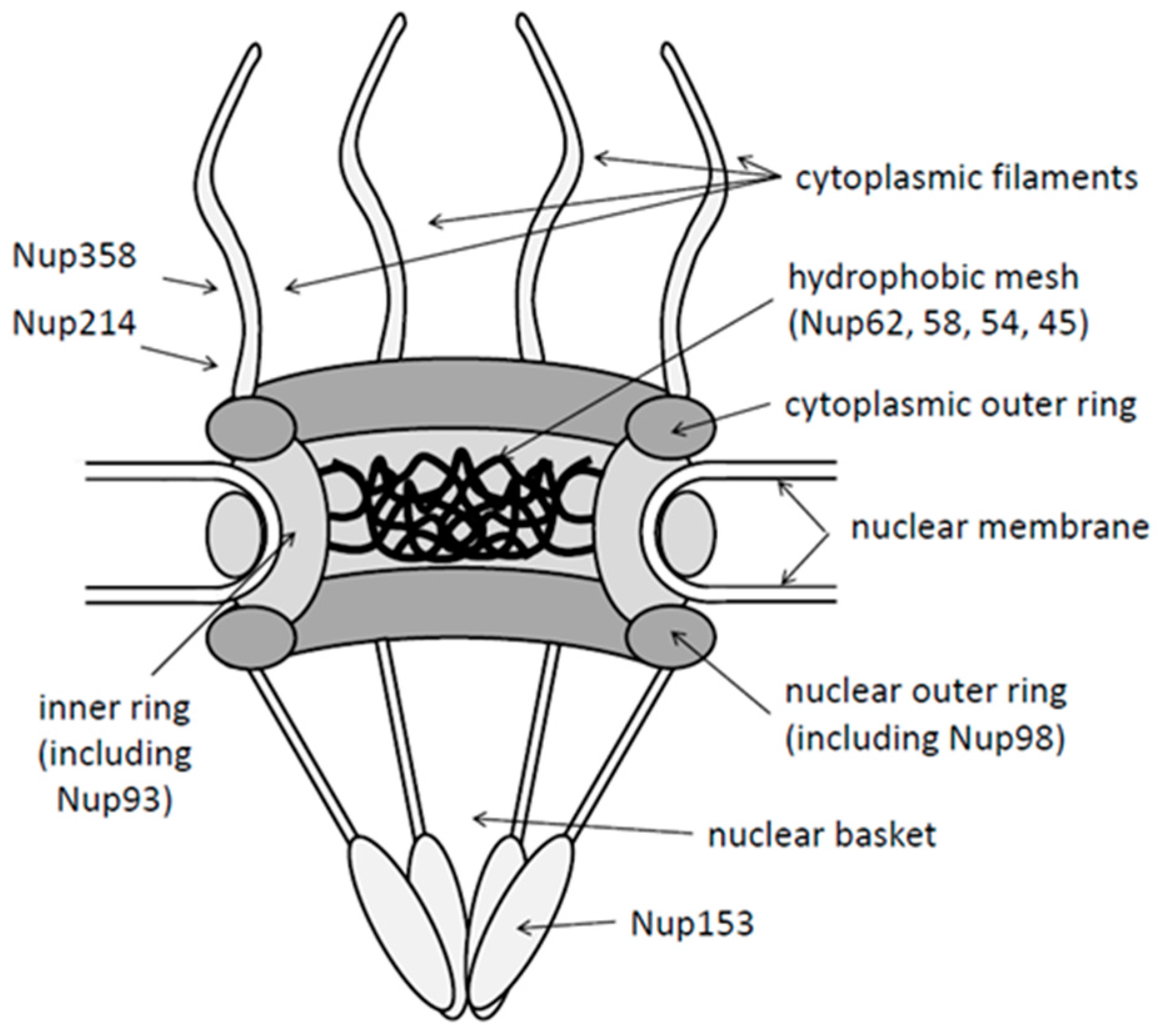
2.1. Structure of Nuclear Pores

NPCs are macromolecular complexes of >125 MDa having an octagonal rotation symmetry. NPCs are phylogenetically well conserved even between distant species like humans and Xenopus leavis. NPCs are composed of ~30 different proteins collectively called nucleoporins (Nups) (reviewed in [15]). Some Nups are organized in a symmetric ring in the nuclear envelope forming the pore. Other Nups exhibit an asymmetric distribution as e.g., Nup358 and Nup214, which are part of eight fibers emanating from the cytoplasmic face of the pore, and Nup153 and Tpr, which make up the so-called nuclear basket (reviewed in [16]). Nups are not stably incorporated into NPCs but vary substantially in their NPC residence times, which ranges between 13 s and 70 h [17,18]. Nups are not only essential for nuclear import and export but also for differentiation during embryogenesis, cell cycle, and intranuclear chromatin distribution [19,20]. One third of the Nups contains phenylalanine-glycine (FG)-repeats, which are essential for cell viability and which form a diffusion barrier inside the pore [21,22,23]. Passive diffusion through the NPC is limited to small molecules of up to 5 nm [24], while active transport of macromolecules, with a few exceptions, depends on nuclear transport receptors, collectively called importins or exportins (karyopherins). Active transport allows the translocation of ~1000 macromolecules per second and NPC [25] and the number of NPCs per nucleus varies upon the metabolic activity of the cell [26] (400 in Purkinje cells and 18,500 in oligodrendrocytes [27]). The NPC copy number is further regulated by the cell cycle, being higher during G2 phase than G1 phase (e.g., 8.5 to 5 NPCs/µm2 nuclear envelope in HeLa cells [28]). In electron microscopy, the channel forming the nuclear pore has a diameter of 40 nm and the maximal cargo size is 39 nm [29], which restricts import and export of large structures as viral capsids.

2.2. Nuclear Import Receptors

Nine nuclear import receptors (also called karyopherins; Kaps) which exhibit different cargo-specificities, have been described so far (reviewed in [30]). Most import receptors bind directly to their cargo as exemplified by transportin-1 (Kapβ2), which interacts with a proline-tyrosine (PY) nuclear localization signal (NLS). This signal, has been first characterized in the C-terminal M9 domain of the heterogeneous nuclear ribonucleoprotein A1 (hnRNP A1) leading to the name M9 signal [31,32]. In contrast, importin β (Kapβ1) interacts either directly with cargos comprising an importin-β-binding domain (IBB) or it binds indirectly to classical NLSs via the adapter protein importin α (Kapα). Importin α comprises an IBB for importin β-interaction and a site for the interaction with classical NLSs. While a canonical IBB comprises 13 basic amino acids in seven clusters distributed over 39 residues [33,34], classical NLSs are formed by clusters of typically (four to) six basic amino acids, which can be mono- or bipartite. The NLS prototype is found on SV40 T-antigen (SV40TAg [35,36]) exhibiting the amino acid sequence PKKKRKV [35]. A certain selectivity of nuclear import is achieved by the diversity of importin α isoforms, which show preferences for some NLSs. There are seven importin α subfamilies, which are all expressed in adult tissues except importin α6, which is testis-specific (reviewed in [37]).

3. Nuclear Import of Macromolecules

3.1. Nuclear Import Using Import Receptors

Classical active nuclear import is initiated by the interaction of the import receptor with the NLS on the cargo. This interaction can be regulated by different mechanisms: post-translational modifications can directly affect import receptor cargo-binding. This was shown for the phosphorylation of threonine 124 on the SV40TAg, which is directly adjacent to the NLS and which inhibits the nuclear import [38]. Another form of inactivation is the intermolecular masking as exemplified by the NLS of nuclear factor κB (NFκB), which is hidden by binding of IκB and which becomes accessible after IκB degradation [39]. Intramolecular NLS masking was described for nuclear factor of activated T-cells (NF-ATc) in which the NLS is hidden by association with phosphorserines [40].
Interaction with the cargo changes the structure of the import receptor [41,42], allowing interaction with Nups. First of the Nups is Nup358 (also known as RanBP2), which localizes in the extremity of cytoplasmic filaments extruding the NPCs. The subsequent step of translocation through the nuclear pore, which is filled by a hydrophobic mesh, is not fully elucidated. A number of mechanisms have been proposed, all involving FG-repeats of Nups in the central channel (Nups 98, 93, 62, 58, 54 and 45) and which interact with transport receptors. The “polymer brush model”, for instance, suggests that movements of the unfolded FG-Nups sweep away macromolecules [43,44,45]; the so called “collapse model” suggests a collapse of FG-repeats [46,47], and the “hydrophobic gel model”, also called “saturated model”, postulates that transport factors bind to the FG-repeats dissolving the FG-repeat cross-links [21,48].
Nuclear import is terminated in the nuclear basket where the cargo-import receptor-complex binds to Nup153 for then being dissociated by interaction between the import receptor and the ras-related nuclear protein (Ran) in its GTP-bound form. While the cargo diffuses deeper into the nucleus, the import receptor-RanGTP-complex is exported through the NPC. On the cytoplasmic face of the NPC, GTP is hydrolyzed to GDP, which dissociates RanGDP from the import receptor, resulting in recycling of the import receptor. Recycling of Ran requires then nuclear import of RanGDP using the nuclear transport factor 2 (NTF2) [49], which is followed by the replacement of GDP by GTP. This exchange reaction is catalyzed by the chromatin-bound Ran guanine nucleotide exchange factor (RanGEF; also termed regulator of chromosome condensation, RCC1). The driving force of the import reactions is the RanGTP concentration, which is 1000 fold higher in nucleus than in the cytoplasm [50].

3.2. Import Receptor-Independent Pathways

There are a few reports describing alternative pathways as the one for the calcium-binding proteins calmodulin and calreticulin [51]. Translocation is mediated by direct interaction with NPC components and is independent of carrier molecules. Nevertheless, a coexistence with an import receptor-dependent pathway is assumed. Moreover, calmodulin and calreticulin themselves can act as nuclear import receptors, as they import the transcription factors SRY and SOX9 [52]. Calreticulin can also act as a nuclear export factor, which was exemplified for the glucocorticoid receptor, the thyroid hormone receptor α1 and some viral proteins as e.g., HTLV-1 protein Tax [53,54,55,56].

4. Capsid Disassembly and Import of Other Viral Genomes

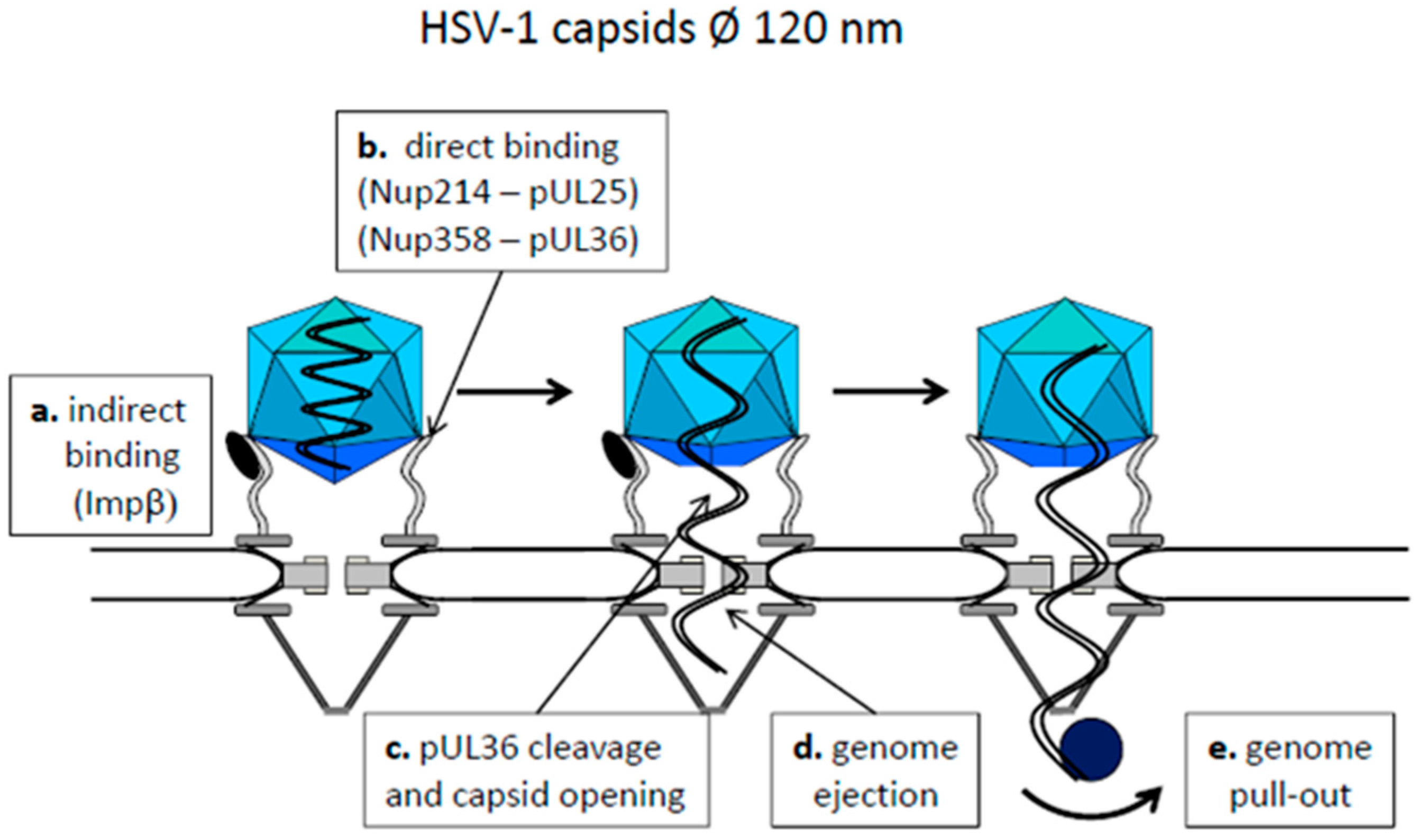
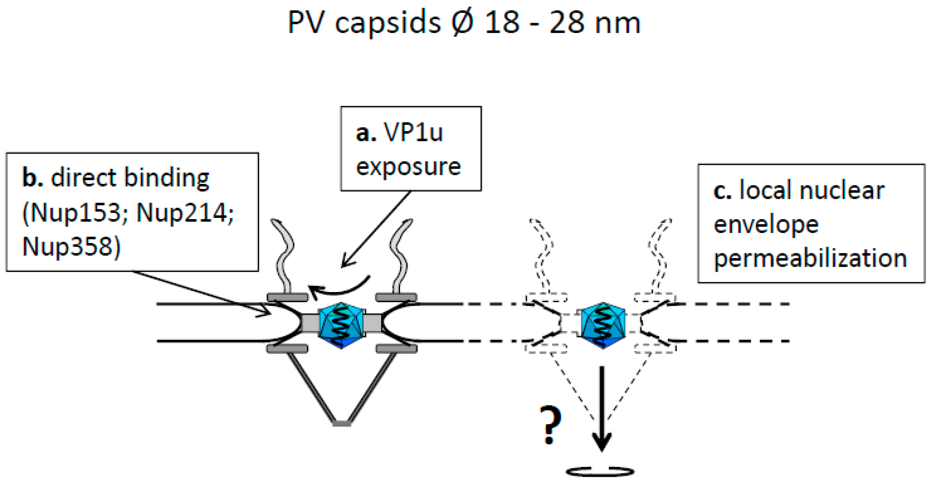
4.1. Capsids Larger than the Maximal Transport Diameter of the NPC

The release of viral genomes with a nuclear replication step largely depends on the surrounding structure, which can have the form of a capsid. As viral genomes comprise pathogen-associated molecular patterns (PAMPs) they will be sensed by cytosolic or membrane-bound pattern recognition receptors (PRRs) [57]. It must be thus assumed that the later the genome is released on its way to the nucleus, the lower the risk of triggering innate immunity is. Thus, most DNA- and retroviruses ensure the transport of their genome within a closed protein shell until their arrival at the nuclear envelope. This is well established for adenoviruses (Ads) and herpes viruses such as the human herpes simplex virus-1 (HSV-1). Moreover, there is growing evidence that also human immunodeficiency virus (HIV) capsids, which are thought to liberate the preintegration complex (PIC) of DNA and attached viral proteins from the capsid in the cytosol, binds to the NPC prior to PIC release [58].
Herpes viruses enter the cell by fusion of the viral membrane with the plasma- or endosomal membrane (reviewed in [59]). This leads to release of the capsid, which stays attached to several tegument proteins [60,61]. Ojala et al., showed that the docking of the capsids to NPCs is mediated by importin β [62] (Figure 2a). Other reports describe a direct HSV capsid NPC-binding via the inner tegument protein pUL25 interacting with the cytoplasmic filament protein Nup214 [63] or by pUL36Nup358-interaction [64,65] (Figure 2b). Herpesviral capsids, being 120 nm in diameter, cannot pass the pore intact but become opened at a site opposed to the nuclear pore (Figure 2c) by a mechanism that remains enigmatic. Using permeabilized cells Ojala et al. showed that not only importin β but also energy and Ran are required [62] and studies of others in cell culture indicate that proteolytic cleavage of pUL36 is needed for uncoating [66] (Figure 2c). The subsequent step of genome passage into the pore also rests largely unknown. However, the mode of HSV capsid assembly favors a mechanism similar to bacteriophages, in which the genome is filled into preformed pre-capsids requiring energy. In fact, herpes viral DNA is packed to near crystalline densities in the capsid [67] and opening of the capsid then causes genome ejection (Figure 2d). This is driven by repulsion of the densely packed DNA through the pore, which was visualized in vitro by atomic force microscopy using capsids and nuclear envelopes from Xenopus laevis oocytes [68]. The last step of genome entry is assumed to be caused by transcribing RNA polymerases pulling out the viral DNA into the nucleus (Figure 2e). Such a mechanism was described for the bacteriophage T7 genome release [69] and the homology to HSV genome entry is supported by the observation that the immediate-early genes access the nucleus first [70]. In summary, HSV capsids show a kind of disrupted nuclear import reaction via transport receptors and additional direct Nup interaction, followed by a defined capsid opening at the cytosolic face of the NPC.
The non-enveloped Ads exhibit diameters of 90 nm and enter cells by clathrin-mediated endocytosis. They need acidification and proteolytic capsid modification for successful infection [71], which leads to removal of the viral fibers and part of the viral protein VI. Protein VI then mediates the release of the partially disassembled capsid into the cytosol and the partially dismantled capsid interacts directly with Nup214 [72,73] (Figure 3a). Using permeabilized cells, partially depleted for Nup358 or Nup214, Cassany et al. showed that Nup214 is required for capsid binding to the NPC but also for genome release and genome transport into the nucleus [74], while Nup358 was dispensable. These results are in conflict to data of Strunze et al. showing that Nup358 is involved in capsid disassembly. The authors further showed that genome liberation is driven by kinesin-1, a motor protein for anterograde cytoplasmic cargo movement [75]. As the authors also observed an increased permeability of the nuclear envelope upon Ad2 infection, they concluded that the kinesin-1 movement results in dissociation of Nups as Nup358 from the NPC. However, kinesin-1 movement raises forces of maximal 5.5 pN [76] but rupture of individual protein molecules interactions mostly need much higher forces (several ten or hundred pN; reviewed in [77]), so that the driving forces leading to genome release need confirmation.
During Ads genome release, the capsid fall apart to pentons and hexons, which stay cytoplasmic [78,79] (Figure 3b). This cytoplasmic localization is consistent with a transport receptor-independent capsid NPC-interaction, as an import receptor would mediate nuclear translocation. The released Ad genome remains attached to several viral proteins as protein VII, protein X and the terminal protein (TP) [80] (Figure 3c). While there is consensus that protein VII is essential for nuclear import of the genome, the molecular import pathway is less clear. As transport receptors importin β, importin-7 and transportin-1 were reported but the requirement of adapter proteins as heat shock protein (hsp)70 and histone H1 was described [72,81,82] (Figure 3d).
In summary, partially disassembled Ad capsids (i) interact directly with Nups; (ii) disassemble at the cytoplasmic face of the NPC into capsid subunits and (iii) the released viral genome becomes imported via genome-attached viral proteins by a not yet unequivocally identified classical nuclear import pathway using nuclear import receptors.

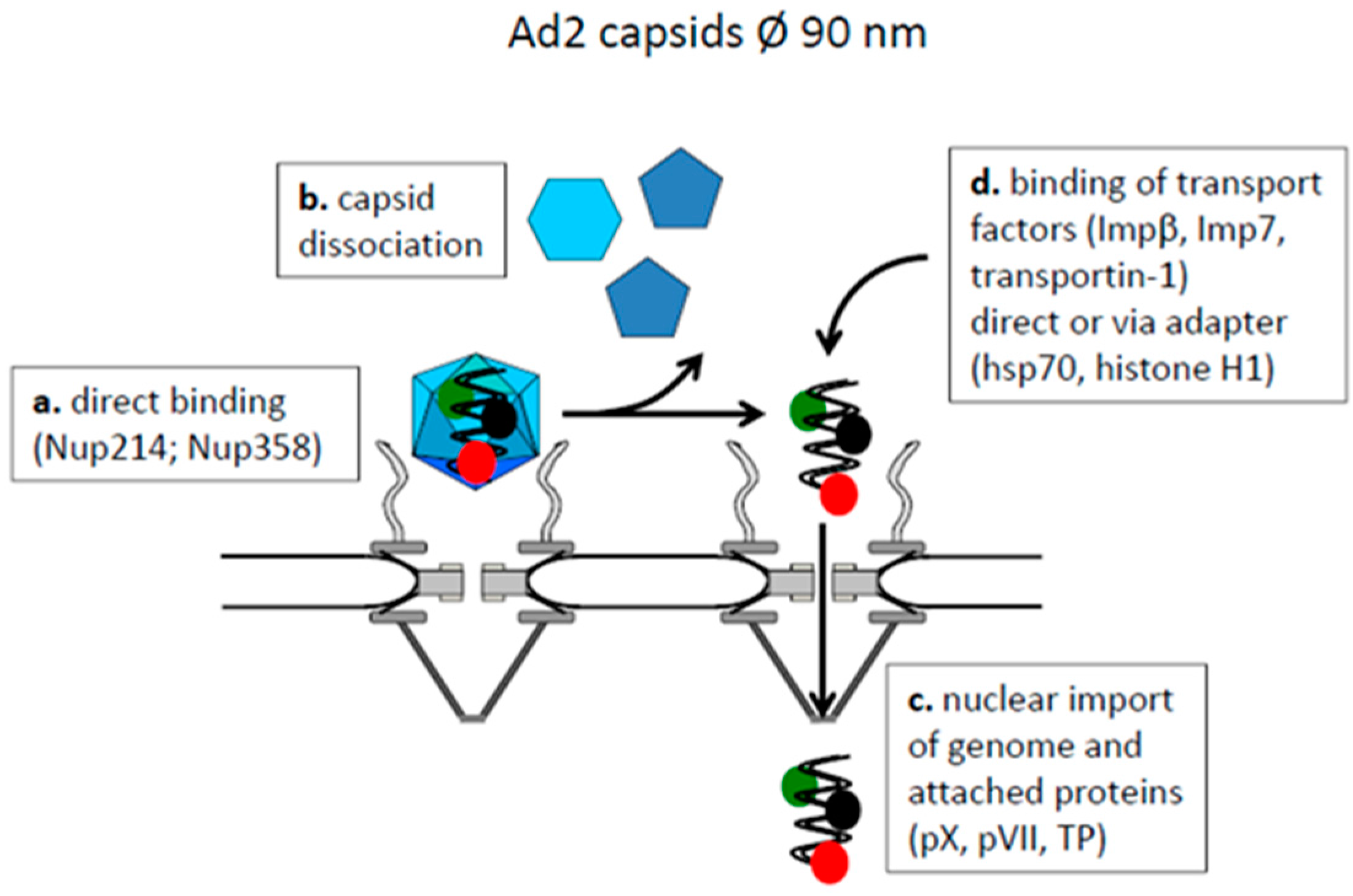
4.2. Capsids Smaller than the Maximal Transport Diameter of the NPC

A few capsids have diameters below the exclusion limit of the NPCs. Circoviridae, non-enveloped viruses with a single stranded DNA genome infecting pigs and birds, have diameters of 15 to 30 nm. They are composed of a single protein species (Cap protein) and exhibit a T=1 symmetry [83]. It was shown that both the Cap protein but also the viral replication protein Rep accumulate in the karyoplasm. Consistently, an NLS was identified on Cap but the requirement of the Cap NLS for nuclear import of the genome remains unknown.
Parvoviridae (PV), also non-enveloped viruses with a single stranded DNA genome and infecting a broad range of animals including human, exhibit capsid diameters of 18 to 28 nm (reviewed in [84]). They could thus pass the NPC without disintegration. This is seemingly supported by observations that parvoviral capsids accumulate in the nucleus shortly after infection, which is in agreement with the presence of an NLS on the large PV capsid protein VP1. However, the NLS which was found in different PV, localizes in the VP1u domain at the N-terminus, which is hidden in the virion (reviewed in [85]). Heat treatment externalizes this domain and it was proposed that such structural change also occurs upon endosomal entry and acidification. N-terminal VP1u exposure was also observed in vitro upon incubation of the parvovirus H1 (PV-H1) and adeno-associated virus 2 (AAV2) with isolated Nups [86] (Figure 4a) so that the mechanism of NLS exposure and its need for capsid interaction with NPCs stays unclear. An alternative NLS function is implied by parvoviral capsid assembly, which occurs within the nucleus requiring nuclear import of the capsid proteins VP1 and VP2. This transport is mediated by VP1, which forms a heterotrimer with two VP2 molecules, which are devoid of NLS [87].
Recent observation showed that the PV-H1, but also adeno-associated virus type 2 (AAV-2) attach directly to the Nup358, Nup214 and Nup153 (Figure 4b). It remains open if Nup153-binding reflects that the parvoviruses passed the nuclear pore in vivo as these assays were performed with disassembled NPCs. Considering the small capsid size, the direct Nup binding could result in an import receptor-independent transport similar to calnexin and calreticulin. However, PV-H1, canine parvovirus and different AAVs disintegrate the nuclear envelope locally [86,88], which was observed in several experimental systems and cell types including somatic cells and Xenopus laevis oocytes (Figure 4c). Local nuclear envelope degradation was not lethal to the cells but correlated to successful infection. It was shown to enhance nuclear entry of papillomaviruses, which are unable to trigger their nuclear entry during infection [8]. Parvoviruses thus combine characteristics of Ad, HSV-1, calnexin and calreticulin in terms of a direct interaction with Nups but they disintegrate the nuclear envelope instead of being imported into the nucleus by classical nuclear import pathways.

5. Organization of HBV Core Proteins and Capsids

HBV capsids are made up from a single protein species called core or capsid protein (Cp). Cp comprises a 140 amino acid-long structural domain and a 34–36 amino acid-long C- terminal domain (CTD), separated by a spacer of nine amino acids. The CTD harbors 16 arginine residues, organized in four clusters and five serine residues, which can be phosphorylated by a cellular protein kinase. The identity of the protein kinase is not unequivocally identified and the extent of Cp phosphorylation seems to be related to genome maturation inside the capsid. Furthermore the CTD comprises at least two overlapping bipartite NLS, which mediate nuclear import after fusion to bovine serum albumin (BSA) [89,90,91,92,93,94].
240 copies of Cp form a capsid with a T=4 symmetry even in vitro in the absence of other proteins. Upon Cp expression in Escherichia coli (coliC), a minor fraction exhibiting a T=3 symmetry was also described [95,96,97,98] but virion-derived capsids comprise the large form [99,100]. Assembly starts with rapid Cp dimerization, followed by a slow formation of hexamers. During the proceeding capsid assembly no further distinct intermediates could be identified [101,102]. Noteworthy, HBV capsids are not stable structures but are subject to permanent dissociation and re-association of Cp subunits, which is called capsid breathing [103].
Aside of their symmetry, HBV capsids can be further distinguished upon their nucleic acid content. (i) mature rcDNA- or double stranded linear (dsl) DNA-containing capsids (matC), which interact with the surface proteins and become secreted [104]. In our hands, these capsids are a minor fraction in transfected cells; (ii) capsids, containing the RNA pregenome and polymerase (Pol) (rnaC), which are also rare and which cannot be enveloped [4,104]; (iii) capsids containing all intermediates of genome replication from pregenome to rcDNA (immatC). As rnaC they do not become enveloped [4,104]. Of note is one exception: snow goose hepatitis B virions comprise a ssDNA genome [105]; (iv) empty capsids being a product of core protein over-expression (empC). These capsids can also be enveloped and secreted as empty virions [106,107].
As implied by their distinct capacity for envelopment, capsids not only differ in their enclosed nucleic acid but also in their structure. Within a resolution limit of 30 Å, coliC, which contain bacterial RNA, are identical to liver-expressed capsids [108] and more recent studies with 16 Å resolution supported that no gross structural changes are linked with genome maturation and envelopment [100]. However, better resolution with 10 Å showed that a hydrophobic pocket is present only on DNA-containing capsids [99].
Digestion with 40 nm gold particles-absorbed trypsin, which cleaves C-terminal arginine and lysine residues [109] was found to remove parts of the CTD [110] from capsids in which genome maturation has occurred. This was not observed when using coliC, which is consistent to image reconstructions from cryo-electron microscopy showing that the CTD is luminal [103]. In vitroCTD phosphorylation of these capsids (PcoliC), requiring dissociation, phosphorylation by protein kinase C (PKC) α/β and subsequent re-assembly, allowed trypsin digestion of the phosphorylated CTDs [110]. This indicates that the CTDs became exposed, which was recently confirmed by tryptic digests and cryo electron microscopy using a capsid mutant in which three serines of the CTD were replaced by the acidic aa glutamate mimicking phosphorylation [111]. The latter study also excludes that trypsin-cleavage of the PcoliC occurred due to the dissociation and re-association reactions.
The same tryptic digests were performed using matC, purified from cell culture supernatants of stably HBV transfected hepatoma cell line and with immatC purified from these cells. The results showed that proceeding of genome maturation was linked to increased CTD-exposure [110]. Digesting immatC from cells in which genome maturation was additionally inhibited, confirmed this finding and further showed that not the secretion process but genome maturation caused the increased CTD exposure. As demonstrated for PcoliC, trypsin digest removed all radioactively phosphorylated CTDs indicating similar or identical structural changes upon phosphorylation and genome maturation.
In summary, these data indicate that the CTDs on capsids have a dual topology—either inside the lumen or exposed to capsids exterior—which is driven by their affinity to the interior. In line with these findings Melegari et al. observed that Cp have a higher affinity to single stranded than to double stranded nucleic acids [112], explaining CTD exposure in mature capsids despite of their low phosphorylation.

6. Intracellular HBV Capsid Localization

Liver histology from HBV infected patients revealed that Cp and/or capsids are found in both cytosol and nucleus. Akiba et al. and Sharma et al. observed more frequent nuclear localization [113,114] while others observed a mainly cytoplasmic localization [115,116,117,118]. Clinically, the majority of studies associate cytoplasmic capsids with high hepatocellular injury [119,120,121,122] and a low level viremia [115]. Consistently, nuclear core dominance is associated with high viral load and minor hepatitis activity [120] but some patients show both core distributions.
Core from a patient with nuclear core stain was found to be assembled to capsids, and isopycnic CsCl ultracentrifugation showed that they were devoid of nucleic acids [123]. Consistently, electron microscopy of nuclei from core-transgenic mice exhibited a high concentration of nuclear capsids [124]. In these mice, cytosolic capsids were only observed in hepatocytes during cell division indicating that the nuclear envelope is impermeable for capsids. It was thus concluded that nuclear capsids are derived from nuclear import of unassembled Cp [124].
In HBV infected HepaRG cells but also in HBV-transfected hepatoma cell lines, core has been mainly detected in the cytoplasm but only occasionally in the nucleus [125,126]. Investigating the molecular mechanism determining capsid localization, Deroubaix et al. found that the relative expression of Cp and pol in the presence of the epsilon signal on the RNA pregenome determine Cp and capsid localization. The loss of interaction between CTD and Pol, which needs epsilon and Cp phosphorylation, caused nuclear capsid and Cp stain [126]. It remains however unknown which partner is rate-limiting in vivo as not only Cp but also Pol of HBV (and Pol the duck hepatitis B virus (DHBV)) is over-expressed [127,128].

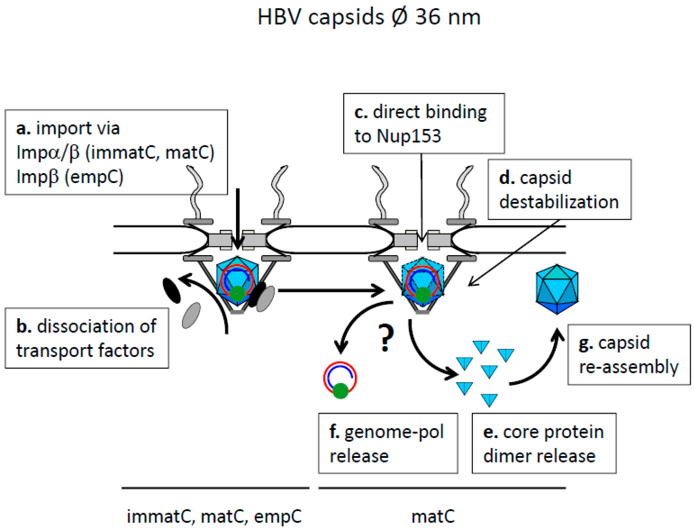
7. Transport of HBV Capsids and Genome

Investigations on nuclear core transport need the specification of the protein structure, which is transported. Non-assembled Cp expose their CTD but phosphorylation may counteract importin α-binding to the NLSs similar to threonine 124 phosphorylation of the SV40TAg [38]. However, recent in vitro binding studies showed that unassembled core proteins exhibit a direct binding to importin β as the CTD also comprises an IBB [129]. As both, importin α-NLS and importin β-IBB interactions are based on electrostatic forces, the importin β-binding is stronger as more amino acids interact. Experimentally, the different affinities are in the µM range for NLS-importin α [130] and in the nM range for IBB-importin β [33]. Further problems in identification of core localization experiments arise from nuclear export of unassembled Cp due to a nuclear export signal found on HBV-but also on DHBV Cp [131,132].
Like Cp, empC also expose the CTD and also interact with importin β directly [129]. At high importin β concentrations, the capsids disassemble leading to the hypothesis that importin β is the key molecule for the removal of unassembled Cp and empC from the cytoplasm. Such removal could be important in infection as cytoplasmic Cp are degraded by proteasomes, which was observed upon addition of Cp-directed antivirals to HBV-expressing cells [133]. Entry of the proteolytic fragments into the major histocompatibility complex (MHC) class I pathway could then proceed via the endoplasmic reticulum (ER) and Golgi-bound TAP (transporter associated with antigen processing). The resulting exposure of core epitope on the surface of hepatocytes is known to be the main target of CD8+ T cells eliminating infected hepatocytes [134], which is in agreement to the inflammatory responses in patients exhibiting cytosolic core.
Consistent with the hidden CTDs, coliC cannot precipitate importin β neither directly nor indirectly via importin α from cytosolic lysates [89]. Consequently, they do not interact with the nuclear envelope in digitonin-permeabilized cells [89] or after microinjection into the cytoplasm of Xenopus laevis oocytes [29]. They thus do not show a significant interaction with outer parts of the NPCs as the capsids of adeno- herpes- and PV.
Adding PcoliC, phosphorylated to 0.5 phosphates per CTD, to permeabilized cells resulted in strong binding to the nuclear envelope and, more specifically, to the NPCs [89]. Surprisingly NPC-binding required not only importin β but also importin α. The need of importin α is supported by inhibition of the interaction by addition of an excess of NLS-peptides to which importin β does not bind [89]. Further, this finding is consistent with the observation that binding of importin β to the capsids needed importin α [89]. Together these findings imply that only the NLS-bearing part of the CTD is exposed but not in entire CTD, which is required for IBB-exposure. The alternative interpretation that phosphorylation interferes with direct importin β-binding but not with importin α-interaction appears unlikely due to the higher affinity of importin β for IBBs [130].
Early studies in cells showed that the NPC can import NLS-coated gold particles that are up to ~26 nm in diameter (including the protein coat; [135]), which was thought to be the threshold for karyophilic macromolecules crossing the NPC. Consequently, HBV capsids were assumed to become arrested on the cytoplasmic face of the NPC. It was thus in contradiction that PcoliC localize also in the nuclear basket after microinjection into the cytoplasm of Xenopus laevis oocytes [29]. Nuclear localization was not limited to the rare NPCs exhibiting a 10 fold symmetry suggesting that either the capsids had disassembled, followed by passage through the pore and subsequent re-assembly or that they had been squeezed, or that the maximal cargo size for macromolecules passing the NPC was seriously underestimated. Microinjection of NLS-coated gold particles confirmed that pore diameter was 39–40 nm, allowing passage of the capsids even after addition of an additional protein layer of importin α/β, which accounts for 1.9 nm [29]. These findings however raised the question why the capsids passed the NPC but failed to diffuse deeper into the karyoplasm.
Despite of several imaging data, showing that the structure of coliC and authentic capsids from patients or capsids from transfected cells are similar if not identical [95,112,136] it cannot be completely excluded that pol or heat shock proteins present in the capsids lumen modify capsids structure. This is in particular true as high resolution data were obtained by image reconstruction after cryo electron microscopy of thousands of capsids during which asymmetrical changes are merged out. Local structure changes e.g., at a site where Cp interact with pol would have been invisible, requiring confirmation of coliC-derived data by functional assays using authentic capsids.
Adding immatC to permeabilized cells showed that they bound to NPCs as PcoliC [110] and that they also entered the nuclear basket. MatC in contrast bound to the NPCs in a CTD- and importin α/β-dependent manner, entered the nuclear basket but were also found inside the karyoplasm [110].
Asking for the factor(s) retaining capsids in the nuclear basket, pull-down assays showed interaction with Nup153. Binding was ~200 times stronger than Nup153 interaction with importin β, which is a natural binding partner. Consistent with such strong interaction, partial silencing of Nup153 followed by nuclear import in permeabilized cells exhibited arrival of just a small proportion of PcoliC inside the nucleus [137]. However, the pull-down experiments with Nup153 revealed that also coliC efficiently bound Nup153. This indicates that importin α/β is just needed for capsid transport into the nuclear basket but that the CTD is not required for arresting the capsid.
The observation that NPCs can transport cargos with the size of HBV capsid into the nucleus does not exclude a more complex scenario: (i) capsids bind to the cytosolic face of the NPC where (ii) they disintegrate to Cp; (iii) followed by Cp translocation into the nuclear basket using nuclear import receptors; (iv) followed by reassembly to capsids after dissociation of the import factors on the nuclear side of the pore. Experiments in permeabilized cells and in Xenopus laevis oocytes in which UV-cross linked matC were used however showed that cross linking prevented arrival of matC in the nucleus although entering the nuclear basket [137]. These findings argued for a mandatory capsid disassembly before diffusing deeper into the karyoplasm.
In comparison to other viruses, HBV capsids thus have a more complex regulation in that there are different capsid forms, which differ in their genome maturation, Cp phosphorylation and structure. While exposure of the CTD is required for NPC interaction via nuclear transport receptors like it was shown for HSV-1 all capsids, direct interaction with Nup153 occurs after passage of the nuclear pore inside the nuclear basket. With regard to the latter interaction HBV capsids thus share also the characteristic of direct Nup binding as it was observed for Ad, HSV-1 and PV capsids.

8. Transport and Release of the Hepadnaviral Genome

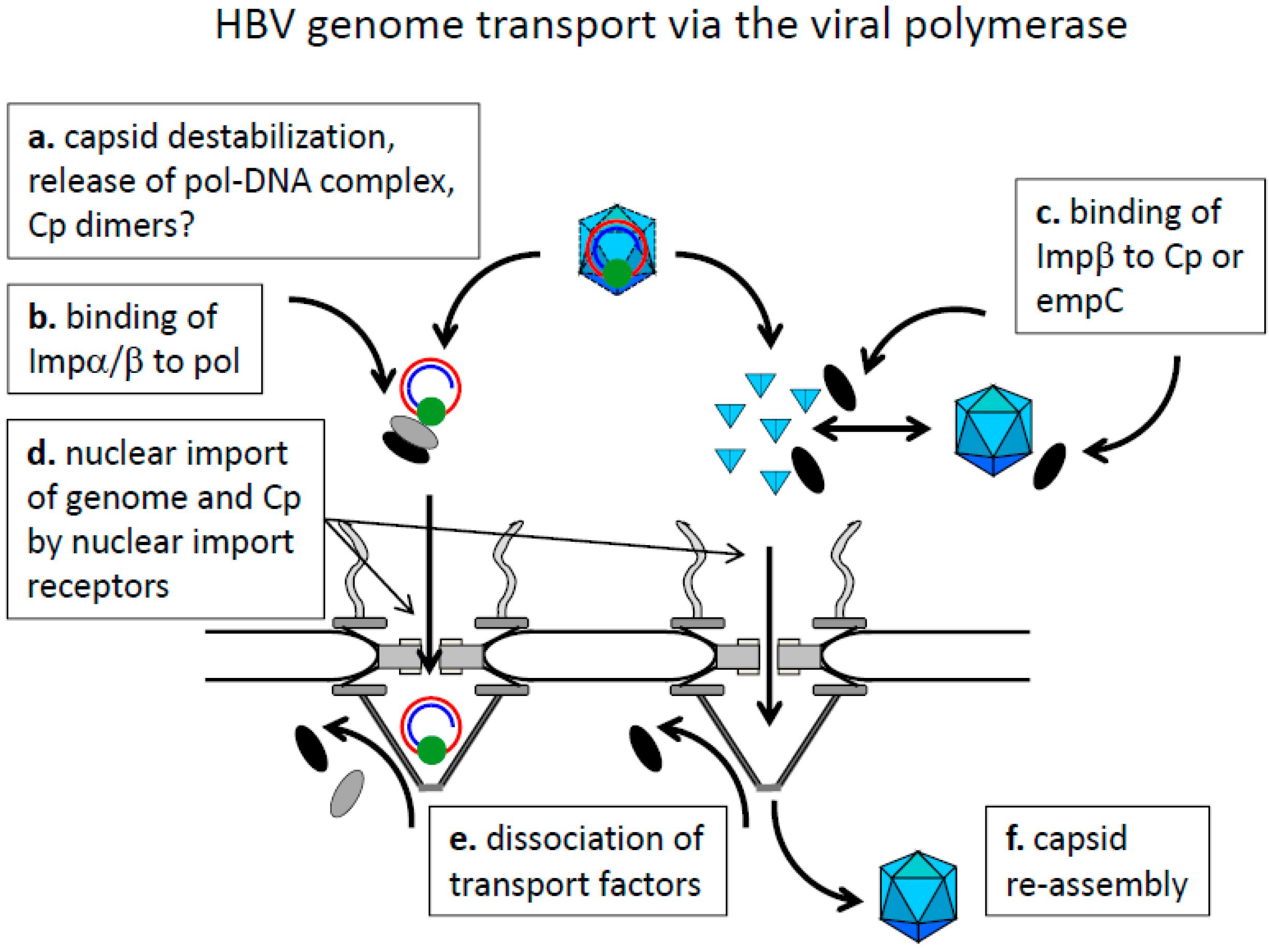
8.1. Cytoplasmic Genome Release and Genome Transport by the Viral Polymerase

There are two possibilities of how hepadnaviral genomes could enter the nucleus, which are summarized in Figure 4 and Figure 5. The first model comprises nuclear genome entry driven by Pol, which is covalently attached to the genome (Figure 5). In fact exposure of Pol-genome complexes from the woodchuck hepatitis virus to isolated nuclei led to nuclear translocation of the genome [138]. However, isolation of the complex from the capsids required treatment with 4 M urea, denaturating Pol. This could lead to exposure of internal Pol domains, which are not exposed in vivo. Seemingly this assumption is supported by the findings of Cao et al., and Yao et al. showing that the Pol of HBV and DHBV are cytoplasmic [127,128]. Aside of fundamental differences to the woodchuck hepatitis virus Pol, the data however do not exclude that Pol undergo structural changes during encapsidation and genome maturation. Consequently, Pol structure released from the capsid could be different and in fact structural changes were reported upon epsilon binding [139,140]. Consistent with a Pol-mediated genome transport are findings of Guo et al., who expressed an envelope-negative HBV mutant in a hepatoma cell line [141]. The authors observed cytoplasmic DNase-sensitive rcDNA devoid of the viral polymerase which co-sedimented with capsids and which could be precipitated by anti-capsid antibodies. This indicates that capsids can open in the cytosol potentially allowing genome release and could result in Pol-mediated transport prior to pol degradation. This possibility remains however hypothetical as the majority of deproteinized genomes stayed capsid-bound favoring nuclear transport by the capsid. Further, it must be considered that the cytoplasm in these experiments was yielded by low speed centrifugation of a homogenate derived from douncing, which damages nuclei to some extent. As there was no contamination control of the cytosolic fraction with nuclear components the results require further confirmation.
There is thus no direct exclusion of a pol-mediated nuclear import of the HBV genome but the fact that HBV does not induce any innate immune response—nor counteract such a response [124,142]—indirectly argues against the release of a protein-rcDNA complex in the cytoplasm. Further support for a genome transport within the capsid comes from native fluorescence in situ hybridizations (native FISH). This technique allows detection of released HBV genomes only and showed no cytoplasmic but exclusively nuclear genomes after capsid-lipofection [143]. This technique induced high efficient cccDNA generation and subsequent virus replication indicating some similarity to the in vivo situation. However, the absence of cytosolic released genomes does not exclude capsid disassembly at the cytoplasmic face of the NPC followed by rapid translocation into the nucleus.

8.2. Nuclear Genome-Translocation in Intact Capsids and Nuclear Genome Release

Despite of the artificial setup, permeabilized cells not only allow the import of HBV capsids but native FISH revealed released intranuclear HBV genomes when matC were added [110]. This observation is in agreement with the disassembly of capsids to Cp dimers, which only occurred when the capsids got contact with the NPC [144]. When nuclear import was inhibited no capsid disintegration was observed with a threshold of detection which was estimated to be <5% of the subjected capsids.
The latter studies, also summarized in Figure 6, showed that capsid transport and disassembly was combined with intranuclear re-assembly of the Cp dimers to genome-free capsids and no Cp degradation. The capsids were mostly filled with cellular RNA, which is in contrast to nuclear capsids isolated from human liver. As the liver capsids were likely derived from over-expressed capsids and not from genome import, a different Cp phosphorylation state could explain the difference. This hypothesis is in line with observations that these capsids contain one PKC molecule per capsid [145] and that PKC-phosphorylated core proteins do not encapsidate RNA upon in vitro assembly [146].
The electron microscopy data showing capsids in the nuclear basket suggest that this is the place where genome release occurs. Following this hypothesis, the disassembly of matC would result in 120 core protein dimers, which outnumber the 16 Nup153 molecules per NPC. Super numerous Cp dimers could then diffuse deeper into the karyoplasm and re-assemble to capsids. The limits of electron microscopy however do not allow to exclude that a part of the capsids leave the nuclear basket intact for disassembling shortly afterwards. Functional evidence against this hypothesis was shown by Rabe et al. [110], who observed that genome release, detected by native FISH, occurred even when Ran was absent, thus before a potential detachment of capsids could happen.
The observation that mature capsids disassemble preferentially can be explained with their lower stability as shown by Cui et al. [147] who used proteinase K resistance and DNase sensitivity as read-out of their in vitro assays. A potential molecular mechanism could be their low phosphorylation. In fact capsids, formed by the glutamate residue-mutant, were more stable to higher NaCl concentration [111]. The driving forces causing that the capsids with a mature genome disassemble in the nuclear basket remain however fully unknown.
Comparing the genome transport across the nuclear envelope of HBV capsids with that of other viruses thus shows obvious differences. Larger capsids as those of Ad and HSV have to disassemble on the cytoplasmic side of the NPC followed by classical nuclear import (Ad) or a mixture of injection and “pulling in” (HSV). Small capsids as that of HBV and PV can pass the NPC but they seem to stay bound by direct interaction with Nups. This raises the hypothesis whether the NPC is the general environment triggering genome release.

Acknowledgments

We thank the Fondation pour la recherche médicale (FRM; www.frm.org; équipe FRM 2011 DEQ20110421299) and the National French Agency for Research against HIV and Hepatitis Viruses (ANRS; http://www.anrs.fr/; AAP 2014-2), which supported the work with the salary of LG.

Author Contributions

Both authors wrote the manuscript and shared the bibliography.

Conflicts of Interest

The authors declare no conflict of interest

References

  1. Königer, C.; Wingert, I.; Marsmann, M.; Rösler, C.; Beck, J.; Nassal, M. Involvement of the host DNA-repair enzyme TDP2 in formation of the covalently closed circular DNA persistence reservoir of hepatitis B viruses. Proc. Natl. Acad. Sci. USA 2014, 111, E4244–E4253. [Google Scholar] [CrossRef] [PubMed]
  2. Qi, Y.; Gao, Z.; Xu, G.; Peng, B.; Liu, C.; Yan, H.; Yao, Q.; Sun, G.; Liu, Y.; Tang, D.; et al. DNA Polymerase κ Is a Key Cellular Factor for the Formation of Covalently Closed Circular DNA of Hepatitis B Virus. PLoS Pathog. 2016, 12, e1005893. [Google Scholar] [CrossRef] [PubMed]
  3. Tong, S.; Revill, P. Overview of hepatitis B viral replication and genetic variability. J. Hepatol. 2016, 64, S4–S16. [Google Scholar] [CrossRef] [PubMed]
  4. Gerelsaikhan, T.; Tavis, J.E.; Bruss, V. Hepatitis B virus nucleocapsid envelopment does not occur without genomic DNA synthesis. J. Virol. 1996, 70, 4269–4274. [Google Scholar] [PubMed]
  5. Tuttleman, J.S.; Pourcel, C.; Summers, J. Formation of the pool of covalently closed circular viral DNA in hepadnavirus-infected cells. Cell 1986, 47, 451–460. [Google Scholar] [CrossRef]
  6. Summers, J.; Smith, P.M.; Horwich, A.L. Hepadnavirus envelope proteins regulate covalently closed circular DNA amplification. J. Virol. 1990, 64, 2819–2824. [Google Scholar] [PubMed]
  7. Goff, S.P. Host factors exploited by retroviruses. Nat. Rev. Microbiol. 2007, 5, 253–263. [Google Scholar] [CrossRef] [PubMed]
  8. Aydin, I.; Weber, S.; Snijder, B.; Ventayol, P.S.; Kühbacher, A.; Becker, M.; Day, P.M.; Schiller, J.T.; Kann, M.; Pelkmans, L.; et al. Large Scale RNAi Reveals the Requirement of Nuclear Envelope Breakdown for Nuclear Import of Human Papillomaviruses. PLoS Pathog. 2014, 10, e1004162. [Google Scholar] [CrossRef] [PubMed]
  9. Clever, J.; Yamada, M.; Kasamatsu, H. Import of simian virus 40 virions through nuclear pore complexes. Proc. Natl. Acad. Sci. USA 1991, 88, 7333–7337. [Google Scholar] [CrossRef] [PubMed]
  10. Yamada, M.; Kasamatsu, H. Role of nuclear pore complex in simian virus 40 nuclear targeting. J. Virol. 1993, 67, 119–130. [Google Scholar] [PubMed]
  11. Nakanishi, A.; Clever, J.; Yamada, M.; Li, P.P.; Kasamatsu, H. Association with capsid proteins promotes nuclear targeting of simian virus 40 DNA. Proc. Natl. Acad. Sci. USA 1996, 93, 96–100. [Google Scholar] [CrossRef] [PubMed]
  12. Nakanishi, A.; Shum, D.; Morioka, H.; Otsuka, E.; Kasamatsu, H. Interaction of the Vp3 Nuclear Localization Signal with the Importin α2/β Heterodimer Directs Nuclear Entry of Infecting Simian Virus 40. J. Virol. 2002, 76, 9368–9377. [Google Scholar] [CrossRef] [PubMed]
  13. Nakanishi, A.; Itoh, N.; Li, P.P.; Handa, H.; Liddington, R.C.; Kasamatsu, H. Minor Capsid Proteins of Simian Virus 40 Are Dispensable for Nucleocapsid Assembly and Cell Entry but Are Required for Nuclear Entry of the Viral Genome. J. Virol. 2007, 81, 3778–3785. [Google Scholar] [CrossRef] [PubMed]
  14. Butin-Israeli, V.; Ben-nun-Shaul, O.; Kopatz, I.; Adam, S.A.; Shimi, T.; Goldman, R.D.; Oppenheim, A. Simian virus 40 induces lamin A/C fluctuations and nuclear envelope deformation during cell entry. Nucleus 2011, 2, 320–330. [Google Scholar] [CrossRef] [PubMed]
  15. Von Appen, A.; Beck, M. Structure Determination of the Nuclear Pore Complex with Three-Dimensional Cryo electron Microscopy. J. Mol. Biol. 2016, 428, 2001–2010. [Google Scholar] [CrossRef] [PubMed]
  16. Alber, F.; Dokudovskaya, S.; Veenhoff, L.M.; Zhang, W.; Kipper, J.; Devos, D.; Suprapto, A.; Karni-Schmidt, O.; Williams, R.; Chait, B.T.; et al. The molecular architecture of the nuclear pore complex. Nature 2007, 450, 695–701. [Google Scholar] [CrossRef] [PubMed]
  17. Rabut, G.; Doye, V.; Ellenberg, J. Mapping the dynamic organization of the nuclear pore complex inside single living cells. Nat. Cell Biol. 2004, 6, 1114–1121. [Google Scholar] [CrossRef] [PubMed]
  18. Tran, E.J.; Wente, S.R. Dynamic Nuclear Pore Complexes: Life on the Edge. Cell 2006, 125, 1041–1053. [Google Scholar] [CrossRef] [PubMed]
  19. D’Angelo, M.A.; Anderson, D.J.; Richard, E.; Hetzer, M.W. Nuclear Pores Form de Novo from Both Sides of the Nuclear Envelope. Science 2006, 312, 440–443. [Google Scholar] [CrossRef] [PubMed]
  20. Raices, M.; D’Angelo, M.A. Nuclear pore complex composition: A new regulator of tissue-specific and developmental functions. Nat. Rev. Mol. Cell Biol. 2012, 13, 687–699. [Google Scholar] [CrossRef] [PubMed]
  21. Frey, S.; Görlich, D. A Saturated FG-Repeat Hydrogel Can Reproduce the Permeability Properties of Nuclear Pore Complexes. Cell 2007, 130, 512–523. [Google Scholar] [CrossRef] [PubMed]
  22. Patel, S.S.; Belmont, B.J.; Sante, J.M.; Rexach, M.F. Natively Unfolded Nucleoporins Gate Protein Diffusion across the Nuclear Pore Complex. Cell 2007, 129, 83–96. [Google Scholar] [CrossRef] [PubMed]
  23. Strawn, L.A.; Shen, T.; Shulga, N.; Goldfarb, D.S.; Wente, S.R. Minimal nuclear pore complexes define FG repeat domains essential for transport. Nat. Cell Biol. 2004, 6, 197–206. [Google Scholar] [CrossRef] [PubMed]
  24. Ghavami, A.; van der Giessen, E.; Onck, P.R. Energetics of Transport through the Nuclear Pore Complex. PLoS ONE 2016, 11, e0148876. [Google Scholar] [CrossRef] [PubMed]
  25. Ribbeck, K.; Görlich, D. Kinetic analysis of translocation through nuclear pore complexes. EMBO J. 2001, 20, 1320–1330. [Google Scholar] [CrossRef] [PubMed]
  26. Doucet, C.M.; Hetzer, M.W. Nuclear pore biogenesis into an intact nuclear envelope. Chromosoma 2010, 119, 469–477. [Google Scholar] [CrossRef] [PubMed]
  27. Garcia-Segura, L.M.; Lafarga, M.; Berciano, M.T.; Hernandez, P.; Andres, M.A. Distribution of nuclear pores and chromatin organization in neurons and glial cells of the rat cerebellar cortex. J. Comp. Neurol. 1989, 290, 440–450. [Google Scholar] [CrossRef] [PubMed]
  28. Maeshima, K.; Yahata, K.; Sasaki, Y.; Nakatomi, R.; Tachibana, T.; Hashikawa, T.; Imamoto, F.; Imamoto, N. Cell-cycle-dependent dynamics of nuclear pores: Pore-free islands and lamins. J. Cell Sci. 2006, 119, 4442–4451. [Google Scholar] [CrossRef] [PubMed]
  29. Panté, N.; Kann, M. Nuclear Pore Complex Is Able to Transport Macromolecules with Diameters of ~39 nm. Mol. Biol. Cell 2002, 13, 425–434. [Google Scholar] [CrossRef] [PubMed]
  30. Cautain, B.; Hill, R.; de Pedro, N.; Link, W. Components and regulation of nuclear transport processes. FEBS J. 2015, 282, 445–462. [Google Scholar] [CrossRef] [PubMed]
  31. Nakielny, S.; Siomi, M.C.; Siomi, H.; Michael, W.M.; Pollard, V.; Dreyfuss, G. Transportin: Nuclear Transport Receptor of a Novel Nuclear Protein Import Pathway. Exp. Cell Res. 1996, 229, 261–266. [Google Scholar] [CrossRef] [PubMed]
  32. Siomi, H.; Dreyfuss, G. A nuclear localization domain in the hnRNP A1 protein. J. Cell Biol. 1995, 129, 551–560. [Google Scholar] [CrossRef] [PubMed]
  33. Lott, K.; Cingolani, G. The importin β binding domain as a master regulator of nucleocytoplasmic transport. Biochim. Biophys. Acta 2011, 1813, 1578–1592. [Google Scholar] [CrossRef] [PubMed]
  34. Palmeri, D.; Malim, M.H. Importin β Can Mediate the Nuclear Import of an Arginine-Rich Nuclear Localization Signal in the Absence of Importin α. Mol. Cell. Biol. 1999, 19, 1218–1225. [Google Scholar] [CrossRef] [PubMed]
  35. Kalderon, D.; Roberts, B.L.; Richardson, W.D.; Smith, A.E. A short amino acid sequence able to specify nuclear location. Cell 1984, 39, 499–509. [Google Scholar] [CrossRef]
  36. Lanford, R.E.; Butel, J.S. Construction and characterization of an SV40 mutant defective in nuclear transport of T antigen. Cell 1984, 37, 801–813. [Google Scholar] [CrossRef]
  37. Pumroy, R.A.; Cingolani, G. Diversification of importin-α isoforms in cellular trafficking and disease states. Biochem. J. 2015, 466, 13–28. [Google Scholar] [CrossRef] [PubMed]
  38. Jans, D.A.; Ackermann, M.J.; Bischoff, J.R.; Beach, D.H.; Peters, R. p34cdc2-mediated phosphorylation at T124 inhibits nuclear import of SV-40 T antigen proteins. J. Cell Biol. 1991, 115, 1203–1212. [Google Scholar] [CrossRef] [PubMed]
  39. Lin, Y.C.; Brown, K.; Siebenlist, U. Activation of NF-kappa B requires proteolysis of the inhibitor I kappa B-alpha: Signal-induced phosphorylation of I kappa B-alpha alone does not release active NF-kappa B. Proc. Natl. Acad. Sci. USA 1995, 92, 552–556. [Google Scholar] [CrossRef] [PubMed]
  40. Beals, C.R.; Clipstone, N.A.; Ho, S.N.; Crabtree, G.R. Nuclear localization of NF-ATc by a calcineurin-dependent, cyclosporin-sensitive intramolecular interaction. Genes Dev. 1997, 11, 824–834. [Google Scholar] [CrossRef] [PubMed]
  41. Cingolani, G.; Lashuel, H.A.; Gerace, L.; Müller, C.W. Nuclear import factors importin α and importin β undergo mutually induced conformational changes upon association. FEBS Lett. 2000, 484, 291–298. [Google Scholar] [CrossRef]
  42. Lee, S.J.; Sekimoto, T.; Yamashita, E.; Nagoshi, E.; Nakagawa, A.; Imamoto, N.; Yoshimura, M.; Sakai, H.; Chong, K.T.; Tsukihara, T.; et al. The Structure of Importin-ß Bound to SREBP-2: Nuclear Import of a Transcription Factor. Science 2003, 302, 1571–1575. [Google Scholar] [CrossRef] [PubMed]
  43. Rout, M.P.; Aitchison, J.D.; Suprapto, A.; Hjertaas, K.; Zhao, Y.; Chait, B.T. The Yeast Nuclear Pore Complex. J. Cell Biol. 2000, 148, 635–652. [Google Scholar] [CrossRef] [PubMed]
  44. Lim, R.Y.H.; Huang, N.-P.; Köser, J.; Deng, J.; Lau, K.H.A.; Schwarz-Herion, K.; Fahrenkrog, B.; Aebi, U. Flexible phenylalanine-glycine nucleoporins as entropic barriers to nucleocytoplasmic transport. Proc. Natl. Acad. Sci. USA 2006, 103, 9512–9517. [Google Scholar] [CrossRef] [PubMed]
  45. Rout, M.P.; Aitchison, J.D.; Magnasco, M.O.; Chait, B.T. Virtual gating and nuclear transport: The hole picture. Trends Cell Biol. 2003, 13, 622–628. [Google Scholar] [CrossRef] [PubMed]
  46. Lim, R.Y.H.; Fahrenkrog, B.; Köser, J.; Schwarz-Herion, K.; Deng, J.; Aebi, U. Nanomechanical Basis of Selective Gating by the Nuclear Pore Complex. Science 2007, 318, 640–643. [Google Scholar] [CrossRef] [PubMed]
  47. Lim, R.Y.H.; Köser, J.; Huang, N.; Schwarz-Herion, K.; Aebi, U. Nanomechanical interactions of phenylalanine–glycine nucleoporins studied by single molecule force–volume spectroscopy. J. Struct. Biol. 2007, 159, 277–289. [Google Scholar] [CrossRef] [PubMed]
  48. Frey, S.; Richter, R.P.; Görlich, D. FG-Rich Repeats of Nuclear Pore Proteins Form a Three-Dimensional Meshwork with Hydrogel-Like Properties. Science 2006, 314, 815–817. [Google Scholar] [CrossRef] [PubMed]
  49. Ribbeck, K.; Görlich, D. The permeability barrier of nuclear pore complexes appears to operate via hydrophobic exclusion. EMBO J. 2002, 21, 2664–2671. [Google Scholar] [CrossRef] [PubMed]
  50. Görlich, D.; Seewald, M.J.; Ribbeck, K. Characterization of Ran-driven cargo transport and the RanGTPase system by kinetic measurements and computer simulation. EMBO J. 2003, 22, 1088–1100. [Google Scholar] [CrossRef] [PubMed]
  51. Wagstaff, K.M.; Jans, D.A. Importins and Beyond: Non-Conventional Nuclear Transport Mechanisms. Traffic 2009, 10, 1188–1198. [Google Scholar] [CrossRef] [PubMed]
  52. Argentaro, A.; Sim, H.; Kelly, S.; Preiss, S.; Clayton, A.; Jans, D.A.; Harley, V.R. A SOX9 Defect of Calmodulin-dependent Nuclear Import in Campomelic Dysplasia/Autosomal Sex Reversal. J. Biol. Chem. 2003, 278, 33839–33847. [Google Scholar] [CrossRef] [PubMed]
  53. Holaska, J.M.; Black, B.E.; Love, D.C.; Hanover, J.A.; Leszyk, J.; Paschal, B.M. Calreticulin Is a Receptor for Nuclear Export. J. Cell Biol. 2001, 152, 127–140. [Google Scholar] [CrossRef] [PubMed]
  54. Alefantis, T.; Flaig, K.E.; Wigdahl, B.; Jain, P. Interaction of HTLV-1 Tax protein with calreticulin: Implications for Tax nuclear export and secretion. Biomed. Pharmacother. 2007, 61, 194–200. [Google Scholar] [CrossRef] [PubMed]
  55. Bunn, C.F.; Neidig, J.A.; Freidinger, K.E.; Stankiewicz, T.A.; Weaver, B.S.; McGrew, J.; Allison, L.A. Nucleocytoplasmic Shuttling of the Thyroid Hormone Receptorα. Mol. Endocrinol. 2008. [Google Scholar] [CrossRef] [PubMed]
  56. Grespin, M.E.; Bonamy, G.M.C.; Roggero, V.R.; Cameron, N.G.; Adam, L.E.; Atchison, A.P.; Fratto, V.M.; Allison, L.A. Thyroid Hormone Receptor α1 Follows a Cooperative CRM1/Calreticulin-mediated Nuclear Export Pathway. J. Biol. Chem. 2008, 283, 25576–25588. [Google Scholar] [CrossRef] [PubMed]
  57. Mogensen, T.H. Pathogen Recognition and Inflammatory Signaling in Innate Immune Defenses. Clin. Microbiol. Rev. 2009, 22, 240–273. [Google Scholar] [CrossRef] [PubMed]
  58. Arhel, N.J.; Souquere-Besse, S.; Munier, S.; Souque, P.; Guadagnini, S.; Rutherford, S.; Prévost, M.-C.; Allen, T.D.; Charneau, P. HIV-1 DNA Flap formation promotes uncoating of the pre-integration complex at the nuclear pore. EMBO J. 2007, 26, 3025–3037. [Google Scholar] [CrossRef] [PubMed]
  59. Campadelli-Fiume, G.; Menotti, L.; Avitabile, E.; Gianni, T. Viral and cellular contributions to herpes simplex virus entry into the cell. Curr. Opin. Virol. 2012, 2, 28–36. [Google Scholar] [CrossRef] [PubMed]
  60. Morrison, E.E.; Stevenson, A.J.; Wang, Y.F.; Meredith, D.M. Differences in the intracellular localization and fate of herpes simplex virus tegument proteins early in the infection of Vero cells. J. Gen. Virol. 1998, 79, 2517–2528. [Google Scholar] [CrossRef] [PubMed]
  61. Sodeik, B.; Ebersold, M.W.; Helenius, A. Microtubule-mediated Transport of Incoming Herpes Simplex Virus 1 Capsids to the Nucleus. J. Cell Biol. 1997, 136, 1007–1021. [Google Scholar] [CrossRef] [PubMed]
  62. Ojala, P.M.; Sodeik, B.; Ebersold, M.W.; Kutay, U.; Helenius, A. Herpes Simplex Virus Type 1 Entry into Host Cells: Reconstitution of Capsid Binding and Uncoating at the Nuclear Pore Complex In Vitro. Mol. Cell. Biol. 2000, 20, 4922–4931. [Google Scholar] [CrossRef] [PubMed]
  63. Pasdeloup, D.; Blondel, D.; Isidro, A.L.; Rixon, F.J. Herpesvirus Capsid Association with the Nuclear Pore Complex and Viral DNA Release Involve the Nucleoporin CAN/Nup214 and the Capsid Protein pUL25. J. Virol. 2009, 83, 6610–6623. [Google Scholar] [CrossRef] [PubMed]
  64. Copeland, A.M.; Newcomb, W.W.; Brown, J.C. Herpes Simplex Virus Replication: Roles of Viral Proteins and Nucleoporins in Capsid-Nucleus Attachment. J. Virol. 2009, 83, 1660–1668. [Google Scholar] [CrossRef] [PubMed]
  65. Liashkovich, I.; Hafezi, W.; Kühn, J.M.; Oberleithner, H.; Shahin, V. Nuclear delivery mechanism of herpes simplex virus type 1 genome. J. Mol. Recognit. 2011, 24, 414–421. [Google Scholar] [CrossRef] [PubMed]
  66. Jovasevic, V.; Liang, L.; Roizman, B. Proteolytic Cleavage of VP1-2 Is Required for Release of Herpes Simplex Virus 1 DNA into the Nucleus. J. Virol. 2008, 82, 3311–3319. [Google Scholar] [CrossRef] [PubMed]
  67. Booy, F.P.; Newcomb, W.W.; Trus, B.L.; Brown, J.C.; Baker, T.S.; Steven, A.C. Liquid-crystalline, phage-like packing of encapsidated DNA in herpes simplex virus. Cell 1991, 64, 1007–1015. [Google Scholar] [CrossRef]
  68. Shahin, V.; Hafezi, W.; Oberleithner, H.; Ludwig, Y.; Windoffer, B.; Schillers, H.; Kühn, J.E. The genome of HSV-1 translocates through the nuclear pore as a condensed rod-like structure. J. Cell Sci. 2006, 119, 23–30. [Google Scholar] [CrossRef] [PubMed]
  69. Molineux, I.J. No syringes please, ejection of phage T7 DNA from the virion is enzyme driven. Mol. Microbiol. 2001, 40, 1–8. [Google Scholar] [CrossRef] [PubMed]
  70. Newcomb, W.W.; Cockrell, S.K.; Homa, F.L.; Brown, J.C. Polarized DNA Ejection from the Herpesvirus Capsid. J. Mol. Biol. 2009, 392, 885–894. [Google Scholar] [CrossRef] [PubMed]
  71. Greber, U.F.; Willetts, M.; Webster, P.; Helenius, A. Stepwise dismantling of adenovirus 2 during entry into cells. Cell 1993, 75, 477–486. [Google Scholar] [CrossRef] [Green Version]
  72. Trotman, L.C.; Mosberger, N.; Fornerod, M.; Stidwill, R.P.; Greber, U.F. Import of adenovirus DNA involves the nuclear pore complex receptor CAN/Nup214 and histone H1. Nat. Cell Biol. 2001, 3, 1092–1100. [Google Scholar] [CrossRef] [PubMed]
  73. Strunze, S.; Trotman, L.C.; Boucke, K.; Greber, U.F. Nuclear Targeting of Adenovirus Type 2 Requires CRM1-mediated Nuclear Export. Mol. Biol. Cell 2005, 16, 2999–3009. [Google Scholar] [CrossRef] [PubMed] [Green Version]
  74. Cassany, A.; Ragues, J.; Guan, T.; Bégu, D.; Wodrich, H.; Kann, M.; Nemerow, G.R.; Gerace, L. Nuclear Import of Adenovirus DNA Involves Direct Interaction of Hexon with an N-Terminal Domain of the Nucleoporin Nup214. J. Virol. 2015, 89, 1719–1730. [Google Scholar] [CrossRef] [PubMed]
  75. Strunze, S.; Engelke, M.F.; Wang, I.-H.; Puntener, D.; Boucke, K.; Schleich, S.; Way, M.; Schoenenberger, P.; Burckhardt, C.J.; Greber, U.F. Kinesin-1-Mediated Capsid Disassembly and Disruption of the Nuclear Pore Complex Promote Virus Infection. Cell Host Microbe 2011, 10, 210–223. [Google Scholar] [CrossRef] [PubMed] [Green Version]
  76. Schnitzer, M.J.; Visscher, K.; Block, S.M. Force production by single kinesin motors. Nat. Cell Biol. 2000, 2, 718–723. [Google Scholar] [PubMed]
  77. Weisel, J.W.; Shuman, H.; Litvinov, R.I. Protein–protein unbinding induced by force: Single-molecule studies. Curr. Opin. Struct. Biol. 2003, 13, 227–235. [Google Scholar] [CrossRef]
  78. Martin-Fernandez, M.; Longshaw, S.V.; Kirby, I.; Santis, G.; Tobin, M.J.; Clarke, D.T.; Jones, G.R. Adenovirus Type-5 Entry and Disassembly Followed in Living Cells by FRET, Fluorescence Anisotropy, and FLIM. Biophys. J. 2004, 87, 1316–1327. [Google Scholar] [CrossRef] [PubMed]
  79. Greber, U.F.; Suomalainen, M.; Stidwill, R.P.; Boucke, K.; Ebersold, M.W.; Helenius, A. The role of the nuclear pore complex in adenovirus DNA entry. EMBO J. 1997, 16, 5998–6007. [Google Scholar] [CrossRef] [PubMed]
  80. Xue, Y.; Johnson, J.S.; Ornelles, D.A.; Lieberman, J.; Engel, D.A. Adenovirus Protein VII Functions throughout Early Phase and Interacts with Cellular Proteins SET and pp32. J. Virol. 2005, 79, 2474–2483. [Google Scholar] [CrossRef] [PubMed]
  81. Saphire, A.C.S.; Guan, T.; Schirmer, E.C.; Nemerow, G.R.; Gerace, L. Nuclear Import of Adenovirus DNA in Vitro Involves the Nuclear Protein Import Pathway and hsc70. J. Biol. Chem. 2000, 275, 4298–4304. [Google Scholar] [CrossRef] [PubMed]
  82. Hindley, C.E.; Lawrence, F.J.; Matthews, D.A. A Role for Transportin in the Nuclear Import of Adenovirus Core Proteins and DNA. Traffic 2007, 8, 1313–1322. [Google Scholar] [CrossRef] [PubMed]
  83. Crowther, R.A.; Berriman, J.A.; Curran, W.L.; Allan, G.M.; Todd, D. Comparison of the Structures of Three Circoviruses: Chicken Anemia Virus, Porcine Circovirus Type 2, and Beak and Feather Disease Virus. J. Virol. 2003, 77, 13036–13041. [Google Scholar] [CrossRef] [PubMed]
  84. Berns, K.I.; Parrish, C.R. Fields Virology, 6th ed.; Lippincott Williams & Wilkins: Philadelphia, PA, USA, 2013; pp. 1768–1791. [Google Scholar]
  85. Snoussi, K.; Kann, M. Interaction of parvoviruses with the nuclear envelope. Adv. Biol. Regul. 2014, 54, 39–49. [Google Scholar] [CrossRef] [PubMed]
  86. Porwal, M.; Cohen, S.; Snoussi, K.; Popa-Wagner, R.; Anderson, F.; Dugot-Senant, N.; Wodrich, H.; Dinsart, C.; Kleinschmidt, J.A.; Panté, N.; et al. Parvoviruses Cause Nuclear Envelope Breakdown by Activating Key Enzymes of Mitosis. PLoS Pathog. 2013, 9, e1003671. [Google Scholar] [CrossRef] [PubMed]
  87. Riolobos, L.; Valle, N.; Hernando, E.; Maroto, B.; Kann, M.; Almendral, J.M. Viral Oncolysis That Targets Raf-1 Signaling Control of Nuclear Transport. J. Virol. 2010, 84, 2090–2099. [Google Scholar] [CrossRef] [PubMed] [Green Version]
  88. Cohen, S.; Marr, A.K.; Garcin, P.; Panté, N. Nuclear Envelope Disruption Involving Host Caspases Plays a Role in the Parvovirus Replication Cycle. J. Virol. 2011, 85, 4863–4874. [Google Scholar] [CrossRef] [PubMed]
  89. Kann, M.; Sodeik, B.; Vlachou, A.; Gerlich, W.H.; Helenius, A. Phosphorylation-dependent binding of hepatitis B virus core particles to the nuclear pore complex. J. Cell Biol. 1999, 145, 45–55. [Google Scholar] [CrossRef] [PubMed]
  90. Haryanto, A.; Schmitz, A.; Rabe, B.; Gassert, E.; Vlachou, A.; Kann, M. Analysis of the nuclear localization signal of the hepatitis B virus capsid. Int. Res. J. Biochem. Bioinform. 2012, 2, 174–185. [Google Scholar]
  91. Yeh, C.T.; Liaw, Y.F.; Ou, J.H. The arginine-rich domain of hepatitis B virus precore and core proteins contains a signal for nuclear transport. J. Virol. 1990, 64, 6141–6147. [Google Scholar] [PubMed]
  92. Eckhardt, S.G.; Milich, D.R.; McLachlan, A. Hepatitis B virus core antigen has two nuclear localization sequences in the arginine-rich carboxyl terminus. J. Virol. 1991, 65, 575–582. [Google Scholar] [PubMed]
  93. Liu, K.; Ludgate, L.; Yuan, Z.; Hu, J. Regulation of Multiple Stages of Hepadnavirus Replication by the Carboxyl-Terminal Domain of Viral Core Protein in trans. J. Virol. 2015, 89, 2918–2930. [Google Scholar] [CrossRef] [PubMed]
  94. Li, H.-C.; Huang, E.-Y.; Su, P.-Y.; Wu, S.-Y.; Yang, C.-C.; Lin, Y.-S.; Chang, W.-C.; Shih, C. Nuclear Export and Import of Human Hepatitis B Virus Capsid Protein and Particles. PLoS Pathog. 2010, 6. [Google Scholar] [CrossRef] [PubMed]
  95. Crowther, R.A.; Kiselev, N.A.; Böttcher, B.; Berriman, J.A.; Borisova, G.P.; Ose, V.; Pumpens, P. Three-dimensional structure of hepatitis B virus core particles determined by electron cryomicroscopy. Cell 1994, 77, 943–950. [Google Scholar] [CrossRef]
  96. Böttcher, B.; Wynne, S.A.; Crowther, R.A. Determination of the fold of the core protein of hepatitis B virus by electron cryomicroscopy. Nature 1997, 386, 88–91. [Google Scholar] [CrossRef] [PubMed]
  97. Conway, J.F.; Cheng, N.; Zlotnick, A.; Wingfield, P.T.; Stahl, S.J.; Steven, A.C. Visualization of a 4-helix bundle in the hepatitis B virus capsid by cryo-electron microscopy. Nature 1997, 386, 91–94. [Google Scholar] [CrossRef] [PubMed]
  98. Wynne, S.A.; Crowther, R.A.; Leslie, A.G.W. The Crystal Structure of the Human Hepatitis B Virus Capsid. Mol. Cell 1999, 3, 771–780. [Google Scholar] [CrossRef]
  99. Roseman, A.M.; Berriman, J.A.; Wynne, S.A.; Butler, P.J.G.; Crowther, R.A. A structural model for maturation of the hepatitis B virus core. Proc. Natl. Acad. Sci. USA 2005, 102, 15821–15826. [Google Scholar] [CrossRef] [PubMed]
  100. Dryden, K.A.; Wieland, S.F.; Whitten-Bauer, C.; Gerin, J.L.; Chisari, F.V.; Yeager, M. Native Hepatitis B Virions and Capsids Visualized by Electron Cryomicroscopy. Mol. Cell 2006, 22, 843–850. [Google Scholar] [CrossRef] [PubMed]
  101. Stray, S.J.; Bourne, C.R.; Punna, S.; Lewis, W.G.; Finn, M.G.; Zlotnick, A. A heteroaryldihydropyrimidine activates and can misdirect hepatitis B virus capsid assembly. Proc. Natl. Acad. Sci. USA 2005, 102, 8138–8143. [Google Scholar] [CrossRef] [PubMed]
  102. Zlotnick, A.; Johnson, J.M.; Wingfield, P.W.; Stahl, S.J.; Endres, D. A Theoretical Model Successfully Identifies Features of Hepatitis B Virus Capsid Assembly. Biochemistry 1999, 38, 14644–14652. [Google Scholar] [CrossRef] [PubMed]
  103. Zlotnick, A.; Cheng, N.; Stahl, S.J.; Conway, J.F.; Steven, A.C.; Wingfield, P.T. Localization of the C terminus of the assembly domain of hepatitis B virus capsid protein: Implications for morphogenesis and organization of encapsidated RNA. Proc. Natl. Acad. Sci. USA 1997, 94, 9556–9561. [Google Scholar] [CrossRef] [PubMed]
  104. Wei, Y.; Tavis, J.E.; Ganem, D. Relationship between viral DNA synthesis and virion envelopment in hepatitis B viruses. J. Virol. 1996, 70, 6455–6458. [Google Scholar] [PubMed]
  105. Greco, N.; Hayes, M.H.; Loeb, D.D. Snow Goose Hepatitis B Virus (SGHBV) Envelope and Capsid Proteins Independently Contribute to the Ability of SGHBV To Package Capsids Containing Single-Stranded DNA in Virions. J. Virol. 2014, 88, 10705–10713. [Google Scholar] [CrossRef] [PubMed]
  106. Ning, X.; Nguyen, D.; Mentzer, L.; Adams, C.; Lee, H.; Ashley, R.; Hafenstein, S.; Hu, J. Secretion of Genome-Free Hepatitis B Virus—Single Strand Blocking Model for Virion Morphogenesis of Para-retrovirus. PLoS Pathog. 2011, 7, e1002255. [Google Scholar] [CrossRef] [PubMed]
  107. Miller, R.H.; Tran, C.-T.; Robinson, W.S. Hepatitis B virus particles of plasma and liver contain viral DNA-RNA hybrid molecules. Virology 1984, 139, 53–63. [Google Scholar] [CrossRef]
  108. Kenney, J.M.; vonBonsdorff, C.-H.; Nassal, M.; Fuller, S.D. Evolutionary conservation in the hepatitis B virus core structure: Comparison of human and duck cores. Structure 1995, 3, 1009–1019. [Google Scholar] [CrossRef]
  109. Olsen, J.V.; Ong, S.-E.; Mann, M. Trypsin Cleaves Exclusively C-terminal to Arginine and Lysine Residues. Mol. Cell. Proteomics 2004, 3, 608–614. [Google Scholar] [CrossRef] [PubMed]
  110. Rabe, B.; Vlachou, A.; Panté, N.; Helenius, A.; Kann, M. Nuclear import of hepatitis B virus capsids and release of the viral genome. Proc. Natl. Acad. Sci. USA 2003, 100, 9849–9854. [Google Scholar] [CrossRef] [PubMed]
  111. Selzer, L.; Kant, R.; Wang, J.C.-Y.; Bothner, B.; Zlotnick, A. Hepatitis B Virus Core Protein Phosphorylation Sites Affect Capsid Stability and Transient Exposure of the C-terminal Domain. J. Biol. Chem. 2015, 290, 28584–28593. [Google Scholar] [CrossRef] [PubMed]
  112. Melegari, M.; Bruss, V.; Gerlich, W.H. Viral Hepatitis and Liver Disease; Williams and Wilkins: Baltimore, MD, USA, 1991; pp. 164–168. [Google Scholar]
  113. Akiba, T.; Nakayama, H.; Miyazaki, Y.; Kanno, A.; Ishii, M.; Ohori, H. Relationship between the Replication of Hepatitis B Virus and the Localization of Virus Nucleocapsid Antigen (HBcAg) in Hepatocytes. J. Gen. Virol. 1987, 68, 871–877. [Google Scholar] [CrossRef] [PubMed]
  114. Sharma, R.R.; Dhiman, R.K.; Chawla, Y.; Vasistha, R.K. Immunohistochemistry for core and surface antigens in chronic hepatitis. Trop. Gastroenterol. 2002, 23, 16–19. [Google Scholar] [PubMed]
  115. Liu, C.-J.; Jeng, Y.-M.; Chen, C.-L.; Cheng, H.-R.; Chen, P.-J.; Chen, T.-C.; Liu, C.-H.; Lai, M.-Y.; Chen, D.-S.; Kao, J.-H. Hepatitis B Virus Basal Core Promoter Mutation and DNA Load Correlate with Expression of Hepatitis B Core Antigen in Patients with Chronic Hepatitis B. J. Infect. Dis. 2009, 199, 742–749. [Google Scholar] [CrossRef] [PubMed]
  116. Michalak, T.; Nowoslawski, A. Crystalline Aggregates of Hepatitis B Core Particles in Cytoplasm of Hepatocytes. Intervirology 1982, 17, 247–252. [Google Scholar] [CrossRef] [PubMed]
  117. Park, Y.N.; Han, K.H.; Kim, K.S.; Chung, J.P.; Kim, S.; Park, C. Cytoplasmic expression of hepatitis B core antigen in chronic hepatitis B virus infection: Role of precore stop mutants. Liver Int. 1999, 19, 199–205. [Google Scholar] [CrossRef]
  118. Petit, M.A.; Pillot, J. HBc and HBe antigenicity and DNA-binding activity of major core protein P22 in hepatitis B virus core particles isolated from the cytoplasm of human liver cells. J. Virol. 1985, 53, 543–551. [Google Scholar] [PubMed]
  119. Chu, C.-M.; Yeh, C.-T.; Sheen, I.-S.; Liaw, Y.-F. Subcellular localization of hepatitis B core antigen in relation to hepatocyte regeneration in chronic hepatitis B. Gastroenterology 1995, 109, 1926–1932. [Google Scholar] [CrossRef]
  120. Chu, C.M.; Yeh, C.T.; Chien, R.N.; Sheen, I.S.; Liaw, Y.F. The degrees of hepatocyte nuclear but not cytoplasmic expression of hepatitis B core antigen reflect the level of viral replication in chronic hepatitis B virus infection. J. Clin. Microbiol. 1997, 35, 102–105. [Google Scholar] [PubMed]
  121. Kim, T.H.; Cho, E.Y.; Oh, H.J.; Choi, C.S.; Kim, J.W.; Moon, H.B.; Kim, H.C. The Degrees of Hepatocyte Cytoplasmic Expression of Hepatitis B Core Antigen correlate with Histologic Activity of Liver Disease in the Young Patients with Chronic Hepatitis B Infection. J. Korean Med. Sci. 2006, 21, 279–283. [Google Scholar] [CrossRef] [PubMed]
  122. Naoumov, N.V.; Portmann, B.C.; Tedder, R.S.; Ferns, B.; Eddleston, A.L.; Alexander, G.J.; Williams, R. Detection of hepatitis B virus antigens in liver tissue. A relation to viral replication and histology in chronic hepatitis B infection. Gastroenterology 1990, 99, 1248–1253. [Google Scholar] [CrossRef]
  123. Gerlich, W.H.; Goldmann, U.; Müller, R.; Stibbe, W.; Wolff, W. Specificity and localization of the hepatitis B virus-associated protein kinase. J. Virol. 1982, 42, 761–766. [Google Scholar] [PubMed]
  124. Guidotti, L.G.; Martinez, V.; Loh, Y.T.; Rogler, C.E.; Chisari, F.V. Hepatitis B virus nucleocapsid particles do not cross the hepatocyte nuclear membrane in transgenic mice. J. Virol. 1994, 68, 5469–5475. [Google Scholar] [PubMed]
  125. Gripon, P.; Rumin, S.; Urban, S.; Seyec, J.L.; Glaise, D.; Cannie, I.; Guyomard, C.; Lucas, J.; Trepo, C.; Guguen-Guillouzo, C. Infection of a human hepatoma cell line by hepatitis B virus. Proc. Natl. Acad. Sci. USA 2002, 99, 15655–15660. [Google Scholar] [CrossRef] [PubMed]
  126. Deroubaix, A.; Osseman, Q.; Cassany, A.; Bégu, D.; Ragues, J.; Kassab, S.; Lainé, S.; Kann, M. Expression of viral polymerase and phosphorylation of core protein determine core and capsid localization of the human hepatitis B virus. J. Gen. Virol. 2015, 96, 183–195. [Google Scholar] [CrossRef] [PubMed]
  127. Cao, F.; Tavis, J.E. Detection and characterization of cytoplasmic hepatitis B virus reverse transcriptase. J. Gen. Virol. 2004, 85, 3353–3360. [Google Scholar] [CrossRef] [PubMed]
  128. Yao, E.; Gong, Y.; Chen, N.; Tavis, J.E. The Majority of Duck Hepatitis B Virus Reverse Transcriptase in Cells Is Nonencapsidated and Is Bound to a Cytoplasmic Structure. J. Virol. 2000, 74, 8648–8657. [Google Scholar] [CrossRef] [PubMed]
  129. Chen, C.; Wang, J.C.-Y.; Pierson, E.E.; Keifer, D.Z.; Delaleau, M.; Gallucci, L.; Cazenave, C.; Kann, M.; Jarrold, M.F.; Zlotnick, A. Importin β Can Bind Hepatitis B Virus Core Protein and Empty Core-Like Particles and Induce Structural Changes. PLoS Pathog. 2016, 12, e1005802. [Google Scholar] [CrossRef] [PubMed]
  130. Cardarelli, F.; Bizzarri, R.; Serresi, M.; Albertazzi, L.; Beltram, F. Probing Nuclear Localization Signal-Importin α Binding Equilibria in Living Cells. J. Biol. Chem. 2009, 284, 36638–36646. [Google Scholar] [CrossRef] [PubMed]
  131. Yang, C.-C.; Huang, E.-Y.; Li, H.-C.; Su, P.-Y.; Shih, C. Nuclear Export of Human Hepatitis B Virus Core Protein and Pregenomic RNA Depends on the Cellular NXF1-p15 Machinery. PLoS ONE 2014, 9, e106683. [Google Scholar] [CrossRef] [PubMed]
  132. Mabit, H.; Knaust, A.; Breiner, K.M.; Schaller, H. Nuclear Localization of the Duck Hepatitis B Virus Capsid Protein: Detection and Functional Implications of Distinct Subnuclear Bodies in a Compartment Associated with RNA Synthesis and Maturation. J. Virol. 2003, 77, 2157–2164. [Google Scholar] [CrossRef] [PubMed]
  133. Deres, K.; Schröder, C.H.; Paessens, A.; Goldmann, S.; Hacker, H.J.; Weber, O.; Krämer, T.; Niewöhner, U.; Pleiss, U.; Stoltefuss, J.; et al. Inhibition of Hepatitis B Virus Replication by Drug-Induced Depletion of Nucleocapsids. Science 2003, 299, 893–896. [Google Scholar] [CrossRef] [PubMed]
  134. Bertoletti, A.; Ferrari, C.; Fiaccadori, F.; Penna, A.; Margolskee, R.; Schlicht, H.J.; Fowler, P.; Guilhot, S.; Chisari, F.V. HLA class I-restricted human cytotoxic T cells recognize endogenously synthesized hepatitis B virus nucleocapsid antigen. Proc. Natl. Acad. Sci. USA 1991, 88, 10445–10449. [Google Scholar] [CrossRef] [PubMed]
  135. Dworetzky, S.I.; Lanford, R.E.; Feldherr, C.M. The effects of variations in the number and sequence of targeting signals on nuclear uptake. J. Cell Biol. 1988, 107, 1279–1287. [Google Scholar] [CrossRef] [PubMed]
  136. Uy, A.; Bruss, V.; Gerlich, W.H.; Köchel, H.G.; Thomssen, R. Precore sequence of hepatitis B virus inducing e antigen and membrane association of the viral core protein. Virology 1986, 155, 89–96. [Google Scholar] [CrossRef]
  137. Schmitz, A.; Schwarz, A.; Foss, M.; Zhou, L.; Rabe, B.; Hoellenriegel, J.; Stoeber, M.; Panté, N.; Kann, M. Nucleoporin 153 Arrests the Nuclear Import of Hepatitis B Virus Capsids in the Nuclear Basket. PLoS Pathog. 2010, 6, e1000741. [Google Scholar] [CrossRef] [PubMed]
  138. Kann, M.; Bischof, A.; Gerlich, W.H. In vitro model for the nuclear transport of the hepadnavirus genome. J. Virol. 1997, 71, 1310–1316. [Google Scholar] [PubMed]
  139. Tavis, J.E.; Ganem, D. Evidence for activation of the hepatitis B virus polymerase by binding of its RNA template. J. Virol. 1996, 70, 5741–5750. [Google Scholar] [PubMed]
  140. Tavis, J.E.; Massey, B.; Gong, Y. The Duck Hepatitis B Virus Polymerase Is Activated by Its RNA Packaging Signal ε. J. Virol. 1998, 72, 5789–5796. [Google Scholar] [PubMed]
  141. Guo, H.; Jiang, D.; Zhou, T.; Cuconati, A.; Block, T.M.; Guo, J.-T. Characterization of the Intracellular Deproteinized Relaxed Circular DNA of Hepatitis B Virus: An Intermediate of Covalently Closed Circular DNA Formation. J. Virol. 2007, 81, 12472–12484. [Google Scholar] [CrossRef] [PubMed]
  142. Wieland, S.; Thimme, R.; Purcell, R.H.; Chisari, F.V. Genomic analysis of the host response to hepatitis B virus infection. Proc. Natl. Acad. Sci. USA 2004, 101, 6669–6674. [Google Scholar] [CrossRef] [PubMed]
  143. Rabe, B.; Glebe, D.; Kann, M. Lipid-Mediated Introduction of Hepatitis B Virus Capsids into Nonsusceptible Cells Allows Highly Efficient Replication and Facilitates the Study of Early Infection Events. J. Virol. 2006, 80, 5465–5473. [Google Scholar] [CrossRef] [PubMed]
  144. Rabe, B.; Delaleau, M.; Bischof, A.; Foss, M.; Sominskaya, I.; Pumpens, P.; Cazenave, C.; Castroviejo, M.; Kann, M. Nuclear entry of hepatitis B virus capsids involves disintegration to protein dimers followed by nuclear reassociation to capsids. PLoS Pathog. 2009, 5, e1000563. [Google Scholar] [CrossRef] [PubMed]
  145. Wittkop, L.; Schwarz, A.; Cassany, A.; Grün-Bernhard, S.; Delaleau, M.; Rabe, B.; Cazenave, C.; Gerlich, W.; Glebe, D.; Kann, M. Inhibition of protein kinase C phosphorylation of hepatitis B virus capsids inhibits virion formation and causes intracellular capsid accumulation: HBV capsid phosphorylation by PKC. Cell. Microbiol. 2010, 12, 962–975. [Google Scholar] [CrossRef] [PubMed]
  146. Kann, M.; Gerlich, W.H. Effect of core protein phosphorylation by protein kinase C on encapsidation of RNA within core particles of hepatitis B virus. J. Virol. 1994, 68, 7993–8000. [Google Scholar] [PubMed]
  147. Cui, X.; Ludgate, L.; Ning, X.; Hu, J. Maturation-Associated Destabilization of Hepatitis B Virus Nucleocapsid. J. Virol. 2013, 87, 11494–11503. [Google Scholar] [CrossRef] [PubMed]
Figure 1. Schematic presentation of the nuclear pore complex (NPC) with key structures. Cytoplasm: top, nucleus: bottom of the figure. Nup: nucleoporins.
Viruses 09 00021 g001
Figure 2. Model of capsid attachment to the NPC and nuclear genome release of herpesviruses. Cytoplasm: top, karyoplasm: bottom. The capsids are shown as blue icosahedra, the genome as a double waved line in black. Importin β (Impβ): black ellipse. The cellular RNA polymerase, transcribing the immediate early genes is depicted as blue sphere.
Viruses 09 00021 g002
Figure 3. Model of capsid attachment to the NPC, capsid dissociation and nuclear genome transport of adenoviruses. Cytoplasm: top, karyoplasm: bottom. The capsids are shown as blue icosahedra, the genome as a double waved line in black. The genome-attached proteins are depicted as red, green and black spheres.
Viruses 09 00021 g003
Figure 4. Model of parvoviral capsid attachment to the NPC and local dissociation of the nuclear envelope. Cytoplasm: top, karyoplasm: bottom. The capsids are shown as blue icosahedra, the genome as a single waved, black line.
Viruses 09 00021 g004
Figure 5. Hypothetical model of HBV genome transport mediated via the viral polymerase. Cytoplasm: top, karyoplasm: bottom. The capsids are shown as blue icosahedra, the genome as red circle (minus strand DNA) and an incomplete blue circle (plus strand DNA) with the covalently attached polymerase (green sphere). Importin β: black ellipse, importin α: grey ellipse. Core protein dimers are shown as light blue triangles. The model is based on a cytoplasmic capsid disassembly, which could also occur at the NPC. Capsid disassembly would result in core protein dimers, which in turn could re-assemble to empty capsids. These capsids would then be dissociated again upon importin β-binding. Import of the genome in complex with the viral polymerase is shown to occur via importin α/β as no IBB can be identified on the polymerase.
Viruses 09 00021 g005
Figure 6. Model of HBV genome transport mediated in intact capsids. Cytoplasm: top, karyoplasm: bottom. The capsids are shown as blue icosahedra, the genome as red circle (minus strand DNA) and an incomplete blue circle (plus strand DNA) with the covalently attached polymerase (green sphere). Importin β: black ellipse, importin α: grey ellipse. Core protein dimers are shown as light blue triangles. The model is based on capsid disassembly in the nuclear basket after capsid Nup153-interaction. Disassembly is restricted to capsids with a mature genome by an unknown mechanism. It leads to release of the polymerase-genome complex and to nuclear core protein dimers, which re-assemble to empty capsids.
Viruses 09 00021 g006

