Review

The Necessity to Investigate In Vivo Fate of Nanoparticle-Loaded Dissolving Microneedles

by
Ziyao Chang
1,
Yuhuan Wu
1,
Ping Hu
2,
Junhuang Jiang
2,*,
Guilan Quan
2,
Chuanbin Wu
2,
Xin Pan
1 and
Zhengwei Huang
2,*
1
School of Pharmaceutical Sciences, Sun Yat-Sen University, Guangzhou 510006, China
2
College of Pharmacy, Jinan University, Guangzhou 511443, China
*
Authors to whom correspondence should be addressed.
Pharmaceutics 2024, 16(2), 286; https://doi.org/10.3390/pharmaceutics16020286
Submission received: 28 December 2023 / Revised: 9 February 2024 / Accepted: 15 February 2024 / Published: 17 February 2024
(This article belongs to the Special Issue Recent Advances in Microneedle-Mediated Drug Delivery, 2nd Edition)

Abstract

:
Transdermal drug delivery systems are rapidly gaining prominence and have found widespread application in the treatment of numerous diseases. However, they encounter the challenge of a low transdermal absorption rate. Microneedles can overcome the stratum corneum barrier to enhance the transdermal absorption rate. Among various types of microneedles, nanoparticle-loaded dissolving microneedles (DMNs) present a unique combination of advantages, leveraging the strengths of DMNs (high payload, good mechanical properties, and easy fabrication) and nanocarriers (satisfactory solubilization capacity and a controlled release profile). Consequently, they hold considerable clinical application potential in the precision medicine era. Despite this promise, no nanoparticle-loaded DMN products have been approved thus far. The lack of understanding regarding their in vivo fate represents a critical bottleneck impeding the clinical translation of relevant products. This review aims to elucidate the current research status of the in vivo fate of nanoparticle-loaded DMNs and elaborate the necessity to investigate the in vivo fate of nanoparticle-loaded DMNs from diverse aspects. Furthermore, it offers insights into potential entry points for research into the in vivo fate of nanoparticle-loaded DMNs, aiming to foster further advancements in this field.

1. Transdermal Drug Delivery System: A Rising Star

A drug delivery system is defined as a formulation or device used to deliver a specific type of drug (chemical, biological, or natural product) to improve its efficacy and safety [1]. It can be classified according to its chemical nature, physical state, and drug delivery route. Among them, classification according to the drug delivery route aligns closely with clinical medication practices and is widely endorsed by researchers [2]. In this classification, drug delivery systems are typically grouped into categories such as oral, injectable, inhalable, transdermal, and others [3]. These diverse drug delivery systems have found extensive application in clinical settings, benefiting patients suffering from multiple various ailments.
Notably, oral drug delivery systems, injectable drug delivery systems, and inhalation drug delivery systems have insurmountable limitations that restrict their further development. (1) The highly acidic environment of the gastric juice (pH 0.9–1.2) and the digestive enzymes in the digestive tract (e.g., pepsin and trypsin) pose severe challenges to the chemical stability of the drug and the excipients in the drug delivery system; acid/enzyme unstable systems are difficult to develop as oral drug delivery systems [4]. Moreover, the first-pass effect in the liver during oral drug delivery causes biotransformation, which will affect bioavailability [5]. (2) The main problem of injectable drug delivery systems is poor patient compliance. The injection procedure must be performed by healthcare professionals and is mostly accompanied by non-negligible pain, inflammation, and infection at the injection site [5]. This problem is further amplified in the occasion of long-term drug administration, which may cause psychological discomfort and affect the treatment effect. (3) Although the inhalable drug delivery system can achieve better patient compliance, the relevant technology has yet to mature globally. In addition, it is difficult to effectively regulate the deposition site of inhaled drug particles under the current preparation technology, resulting in a relatively low drug delivery efficiency [5].
The transdermal drug delivery system mainly involves delivering drugs painlessly to the blood circulation through the skin [6]. The transdermal drug delivery system has obvious advantages compared to the other three drug delivery systems. Firstly, delivering drugs through the skin can avoid the first-pass effect in the liver. The pH of human skin is nearly neutral, which avoids the pH changes in gastrointestinal transport. Drug absorption is not affected by pH, food, and transit time in the digestive tract, reducing the adverse effects [7]. Secondly, the transdermal drug delivery system exhibits good patient compliance and can be self-administrated [8]. Thirdly, the manufacturing methods of transdermal drug delivery systems are relatively mature. Fourthly, the drug delivery sites are flexible and can be adjusted on demand [9]. Due to these advantages, transdermal drug delivery systems have gained significant interest in the pharmaceutics arena and related fields.

2. Challenge of Transdermal Drug Delivery System: Low Transdermal Absorption Rate

With the increase in transdermal drug delivery systems’ clinical applications, physicians, pharmacists, and scientists are concerned about the low transdermal absorption rate, which is commonly found in commercially available products. It is necessary to understand the absorption mechanism to investigate how to improve the transdermal absorption rate. The transdermal absorption rate refers to the proportion of drug molecules that can cross the internal structure of the skin and be absorbed into the blood circulation in a certain period [5]. Studies have revealed that factors influencing transdermal absorption primarily fall into physiological factors, drug properties, and delivery systems [10]. Due to the problem of low drug absorption rate, it is difficult for transdermal drug delivery systems to achieve effective blood concentration, thereby hindering the development of their clinical practice [11].
At the physiological level, skin consists of the epidermis, dermis, hypodermis, and skin appendage [12]. The epidermal pathway is the main one used in drug transdermal absorption [13]. The epidermis includes the stratum corneum, stratum pellucidum, stratum granulosum, stratum spinosum, and stratum basale [14]. It has been reported that the stratum corneum barrier is a crucial factor leading to the low transdermal absorption rate in transdermal drug delivery systems [15]. The stratum corneum is a lipophilic dense layer of dead cells with thickness ranging from 10 to 20 μm [16]. Because of its composition of dead cells and insufficient active uptake ability, only molecules with appropriate lipid solubility and molecular weight can be passively transported across the stratum corneum, and this passive transport process is driven by a concentration gradient with relatively low efficiency [17,18]. Therefore, the presence of the stratum corneum significantly limits the drug absorption rate through the epidermal pathway and the macroscopic transdermal absorption rate.
In addition, drug properties play an important role in transdermal delivery. Specifically, the drug must have certain physicochemical properties to facilitate its absorption through the skin and into the microcirculation. Their properties determine their transport speed within the skin. In general, drugs with low molecular weight, low melting point, and strong pharmacological effects can easily permeate into the skin [19]. In addition, since the stratum corneum has the permeability of a lipid-like membrane and the active epidermis underneath is aqueous tissue, drugs with moderate oil/water partition coefficients have better transdermal permeability and greater penetration. If the drug is insoluble in both oil and water, it struggles to absorb into the skin [20]. Very lipophilic drugs may accumulate in the stratum corneum and cannot be absorbed. For water-soluble drugs, although the transdermal permeability coefficient is small, there may be a high rate of skin penetration when the solubility is high [11].
For delivery systems in recent years, transdermal drug delivery technology has evolved from simplistic methods reliant solely on passive diffusion to sophisticated drug release systems that respond to external stimuli. Transdermal drug delivery has now become a mature technology that can release drugs on demand by controlling the release behaviors [21]. The first generation of the transdermal drug delivery system was used to deliver small molecular, lipophilic, and low-dose drugs. To improve delivering efficacy, the second generation of transdermal drug delivery used techniques such as ultrasound and iontophoresis to deliver the drugs. The third generation of transdermal drug delivery employs technologies such as microdermabrasion, electroporation, thermal ablation, and microneedles to effectively breach the stratum corneum barrier [22].

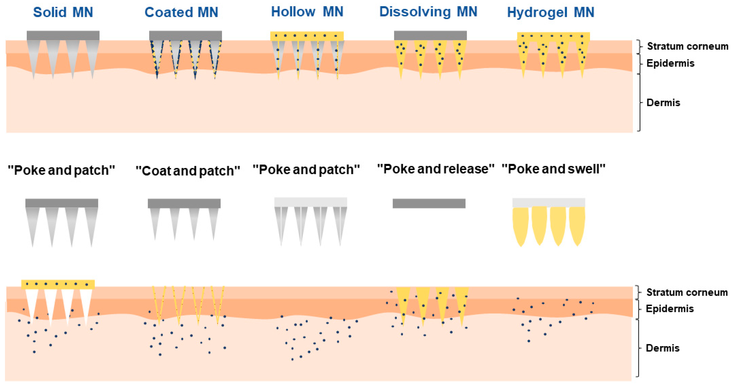
3. Overcoming the Challenge with Dissolving Microneedles

The emergence of microneedle technology has provided potential insights for overcoming the stratum corneum barrier, resulting in the enhancement of the transdermal absorption rate. The microneedle is a chip-type transdermal drug delivery system fabricated by integrating hundreds of needle tips with a length of 25~2000 μm (mostly 100~1500 μm) on a matrix of ~1 cm2 area [23]. It can create a large number of tiny pores within the skin as delivery channels to promote the penetration of drugs through physical penetration with the needles [24]. Thus, they can greatly increase the transdermal absorption rate compared with transdermal drug formulations [25]. In addition, delivering drugs through microneedles can achieve a rapid transdermal absorption rate and high bioavailability, which is comparable to subcutaneous injection; the former may even exceed the latter when delivering protein or other biomacromolecules [26]. Furthermore, it can be applied to most of the body’s skin positions [27] and improve patient compliance. It is worth mentioning that during subcutaneous injection, the tip of the syringe always inevitably touches the nociceptive nerves that perceive pain in the skin, resulting in severe pain and low patient compliance [28]. However, by regulating the length of microneedles, it is possible to avoid touching the nociceptive nerves distributed in the deeper dermis and subcutaneous tissues, which improves patient compliance by mitigating pain [29].
Over the past few years, microneedles have evolved with significant development, and the current microneedles can be categorized into solid microneedles, coated microneedles, hollow microneedles, dissolving microneedles (DMNs), and swellable microneedles [30] (Figure 1). (1) Solid microneedles: based on the principle of “poke and patch”, firstly, the metal, ceramic, or silicone-based microneedles without loading drugs puncture the stratum corneum and form delivery channels. Then, the drug formulations will be administrated after the microneedle is removed so that the drugs can diffuse through the delivery channels [31]. However, during the actual drug delivery process, the delivery channels generated by solid microneedles are temporary and easy to close, affecting the transdermal absorption rate. (2) Coated microneedles: based on the principle of “coat and poke”, the drugs are coated at the tip of the needles in the form of membranes; then, the drugs dissolve from the surface of the needle tip after administration [32]. This type of microneedle can only be used for drugs with high efficacy per unit dose and cannot be used for high-dose delivery. (3) Hollow microneedles: based on the principle of “poke and flow”, the drug solution’s flow into the skin is facilitated by the driving pressure after applying microneedles [33]. The hollow structure of this kind of microneedle has poor mechanical properties and is prone to fracture during drug delivery. (4) DMNs: based on the principle of “poke and release”, the drugs are loaded in the tip of the needle, and the tip will be degraded or dissolved, releasing the drugs [34]. (5) Swellable microneedles: based on the principle of “poke and swell”, the gel materials in the microneedle dissolve and swell after administration and then squeeze the skin to form a pore, which leads to drug diffusion [35]. However, this type of microneedle has a complicated preparation process and it is difficult to regulate the drug release behavior.
Compared with the other microneedles, DMNs display several advantages: (1) The needle tips exhibit an occupancy effect, where the formed delivery channels will not close before the needle tips are completely dissolved. Unlike DMNs, solid microneedles do not utilize such an effect, which causes a decrease in the transdermal absorption rate [31]. (2) The whole parts of needles can be loaded with drugs, and therefore, the drug-carrying capacity is higher than that of the coated microneedles [36]. (3) The solid structures of the needles have better mechanical properties than hollow microneedles. Hence, they do not fracture easily in the clinical drug delivery process [33]. (4) The preparation process of DMNs is relatively straightforward compared with swellable microneedles, and the drug release behavior can be regulated more easily [33]. In addition, DMNs are prepared with biodegradable and water-soluble materials, which can avoid the generation of harmful medical wastes [37].
Based on the above-mentioned advantages, DMNs are expected to be a plausible technique to overcome the challenge of low transdermal absorption rates. Moreover, they show great potential for accelerating the development of the global transdermal drug delivery system markets.

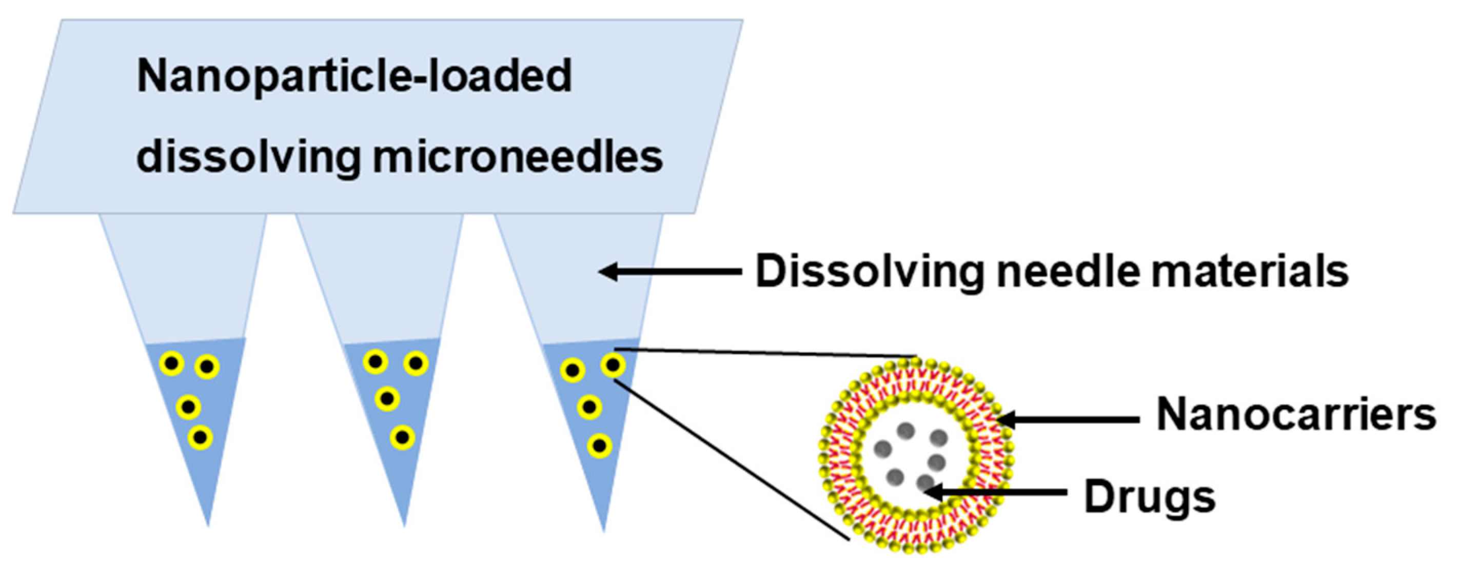
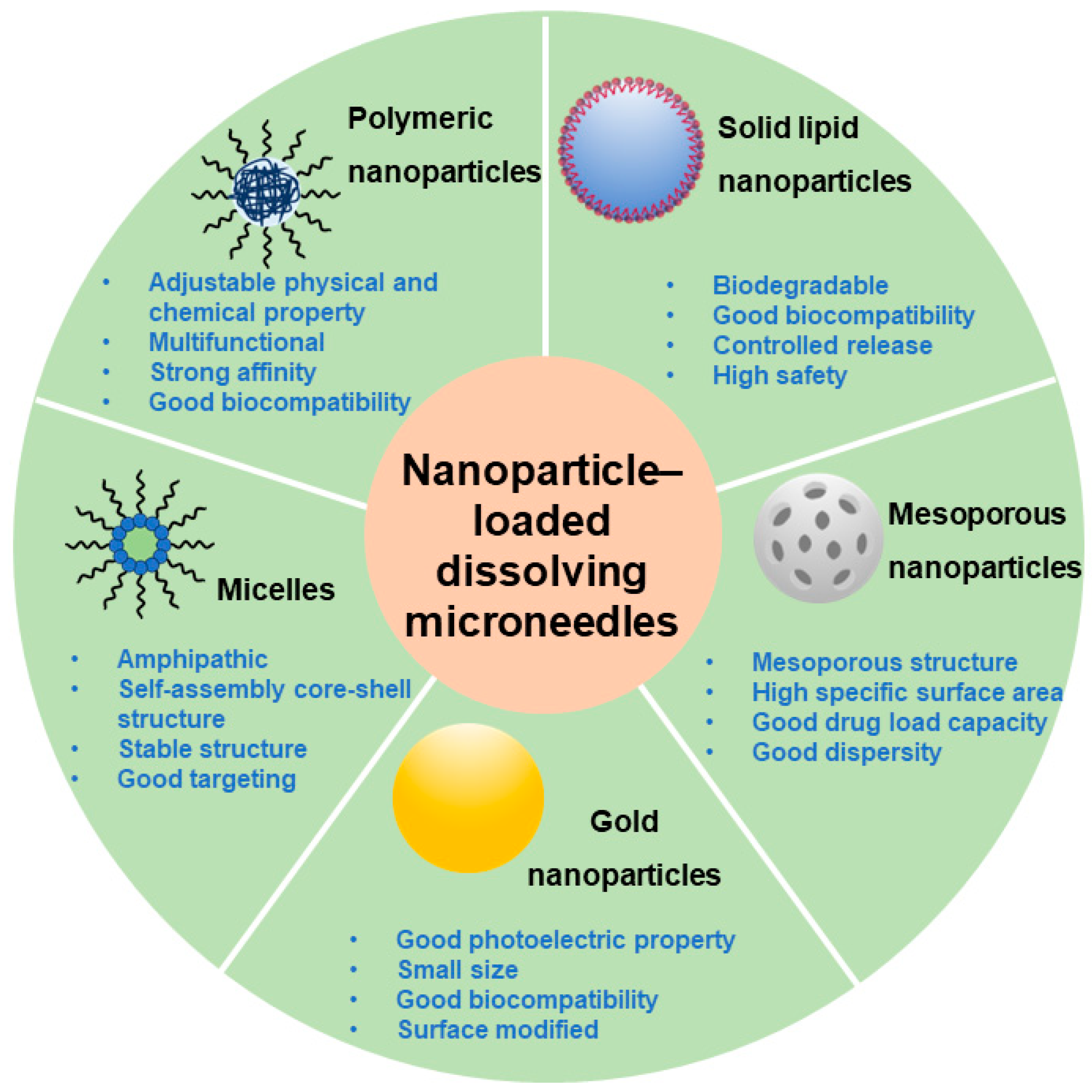
4. Nanoparticle-Loaded DMNs: A Better Choice for Precision Medication

In contemporary medicine, there is a discernible shift towards precision medicine. This evolving paradigm emphasizes the individualization, refinement, and intelligence of clinical treatments, thus setting forth a new and heightened demand within the medical field.
Although DMNs exhibit several advantages, some unmet needs for precision medicine remain. Specifically, most of the materials used in DMNs show strong hydrophilicity, such as hyaluronic acid (HA) [18], polyvinyl alcohol (PVA) [38], and povidone K30 (PVP K30) [39], while about 75% of marketed products and 90% of drugs in development pipelines are hydrophobic [40]. Therefore, hydrophobic drugs display low solubility in hydrophilic microneedle materials [41], which is attributed to a poor drug loading capacity. In addition, the drug release profiles cannot be controlled by merely using hydrophilic materials, and DMNs consisting of such materials cannot achieve precise and intelligent treatments [42]. Therefore, these two main challenges must be addressed when developing DMNs to fulfill the requirements of precision medicine.
Applying nanocarriers is a novel approach to improve the drug loading and controlled-release capacities of DMNs. Nanocarriers are drug delivery carriers with a diameter ranging from 1 to 1000 nm (mostly 100 to 500 nm) in single or multiple dimensions [43]. On one hand, nanocarriers can enhance the drug loading capacity of DMNs, and the proposed mechanisms are provided below. Firstly, the hydrophobic regions of the matrix materials serve as suitable accommodation media for hydrophobic drugs to improve the drug solubility of nanocarriers [44]. Secondly, at the microscopic level, nanocarriers have a subtle “lattice-like” structure in which the cavities of the “lattice” can accommodate drug monomers or oligomers, thereby facilitating drug dispersion [45]. Moreover, at the nanoscale, the solubility of the drugs will increase significantly as the size of the drugs decreases. The detailed mechanism of this phenomenon can be explained by the Ostwald–Freundlich equation [45], which illustrates that the solubility of a drug is related to its particle size:
ln S 2 S 1 = 2 σ M ρ R T 1 r 2 1 r 1
where r1 is the radius of drug particle 1, r2 is the radius of drug particle 2, S1 is for the solubility of drug particle 1, S2 is the solubility of drug particle 2, σ is the surface energy of the drug particles, M is the molecular weight of the drug, ρ is the density of the drug particles, R is the ideal gas constant, and T is the absolute temperature. According to Equation (1), the smaller the particle size of the drug molecules, the higher the solubility. Meanwhile, due to the domain-limiting effect of nanocarriers, the distribution diameter of the drug is within the nanoscale range [46]. Therefore, the solubility of the drugs in the nanocarrier can be effectively enhanced.
On the other hand, nanocarriers can achieve a controlled release profile. Firstly, hydrophobic carrier materials act as retardants, which prolong the drug release cycles and reduce uncontrollable burst releases [47]. Secondly, the physical and chemical properties of nanocarrier materials play an important role in the nanoparticle penetration rate and diffusion range. The drug release behavior of nanomaterials can be modulated by physicochemical modification [48]. Finally, some nanocarriers have active or passive targeting effects for specific organs, which can potentially control drug release at specific sites [49].
In addition, applying nanoparticle-loaded DMNs can improve the bioavailability and retention time of drugs, increasing therapeutic efficacy and reducing the toxicity of some drugs [50]. Furthermore, some nanoparticles were designed to construct smart-responsive MNs, which have become a research hotspot. This type of microneedle can intelligently and dynamically regulate drug release in response to changes in pH, glucose, and enzyme concentrations in the internal microenvironment of the tumor or can be stimulated by external temperature, electric field, or light radiation [51]. Nowadays, nanoparticle-loaded DMNs have been applied to cure diseases through chemotherapy [52], gene therapy [53], immunotherapy [54], photothermal [55], and photodynamic therapies [56]. Some drug formulations for treating specific diseases (e.g., cancer, diabetes, and cardiovascular diseases) are in different stages of clinical trials or have been commercialized [57]. In addition, multiple factors that affect the drug loading and release efficiency of NPs have been extensively studied [58,59].
Based on the above statements, developing composite DMNs with nanocarriers for further clinical application is necessary and significant. Combining DMNs with nanocarriers via enriching nanocarriers at the tips of the DMNs can fabricate a new type of nanoparticle-loaded DMN (Figure 2), which possesses the advantages of both DMNs and nanocarriers such as high drug loading capacity, flexible drug release behavior, good mechanical properties, and simple preparation steps.
Currently, formulation scientists have attempted to apply multiple types of nanocarriers including solid lipid nanoparticles (SLNs), mesoporous nanoparticles, gold nanoparticles, micelles, and polymer nanoparticles into the needles of DMNs to prepare nanoparticle-loaded DMNs (Figure 3). It has been reported that these nanocarriers have been used to treat diseases such as diabetes, infections, cancers, and skin diseases topically and systemically [60]. Some representative examples are summarized in Table 1. Herein, nanoparticle-loaded DMNs have a broad clinical application prospect.
However, nanoparticle-loaded DMNs still face some challenges that hinder their further development and clinical application. The incorporation of nanoparticles into MNs increases the complexity of the MN manufacturing process, and additional manufacturing steps are required. Moreover, the compatibility between the nanoparticles and microneedle materials should be fully investigated to maintain the properties of microneedles such as mechanical strength [81]. In addition, the nanoparticles undergo complex processes in the body. Therefore, the in vivo fate of nanoparticle-loaded DMNs must be investigated comprehensively.

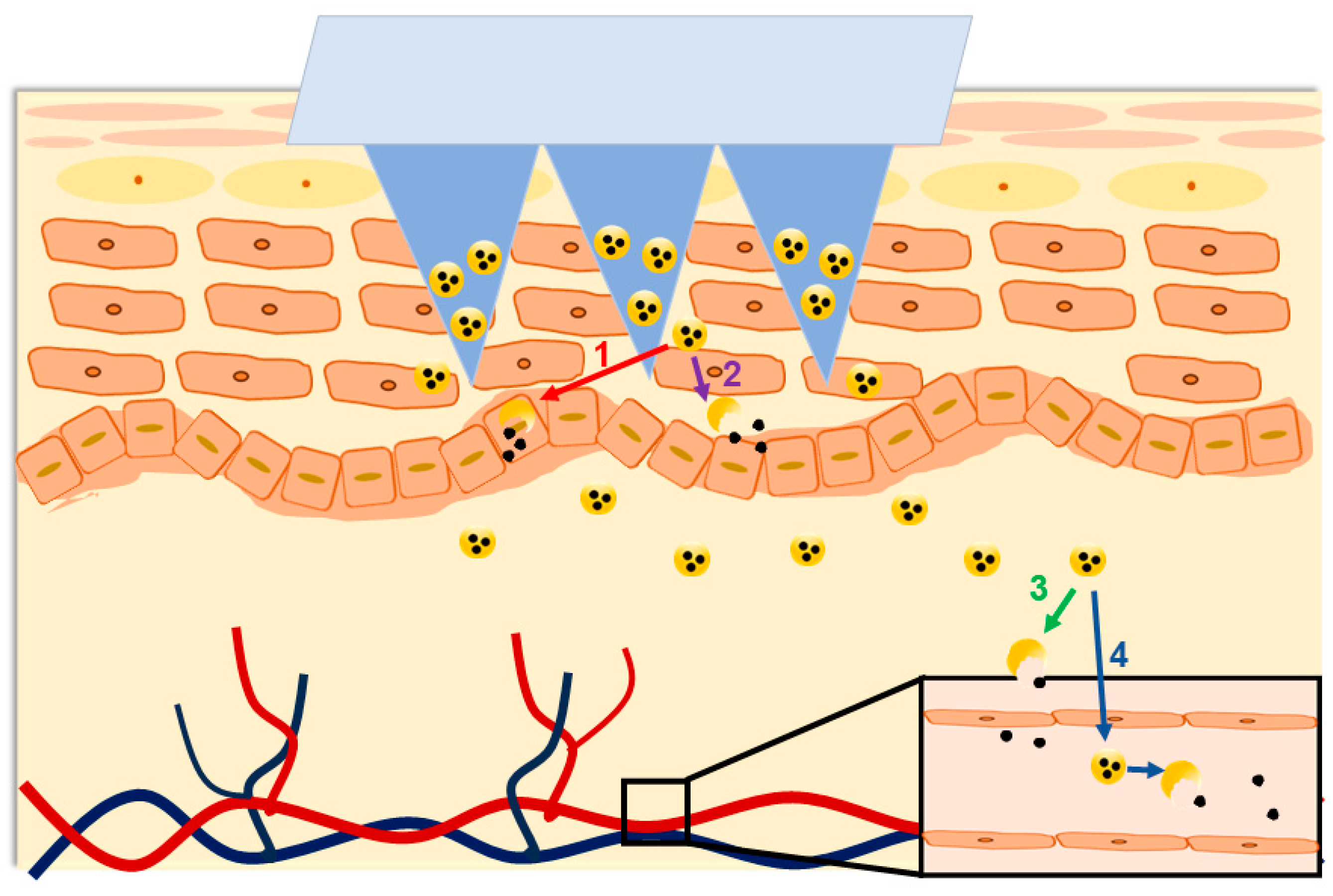
5. Necessity of In Vivo Fate Study

The in vivo fate of a formulation refers to all the disposition processes encountered in the body by the components of the formulation, including absorption, distribution, metabolism, and excretion (ADME) at the level of major cells, tissues, organs, and systems [82]. In the DMN formulation, the components include the drugs, the needle materials, and the nanocarriers. In the past few years, researchers have focused mainly on the in vivo fate of drugs [83,84,85]. However, with the continuous development of pharmaceutics, scientists realize that multiple types of excipients entering the body also undergo ADME processes [86]. Therefore, the in vivo fate of the drugs, needle materials, and nanocarriers, constituting the in vivo fate of nanoparticle-loaded DMNs, should be considered during formulation design and development.

5.1. Necessity Analysis from Clinical Perspective

Although nanoparticle-loaded DMNs show potential for clinical application, as of December 2023, no related products have been approved for clinical application so far. It is presumed that the lack of in vivo fate studies is the main challenge preventing their clinical translation. After transdermal delivery, nanocarriers may be absorbed via the transdermal pathway or the transdermal appendages pathway, and enzymatic degradation may occur during their transfer across epidermal cells, dermal stroma, and vascular endothelial cells [87]. Although DMNs can break through the stratum corneum barrier, the nanocarriers still face certain transfer processes after release [88]. It may be speculated that nanoparticles would undergo these possible in vivo fates: (1) intact nanoparticles are taken up by skin cells and degraded, so do not enter the blood circulation; (2) nanoparticles are degraded before they are taken up by skin cells, so do not enter the blood circulation; (3) nanoparticles travel across the skin cells but degrade before they are taken up into the blood vessels by the vascular endothelial cells; or (4) intact nanoparticles are taken up by the vascular endothelial cells and enter into the blood circulation (Figure 4). In addition, some properties of the nanocarriers remain unknown, including the transdermal absorption rate, degradation rate, the percentage of complete nanocarrier absorption into the blood and the organs, and the excretion kinetics. These will cause difficulties in prescription, administration, therapeutic monitoring, and pharmacovigilance. We hypothesized that only comprehensive in vivo fate studies can illustrate the in vivo fate of nanoparticle-loaded DMNs. Most importantly, understanding the in vivo fate studies can largely avoid therapeutic failures in clinical applications and potentially promote clinical translation.
Numerous studies have endeavored to explore the in vivo fate of drugs and nanoparticles. The drugs and nanocarriers may undergo dependent or independent in vivo fates. Therefore, understanding the in vivo properties and biodistribution of both drugs and nanocarriers is critical to facilitate the development of nanoparticle-loaded DMNs.
Among all drug modalities, small molecules are usually low-molecular-weight compounds with a defined chemical structure [89]. They exhibit specificity depending on the chemical structures and the targets they interact with in vivo. When small molecules are absorbed in the blood, they are distributed through the body’s circulation to various organs and tissues, where they interact with cellular components. Small molecules tend to have a shorter half-life and are easily metabolized by the body, thus limiting their duration in the body [90]. To explore their in vivo fate, Li et al. developed clearing-assisted tissue click chemistry (CATCH) to optically image covalent drug targets in intact mammalian tissues. This study provided a valuable platform for visualizing the in vivo interactions of small molecules in tissue [91]. Pires et al. constructed a novel approach (pkCSM) that uses graph-based signatures to predict central ADMET [92]. Biologics such as proteins and peptides are macromolecular drugs that display different pharmacokinetics compared with small molecules. The high specificity and strong pharmacological activity of biologics contribute to their application in various diseases [93]. Their immunogenicity, complex structures, and spatial conformation are important factors in their in vivo fates [94]. In addition, some single-stranded nucleic acids are absorbed by the liver and kidneys. Absorption of double-stranded nucleic acids can be more challenging, as their double-stranded structure inhibits the ability of the phosphorothioate backbone to enhance absorption [95]. The pharmacokinetic properties of nucleic acid drugs largely depend on the type of chemical modification of the phosphate backbone and ribose of the nucleic acid, as the chemical modification strategy will have a direct impact on the biostability of the nucleic acid drug and its ability to bind to plasma proteins [95]. For example, phosphorothioate modifications can induce nonspecific binding of nucleic acids to plasma proteins, resulting in beneficial effects on blood clearance, biodistribution, and cellular uptake [96]. Therefore, all of the above factors are worth investigating.
Regarding nanocarriers, a thorough investigation into the myriad factors influencing in vivo fates is imperative. It has been reported that the protein corona is an important factor affecting the interaction between nanoparticles and organisms and the in vivo delivery process. The nature of nanoparticles themselves and the environment of the organism can regulate the in vivo delivery process of nanoparticles by influencing the composition of protein coronas [97]. Therefore, paying attention to protein coronas in the in vivo fate studies is necessary. In addition, the size of the nanoparticles was found to determine their elimination and targeting. Du et al. provided a systematic summary of the size-scale laws governing the transport of nanoparticles in the human body and how size-dependent transport can be used to address the main challenges in translational nanomedicine [98]. Furthermore, the shape, hardness [99], surface charge, and surface hydrophilicity of nanoparticles could influence their clearance and translocation [100], which are also worth investigating.

5.2. Necessity Analysis from the Industrial Perspective

Clinical application is the landing point of industrialization, and in vivo fate research is conducive to the industrialization of sophisticated formulations. Firstly, the approval of such formulations by most global regulatory agencies requires clinical trials, and in vivo fate research may provide valuable insights for these trials. Secondly, studying the in vivo fate can effectively save research and development (R&D) spending. Currently, the cost of the existing preclinical research stage is relatively low. It is suggested to increase the investment at the preclinical research stage to avoid some risks for clinical trials in advance and to reduce the trial-and-error cost in the clinical stage as a result. Regarding nanoparticle-loaded DMNs, in-depth in vivo fate studies can be applied in the official preclinical research stage to reduce total R&D costs. Thirdly, it is beneficial for the optimization of formulation. The “quality by design (QbD)” strategy proposed by the U.S. FDA early this century is commonly used for formulations in industrialized R&D [101]. In QbD, the critical quality attribute (CQA) is one of the most important factors. Physicochemical properties such as dissolution and stability are often used as CQAs in conventional formulation industrialization (e.g., industrial transformation of oral drug delivery systems represented by tablets and transdermal drug delivery systems represented by patches). It is rational to some extent because traditional formulations have better in vivo/in vitro correlation (IVIVC), and excellent physicochemical properties usually imply good in vivo pharmacokinetics [102]. Therefore, CQAs can reflect the R&D success rate. However, nanocarrier-loaded DMNs are emerging, as well as other complex formulations, and the mere use of physicochemical properties as a CQA cannot predict clinical performance, which makes it challenging to ensure the success of R&D. For this reason, the development strategies should be modified and optimized by the addition of the in vivo fate investigation as a CQA. For example, the transdermal absorption rate should be used as a CQA for the R&D QbD of nanocarrier-loaded DMNs to promote the industrialization of the potential products.

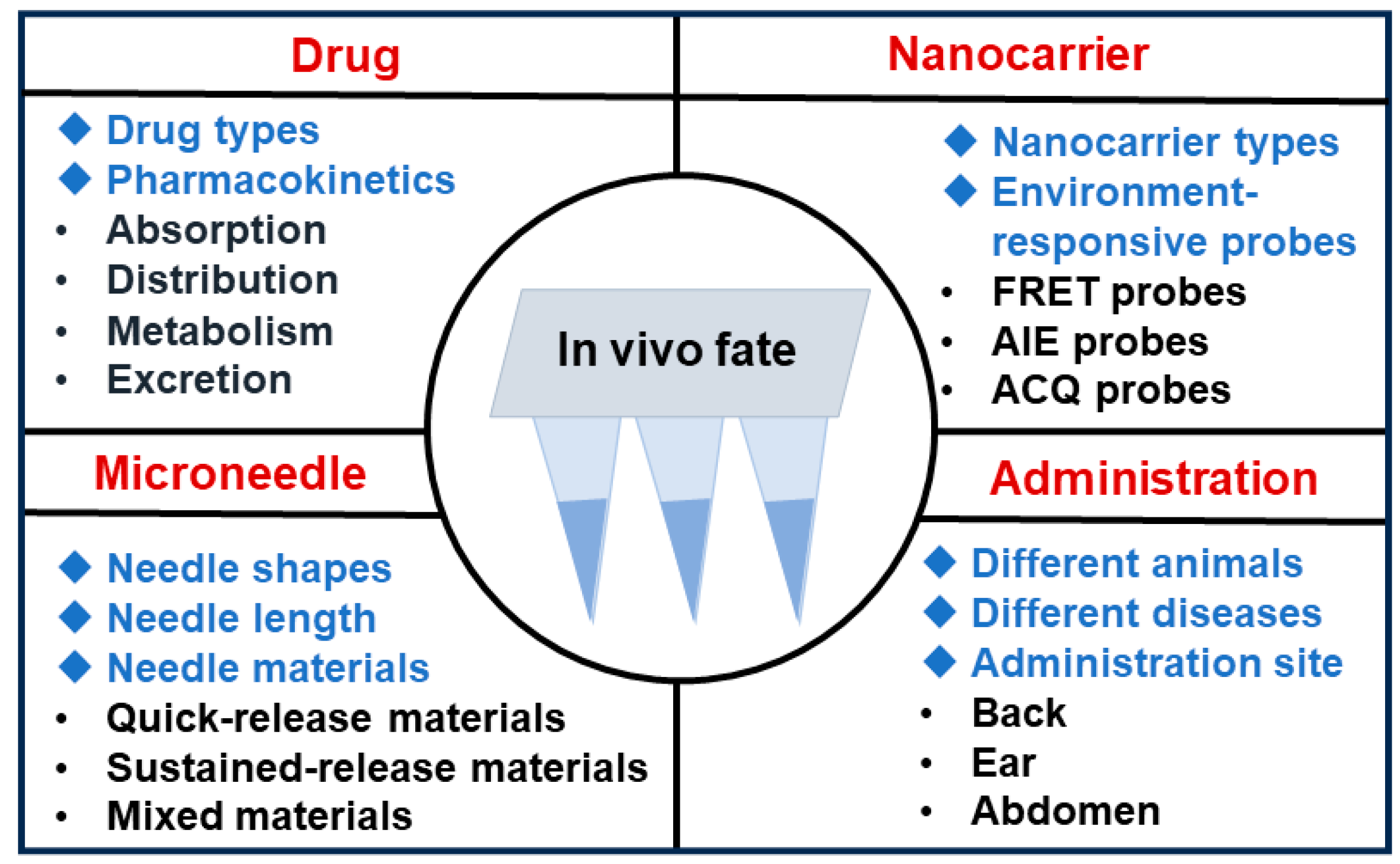
6. Entry Points for In Vivo Fate Studies

As we have demonstrated the necessity to carry out in vivo fate studies on nanoparticle-loaded DMNs, the next task is to investigate some entry points. The in vivo fate studies of nanoparticle-loaded DMNs can be designed according to the following sectors (Figure 5).
(1) Drugs: Multiple types of drug modalities, such as small molecules [103], proteins [104], peptides [105], nucleic acids [106], and cells [107], have been loaded into DMNs. The conventional discipline of pharmacokinetics mainly focuses on the ADME processes and has established a relatively systematic research pattern. The in vivo fate of drugs can be elucidated with the assistance of existing technologies [108].
(2) Nanocarriers: Multiple nanocarriers with different structures could be investigated for the in vivo fates. The fluorescent probe tracing method is a reliable, convenient, and widely used method for in vivo fate studies [109]. Nanocarriers can be labeled with different probes to investigate their in vivo fates. However, conventional fluorescent probes will still emit signals after separating from the labeled parent particles, which will interfere with the results. To solve this problem, environment-responsive probes in response to environmental changes have attracted widespread attention [110]. Such probes can distinguish in-carrier signals from free-probe signals effectively. There are three main categories of environment-responsive fluorescent probes, whose principles are based on fluorescence resonance energy transfer (FRET) [111], aggregation-induced emission (AIE) [112], and aggregation-caused quenching (ACQ) effects [113], respectively. The FRET probes are pairs of fluorescent probe molecules with overlay fluorescence emission spectra that can undergo dipole–dipole interactions [114]. The FRET effect refers to the transfer of radiant energy from one probe molecule in the excited state to another in the ground state upon excitation, which can only occur when the distance between pairs of fluorescent probe molecules is less than 10 nm [115]. When FRET probes are loaded in an intact nanoparticle delivery system, they are restricted by the microstructure of the nanodrug delivery system, and the distance between pairs of probe molecules is limited to produce the FRET effect. Meanwhile, when FRET probes are released from the incomplete nanoparticle delivery system into an aqueous biomatrix, the distance between pairs of probe molecules will be significantly larger and the FRET effect will disappear. The AIE and ACQ probes utilize the AIE and ACQ effects, respectively. AIE probe and ACQ probe molecules have large conjugated structures (aromatic groups), which will undergo aggregation in aqueous biological matrices due to hydrophobic interactions, such as π-π stacking, and form aggregates [116]. The ACQ probes emit fluorescence when encapsulated in nanoparticles. However, after experiencing the degradation or destruction of the nanocarriers, the molecules are released and undergo signal switching or fluorescence quenching due to environmental changes. In contrast, AIE probes show exactly opposite properties. They suffer enhanced fluorescence signals when released from the nanoparticles [117]. Therefore, the status of the nanoparticles in the body can be monitored through the signal changes from these probes.
(3) Microneedle: When the DMNs are inserted into the skin, the hydrophilic microneedle will dissolve in the skin interstitial fluid [118]. Nanoparticles are always loaded into the needle of DMNs. Therefore, nanoparticles will be released into the skin and then transported into circulation when the needles of the DMNs are dissolved [81]. The individual DMN degradation behaviors are mainly governed by the needle shape, length, material, etc. Therefore, the in vivo fate studies of nanoparticle-loaded DMNs can be explored from these aspects. Based on this scale, a series of studies have been performed. For example, alternative lengths of the needle span a wide range (25–2000 μm) [119], and the needle length significantly affects the in vivo fate of nanoparticle-loaded DMNs. This is because the dermal region consists of various cells and matrices that interact with the nanocarriers, and the needle length determines the penetration depth of the formulation in the skin [120]. Specifically, at different depths of the dermis, different types and compositions of cells and matrices are likely to affect the transdermal absorption process, ultimately resulting in different therapeutic effects. Therefore, it is essential to investigate the role of needle length on the transdermal absorption rate. Shi et al. investigated the in vivo fate of nanocarrier-loaded DMNs with different lengths of needles. They found that in the spatial dimension, DMNs showed a length-dependent diffusion depth, while in the temporal dimension, the diffusion rates of DMNs with different lengths (400, 800, and 1200 μm) were similar within 24 h of insertion [121].
In addition, the shapes and materials of microneedles also have an influence on the performance. Li et al. investigated DMNs with different needle geometries, including cone, cone–cylinder, rectangular pyramid, and hexagonal pyramid. The in vivo studies demonstrated that cone MNs exhibited the highest dissolution ratio of 80%, whereas the cone–cylindrical MNs had the lowest dissolution ratio of 40% [122]. Aoyagi et al. systematically examined the impact of tip geometry (tip angle, width) on the mechanical properties of MNs. The results revealed that MNs with low tip angles (15–30°) and thin needle shafts (120 μm) effectively enhanced microneedle insertion for efficient drug delivery [123]. The chemical composition of different needle materials determines their water solubility, swelling, and degradation, affecting the drug release from microneedles [124]. As DMNs typically dissolve entirely within seconds to minutes, they typically manifest a sudden release profile, which is advantageous for scenarios requiring rapid onset of action, such as pain relief. To accelerate the dissolving process, an effervescing agent could be added to the needle materials [125]. However, for vaccines or insulin delivery, a sustained payload release profile is desirable to mitigate side effects and reduce the frequency of MN administration. To achieve prolonged release, needle materials with slower dissolution are preferred [126]. Utilizing chitosan as a needle material enables the sustained release of payloads for up to 28 days [127]. Different needle materials afford DMNs varying dissolving properties, thereby offering flexibility in tailoring release kinetics to specific therapeutic requirements.
(4) Administration: The nanoparticle-loaded DMNs may be used for different administration sites, different diseases, or different animals, and the corresponding in vivo fates can be investigated. Different administration sites are likely to affect the in vivo fate of nanoparticle-loaded DMNs, and the reasons are as follows: Firstly, the thicknesses of the epidermis and dermis are different, which implies variable depths of needle entry into the dermal region after microneedle administration. Secondly, different types and contents of cells and matrices in the dermal layer may interact differently with the nanocarriers. Thirdly, different mechanical strengths of skins contribute different shear forces on the microneedles. Fu et al. explored the influence of nanoparticle-loaded DMNs’ application sites with ACQ probes and demonstrated that the transdermal diffusion rate of nanoparticle-loaded DMNs was positively correlated with skin thickness. Ear skin showed the highest transdermal diffusion rate, followed by abdomen and back skin [128]. In addition to the common target tissue skin, MN can still target other tissues and organs, including the eye [129], mouth [130], heart [131], gastrointestinal tract [132], and tumor [133]. Different target tissues may undergo various in vivo fates.

7. Outlook

Many factors may affect the in vivo study and are worth investigating. Based on the components of microneedles, drug types and pharmacokinetics, nanocarriers (size, structure, type), needle (material, composition, shape), fluorescence probe (AIE, ACQ, FRET), administration (animals, disease, site, time), etc., can be investigated.
In addition, many new types of DMNs have been developed and gained more attention. For example, gas-propelled DMNs were fabricated to improve drug transdermal efficiency, which utilizes gas as the driving force for skin permeation [134]. Moreover, to achieve transmucosal sequential delivery of multiple drugs, double-layer DMNs were developed to treat oral mucosa diseases [130]. The in vivo fate of these novel DMNs is also suggested to be investigated, and relevant research will further be conducted. Most importantly, the in vivo fate studies of nanoparticle-loaded DMNs will significantly facilitate the development of clinical practice and industrialization.
In the foreseeable future, it is anticipated that in vivo fate studies of nanoparticle-loaded DMNs will emerge as a focal point of research interest. There is a pressing need for more precise tools and methods to reveal the in vivo fate studies of nanoparticle-loaded DMNs. To address a diverse array of therapeutic requirements and cater to the delivery demands of different types of drugs, multiple types of nanoparticle-loaded DMNs will be constructed with enhanced properties. This trajectory is poised to significantly advance the understanding of in vivo fate studies, thereby setting the stage for future exploration in this domain.
Regarding clinical translation, several imperative considerations come to the forefront. Firstly, essential evaluation criteria are in demand for evaluating the in vivo fate studies of nanoparticle-loaded DMNs. Secondly, achieving large-scale industrialized production of microneedle products is a significant challenge. Thirdly, due attention must be accorded to ensuring the safety and stability of both the MNs and the NPs. Finally, the cost implications pertaining to APIs, excipients, and storage conditions emerge as pivotal factors influencing the clinical application.

8. Conclusions

Transdermal drug delivery systems have gained great attention in pharmaceutics. As a novel transdermal drug delivery system, nanoparticle-loaded DMNs possess the advantages of DMNs like high drug loading, good mechanical properties, and easy fabrication. Therefore, they demonstrate outstanding potential in clinical application, particularly precision medication. Nevertheless, as of December 2023, no nanoparticle-loaded DMN products have received official approval from the FDA. It is predicted that the lack of in vivo fate studies is the critical bottleneck issue hampering the clinical translation of relevant products. The in vivo fate studies of nanoparticle-loaded DMNs, which will fulfill the requirements of their clinical application and industry transformation, are necessary.

Author Contributions

Conceptualization, Z.C., X.P. and Z.H.; writing—original draft preparation, Z.C., Y.W. and J.J.; supervision, G.Q. and Z.H.; methodology, P.H.; visualization; J.J. and G.Q.; validation, C.W. and X.P.; project administration, G.Q., C.W. and X.P.; funding acquisition, P.H. and C.W.; writing—review and editing, Z.C., Y.W. and Z.H. All authors have read and agreed to the published version of the manuscript.

Funding

This work was supported by the National Natural Science Foundation of China (Grant No. 82373800, 82104070), and the Natural Science Foundation of Guangdong Province of China (Grant No. 2021A1515220047, 2023A1515011135).

Data Availability Statement

Details are available from authors.

Conflicts of Interest

The authors declare no conflicts of interest.

References

  1. Jain, K.K. An Overview of Drug Delivery Systems. Methods Mol. Biol. 2020, 2059, 1–54. [Google Scholar]
  2. Park, H.; Otte, A.; Park, K. Evolution of drug delivery systems: From 1950 to 2020 and beyond. J. Control. Release 2022, 342, 53–65. [Google Scholar] [CrossRef] [PubMed]
  3. Anselmo, A.C.; Mitragotri, S. An overview of clinical and commercial impact of drug delivery systems. J. Control. Release 2014, 190, 15–28. [Google Scholar] [CrossRef] [PubMed]
  4. Liu, L.; Yao, W.; Rao, Y.; Lu, X.; Gao, J. pH-Responsive carriers for oral drug delivery: Challenges and opportunities of current platforms. Drug Delivery 2017, 24, 569–581. [Google Scholar] [CrossRef] [PubMed]
  5. Dai, T.; Jiang, W.; Guo, Z.; Wang, Z.; Huang, M.; Zhong, G.; Liang, C.; Pei, X.; Dai, R. Studies on oral bioavailability and first-pass metabolism of withaferin A in rats using LC-MS/MS and Q-TRAP. Biomed. Chromatogr. 2019, 33, e4573. [Google Scholar] [CrossRef] [PubMed]
  6. Marwah, H.; Garg, T.; Goyal, A.K.; Rath, G. Permeation enhancer strategies in transdermal drug delivery. Drug Delivery 2016, 23, 564–578. [Google Scholar] [CrossRef]
  7. Lam, P.L.; Gambari, R. Advanced progress of microencapsulation technologies: In Vivo and In Vitro models for studying oral and transdermal drug deliveries. J. Control. Release 2014, 178, 25–45. [Google Scholar] [CrossRef]
  8. Ramadon, D.; McCrudden, M.T.C.; Courtenay, A.J.; Donnelly, R.F. Enhancement strategies for transdermal drug delivery systems: Current trends and applications. Drug Delivery Transl. Res. 2022, 12, 758–791. [Google Scholar] [CrossRef]
  9. Dasht Bozorg, B.; Bhattaccharjee, S.A.; Somayaji, M.R.; Banga, A.K. Topical and transdermal delivery with diseased human skin: Passive and iontophoretic delivery of hydrocortisone into psoriatic and eczematous skin. Drug Delivery Transl. Res. 2022, 12, 197–212. [Google Scholar] [CrossRef]
  10. Bird, D.; Ravindra, N.M. Transdermal drug delivery and patches-An overview. Med. Devices Sens. 2020, 3, e10069. [Google Scholar] [CrossRef]
  11. Carter, P.; Narasimhan, B.; Wang, Q. Biocompatible nanoparticles and vesicular systems in transdermal drug delivery for various skin diseases. Int. J. Pharm. 2019, 555, 49–62. [Google Scholar] [CrossRef] [PubMed]
  12. Jorgensen, A.M.; Gorkun, A.; Mahajan, N.; Willson, K.; Clouse, C.; Jeong, C.G.; Varkey, M.; Wu, M.; Walker, S.J.; Molnar, J.A.; et al. Multicellular bioprinted skin facilitates human-like skin architecture in vivo. Sci. Transl. Med. 2023, 15, eadf7547. [Google Scholar] [CrossRef] [PubMed]
  13. Manikkath, J.; Sumathy, T.K.; Manikkath, A.; Mutalik, S. Delving Deeper into Dermal and Transdermal Drug Delivery: Factors and Mechanisms Associated with Nanocarrier-mediated Strategies. Curr. Pharm. Des. 2018, 24, 3210–3222. [Google Scholar] [CrossRef] [PubMed]
  14. Schmitt, T.; Neubert, R.H.H. State of the Art in Stratum Corneum Research. Part II: Hypothetical Stratum Corneum Lipid Matrix Models. Skin Pharmacol. Physiol. 2020, 33, 213–230. [Google Scholar] [CrossRef] [PubMed]
  15. Gupta, M.; Agrawal, U.; Vyas, S.P. Nanocarrier-based topical drug delivery for the treatment of skin diseases. Expert Opin. Drug Delivery 2012, 9, 783–804. [Google Scholar] [CrossRef]
  16. Iwai, I.; Han, H.; den Hollander, L.; Svensson, S.; Oefverstedt, L.-G.; Anwar, J.; Brewer, J.; Bloksgaard, M.; Laloeuf, A.; Nosek, D.; et al. The Human Skin Barrier Is Organized as Stacked Bilayers of Fully Extended Ceramides with Cholesterol Molecules Associated with the Ceramide Sphingoid Moiety. J. Investig. Dermatol. 2012, 132, 2215–2225. [Google Scholar] [CrossRef]
  17. Sa, G.F.F.; Serpa, C.; Arnaut, L.G. Stratum corneum permeabilization with photoacoustic waves generated by piezophotonic materials. J. Control. Release 2013, 167, 290–300. [Google Scholar] [CrossRef]
  18. Anantrao, J.H.; Nath, P.A.; Nivrutti, P.R. Drug Penetration Enhancement Techniques in Transdermal Drug Delivery System: A Review. J. Pharm. Res. Int. 2021, 33, 46–61. [Google Scholar] [CrossRef]
  19. Yu, Y.-Q.; Yang, X.; Wu, X.-F.; Fan, Y.-B. Enhancing Permeation of Drug Molecules Across the Skin via Delivery in Nanocarriers: Novel Strategies for Effective Transdermal Applications. Front. Bioeng. Biotechnol. 2021, 9, 646554. [Google Scholar] [CrossRef]
  20. Alkilani, A.Z.; Nasereddin, J.; Hamed, R.; Nimrawi, S.; Hussein, G.; Abo-Zour, H.; Donnelly, R.F. Beneath the Skin: A Review of Current Trends and Future Prospects of Transdermal Drug Delivery Systems. Pharmaceutics 2022, 14, 1152. [Google Scholar] [CrossRef] [PubMed]
  21. Jeong, W.Y.; Kwon, M.; Choi, H.E.; Kim, K.S. Recent advances in transdermal drug delivery systems: A review. Biomater. Res. 2021, 25, 24. [Google Scholar] [CrossRef]
  22. Qindeel, M.; Ullah, M.H.; Fakharud, D.; Ahmed, N.; Rehman, A.U. Recent trends, challenges and future outlook of transdermal drug delivery systems for rheumatoid arthritis therapy. J. Control. Release 2020, 327, 595–615. [Google Scholar] [CrossRef]
  23. Chakraborty, C.; Bhattacharya, M.; Lee, S.-S. Current Status of Microneedle Array Technology for Therapeutic Delivery: From Bench to Clinic. Mol. Biotechnol. 2023. [Google Scholar] [CrossRef]
  24. Ma, G.; Wu, C. Microneedle, bio-microneedle and bio-inspired microneedle: A review. J. Control. Release 2017, 251, 11–23. [Google Scholar] [CrossRef] [PubMed]
  25. Zhou, Y.; Jin, Y.; Jiang, M.; Yang, F.; Luo, J.; Wang, Y.; Tao, X. Research Progress on Combination of Microneedle and New Transdermal Drug Delivery Carrier. Chin. J. Mod. Appl. Pharm. 2020, 37, 2170–2176. [Google Scholar]
  26. Harvey, A.J.; Kaestner, S.A.; Sutter, D.E.; Harvey, N.G.; Mikszta, J.A.; Pettis, R.J. Microneedle-Based Intradermal Delivery Enables Rapid Lymphatic Uptake and Distribution of Protein Drugs. Pharm. Res. 2011, 28, 107–116. [Google Scholar] [CrossRef] [PubMed]
  27. Donnelly, R.F.; Singh, T.R.R.; Woolfson, A.D. Microneedle-based drug delivery systems: Microfabrication, drug delivery, and safety. Drug Delivery 2010, 17, 187–207. [Google Scholar] [CrossRef] [PubMed]
  28. Vora, L.K.; Moffatt, K.; Tekko, I.A.; Paredes, A.J.; Volpe-Zanutto, F.; Mishra, D.; Peng, K.; Thakur, R.R.S.; Donnelly, R.F. Microneedle array systems for long-acting drug delivery. Eur. J. Pharm. Biopharm. 2021, 159, 44–76. [Google Scholar] [CrossRef] [PubMed]
  29. Oh, S.J.; Jung, J.H. Sustainable Drug Release Using Nanoparticle Encapsulated Microneedles. Chem. Asian J. 2022, 17, e202200333. [Google Scholar] [CrossRef] [PubMed]
  30. McCrudden, M.T.C.; McAlister, E.; Courtenay, A.J.; Gonzalez-Vazquez, P.; Singh, T.R.R.; Donnelly, R.F. Microneedle applications in improving skin appearance. Exp. Dermatol. 2015, 24, 561–566. [Google Scholar] [CrossRef] [PubMed]
  31. van der Maaden, K.; Jiskoot, W.; Bouwstra, J. Microneedle technologies for (trans)dermal drug and vaccine delivery. J. Control. Release 2012, 161, 645–655. [Google Scholar] [CrossRef]
  32. Lu, Z.; Du, S.; Li, J.; Zhang, M.; Nie, H.; Zhou, X.; Li, F.; Wei, X.; Wang, J.; Liu, F.; et al. Langmuir-Blodgett-Mediated Formation of Antibacterial Microneedles for Long-Term Transdermal Drug Delivery. Adv. Mater. 2023, 35, e2303388. [Google Scholar] [CrossRef]
  33. Chen, Y.-H.; Wang, F.-Y.; Chan, Y.-S.; Huang, C.; Huang, Y.-Y. Biofabricating hollow microneedle array with controllable microstructure for cell transplantation. J. Biomed. Mater. Res. Part B 2022, 110, 1997–2005. [Google Scholar] [CrossRef] [PubMed]
  34. Sun, Y.; Chen, M.; Yang, D.; Qin, W.; Quan, G.; Wu, C.; Pan, X. Self-assembly nanomicelle-microneedle patches with enhanced tumor penetration for superior chemo-photothermal therapy. Nano Res. 2022, 15, 2335–2346. [Google Scholar] [CrossRef]
  35. Liu, D.; Yu, B.; Jiang, G.; Yu, W.; Zhang, Y.; Xu, B. Fabrication of composite microneedles integrated with insulin-loaded CaCO3 microparticles and PVP for transdermal delivery in diabetic rats. Mater. Sci. Eng., C 2018, 90, 180–188. [Google Scholar] [CrossRef]
  36. Kim, Y.-C.; Park, J.-H.; Prausnitz, M.R. Microneedles for drug and vaccine delivery. Adv. Drug Delivery Rev. 2012, 64, 1547–1568. [Google Scholar] [CrossRef] [PubMed]
  37. Chen, M.-C.; Lai, K.-Y.; Ling, M.-H.; Lin, C.-W. Enhancing immunogenicity of antigens through sustained intradermal delivery using chitosan microneedles with a patch-dissolvable design. Acta Biomater. 2018, 65, 66–75. [Google Scholar] [CrossRef]
  38. Shi, Y.; Zhao, J.; Li, H.; Yu, M.; Zhang, W.; Qin, D.; Qiu, K.; Chen, X.; Kong, M. A Drug-Free, Hair Follicle Cycling Regulatable, Separable, Antibacterial Microneedle Patch for Hair Regeneration Therapy. Adv. Healthcare Mater. 2022, 11, e2200908. [Google Scholar] [CrossRef]
  39. Ping, Y.; Gao, Q.; Li, C.X.; Wang, Y.; Wang, Y.L.; Li, S.; Qiu, M.J.; Zhang, L.Q.; Tu, A.L.; Tian, Y.; et al. Construction of microneedle of Atractylodes macrocephala Rhizoma aqueous extract and effect on mammary gland hyperplasia based on intestinal flora. Front. Endocrinol. 2023, 14, 1158318. [Google Scholar] [CrossRef]
  40. Fang, Z.; Zhang, B.; Xing, W.; Yu, H.; Xing, C.; Gong, N.; Lu, Y.; Du, G. An Evolving Role of Aqueous Piperazine to Improve the Solubility of Non-Steroidal Anti-Inflammatory Drugs. J. Pharm. Sci. 2022, 111, 2839–2847. [Google Scholar] [CrossRef]
  41. Du, M.-X.; Yuan, Y.-F.; Zhang, J.-M.; Liu, C.-Y. Hydrogen-Bonding Interactions in Polymer-Organic Solvent Mixtures. Macromolecules 2022, 55, 4578–4588. [Google Scholar] [CrossRef]
  42. Zaitoon, A.; Luo, X.; Lim, L.-T. Triggered and controlled release of active gaseous/volatile compounds for active packaging applications of agri-food products: A review. Compr. Rev. Food Sci. Food Saf. 2022, 21, 541–579. [Google Scholar] [CrossRef]
  43. Hueppe, N.; Wurm, F.R.; Landfester, K. Nanocarriers with Multiple Cargo Load—A Comprehensive Preparation Guideline Using Orthogonal Strategies. Macromol. Rapid Commun. 2022, 44, e2200611. [Google Scholar] [CrossRef]
  44. Ko, C.-N.; Zang, S.; Zhou, Y.; Zhong, Z.; Yang, C. Nanocarriers for effective delivery: Modulation of innate immunity for the management of infections and the associated complications. J. Nanobiotechnol. 2022, 20, 380. [Google Scholar] [CrossRef]
  45. Chountoulesi, M.; Pispas, S.; Tseti, I.K.; Demetzos, C. Lyotropic Liquid Crystalline Nanostructures as Drug Delivery Systems and Vaccine Platforms. Pharmaceuticals 2022, 15, 429. [Google Scholar] [CrossRef] [PubMed]
  46. Ran, Q.; Wang, M.; Kuang, W.; Ouyang, J.; Han, D.; Gao, Z.; Gong, J. Advances of Combinative Nanocrystal Preparation Technology for Improving the Insoluble Drug Solubility and Bioavailability. Crystals 2022, 12, 1200. [Google Scholar] [CrossRef]
  47. Machado, T.O.; Grabow, J.; Sayer, C.; de Araujo, P.H.H.; Ehrenhard, M.L.; Wurm, F.R. Biopolymer-based nanocarriers for sustained release of agrochemicals: A review on materials and social science perspectives for a sustainable future of agri- and horticulture. Adv. Colloid Interface Sci. 2022, 303, 102645. [Google Scholar] [CrossRef]
  48. De, R.; Mahata, M.K.; Kim, K.-T. Structure-Based Varieties of Polymeric Nanocarriers and Influences of Their Physicochemical Properties on Drug Delivery Profiles. Adv. Sci. 2022, 9, 2105373. [Google Scholar] [CrossRef] [PubMed]
  49. Wang, S.; Lv, Q.; Li, X. Recent Advances in Drug Delivery Nanocarriers for Targeting Hepatocellular Carcinoma. J. Nanomater. 2022, 2022, 7798919. [Google Scholar] [CrossRef]
  50. Moreira, A.F.; Rodrigues, C.F.; Jacinto, T.A.; Miguel, S.P.; Costa, E.C.; Correia, I.J. Microneedle-based delivery devices for cancer therapy: A review. Pharmacol. Res. 2019, 148, 104438. [Google Scholar] [CrossRef]
  51. Liu, G.-S.; Kong, Y.; Wang, Y.; Luo, Y.; Fan, X.; Xie, X.; Yang, B.-R.; Wu, M.X. Microneedles for transdermal diagnostics: Recent advances and new horizons. Biomaterials 2020, 232, 119740. [Google Scholar] [CrossRef]
  52. Chen, Z.; Li, H.; Bian, Y.; Wang, Z.; Chen, G.; Zhang, X.; Miao, Y.; Wen, D.; Wang, J.; Wan, G.; et al. Bioorthogonal catalytic patch. Nat. Nanotechnol. 2021, 16, 933. [Google Scholar] [CrossRef] [PubMed]
  53. Qu, M.; Kim, H.-J.; Zhou, X.; Wang, C.; Jiang, X.; Zhu, J.; Xue, Y.; Tebon, P.; Sarabi, S.A.; Ahadian, S.; et al. Biodegradable microneedle patch for transdermal gene delivery. Nanoscale 2020, 12, 16724–16729. [Google Scholar] [CrossRef]
  54. Huang, S.; Wen, T.; Wang, J.; Wei, H.; Xiao, Z.; Li, B.; Shuai, X. Nanoparticle-integrated dissolving microneedles for the co-delivery of R848/aPD-1 to synergistically reverse the immunosuppressive microenvironment of triple-negative breast cancer. Acta Biomater. 2024, 176, 344–355. [Google Scholar] [CrossRef]
  55. Lei, Q.; He, D.; Ding, L.; Kong, F.; He, P.; Huang, J.; Guo, J.; Brinker, C.J.; Luo, G.; Zhu, W.; et al. Microneedle Patches Integrated with Biomineralized Melanin Nanoparticles for Simultaneous Skin Tumor Photothermal Therapy and Wound Healing. Adv. Funct. Mater. 2022, 32, 2113269. [Google Scholar] [CrossRef]
  56. He, G.; Li, Y.; Younis, M.R.; Fu, L.-H.; He, T.; Lei, S.; Lin, J.; Huang, P. Synthetic biology-instructed transdermal microneedle patch for traceable photodynamic therapy. Nat. Commun. 2022, 13, 6238. [Google Scholar] [CrossRef]
  57. Bobo, D.; Robinson, K.J.; Islam, J.; Thurecht, K.J.; Corrie, S.R. Nanoparticle-Based Medicines: A Review of FDA-Approved Materials and Clinical Trials to Date. Pharm. Res. 2016, 33, 2373–2387. [Google Scholar] [CrossRef] [PubMed]
  58. Zhou, Q.; Shao, S.; Wang, J.; Xu, C.; Xiang, J.; Piao, Y.; Zhou, Z.; Yu, Q.; Tang, J.; Liu, X.; et al. Enzyme-activatable polymer-drug conjugate augments tumour penetration and treatment efficacy. Nat. Nanotechnol. 2019, 14, 799–809. [Google Scholar] [CrossRef]
  59. Blanco, E.; Shen, H.; Ferrari, M. Principles of nanoparticle design for overcoming biological barriers to drug delivery. Nat. Biotechnol. 2015, 33, 941–951. [Google Scholar] [CrossRef]
  60. Chen, M.; Quan, G.; Sun, Y.; Yang, D.; Pan, X.; Wu, C. Nanoparticles-encapsulated polymeric microneedles for transdermal drug delivery. J. Control. Release 2020, 325, 163–175. [Google Scholar] [CrossRef]
  61. Prabhu, A.; Jose, J.; Kumar, L.; Salwa, S.; Vijay Kumar, M.; Nabavi, S.M. Transdermal Delivery of Curcumin-Loaded Solid Lipid Nanoparticles as Microneedle Patch: An In Vitro and In Vivo Study. AAPS PharmSciTech 2022, 23, 49. [Google Scholar] [CrossRef] [PubMed]
  62. Qin, W.; Quan, G.; Sun, Y.; Chen, M.; Yang, P.; Feng, D.; Wen, T.; Hu, X.; Pan, X.; Wu, C. Dissolving Microneedles with Spatiotemporally controlled pulsatile release Nanosystem for Synergistic Chemo-photothermal Therapy of Melanoma. Theranostics 2020, 10, 8179–8196. [Google Scholar] [CrossRef] [PubMed]
  63. Permana, A.D.; Tekko, I.A.; McCrudden, M.T.C.; Anjani, Q.K.; Ramadon, D.; McCarthy, H.O.; Donnelly, R.F. Solid lipid nanoparticle-based dissolving microneedles: A promising intradermal lymph targeting drug delivery system with potential for enhanced treatment of lymphatic filariasis. J. Control. Release 2019, 316, 34–52. [Google Scholar] [CrossRef] [PubMed]
  64. Zhang, Z.W.B.; Li, W.B.; Chang, D.; Wei, Z.Q.; Wang, E.D.; Yu, J.; Xu, Y.Z.; Que, Y.M.; Chen, Y.X.; Fan, C.; et al. A combination therapy for androgenic alopecia based on quercetin and zinc/copper dual-doped mesoporous silica nanocomposite microneedle patch. Bioact. Mater. 2023, 24, 81–95. [Google Scholar] [CrossRef] [PubMed]
  65. Qu, X.Y.; Guo, X.L.; Zhu, T.T.; Zhang, Z.; Wang, W.C.; Hao, Y.P. Microneedle patches containing mesoporous polydopamine nanoparticles loaded with triamcinolone acetonide for the treatment of oral mucositis. Front. Bioeng. Biotechnol. 2023, 11, 1203709. [Google Scholar] [CrossRef] [PubMed]
  66. Hao, Y.; Chen, Y.W.; Lei, M.Y.; Zhang, T.Y.; Cao, Y.P.; Peng, J.R.; Chen, L.J.; Qian, Z.Y. Near-Infrared Responsive PEGylated Gold Nanorod and Doxorubicin Loaded Dissolvable Hyaluronic Acid Microneedles for Human Epidermoid Cancer Therapy. Adv. Ther. 2018, 1, 800008. [Google Scholar] [CrossRef]
  67. Chen, M.L.; Yang, D.; Sun, Y.; Liu, T.; Wang, W.H.; Fu, J.T.; Wang, Q.Q.; Bai, X.Q.; Quan, G.L.; Pan, X.; et al. In Situ Self-Assembly Nanomicelle Microneedles for Enhanced Photoimmunotherapy via Autophagy Regulation Strategy. ACS Nano 2021, 15, 3387–3401. [Google Scholar] [CrossRef]
  68. Wei, S.H.; Quan, G.L.; Lu, C.; Pan, X.; Wu, C.B. Dissolving microneedles integrated with pH-responsive micelles containing AIEgen with ultra-photostability for enhancing melanoma photothermal therapy. Biomater. Sci. 2020, 8, 5739–5750. [Google Scholar] [CrossRef]
  69. Hu, H.M.; Ruan, H.; Ruan, S.Y.; Pei, L.X.; Jing, Q.; Wu, T.; Hou, X.L.; Xu, H.; Wang, Y.J.; Feng, N.P.; et al. Acid-responsive PEGylated branching PLGA nanoparticles integrated into dissolving microneedles enhance local treatment of arthritis. Chem. Eng. J. 2022, 431, 134196. [Google Scholar] [CrossRef]
  70. Permana, A.D.; Mir, M.; Utomo, E.; Donnelly, R.F. Bacterially sensitive nanoparticle-based dissolving microneedles of doxycycline for enhanced treatment of bacterial biofilm skin infection: A proof of concept study. Int. J. Pharm. X 2020, 2, 18. [Google Scholar] [CrossRef]
  71. Menon, I.; Patil, S.; Bagwe, P.; Vijayanand, S.; Kale, A.; Gomes, K.B.; Kang, S.M.; D’Souza, M. Dissolving Microneedles Loaded with Nanoparticle Formulation of Respiratory Syncytial Virus Fusion Protein Virus-like Particles (F-VLPs) Elicits Cellular and Humoral Immune Responses. Vaccines 2023, 11, 13. [Google Scholar] [CrossRef]
  72. Gomes, K.B.; D’Souza, B.; Vijayanand, S.; Menon, I.; D’Souza, M.J. A dual-delivery platform for vaccination using antigen-loaded nanoparticles in dissolving microneedles. Int. J. Pharm. 2022, 613, 9. [Google Scholar] [CrossRef]
  73. Vora, L.K.; Donnelly, R.F.; Larrañeta, E.; González-Vázquez, P.; Thakur, R.R.S.; Vavia, P.R. Novel bilayer dissolving microneedle arrays with concentrated PLGA nanomicroparticles for targeted intradermal delivery: Proof of concept. J. Control. Release 2017, 265, 93–101. [Google Scholar] [CrossRef] [PubMed]
  74. Cheng, Z.T.; Lin, H.; Wang, Z.; Yang, X.Y.; Zhang, M.; Liu, X.C.; Wang, B.J.; Wu, Z.F.; Chen, D.Q. Preparation and characterization of dissolving hyaluronic acid composite microneedles loaded micelles for delivery of curcumin. Drug Deliv. Transl. Res. 2020, 10, 1520–1530. [Google Scholar] [CrossRef] [PubMed]
  75. Waghchaure, M.; Govardhane, S.; Shende, P. Enhancement of immunopotentiation using tetanus toxoid-based nanoparticulate dissolvable microneedles. Biomed. Microdevices 2021, 23, 9. [Google Scholar] [CrossRef] [PubMed]
  76. Dawud, H.; Abu Ammar, A. Rapidly Dissolving Microneedles for the Delivery of Steroid-Loaded Nanoparticles Intended for the Treatment of Inflammatory Skin Diseases. Pharmaceutics 2023, 15, 15. [Google Scholar] [CrossRef] [PubMed]
  77. Pu, X.Q.; Ju, X.J.; Liu, W.Y.; Liu, Y.Q.; Li, X.J.; Li, Y.; Xie, R.; Wang, W.; Liu, Z.; Chu, L.Y. Stimulus-Responsive Nanoparticle-Integrated Dissolving Microneedles for Synergetic Chemo-Photothermal Therapy of Superficial Skin Tumors. Ind. Eng. Chem. Res. 2022, 61, 7982–7995. [Google Scholar] [CrossRef]
  78. Mir, M.; Permana, A.D.; Ahmed, N.; Khan, G.M.; Rehman, A.U.; Donnelly, R.F. Enhancement in site-specific delivery of carvacrol for potential treatment of infected wounds using infection responsive nanoparticles loaded into dissolving microneedles: A proof of concept study. Eur. J. Pharm. Biopharm. 2020, 147, 57–68. [Google Scholar] [CrossRef] [PubMed]
  79. Peng, T.T.; Huang, Y.; Feng, X.Q.; Zhu, C.N.; Yin, S.; Wang, X.Y.; Bai, X.Q.; Pan, X.; Wu, C.B. TPGS/hyaluronic acid dual-functionalized PLGA nanoparticles delivered through dissolving microneedles for markedly improved chemo-photothermal combined therapy of superficial tumor. Acta Pharm. Sin. B 2021, 11, 3297–3309. [Google Scholar] [CrossRef] [PubMed]
  80. Tripathy, N.; Wang, J.; Tung, M.; Conway, C.; Chung, E.J. Transdermal Delivery of Kidney-Targeting Nanoparticles Using Dissolvable Microneedles. Cell. Mol. Bioeng. 2020, 13, 475–486. [Google Scholar] [CrossRef]
  81. Hu, X.; Zhang, H.; Wang, Z.; Shiu, C.Y.A.; Gu, Z. Microneedle Array Patches Integrated with Nanoparticles for Therapy and Diagnosis. Small Struct. 2021, 2, 2000097. [Google Scholar] [CrossRef]
  82. Schupp, T.; Plehiers, P.M. Absorption, distribution, metabolism, and excretion of methylene diphenyl diisocyanate and toluene diisocyanate: Many similarities and few differences. Toxicol. Ind. Health 2022, 38, 500–528. [Google Scholar] [CrossRef]
  83. Mueck, W.; Stampfuss, J.; Kubitza, D.; Becka, M. Clinical Pharmacokinetic and Pharmacodynamic Profile of Rivaroxaban. Clin. Pharmacokinet. 2014, 53, 1–16. [Google Scholar] [CrossRef] [PubMed]
  84. Patra, S.; Pradhan, B.; Nayak, R.; Behera, C.; Rout, L.; Jena, M.; Efferth, T.; Bhutia, S.K. Chemotherapeutic efficacy of curcumin and resveratrol against cancer: Chemoprevention, chemoprotection, drug synergism and clinical pharmacokinetics. Semin. Cancer Biol. 2021, 73, 310–320. [Google Scholar] [CrossRef]
  85. van den Anker, J.; Reed, M.D.; Allegaert, K.; Kearns, G.L. Developmental Changes in Pharmacokinetics and Pharmacodynamics. J. Clin. Pharmacol. 2018, 58, S10–S25. [Google Scholar] [CrossRef]
  86. Martinez, M.N.; Sinko, B.; Wu, F.; Flanagan, T.; Borbas, E.; Tsakalozou, E.; Giacomini, K.M. A Critical Overview of the Biological Effects of Excipients (Part I): Impact on Gastrointestinal Absorption. AAPS J. 2022, 24, 60. [Google Scholar] [CrossRef] [PubMed]
  87. Lv, Y.; Wu, W.; Corpstein, C.D.; Li, T.; Lu, Y. Biological and intracellular fates of drug nanocrystals through different delivery routes: Recent development enabled by bioimaging and PK modeling. Adv. Drug Delivery Rev. 2022, 188, 114466. [Google Scholar] [CrossRef] [PubMed]
  88. Xu, M.; Qi, Y.; Liu, G.; Song, Y.; Jiang, X.; Du, B. Size-Dependent In Vivo Transport of Nanoparticles: Implications for Delivery, Targeting, and Clearance. ACS Nano 2023, 17, 20825–20849. [Google Scholar] [CrossRef]
  89. Offringa, R.; Kotzner, L.; Huck, B.; Urbahns, K. The expanding role for small molecules in immuno-oncology. Nat. Rev. Drug Discovery 2022, 21, 821–840. [Google Scholar] [CrossRef] [PubMed]
  90. Shi, Y.; Lu, A.; Wang, X.; Belhadj, Z.; Wang, J.; Zhang, Q. A review of existing strategies for designing long-acting parenteral formulations: Focus on underlying mechanisms, and future perspectives. Acta Pharm. Sin. B 2021, 11, 2396–2415. [Google Scholar] [CrossRef]
  91. Pang, Z.Y.; Schafroth, M.A.; Ogasawara, D.; Wang, Y.; Nudell, V.; Lal, N.K.; Yang, D.; Wang, K.; Herbst, D.M.; Ha, J.; et al. In Situ identification of cellular in mammalian tissue. Cell 2022, 185, 1793–1805.e17. [Google Scholar] [CrossRef]
  92. Pires, D.E.V.; Blundell, T.L.; Ascher, D.B. pkCSM: Predicting Small-Molecule Pharmacokinetic and Toxicity Properties Using Graph-Based Signatures. J. Med. Chem. 2015, 58, 4066–4072. [Google Scholar] [CrossRef] [PubMed]
  93. Anselmo, A.C.; Gokarn, Y.; Mitragotri, S. Non-invasive delivery strategies for biologics. Nat. Rev. Drug Discovery 2019, 18, 19–40. [Google Scholar] [CrossRef] [PubMed]
  94. Cao, S.-J.; Xu, S.; Wang, H.-M.; Ling, Y.; Dong, J.; Xia, R.-D.; Sun, X.-H. Nanoparticles: Oral Delivery for Protein and Peptide Drugs. AAPS PharmSciTech 2019, 20, 190. [Google Scholar] [CrossRef] [PubMed]
  95. Kuijper, E.C.; Bergsma, A.J.; Pijnappel, W.W.M.P.; Aartsma-Rus, A. Opportunities and challenges for antisense oligonucleotide therapies. J. Inherit. Metab. Dis. 2021, 44, 72–87. [Google Scholar] [CrossRef] [PubMed]
  96. Hyjek-Skladanowska, M.; Vickers, T.A.; Napiorkowska, A.; Anderson, B.A.; Tanowitz, M.; Crooke, S.T.; Liang, X.-h.; Seth, P.P.; Nowotny, M. Origins of the Increased Affinity of Phosphorothioate-Modified Therapeutic Nucleic Acids for Proteins. J. Am. Chem. Soc. 2020, 142, 7456–7468. [Google Scholar] [CrossRef]
  97. Wang, S.; Zhang, J.; Zhou, H.; Lu, Y.C.; Jin, X.; Luo, L.; You, J. The role of protein corona on nanodrugs for organ-targeting and its prospects of application. J. Control. Release 2023, 360, 15–43. [Google Scholar] [CrossRef] [PubMed]
  98. Du, B.Z. Size-Dependent in vivo Transport and Interactions of Ultrasmall Nanoparticles. ACS Nano 2019, 17, 20825–20849. [Google Scholar]
  99. Li, X.; Jafari, S.M.; Zhou, F.; Hong, H.; Jia, X.; Mei, X.; Hou, G.; Yuan, Y.; Liu, B.; Chen, S.; et al. The intracellular fate and transport mechanism of shape, size and rigidity varied nanocarriers for understanding their oral delivery efficiency. Biomaterials 2023, 294, 121995. [Google Scholar] [CrossRef]
  100. Liu, Q.; Guan, J.; Qin, L.; Zhang, X.; Mao, S. Physicochemical properties affecting the fate of nanoparticles in pulmonary drug delivery. Drug Discov. Today 2020, 25, 150–159. [Google Scholar] [CrossRef]
  101. Lee, S.-H.; Kim, J.-K.; Jee, J.-P.; Jang, D.-J.; Park, Y.-J.; Kim, J.-E. Quality by Design (QbD) application for the pharmaceutical development process. J. Pharm. Investig. 2022, 52, 649–682. [Google Scholar] [CrossRef]
  102. Camara-Martinez, I.; Blechar, J.A.; Ruiz-Picazo, A.; Garcia-Arieta, A.; Calandria, C.; Merino-Sanjuan, V.; Langguth, P.; Gonzalez-Alvarez, M.; Bermejo, M.; Al-Gousous, J.; et al. Level A IVIVC for immediate release tablets confirms in vivo predictive dissolution testing for ibuprofen. Int. J. Pharm. 2022, 614, 121415. [Google Scholar] [CrossRef]
  103. Wang, C.; Zeng, Y.; Chen, K.-F.; Lin, J.; Yuan, Q.; Jiang, X.; Wu, G.; Wang, F.; Jia, Y.-G.; Li, W. A self-monitoring microneedle patch for light-controlled synergistic treatment of melanoma. Bioact. Mater. 2023, 27, 58–71. [Google Scholar] [CrossRef]
  104. Liu, T.; Chen, M.; Fu, J.; Sun, Y.; Lu, C.; Quan, G.; Pan, X.; Wu, C. Recent advances in microneedles-mediated transdermal delivery of protein and peptide drugs. Acta Pharm. Sin. B 2021, 11, 2326–2343. [Google Scholar] [CrossRef] [PubMed]
  105. Li, M.; Wang, M.; Li, L.; Zhang, L.; Ma, B.; Wang, W. A composite peptide-supramolecular microneedle system for melanoma immunotherapy. Nano Res. 2023, 16, 5335–5345. [Google Scholar] [CrossRef]
  106. Yin, Y.; Su, W.; Zhang, J.; Huang, W.; Li, X.; Ma, H.; Tan, M.; Song, H.; Cao, G.; Yu, S.; et al. Separable Microneedle Patch to Protect and Deliver DNA Nanovaccines Against COVID-19. ACS Nano 2021, 15, 14347–14359. [Google Scholar] [CrossRef] [PubMed]
  107. Li, H.; Wang, Z.; Archibong, E.; Wu, Q.; Chen, G.; Hu, Q.; Ci, T.; Chen, Z.; Wang, J.; Wen, D.; et al. Scattered seeding of CAR T cells in solid tumors augments anticancer efficacy. Natl. Sci. Rev. 2022, 9, nwab172. [Google Scholar] [CrossRef] [PubMed]
  108. Zou, P. Does Food Affect the Pharmacokinetics of Non-orally Delivered Drugs? A Review of Currently Available Evidence. AAPS J. 2022, 24, 1–9. [Google Scholar] [CrossRef] [PubMed]
  109. Harish, V.; Tewari, D.; Gaur, M.; Yadav, A.B.; Swaroop, S.; Bechelany, M.; Barhoum, A. Review on Nanoparticles and Nanostructured Materials: Bioimaging, Biosensing, Drug Delivery, Tissue Engineering, Antimicrobial, and Agro-Food Applications. Nanomater 2022, 12, 457. [Google Scholar] [CrossRef] [PubMed]
  110. Dong, Y.; Li, X.-R.; Chen, Q.; Guo, R.-Y.; Tang, B.-X.; Kan, W.-J.; Zhang, W.; Hu, Y.; Li, J.; Zang, Y.; et al. Microenvironment-Responsive Small-Molecule Probe for Pulmonary Fibrosis Detection. Anal. Chem. 2020, 92, 699–706. [Google Scholar] [CrossRef] [PubMed]
  111. Sidhu, J.S.; Singh, A.; Garg, N.; Kaur, N.; Singh, N. Gold conjugated carbon dots nano assembly: FRET paired fluorescence probe for cysteine recognition. Sens. Actuators, B 2019, 282, 515–522. [Google Scholar] [CrossRef]
  112. Wu, H.; Wang, Y.; Wu, W.-N.; Xu, Z.-Q.; Xu, Z.-H.; Zhao, X.-L.; Fan, Y.-C. A novel ‘turn-on’ coumarin-based fluorescence probe with aggregation-induced emission (AIE) for sensitive detection of hydrazine and its imaging in living cells. Spectrochim. Acta Part A 2019, 222, 117272. [Google Scholar] [CrossRef]
  113. Hu, X.; Zhang, J.; Yu, Z.; Xie, Y.; He, H.; Qi, J.; Dong, X.; Lu, Y.; Zhao, W.; Wu, W. Environment-responsive aza-BODIPY dyes quenching in water as potential probes to visualize the in vivo fate of lipid-based nanocarriers. Nanomedicine 2015, 11, 1939–1948. [Google Scholar] [CrossRef]
  114. Li, D.; Zhuang, J.; He, H.; Jiang, S.; Banerjee, A.; Lu, Y.; Wu, W.; Mitragotri, S.; Gan, L.; Qi, J. Influence of Particle Geometry on Gastrointestinal Transit and Absorption following Oral Administration. ACS Appl. Mater. Interfaces 2017, 9, 42492–42502. [Google Scholar] [CrossRef]
  115. Krainer, G.; Hartmann, A.; Schlierf, M. farFRET: Extending the Range in Single-Molecule FRET Experiments beyond 10 nm. Nano Lett. 2015, 15, 5826–5829. [Google Scholar] [CrossRef] [PubMed]
  116. Wang, H.; Zhao, E.; Lam, J.W.Y.; Tang, B.Z. AIE luminogens: Emission brightened by aggregation. Mater. Today 2015, 18, 365–377. [Google Scholar] [CrossRef]
  117. Duo, Y.; Luo, G.; Zhang, W.; Wang, R.; Xiao, G.G.; Li, Z.; Li, X.; Chen, M.; Yoon, J.; Tang, B.Z. Noncancerous disease-targeting AIEgens. Chem. Soc. Rev. 2023, 52, 1024–1067. [Google Scholar] [CrossRef] [PubMed]
  118. Sartawi, Z.; Blackshields, C.; Faisal, W. Dissolving microneedles: Applications and growing therapeutic potential. J. Control. Release 2022, 348, 186–205. [Google Scholar] [CrossRef] [PubMed]
  119. Hong, X.; Wu, Z.; Chen, L.; Wu, F.; Wei, L.; Yuan, W. Hydrogel Microneedle Arrays for Transdermal Drug Delivery. Nano-Micro Lett. 2014, 6, 191–199. [Google Scholar] [CrossRef]
  120. Sunoqrot, S.; Mahmoud, N.N.; Ibrahim, L.H.; Al-Dabash, S.a.; Raschke, H.; Hergenroder, R. Tuning the Surface Chemistry of Melanin-Mimetic Polydopamine Nanoparticles Drastically Enhances Their Accumulation into Excised Human Skin. ACS Biomater. Sci. Eng. 2020, 6, 4424–4432. [Google Scholar] [CrossRef] [PubMed]
  121. Shi, C.; Yang, D.; Zhao, Y.; Wen, T.; Zhao, W.; Hu, P.; Huang, Z.; Quan, G.; Wu, C.; Pan, X. The spatial-dimensional and temporal-dimensional fate of nanocarrier-loaded dissolving microneedles with different lengths of needles. Med. Drug Discov. 2022, 14, 100124. [Google Scholar] [CrossRef]
  122. Li, Y.; Hu, X.; Dong, Z.; Chen, Y.; Zhao, W.; Wang, Y.; Zhang, L.; Chen, M.; Wu, C.; Wang, Q. Dissolving Microneedle Arrays with Optimized Needle Geometry for Transcutaneous Immunization. Eur. J. Pharm. Sci. 2020, 151, 105361. [Google Scholar] [CrossRef]
  123. Aoyagi, S.; Izumi, H.; Fukuda, M. Biodegradable polymer needle with various tip angles and consideration on insertion mechanism of mosquito’s proboscis. Sens. Actuators A 2008, 143, 20–28. [Google Scholar] [CrossRef]
  124. Zheng, M.; Sheng, T.; Yu, J.; Gu, Z.; Xu, C. Microneedle biomedical devices. Nat. Rev. Bioeng. 2023. [Google Scholar] [CrossRef]
  125. You, J.; Yang, C.; Han, J.; Wang, H.; Zhang, W.; Zhang, Y.; Lu, Z.; Wang, S.; Cai, R.; Li, H.; et al. Ultrarapid-Acting Microneedles for Immediate Delivery of Biotherapeutics. Adv. Mater. 2023, 35, e2304582. [Google Scholar] [CrossRef] [PubMed]
  126. Du, G.; Sun, X. Current Advances in Sustained Release Microneedles. Pharm. Front. 2020, 02, e11–e22. [Google Scholar] [CrossRef]
  127. Bauleth-Ramos, T.; El-Sayed, N.; Fontana, F.; Lobita, M.; Shahbazi, M.A.; Santos, H.A. Recent approaches for enhancing the performance of dissolving microneedles in drug delivery applications. Mater. Today 2023, 63, 239–287. [Google Scholar] [CrossRef]
  128. Fu, Y.; Shi, C.; Li, X.; Wen, T.; Wu, Q.; Zhang, A.; Hu, P.; Wu, C.; Pan, X.; Huang, Z.; et al. Demonstrating Biological Fate of Nanoparticle-Loaded Dissolving Microneedles with Aggregation-Caused Quenching Probes: Influence of Application Sites. Pharmaceutics 2023, 15, 169. [Google Scholar] [CrossRef]
  129. Liu, S.; Bai, Q.; Jiang, Y.; Gao, Y.; Chen, Z.; Shang, L.; Zhang, S.; Yu, L.; Yang, D.; Sui, N.; et al. Multienzyme-Like Nanozyme Encapsulated Ocular Microneedles for Keratitis Treatment. SMALL 2023, 2308403. [Google Scholar] [CrossRef]
  130. Meng, Y.; Li, X.J.; Li, Y.; Zhang, T.Y.; Liu, D.; Wu, Y.Q.; Hou, F.F.; Ye, L.; Wu, C.J.; Feng, X.D.; et al. Novel Double-Layer Dissolving Microneedles for Transmucosal Sequential Delivery of Multiple Drugs in the Treatment of Oral Mucosa Diseases. ACS Appl. Mater. Interfaces 2023, 15, 13892–13906. [Google Scholar] [CrossRef]
  131. Hu, S.; Zhu, D.; Li, Z.; Cheng, K. Detachable Microneedle Patches Deliver Mesenchymal Stromal Cell Factor-Loaded Nanoparticles for Cardiac Repair. ACS Nano 2022, 16, 15935–15945. [Google Scholar] [CrossRef] [PubMed]
  132. Gao, X.; Li, J.; Li, J.; Zhang, M.; Xu, J. Pain-free oral delivery of biologic drugs using intestinal peristalsis-actuated microneedle robots. Sci. Adv. 2024, 10, 1–14. [Google Scholar] [CrossRef] [PubMed]
  133. Panda, A.; Matadh, V.A.; Suresh, S.; Shivakumar, H.N.; Murthy, S.N. Non-dermal applications of microneedle drug delivery systems. Drug Delivery Transl. Res. 2022, 12, 67–78. [Google Scholar] [CrossRef] [PubMed]
  134. Tao, J.; Wang, B.; Dong, Y.; Chen, X.; Li, S.; Jiang, T.; Zhao, X. Photothermal and Acid-Responsive Fucoidan-CuS Bubble Pump Microneedles for Combined CDT/PTT/CT Treatment of Melanoma. ACS Appl. Mater. Interfaces 2023, 15, 40267–40279. [Google Scholar] [CrossRef]
Figure 1. Different types of microneedle.
Pharmaceutics 16 00286 g001
Figure 2. The composition of nanoparticle-loaded DMNs.
Pharmaceutics 16 00286 g002
Figure 3. Representative nanoparticles in nanoparticle-loaded DMNs.
Pharmaceutics 16 00286 g003
Figure 4. Possible in vivo fates of nanoparticle-loaded DMNs (1 represents that intact nanoparticles are taken up by skin cells and degraded, so do not enter the blood circulation; 2 represents that nanoparticles are degraded before they are taken up by skin cells, so do not enter the blood circulation; 3 represents that nanoparticles travel across the skin cells but degrade before they are taken up into the blood vessels by the vascular endothelial cells; 4 represents that intact nanoparticles are taken up by the vascular endothelial cells and enter into the blood circulation).
Pharmaceutics 16 00286 g004
Figure 5. Entry points for in vivo fate studies of nanoparticle-loaded DMNs.
Pharmaceutics 16 00286 g005
Table 1. Representative studies on nanoparticle-loaded DMNs.
NanoparticleNeedle MaterialNeedle LengthIndicationTherapeutic EffectsReference
Curcumin SLNsPVA416 μmParkinson’s diseaseEnhancing the lipophilicity of curcumin and the treatment of Parkinson’s disease[61]
Paclitaxel (PTX)/IR-780 SLNsHA800 μmMelanomaA curable rate of 100% for primary tumor in 30 days[62]
Doxycycline, diethylcarbamazine, and albendazole sulfone-loaded SLNsPVA, PVP500 μmLymphatic filariasisEffective increase in drug concentration in the lymphatic system for the treatment of lymphatic filariasis[63]
Quercetin and zinc/copper dual-doped mesoporous silica nanoparticlesSodium hyaluronate, gelatin, sodium alginate600 μmAndrogenic alopeciaEfficient enhancement of hair growth[64]
Mesoporous polydopamine nanoparticles loaded with triamcinolone acetonideHA700 μmOral mucositisExcellent anti-inflammatory properties[65]
PEGylated gold nanorod-loaded doxorubicinHA600 μmHuman epidermoid cancerRemarkable antitumor efficacy[66]
Chloroquine (CQ)/IR780 micellesHA800 μmMelanomaEffective elimination of primary and distant tumors[67]
Aggregation-induced emission luminogens (AIEgens) NIR950-loaded polymeric micellesPVA 103, PVP K30800 μmMelanomaNotable elimination of melanoma tumors with a low dose of NIR950[68]
PEGylated star-shaped PLGAPeach gum polysaccharide, PVA, HA500 μmArthritisRemarkable increase in synovial uptake of Tet and improvement of arthritis[69]
Polymer nanoparticles loaded with doxycycline (DOX) PVA, PVP600 μmBacterial biofilm skin infectionSuperior antimicrobial and antibiofilm activity[70]
Polymer nanoparticles loaded with RSV fusion proteinHA, trehalose520 μmRespiratory syncytial virus (RSV) vaccineInduction of robust humoral and cellular immune responses in vivo[71]
PLGA nanoparticles loaded with influenza matrix 2 (M2) protein antigenHA, trehalose dihydrate520 μmVaccineDelivery of antigens into the body with potential for development into vaccines[72]
PLGA nanoparticles loaded with vitamin D3PVA, PVP600 μmMicronutrientExcellent delivery of vitamin D3 into the body[73]
Curcumin (Cur)-loaded micellesHA, sodium carboxymethyl starch (CMS-Na)600 μmMelanomaHigh drug delivery efficiency and promising applications in the treatment of melanoma[74]
Polymer nanoparticles loaded with tetanus toxoidPVP250 μmVaccineHighest production of antibodies[75]
PLGA nanoparticles loaded with dexamethasone (DEX)Sodium alginate (SA)500 μmInflammatory skin diseasesExcellent delivery of DEX to inflamed areas of the skin[76]
Polymer nanoparticles loaded with doxorubicin (DOX)HA450 μmSuperficial skin tumorsSignificant killing effect on superficial epidermal tumor cells[77]
Poly(caprolactone)(PCL) nanoparticles loaded with Carvacrol (CAR)PVA, PVP850 μm/600 μmInfected woundsAntimicrobial potential at lower drug concentrations at the site of infection, with considerable antimicrobial activity against Gram-negative bacteria[78]
PLGA nanoparticles loaded with paclitaxel and indocyanine greenPVA, PVP K30800 μmSuperficial tumorFavorable anticancer effect and great potential in cancer treatment[79]
Micelle nanoparticles loaded with rhodamine B (RhB)PVA600 μmChronic kidney diseaseRaised drug concentrations in the kidneys and excellent preservation of kidney health[80]
Disclaimer/Publisher’s Note: The statements, opinions and data contained in all publications are solely those of the individual author(s) and contributor(s) and not of MDPI and/or the editor(s). MDPI and/or the editor(s) disclaim responsibility for any injury to people or property resulting from any ideas, methods, instructions or products referred to in the content.

