
Evaluation of Extrusion Technique for Nanosizing Liposomes


 and
and
Abstract
:1. Introduction
2. Materials and Methods
2.1. Materials
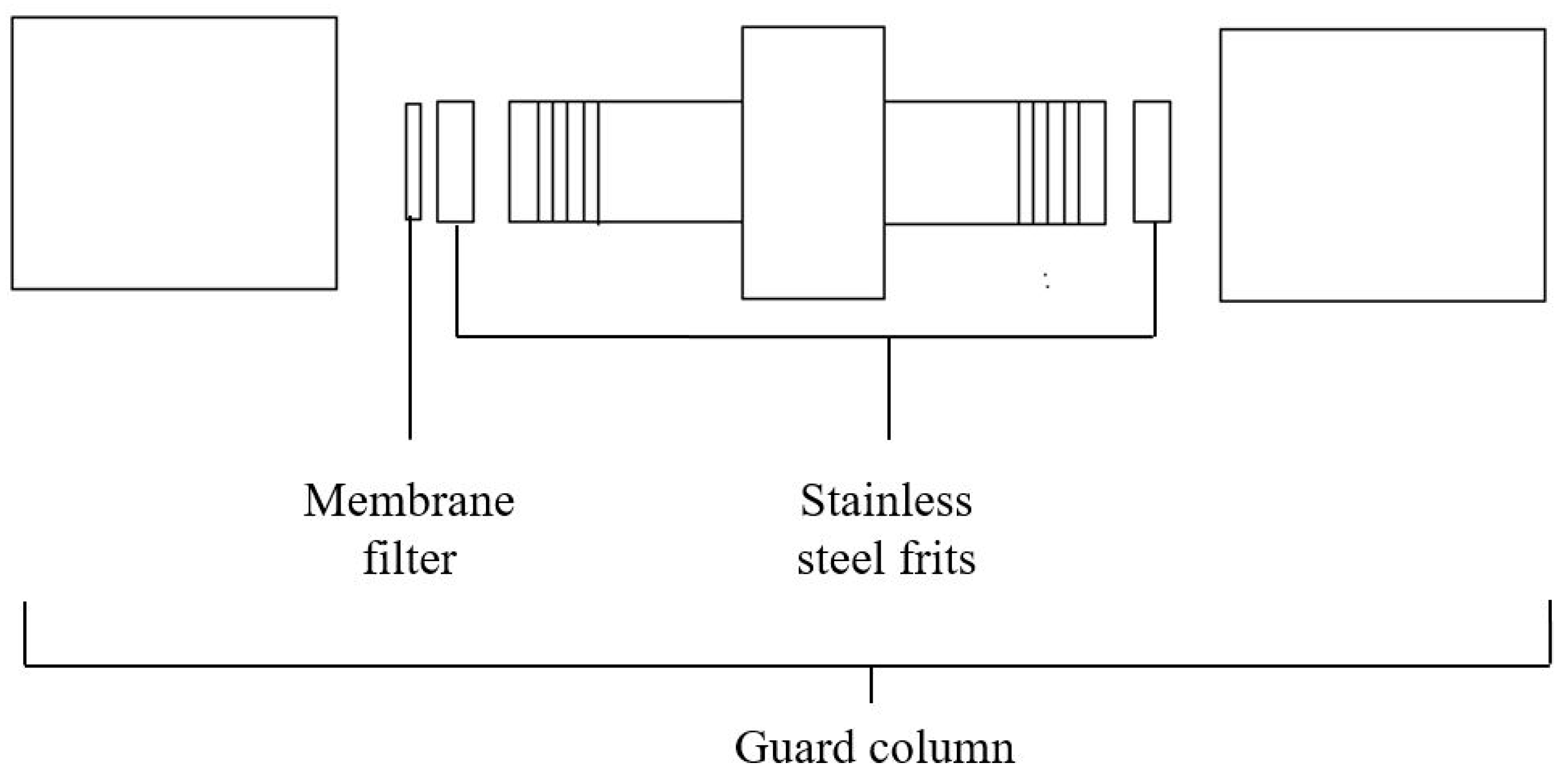
2.2. Instruments
2.3. Preparation of Liposome Suspension
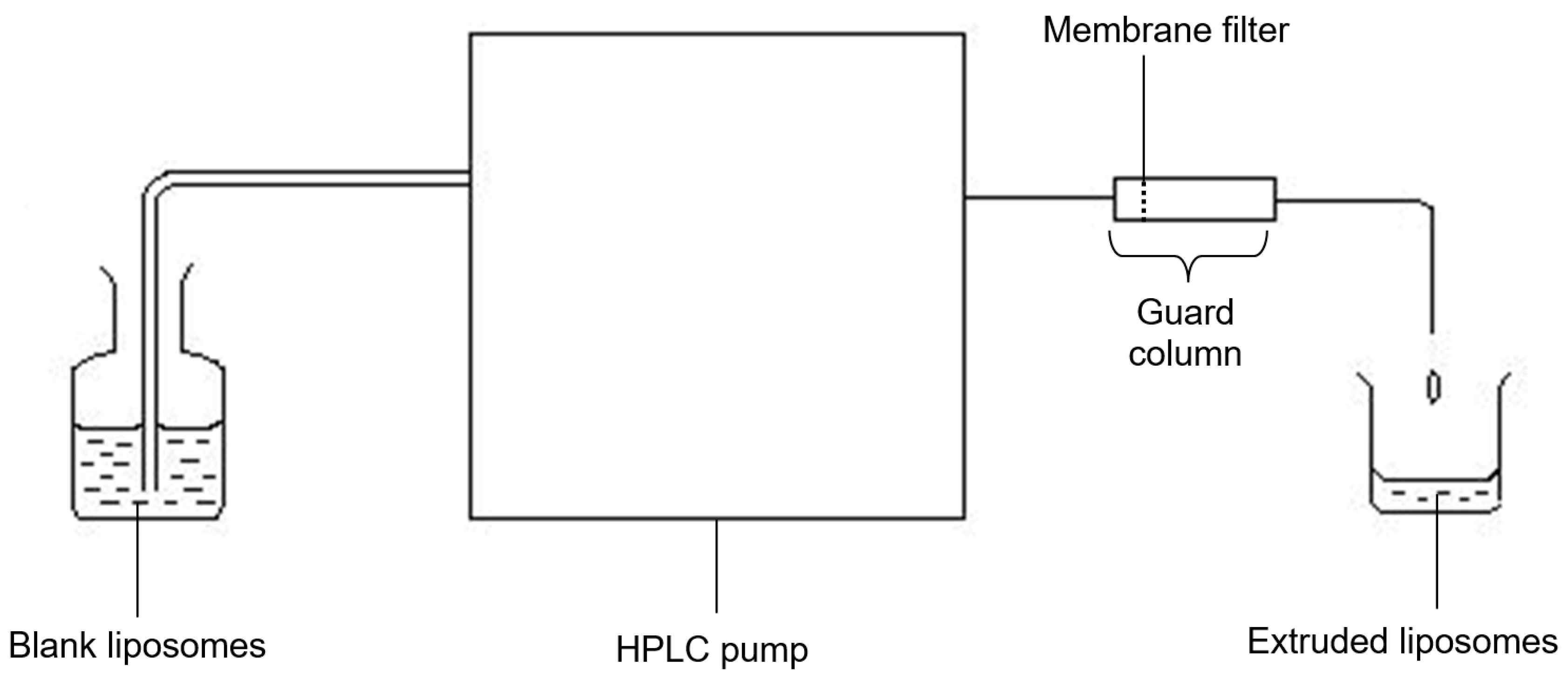
2.4. Extrusion Procedure and Influence of Process Parameters
2.5. Comparison with Size Reducing Methods
2.6. Particle Size Analysis
2.7. Statiscal Analysis
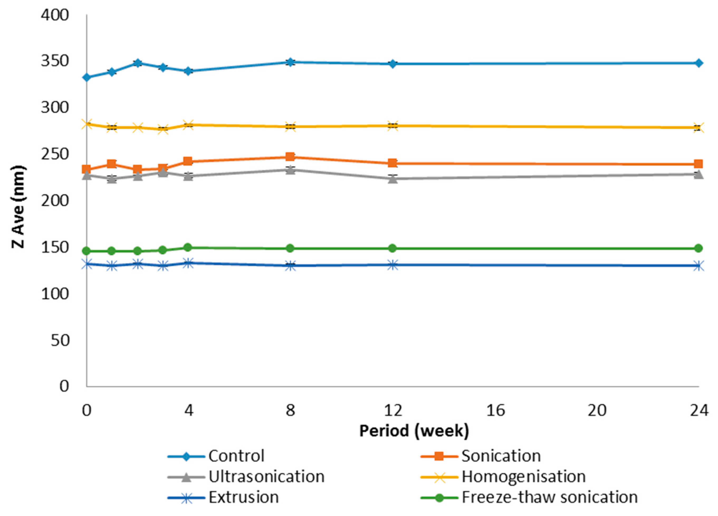
3. Results
3.1. Influence of Process Parameters
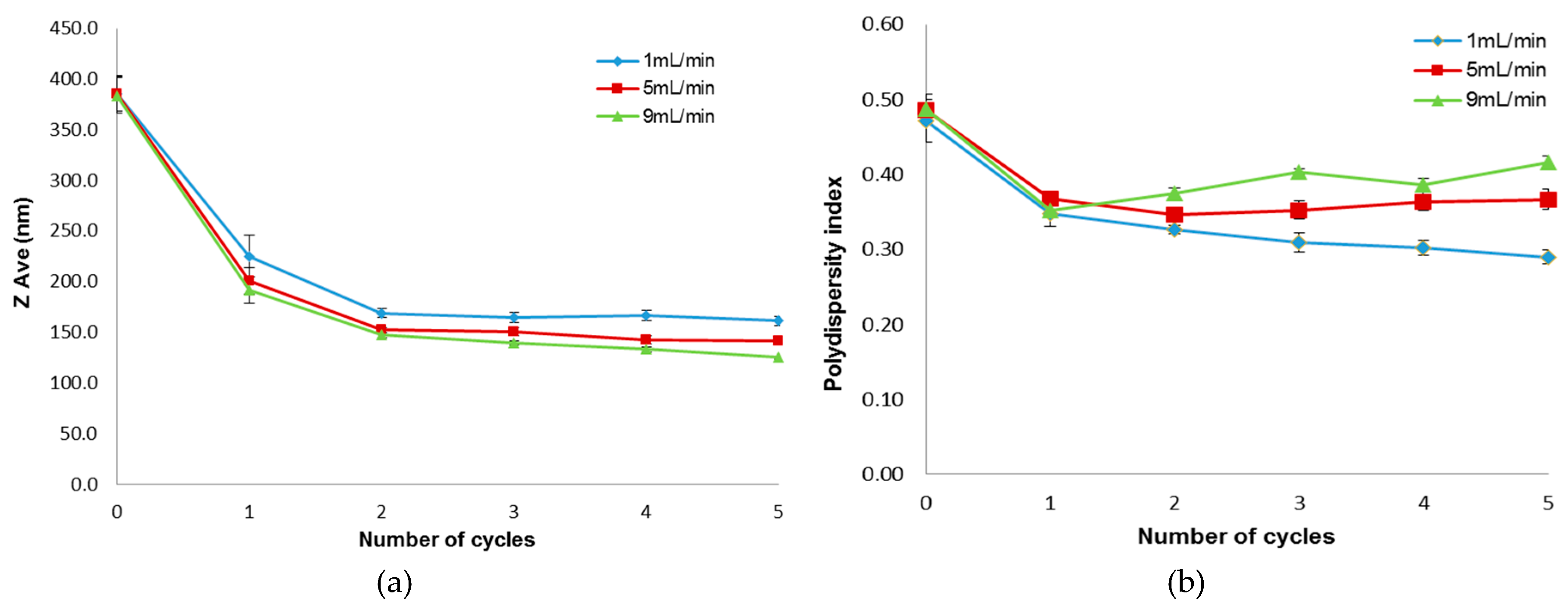
3.1.1. Flow Rate
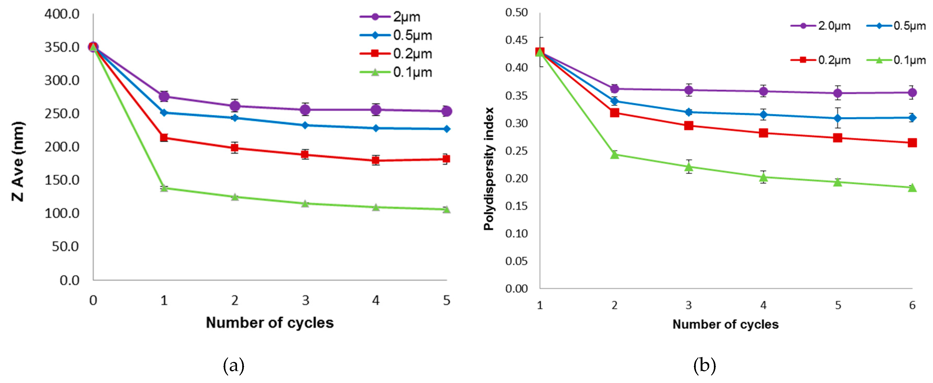
3.1.2. Membrane Pore Size
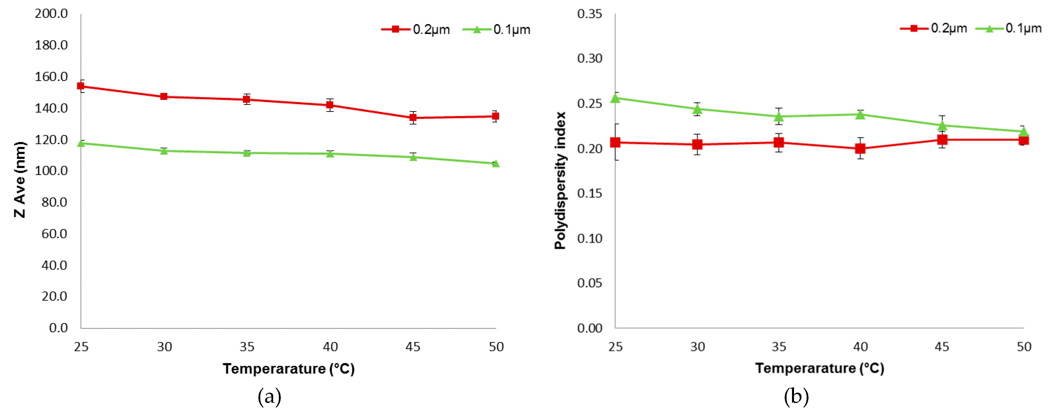
3.1.3. Temperature
3.2. Comparison with Other Size Reducing Methods
4. Discussion
5. Conclusions
Acknowledgments
Author Contributions
Conflicts of Interest
References
- Harashima, H.; Sakata, K.; Funato, K.; Kiwada, H. Enhanced hepatic uptake of liposomes through complement activation depending on the size of liposomes. Pharm. Res. 1994, 11, 402–406. [Google Scholar] [CrossRef] [PubMed]
- Szoka, F.C. The future of liposomal drug delivery. Biotechnol. Appl. Biochem. 1990, 12, 496–500. [Google Scholar] [PubMed]
- Cullis, P.R.; Hope, M.J.; Bally, M.B.; Madden, T.D.; Mayer, L.D.; Janoff, A.S. Liposomes as pharmaceuticals. In Liposomes: From Biophysics to Therapeutics; Ostro, M., Ed.; Marcel Dekker: New York, NY, USA, 1987; pp. 46–47. [Google Scholar]
- Fatouros, D.; Gortzi, O.; Klepetsanis, P.; Antimisiaris, S.G.; Stuart, M.C.A.; Brisson, A.; Ioannou, P.V. Preparation and properties of arsonolipid containing liposomes. Chem. Phys. Lipids 2001, 109, 75–89. [Google Scholar] [CrossRef]
- Hope, M.J.; Nayar, R.; Mayer, L.D.; Cullis, P.R. Reduction of liposome size and preparation of unilamellar vesicles by extrusion techniques. In Liposome Technology: Interactions of Liposomes with the Biological Milieu; Gregoriadis, G., Ed.; CRC Press, Inc.: Boca Raton, FL, USA, 1993; pp. 123–139. [Google Scholar]
- Jousma, H.; Talsma, H.; Spies, F.; Joosten, J.G.H.; Junginger, H.E.; Crommelin, D.J.A. Characterization of liposomes. The influence of extrusion of multilamellar vesicles through polycarbonate membranes on particle size, particle size distribution and number of bilayers. Int. J. Pharm. 1987, 35, 263–274. [Google Scholar] [CrossRef]
- Barnadas-Rodríguez, R.; Sabés, M. Factors involved in the production of liposomes with a high-pressure homogenizer. Int. J. Pharm. 2001, 213, 175–186. [Google Scholar] [CrossRef]
- Talsma, H.; Özer, A.Y.; Bloois, L.V.; Crommelin, D.J.A. The size reduction of liposomes with a high pressure homogenizer (Microfluidizer™). Characterization of Prepared Dispersions and Comparison with Conventional Methods. Drug Dev. Ind. Pharm. 1989, 15, 197–207. [Google Scholar] [CrossRef]
- Mayer, L.D.; Hope, M.J.; Cullis, P.R. Vesicles of variable sizes produced by a rapid extrusion procedure. Biochim. Biophys. Acta 1986, 858, 161–168. [Google Scholar] [CrossRef]
- Olson, F.; Hunt, C.; Szoka, F.; Vail, W.; Papahadjopoulos, D. Preparation of liposomes of defined size distribution by extrusion through polycarbonate membranes. Biochim. Biophys. Acta 1979, 557, 9–23. [Google Scholar] [CrossRef]
- Liu, R.; Cannon, J.B.; Li, Y. Liposomes in solubilization. In Water-Insoluble Drug Formulation; Liu, R., Ed.; Interpharm/CRC: Boca Raton, FL, USA, 2000; pp. 355–404. [Google Scholar]
- Frisken, B.J.; Asman, C.; Patty, P.J. Studies of vesicle extrusion. Langmuir 2000, 16, 928–933. [Google Scholar] [CrossRef]
- Nayar, R.; Hope, M.J.; Cullis, P.R. Generation of large unilamellar vesicles from long-chain saturated phosphatidylcholines by extrusion technique. Biochim. Biophys. Acta 1989, 986, 200–206. [Google Scholar] [CrossRef]
- Hunter, D.G.; Frisken, B.J. Effect of extrusion pressure and lipid properties on the size and polydispersity of lipid vesicles. Biophys. J. 1998, 74, 2996–3002. [Google Scholar] [CrossRef]
- Mui, B.L.-S.; Cullis, P.R.; Evans, E.A.; Madden, T.D. Osmotic properties of large unilamellar vesicles prepared by extrusion. Biophys. J. 1993, 64, 443–453. [Google Scholar] [CrossRef]
- Armengol, X.; Estelrich, J. Physical stability of different liposome compositions obtained by extrusion method. J. Microencapsul. 1995, 12, 525–535. [Google Scholar] [CrossRef] [PubMed]
- Berger, N.; Sachse, A.; Bender, J.; Schubert, R.; Brandl, M. Filter extrusion of liposomes using different devices: Comparison of liposome size, encapsulation efficiencey and process characteristics. Int. J. Pharm. 2001, 223, 55–68. [Google Scholar] [CrossRef]
- Schneider, T.; Sachse, A.; Rößling, G.; Brandl, M. Generation of contrast-carrying liposomes of defined size with a new continuous high pressure extrusion method. Int. J. Pharm. 1995, 117, 1–12. [Google Scholar] [CrossRef]
- Szoka, F.; Olson, F.; Heath, T.; Vail, W.; Mayhew, E.; Papahadjopoulos, D. Preparation of unilamellar liposomes of intermediate size (0.1–0.2 µm) by a combination of reverse phase evaporation and extrusion through polycarbonate membranes. Biochim. Biophys. Acta 1980, 601, 559–571. [Google Scholar] [CrossRef]
- Chapman, C.; Erdahl, W.; Taylor, R.; Pfeiffer, D. Effects of solute concentration on the entrapment of solutes in phospholipid vesicles prepared by freeze-thaw extrusion. Chem. Phys. Lipids 1991, 60, 201–208. [Google Scholar] [CrossRef]
- MacDonald, R.I.; Menco, B.P.M.; Takeshita, K.; Subbarao, N.K.; Hu, L.-R. Small-volume extrusion apparatus for preparation of large, unilamellar vesicles. Biochim. Biophys. Acta (Biomembr.) 1991, 1061, 297–303. [Google Scholar] [CrossRef]
- Hunter, D.G. The Effects of Lipid Composition and Extrusion Pressure and Temperature on the Properties of Phospholipid Vesicles; Simon Fraser University: Burnaby, BC, Canada, 1997. [Google Scholar]
- Mason, T.G.; Bibette, J. Emulsification of Viscoelastic Media. Phys. Rev. Lett. 1996, 77, 3481–3484. [Google Scholar] [CrossRef] [PubMed]
- Martin, F.J.; Morano, J.K. Liposome Extrusion Method. U.S. Patent 06/829,710, 12 April 1988. [Google Scholar]
- Cullis, P.R.; Hope, M.J.; Bally, M.B. Extrusion Technique for Producing Unilamellar Vesicles. U.S. Patent 07/310,495, 16 April 1991. [Google Scholar]
- Woodle, M.C.; Newman, M.S.; Working, P.K. Biological properties of sterically stabilized liposomes. In Stealth Liposomes; Lasic, D.D., Martin, F., Eds.; CRC Press: Boca Raton, FL, USA, 1995; p. 106. [Google Scholar]
- New, R.R.C. Preparation of liposomes. In Liposomes: A Practical Approach; New, R.R.C., Ed.; Oxford University Press: New York, NY, USA, 1990; pp. 33–104. [Google Scholar]
- Diat, O.; Roux, D.; Nallet, F. Effect of shear on a lyotropic lamellar phase. J. Phys. II 1993, 3, 1427–1452. [Google Scholar] [CrossRef]
- Lesieur, S.; Grabielle-Madelmont, C.; Paternostre, M.T.; Olivon, M. Size analysis and stability study of lipid vesicles by high performance gel exclusion chromatography, turbidity and dynamic scattering. Anal. Biochem. 1991, 192, 334–343. [Google Scholar] [CrossRef]
- Clerc, S.G.; Thompson, T.E. A possible mechanism for vesicle formation by extrusion. Biophys. J. 1994, 67, 475–477. [Google Scholar] [CrossRef]
- Gompper, G.; Kroll, D.M. Driven transport of fluid vesicles through narrow pores. Phys. Rev. E 1995, 52, 4198–4208. [Google Scholar] [CrossRef]
- Bruinsma, R. Rheology and shape transitions of vesicles under capillary flow. Phys. A 1996, 234, 249–270. [Google Scholar] [CrossRef]
- Patty, P.J.; Frisken, B.J. The pressure-dependence of the size of extruded vesicles. Biophys. J. 2003, 85, 996–1004. [Google Scholar] [CrossRef]
- Jana, A.K.; Agarwal, S.; Chatterjee, S.N. Ultrasonic radiation induced lipid peroxidation in liposomal membrane. Radiat. Environ. Biophys. 1986, 25, 309–314. [Google Scholar] [CrossRef]
- Castile, J.D.; Taylor, K.M.G. Factors affecting the size distribution of liposomes produced by freeze-thaw extrusion. Int. J. Pharm. 1999, 188, 87–95. [Google Scholar] [CrossRef]
- Ong, S.; Ming, L.; Lee, K.; Yuen, K. Influence of the encapsulation efficiency and size of liposome on the oral bioavailability of griseofulvin-loaded liposomes. Pharmaceutics 2016, 8, 25. [Google Scholar] [CrossRef] [PubMed]


| Size Reduction Method | ZAve (nm) | Polydispersity Index |
|---|---|---|
| Control | 321.6 ± 12.9 | 0.4 ± 0.0 |
| Homogenization | 279.1 ± 15.8 | 0.3 ± 0.0 |
| Sonication | 273.5 ± 15.3 | 0.4 ± 0.1 |
| Ultrasonication | 235.8 ± 8.1 | 0.4 ± 0.0 |
| Freeze-thaw sonication (FTS) | 124.7 ± 0.7 | 0.4 ± 0.0 |
| Extrusion | 103.3 ± 13.5 | 0.2 ± 0.2 |
© 2016 by the authors; licensee MDPI, Basel, Switzerland. This article is an open access article distributed under the terms and conditions of the Creative Commons Attribution (CC-BY) license (http://creativecommons.org/licenses/by/4.0/).
Share and Cite
Ong, S.G.M.; Chitneni, M.; Lee, K.S.; Ming, L.C.; Yuen, K.H. Evaluation of Extrusion Technique for Nanosizing Liposomes. Pharmaceutics 2016, 8, 36. https://doi.org/10.3390/pharmaceutics8040036
Ong SGM, Chitneni M, Lee KS, Ming LC, Yuen KH. Evaluation of Extrusion Technique for Nanosizing Liposomes. Pharmaceutics. 2016; 8(4):36. https://doi.org/10.3390/pharmaceutics8040036
Chicago/Turabian StyleOng, Sandy Gim Ming, Mallikarjun Chitneni, Kah Seng Lee, Long Chiau Ming, and Kah Hay Yuen. 2016. "Evaluation of Extrusion Technique for Nanosizing Liposomes" Pharmaceutics 8, no. 4: 36. https://doi.org/10.3390/pharmaceutics8040036
APA StyleOng, S. G. M., Chitneni, M., Lee, K. S., Ming, L. C., & Yuen, K. H. (2016). Evaluation of Extrusion Technique for Nanosizing Liposomes. Pharmaceutics, 8(4), 36. https://doi.org/10.3390/pharmaceutics8040036