Review

Bacterial Heat-Stable Enterotoxins: Translation of Pathogenic Peptides into Novel Targeted Diagnostics and Therapeutics

1
Department of Pharmacology and Experimental Therapeutics, Thomas Jefferson University, 132 South 10th Street, 1170 Main, Philadelphia, PA 19107, USA
2
School of Biomedical Engineering, Science & Health Systems, Drexel University, Philadelphia, PA 19104, USA
3
Department of Radiation Oncology, University of Pennsylvania, Philadelphia, PA, 19104, USA
*
Author to whom correspondence should be addressed.
These authors contributed equally to this work.
Toxins 2010, 2(8), 2028-2054; https://doi.org/10.3390/toxins2082028
Submission received: 18 July 2010 / Accepted: 3 August 2010 / Published: 5 August 2010
(This article belongs to the Special Issue Enterotoxins)

Abstract

:
Heat-stable toxins (STs) produced by enterotoxigenic bacteria cause endemic and traveler’s diarrhea by binding to and activating the intestinal receptor guanylyl cyclase C (GC-C). Advances in understanding the biology of GC-C have extended ST from a diarrheagenic peptide to a novel therapeutic agent. Here, we summarize the physiological and pathophysiological role of GC-C in fluid-electrolyte regulation and intestinal crypt-villus homeostasis, as well as describe translational opportunities offered by STs, reflecting the unique characteristics of GC-C, in treating irritable bowel syndrome and chronic constipation, and in preventing and treating colorectal cancer.

Abbreviations

ABC:
Avidin Biotin Complex
CFTR:
cystic fibrosis transmembrane conductance regulator
cGMP:
Cyclic guanosine monophosphate
CMA:
cancer mucosa antigens
CNG:
cyclic nucleotide-gated channel
COX-2:
cyclooxygenase-2
GC-C:
guanylyl cyclase C
IBD:
inflammatory bowel disease
IBS:
irritable bowel syndrome
MRP:
multidrug resistance proteins
PDE:
phosphodiesterase
PGE:
prostaglandin
PKA:
cAMP-dependent protein kinase
PKG:
cGMP-dependent protein kinase
PLA2:
phospholipase A2
pRb:
phosphorylated retinoblastoma
SBM:
spontaneous bowel movement
ST:
heat-stable enterotoxin

1. Introduction

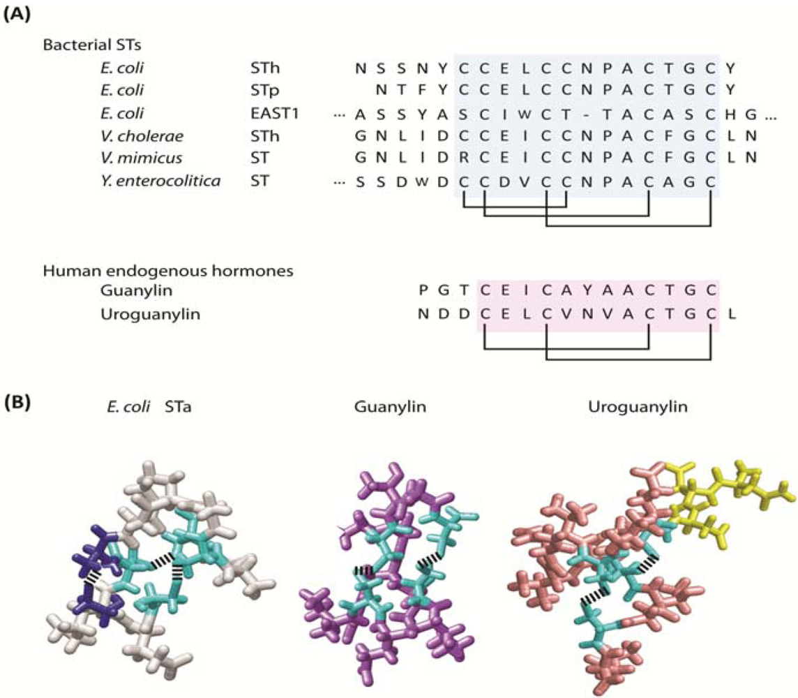
1.1. Bacterial heat-stable enterotoxins

Bacterial heat-stable enterotoxins (STs) first came to attention in the 1970s after heat-inactivation of cultures of bacteria isolated from patients suffering from diarrhea failed to eliminate enterotoxigenic activity [1,2]. Two families of heat-stable enterotoxins have been identified: STa (or STI) and STb (or STII), which differ by their physicochemical and biologic characteristics [1,3]. Although STa and STb were both discovered in intestinal bacterial strains isolated from humans, STb is produced by bacterial strains that preferentially inhabit pigs [4]. Further, STa induces diarrhea through a cyclic nucleotide-dependent mechanism, while STb induces secretion through a cyclic nucleotide-independent mechansim [5]. STa, hereon referred to as ST, will be the only heat-stable enterotoxin discussed in this review.
STs are produced by a variety of enteric pathogenic organisms, including diarrheagenic Escherichia coli (E. coli), Vibrio cholerae, Vibrio mimicus, Yersinia enterocolitica, Citrobacter freundii, and Klebsiella pneumoniae [1,2,6,7,8,9,10]. STs are translated as precursor peptides, which undergo intracellular proteolytic processing to active peptides from 17 to 53 amino acids [11]. Isoforms of ST share a conserved C-terminal region of 13 amino acids containing three disulfide bonds responsible for heat stability and biological activity (Figure 1) [11]. Consistent with the proposed function of ST as an essential survival factor facilitating escape of bacteria from nutrient-poor to nutrient-rich environments, synthesis and secretion of ST is reduced in a milieu enriched in glucose while depletion of this sugar stimulates ST production and secretion [12,13]. Investigation of the pathogenesis underlying diarrhea produced by ST ultimately revealed two intestinal paracrine hormones, guanylin and uroguanylin, and the receptor for these homologous peptides, guanylyl cyclase C (GC-C), encoded by the gene GUCY2C [14].

1.2. Molecular mimicry, convergent evolution and the guanylyl cyclase C paracrine hormone axis

Guanylyl cyclase C (GC-C), the only identified receptor for ST, belongs to the guanylyl cyclase family of receptors that catalyze the conversion of GTP to cGMP upon activation [14]. Guanylyl cyclases are found in two subcellular compartments in mammalian cells: soluble guanylyl cyclases (sGC) are completely intracellular and particulate guanylyl cyclases (pGC), also known as receptor-linked guanylyl cyclase, span the plasma membrane [14]. GC-C is one of the seven isotypes of particulate guanylyl cyclases (GCA to GCG) that exhibit conserved domain structures including an extracellular ligand binding domain, a single transmembrane domain, an intracellular kinase homology domain, and a catalytic domain that produces cGMP [14].
GC-C is primarily expressed in intestinal mucosal cells from the duodenum to the rectum [15,16] where it exists as a pre-formed homo -dimer or -trimer [17] located within apical membranes of epithelial cells populating the crypt-villus axis. The two defined endogenous ligands of GC-C, the hormones guanylin and uroguanylin, share significant homology with ST (Figure 1) [18,19]. Similar to ST, the tertiary structure of these peptides is stabilized by intra-chain disulfide bonds, which are essential for biological activity [20,21,22]. Additionally, guanylin and uroguanylin also are synthesized as pro-peptides [23], but unlike ST, these endogenous peptides undergo proteolytic processing following secretion [22,24,25]. These considerations suggest that ST and enterotoxigenic diarrhea are prime examples of molecular mimicry and convergent evolution. Here, bacteria have co-opted a normal mammalian physiologic function, the regulation of intestinal fluid and electrolyte homeostasis, to produce an evolutionary population survival scheme that guarantees the adequacy of nutrient resources and dissemination into new environments and hosts.
Figure 1. Bacterial heat-stable enterotoxin, guanylin and uroguanylin. (A) Primary structures of heat-stable enterotoxins produced by various pathogenic bacteria and human homologs, guanylin and uroguanylin. Conserved core regions are highlighted (blue for STs; pink for hormones) with disulfide bridges indicated; (B) Three-dimensional structures of E. coli STa (grey), guanylin (purple) and uroguanylin (pink) conserved regions. Non-conserved regions in uroguanylin are labeled in yellow. Cysteines linked by disulfide bonds conserved among three peptides are colored in cyan. Cysteines linked by the extra-disulfide bond, providing ST greater potency than guanylin and uroguanylin, are colored in blue.
Toxins 02 02028 g001

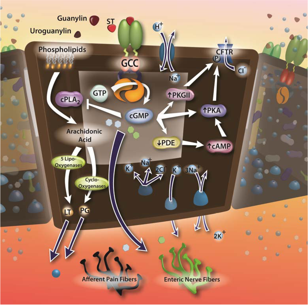
1.3. Guanylyl cyclase C and enterotoxin signaling circuits

Activation of GC-C stimulates a rise in intracellular cGMP, which binds and activates its three downstream effectors [17]: cGMP-dependent protein kinases (PKGs), phosphodiesterases (PDEs) and cyclic nucleotide-gated (CNG) channels. High intracellular levels of cGMP can also cross-activate cAMP-dependent protein kinases (PKA)[26]. PKGs are the principle intracellular mediators for cGMP signaling [27]. PKG is expressed in nearly all tissues, with highest expression in lung, cerebellum, smooth muscle, platelets, and intestinal mucosa [28,29]. PKG consists of two distinct isoforms: PKG type I and type II. PKG I is located within the cytoplasm of most cell types, whereas PKG II is exclusively expressed within plasma membranes in bone, kidney, brain, and intestine [28,29,30,31]. Within the intestinal epithelium, PKG II displays a rostral-caudal gradient of expression, with highest levels found in small intestine and lowest levels found in the distal colon. Intestinal PKG II expression also displays a crypt-villus gradient with highest expression in the villi and lowest expression in crypts [32]. Two closely related PKG I isoforms (type Iα and Iβ), which arise from alternative splicing of the N-terminal region of the PKG I gene have been purified, cloned, and expressed [33,34,35,36,37,38]. Type I and type II PKG isoforms are homodimers of ~75 and 86 kDa monomers, respectively, consisting of (1) an N-terminal domain regulating kinase autoinhibition, autophosphorylation and subcellular localization; (2) regulatory domains with allosteric cGMP binding sites and (3) catalytic domains catalyzing the transfer of the γ-phosphoryl group of ATP to various protein substrates [39]. Beyond the primary signal carrying and amplification capacity of PKGs, cGMP signals also are shaped by PDEs, which degrade cGMPs to 5’ GMPs, and multidrug resistance proteins (MRPs), which are ATP-dependent active transporters pumping out cGMP. These enzymes maintain intracellular cGMP concentrations within a narrow physiologic range, essentially serving as key terminators of guanylyl cyclase signaling [40].

2. Enterotoxin Signaling, Irritable Bowel Syndrome and Chronic Constipation

2.1. Enterotoxins, GC-C and fluid-electrolyte homeostasis

Maintenance of fluid and electrolyte homeostasis is critical for cellular biochemical processes and organ function, and the intestine is one of the major organs controlling this balance. Discovery of the role of GC-C in fluid-electrolyte homeostasis is attributed to the observation that ST is a principal cause of enterotoxigenic diarrheal disease in humans and animals worldwide [38,39,40]. Physiologically, guanylin and uroguanylin regulate intestinal fluid and electrolyte homeostasis through cGMP accumulation, which activates PKGII leading to the phosphorylation of the cystic fibrosis transmembrane conductance regulator (CFTR), producing Cl-, HCO3-, and water secretion [14]. ST induces secretory diarrhea by exploiting this physiologic mechanism, and the enhanced affinity of ST for GC-C produces supra-physiologic induction of cGMP accumulation and extensive water and electrolyte secretion (Figure 2) [14,41,42]. Notably, systemic fluid and electrolyte homeostasis is maintained beyond intestine. The natriuretic and diuretic effects induced by uroguanylin and guanylin in the kidney still remain intact in GC-C deficient mice [43,44]. Therefore, GC-C-deficient mice do not exhibit significant systemic fluid-electrolyte imbalance [45].
Figure 2. GC-C signaling for fluid/electrolyte secretion, inflammation, gut motility, and nociception. GC-C is activated by binding of bacterial heat-stable enterotoxins (STs) or the endogenous ligands, guanylin and uroguanylin. Ligand binding to the extracellular domain of GC-C activates the intracellular catalytic domain converting GTP to cGMP. Intracellular cGMP accumulation leads to fluid and electrolyte secretion through direct activation of cGMP-dependent protein kinase II (PKG II) and downregulation of phosphodiesterase (PDE)-driven cAMP hydrolysis leading to increased activation of cAMP-dependent protein kinase (PKA). PKG II and PKA phosphorylate the cystic fibrosis transmembrane conductance regulator (CFTR) producing channel opening, leading to an efflux of Cl ions and secondary efflux of water. Intracellular cGMP accumulation also decreases pro-inflammatory signaling by inhibiting phospholipase A2, decreasing the liberation of the leukotriene (LT) and prostaglandin (PG) precursor, arachidonic acid, from membrane phospholipids. GC-C activation also enhances intestinal myoelectric activity and decreases afferent pain fiber firing, presumably through the release of specific soluble mediators, which stimulate surrounding dendritic nerve endings and alter neuronal firing rates.
Toxins 02 02028 g002

2.2. Enterotoxins and irritable bowel syndrome

Besides enhancing gut fluid and electrolyte secretion, infections with enterotoxigenic bacteria, including V. cholerae and E. coli, alter gut motility [46,47,48], and administration of enterotoxins produced by these bacteria, including STs, stimulate intestinal myoelectric activity [46,49,50]. Indeed, downstream effects of ST signaling are mediated, in part, by stimulation of the enteric nervous system. Mechanical (vagotomy) or neuropharmacological (tetrodotoxin, lidocaine, etc.) disturbance of enteric neuronal activation, as well as antagonism of gut acetylcholine, serotonin, prostaglandin, and nitric oxide signaling, significantly reduces ST-induced GI secretion [51,52,53,54,55,56,57]. While the mechanism by which ST signaling stimulates enteric neurons is still unclear, GC-C activation by STs in apical epithelial cells may release specific mediators, which stimulate surrounding nerve endings and alter neuronal firing rates (Figure 2) [58,59].
Due to its ability to induce secretion and enhance gut motility, pharmacological modulation of GC-C signaling has been investigated as a possible treatment paradigm for chronic constipation and constipation-predominant irritable bowel syndrome (IBS-C). Irritable bowel syndrome (IBS) is a functional bowel disorder, which is associated with abdominal pain and altered bowel activity. Unlike inflammatory bowel disease (IBD), IBS is not associated with increased inflammation or other pathologic organic changes within the GI tract [60]. Rather, a diagnosis of IBS is based on specific criteria including recurrent abdominal pain or discomfort, relief with defecation, and change in stool frequency and appearance [61]. Diarrhea- (IBS-D) and constipation-predominant IBS (IBS-C) are the two primary subtypes of IBS, with a mixed subtype (IBS-M) occurring less frequently [60].
Constipation and IBS are prevalent in North America, where they have been estimated to affect 12–19% and 5–10% of the population, respectively, with a higher prevalence among females [62,63,64,65,66]. Both conditions are associated with a considerably reduced quality of life [67,68], elevated healthcare costs [69,70,71], and increased disability claims and work absenteeism, which place an economic burden on both patients and employers [72]. The first approach to treating these conditions is diet and lifestyle changes, including increasing exercise and dietary fiber intake [73,74]. Pharmacotherapy with prokinetic and antispasmodic agents are often utilized when lifestyle and dietary changes fail to alleviate symptoms [60]. However, while these agents provide brief symptomatic relief, they do not treat the underlying pathophysiology.

2.3. Enterotoxin analogs for chronic constipation

Linaclotide (MD-1100 acetate) is a synthetic 14-amino acid analogue of ST that binds to and activates GC-C and dose-dependently enhances gut secretion and motility in rodent models [75]. Furthermore, linaclotide has anti-nociceptive properties in rodent models of visceral hypersensitivity, indicating that the drug may both improve intestinal motility and discomfort in patients suffering from constipation [76]. In Phase I trials, linaclotide at single oral doses of 30–3000 μg or multiple doses of 30–1000 μg was safe and well-tolerated with no evidence of systemic exposure [77,78]. In the first phase II randomized clinical trial (RCT) of linaclotide utility in the treatment of constipation, 36 women with IBS-C were treated with oral linaclotide (100–1000 μg once daily). At a daily dose of 1000 μg, linaclotide significantly decreased ascending colon emptying half-time (p = 0.015) and enhanced overall colonic transit at 48 hours (p = 0.02), while both doses accelerated the time to first bowel movement (p = 0.013), increased stool frequency (p = 0.037), decreased stool consistency (p < 0.001), and improved ease of stool passage (p < 0.001) without serious adverse effects [79]. In a follow-up study, 42 patients with chronic constipation were randomized to linaclotide (100, 300, or 1,000 μg) or placebo once daily for two weeks. In this study, linaclotide produced a dose-dependent improvement in spontaneous bowel movement (SBM) frequency (p < 0.05), stool consistency scores, and straining scores, in addition to improving abdominal discomfort, severity of constipation, and overall relief with only mild-to-moderate GI side effects, predominantly diarrhea, being reported [80].
In a subsequent large, multi-center trial of 310 patients with chronic constipation treated with linaclotide (75–600 μg daily) for four weeks, all doses improved the weekly rate of spontaneous bowel movements (SBM) and complete spontaneous bowel movements (CSBM) in addition to improving stool consistency, straining, abdominal discomfort, bloating, and quality of life [81]. Finally, in a recent press release, Ironwood Pharmaceuticals, Inc. and Forest Laboratories, Inc announced that, in two phase III trials of >600 patients with chronic constipation, 12 weeks of linaclotide therapy (133–266 μg per day) doubled average weekly CSBMs, tripled average weekly SBMs, and significantly improved bloating, abdominal discomfort, stool consistency, straining, and constipation severity (p < 0.001), with only minor GI side effects [38].
Currently, further trials of linaclotide therapy for the treatment of constipation are scheduled, and new drugs targeting the GC-C signaling axis for the treatment of chronic constipation and IBS-C are in preclinical development. Among these newer agents is SP-304, a synthetic analogue of the endogenous GC-C ligand, uroguanylin, and SP-333, a synthetic GC-C agonist (Synergy Pharmaceuticals Inc.). These agents are currently in preclinical trials for the treatment of GI diseases, including chronic constipation, IBS-C, and inflammatory bowel disease, and oral administration of SP-304 to rodents has been observed to both promote intestinal secretion and ameliorate GI inflammation [82].

2.4. Targeting GC-C in chronic inflammation

Nitric oxide has anti-inflammatory effects, inhibiting leukocyte adhesion and oxidant production [83]. In that context, pharmacologic inhibitors of phosphodiesterases, which degrade cGMP, have beneficial effects in mouse models of inflammatory bowel disease [84,85]. Similarly, the uroguanylin analogue, SP-304, produces anti-inflammatory effects, associated with downregulation of pro-inflammatory cytokines including IL-4, IL-5, IL-23, and TNF, in mouse models of ulcerative colitis. Moreover, SP-304 also down-regulated cyclooxygenase-2 (COX-2) production of the inflammatory mediator prostaglandin E2 (PGE2)[82]. The inhibitory function of cGMP on inflammatory prostaglandin production may due to a decrease in intracellular arachidonic acid levels. Arachidonic acid, the precursor for prostaglandin synthesis, is liberated from membrane phospholipids via cleavage by phospholipase A2 (PLA2). Interestingly, PKG phosphorylates and inhibits PLA2 [86]. However, the precise mechanisms underlying the anti-inflammatory activity of SP-304 activation of GC-C remain to be determined (Figure 2).

3. Targeting GC-C to Prevent Colorectal Cancer

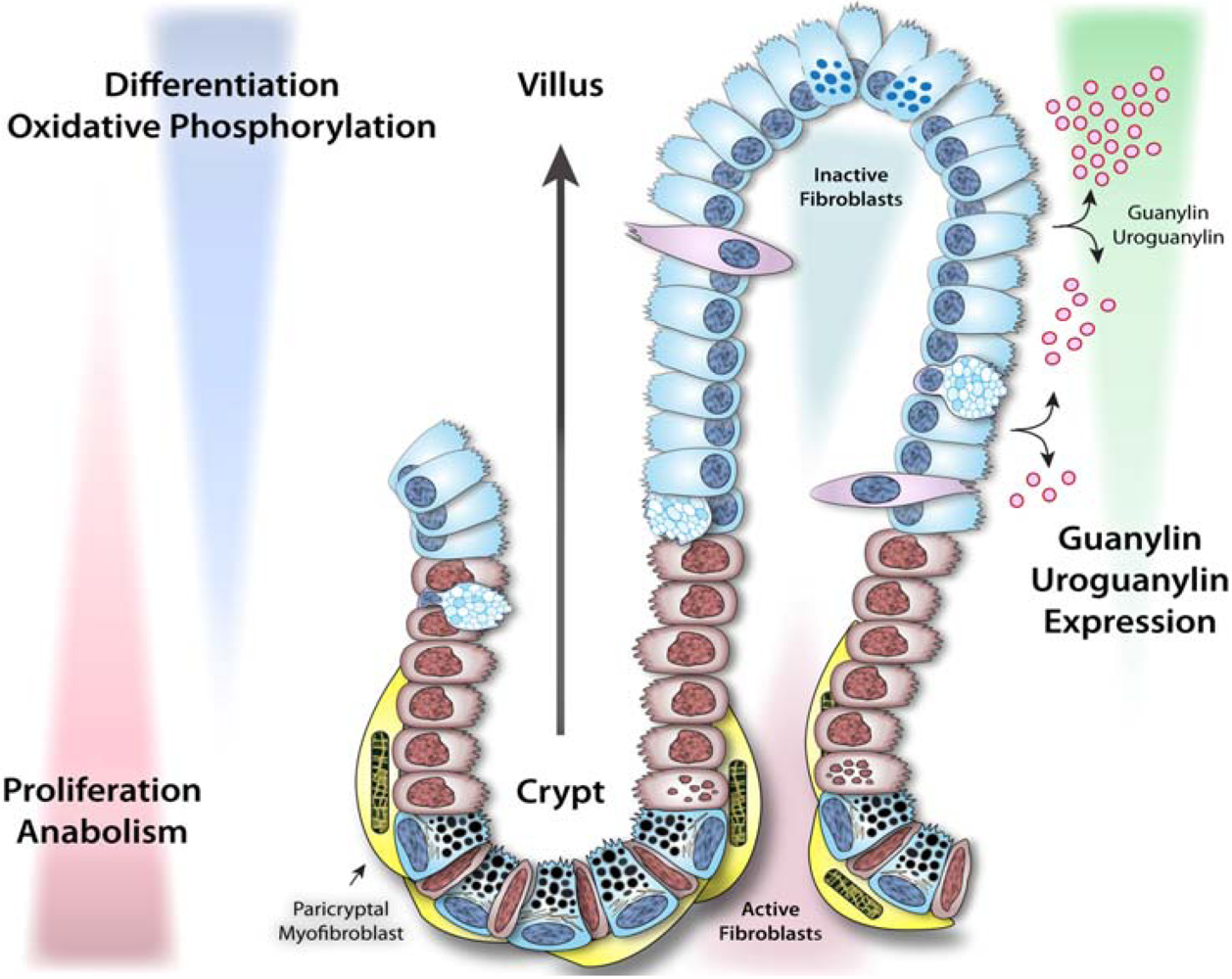
3.1. Dynamics of intestinal epithelial cells and crypt-villus homeostasis

The intestinal epithelium lining the gastrointestinal tract is characterized by coordinated homeostatic programs comprising a developmental continuum integrating proliferation, differentiation, metabolic maturation and apoptosis along the crypt-villus axis. Crypts harbor stem cells at their base, which continuously regenerate progenitor cells destined to differentiate along secretory (goblet, Paneth, and enteroendocrine cells) or absorptive (enterocytes) lineages. This transition from proliferation to lineage commitment is associated with metabolic reprogramming from glycolysis to oxidative phosphorylation as the cells migrate toward the surface [87]. Metabolic reprogramming reflects the energy demands for different compartments, subserving the specific balance between proliferation and differentiation. In the crypt, cell division requires rapidly available energy supplies from glycolysis, while mitochondrially-mediated metabolism exploits the efficiency of ATP production by oxidative phosphorylation, supporting catabolic demands in mature cells in the differentiated compartment [88,89]. Following this transition, goblet, enteroendocrine cells and enterocytes continue to migrate to the tips of the villi and ultimately undergo apoptosis or anoikis [90,91,92]. In contrast, Paneth cells migrate down to the base of the crypt where they reside. The distinctive regenerative characteristic of the intestinal epithelium establishes a vertical axis representing a life cycle continuum, from cell birth to death. Dysregulation of this homeostatic process is intimately linked with intestinal tumorigenesis [89].
Enterocytes, comprising the majority of the intestinal epithelial monolayer, develop well-organized microvilli brush borders, containing key functional proteins mediating cognate digestive and absorptive functions [91]. Interspersed among enterocytes are the hormone-producing enteroendocrine cells, which comprise the largest endocrine system in the body in terms of both cell quantity and variety of hormones produced. Although enteroendocrine cells share biochemical similarities with neurons, they are derived from the same progenitor cells as other epithelial cells, originating from the endoderm. These cells secrete hormones supporting intestinal neuromuscular function, digestion, secretion, and central regulation of food intake as well as other systemic processes [93,94,95,96,97,98]. Goblet cells secrete mucin, which forms a mucus layer protecting intestinal surfaces and facilitating nutrient digestion and absorption by enterocytes [97]. Paneth cells, located only in small intestine, secrete antimicrobial peptides and growth factors into the lumen [99], forming a physical and functional barrier defending against bacterial invasion and intestinal tumorigenesis through innate immune responses [99].

3.2. Enterotoxigenic signaling pathways and intestinal tumorigenesis

There is an important functional relationship between circuits mediating enterotoxigenic signaling, intestinal homeostasis, and colorectal tumorigenesis that reflects co-option of critical physiologic cell pathways by bacteria. There is an under-appreciated inverse epidemiologic relationship between the prevalence of enterotoxigenic infections and colorectal cancer worldwide, and geographic regions that have the highest chronic colonization rates have the lowest rates of intestinal tumorigenesis (Figure 3) [100,101]. Also, guanylin and uroguanylin exhibit a pattern of expression along the crypt-villus axis that is associated with the transition from proliferating to differentiated compartments. These gene products are absent in the bottom of crypts (except Paneth cells in the small intestine) but present in the villus compartment in the small intestine and colonic surface epithelial cells (the mature enterocytes) (Figure 4) [14,59,102,103,104,105]. Additionally, guanylin and uroguanylin are the most commonly lost gene products in colorectal cancer, and their loss occurs early along the continuum of transformation [106,107,108,109]. Moreover, elimination of GC-C signaling increases the susceptibility of mice to intestinal tumorigenesis induced by carcinogens or inherited germline mutations [89,110] while, conversely, supplementation with uroguanylin decreases intestinal tumorigenesis in mouse models [101].
Figure 3. World map comparing the prevalence of enterotoxigenic bacterial infections and colorectal cancer. This map highlights the inverse relationship between the prevalence of enterotoxigenic bacterial infections and incidence of colorectal cancer worldwide. The prevalence of enterotoxigenic infections is represented by the intensity of dots while the incidence of colorectal cancer is represented by color. Adapted from [97].
Toxins 02 02028 g003
The effects of GC-C on normal crypt-villus dynamics and corruption of these mechanisms in tumorigenesis reflect a central role for cGMP and downstream effectors in coordinating intestinal epithelial cell homeostasis. GC-C activation inhibits proliferation of intestinal cells by prolonging the cell cycle through a cGMP-dependent mechanism. Hyperproliferation and acceleration of the epithelial cell cycle in GC-C deficient mice is associated with an increase in mediators promoting the G1/S transition (cyclin D1, pRb) and a decrease in cell cycle suppressors (p27), accompanied by hyperplasia of the crypt compartment, reflecting an increase in the number of progenitor cells. Also, mice deficient in GC-C signaling demonstrate an expansion of the proliferating crypt compartment [89] associated with a defect in differentiation with preferential commitment along the enterocytic, compared to the secretory, lineage [111]. Selective impairment in maturation of the secretory lineage reflects a role for GC-C in regulating intestinal cell differentiation by discrete molecular mechanisms, including interaction with transcription factors specifying secretory lineage commitment including Hes-1 and Math [111,112]. Elimination of GC-C expression also induces genomic instability in intestinal epithelial cells, increasing DNA double strand breaks, loss of heterozygosity, and point mutations in genes central to tumorigenesis, including APC and β-catenin [110]. Beyond accelerating the cell cycle and corrupting DNA damage sensing and repair, loss of GC-C signaling reprograms metabolism to the Warburg phenotype, directing ATP generation through aerobic glycolysis associated with a reduction in mitochondria and increases in reactive oxygen species which directly damage DNA [87,89,113].
Figure 4. GC-C signaling regulates homeostatic programs along the crypt-villus axis in the intestinal epithelium. The intestinal epithelium undergoes continuous regeneration and differentiation along the crypt-villus axis. Stem cells residing near the bottom of crypts give rise to rapidly proliferating progenitor cells, which subsequently differentiate into functional enterocytes. Guanylin and uroguanylin are secreted in a gradient along the crypt-villus axis, while guanylin is primarily secreted in colon and uroguanylin is mainly secreted in small intestine. Ligand activation of GC-C signaling reprograms proliferative and metabolic circuits in intestinal epithelial cells and ensures a homeostatic balance between proliferation and differentiation. Adapted from [84].
Toxins 02 02028 g004
In the context of the established role of accumulated genetic alterations reinforcing genomic instability in carcinogenesis, loss of GC-C ligands early in tumorigenesis underscores the mechanistic contribution of dysregulated GC-C signaling in colorectal cancer. Mechanisms by which GC-C contributes to genomic integrity, including damage protection, detection and assessment, mutation repair, and the associated coordination of replicative decision-making are currently being explored. However, proliferative restriction and genomic quality control reflect reinforcing mechanisms by which GC-C opposes tumorigenesis [110]. Indeed, accelerated progression through G1 phase and premature entry into S phase are necessary for heritability and amplification of genetic instability [113,114]. Together, these observations suggest that GC-C is a lineage-specific tumor suppressor normally involved in the spatiotemporal patterning of the intestinal crypt-villus axis whose silencing, reflecting loss of expression of paracrine hormones, corrupts downstream processes universally underlying neoplastic transformation [87,89,100,110,115,116,117] Accordingly, colorectal tumorigenesis regulated by GC-C signaling suggests a novel pathophysiological paradigm in which colorectal cancer initiates, in part, as a disease of paracrine hormone insufficiency.

3.3. GC-C paracrine hormone replacement to prevent colorectal cancer

The novel roles of GC-C signaling in maintaining intestinal proliferative and metabolic homeostasis and suppressing intestinal tumorigenesis combined with the loss of GC-C ligands as an early event in intestinal neoplasia underscores a novel therapeutic paradigm for targeted colon cancer prevention and treatment through oral supplementation of GC-C ligands. Activation of the dormant tumor-suppressing receptor is anticipated to coordinately rescue cell cycle restriction and reprogram the Warburg glycolytic metabolic phenotype to inhibit intestinal tumorigenesis. Indeed, activation of GC-C signaling in human colon cancer cells inhibits cell proliferation by transient arrest of cell cycle and DNA synthesis, quantified by cell growth, colony formation, and 3H-thymidine incorporation [100,111,117,118]. GC-C signaling also induces a G1-S transition delay, quantified by flow cytometry and BrdU incorporation. This delay in cell cycle progression occurs in the absence of apoptosis, measured by TUNEL analysis and DNA laddering, or necrosis, determined by trypan blue exclusion and lactate dehydrogenase release. Cytostasis induced by GC-C ligands is specifically mediated by the accumulation of cGMP, as it is mimicked by the cell-permeant analog 8-Br-cGMP and reproduced and potentiated by the cGMP-specific phosphodiesterase inhibitor zaprinast, but not the inactive ST analog TJU1-103 [100,101,102,103,104,105,106,107,108,109,110,111,117]. Cytostasis induced by GC-C signaling is associated with altered expression of cell cycle mediators including cyclin D, pRb, and p27 regulating the transition through G1-S phase [89,100,111,117].
Activation of GC-C signaling also reverts the tumorigenic Warburg metabolic phenotype in human and murine colon cancer cells. GC-C signaling inhibits glycolysis and fatty acid synthesis by reducing rate-limiting enzymes including glucose transporter 1, hexokinase, pyruvate kinase, acetyl-CoA carboxylase and acid citrate lyase, associated with a decrease in glucose uptake and lactate production. Further, activation of GC-C signaling in human colon cancer cells induces expression of critical transcription factors required for mitochondrial biogenesis, including PGC1α, mtTFA, and NRF1 [89]. Moreover, GC-C signaling promotes mitochondrial biogenesis by increasing mitochondrial content, associated with enhanced mitochondrial oxygen consumption, dehydrogenase activity and ATP production [89]. Reversion of the tumorigenic phenotype by GC-C is generalizable, reproduced in numerous human and mouse colon cancer cell lines [89]. Of significance, activation of GC-C signaling suppresses ROS (reactive oxygen species) production, reflecting increased function of the electron transport chain [119,120] or increased production of ROS scavengers [121,122], promoting genetic stability [89].
Interestingly, a study [101] using ApcMin/+ mice suggests that supplementation of an endogenous GC-C hormone, uroguanylin, in food and drinking water significantly suppresses intestinal tumorigenesis, including tumor multiplicity and tumor size in both small intestine and colon [101]. Of significance, this study suggests that induction of GC-C signaling prevents tumor initiation and progression by promoting apoptosis, rather than restricting cell proliferation. In that context, it is noteworthy that GC-C signaling prevents, rather than induces apoptosis in human colon cancer cells [123]. Thus, mechanisms underlying inhibition of tumorigenesis by uroguanylin administration remain unresolved [101,116,117].
In summary, these observations suggest that reconstitution of GC-C signaling in human colorectal cancer cells by replacing GC-C ligands inhibits cell proliferation through cGMP-dependent mechanisms by coordinated regulation of the cell cycle, metabolic circuits, chromosomal instability and/or cell death. In the context of universal hormone loss early in colorectal neoplasia, reconstitution of dormant receptor signaling by oral ligand supplementation may prevent initiation and progression of colon cancer [101] by opposing proliferation, metabolic reprogramming, and genomic instability [89,110].

4. Diagnostics and Therapeutics Targeted to GC-C for Metastatic Colorectal Cancer

4.1. GC-C as a marker of metastatic colorectal cancer

Constitutive expression of GC-C mRNA is universally retained throughout the transformational continuum in intestine, from adenoma to metastatic carcinoma [124]. Primary and metastatic tumors universally retain ST binding and cGMP production, in contrast to extra-intestinal tissues and tumors which do not express GC-C [15,125]. Further, GC-C expression in metastatic colorectal tumors was quantitatively similar to that in primary tumors, but in excess of normal colonic mucosa, quantified by immunohistochemistry [15,125]. Moreover, GC-C mRNA was identified in all primary and metastatic colorectal tumors, but not in extra-gastrointestinal tumors examined to date [124]. Universal over-expression of GC-C, at transcriptional and translational levels, by all primary and metastatic tumors arising from the colon and rectum, but not by extra-intestinal tumors, suggest that GC-C may be a unique biomarker for identifying and targeting metastatic colorectal cancer cells. Similarly, Gold and Freedman demonstrated in their seminal paper [126] that increased carcinoembryonic antigen (CEA) can be utilized as a biomarker for primary colon tumors. This elevated expression of CEA facilitates targeting with CEA-specific antibodies to colorectal tumors [127]. However, CEA is detectable in serum, while GC-C is normally absent in the circulation. Further, circulating CEA levels have been found in cigarette smokers, in patients with benign neoplasms, and in 15–20% of subjects with inflammatory disorders such as ulcerative colitis, Crohn's disease, pancreatitis, liver disease, and pulmonary infections. Such non-specificity renders CEA a less useful biomarker [128].

4.2. GC-C qRT-PCR as a molecular marker to stage patients with colorectal cancer

The presence of tumor cells in regional lymph nodes is the single most important prognostic marker of survival in colorectal cancer patients [129,130]. Despite this established relationship, standard histopathologic lymph node screening remains imperfect and patients with node-negative (pN0, stage I and II) disease exhibit five year recurrence rates of ~25%, suggesting the presence of occult metastases in some patients [131]. Also, metastases in lymph nodes represent the principle predictive marker for identifying patients who benefit from adjuvant chemotherapy [132,133,134,135,136]. However, while treatment of stage III patients has been associated with improved survival, its utility in the pN0 population remains uncertain [131,132,133,134,135,136,137,138,139]. These observations emphasize the need for a more accurate assessment of occult metastases in the regional lymph nodes of pN0 colorectal cancer patients to improve current prognostic and therapeutic outcomes.
The expression of GC-C is normally restricted to intestinal epithelia cells. However, its universal over-expression by colorectal cancer cells creates a unique opportunity to utilize the receptor as a biomarker for metastases [15,140,141]. Previous retrospective analyses demonstrated GC-C expression by quantitative reverse transcriptase-polymerase chain reaction (qRT-PCR) and its association with disease recurrence in pN0 patients [142]. These studies formed the basis for a recent trial prospectively exploring molecular analysis of GC-C by qRT-PCR in pN0 colorectal cancer patients to identify occult metastases and define prognostic risk [142]. Indeed, GC-C expression was detected in 87% of pN0 patients, while 13% remained free of tumor cells. Further, 21% of GC-C-positive patients, but only 6% of GC-C-negative patients, developed recurrent disease. Indeed, patients who were GC-C-positive experienced a shorter time to recurrence and a decrease in disease-free survival compared to GC-C-negative patients. Moreover, GC-C qRT-PCR was the most powerful independent prognostic marker of risk for pN0 patients. This study suggests that molecular detection of occult metastases in lymph nodes from pN0 colorectal cancer patients employing GC-C qRT-PCR may improve staging precision and represent an important advancement for the prognostic and predictive management of patients with colorectal cancer. Indeed, the use of this technique in conjunction with established clinical staging modalities will enhance the treatment of patients by better defining individual risk and predicting the benefits of adjuvant chemotherapy.

4.3. GC-C to deliver targeted diagnostics and therapeutics to metastatic colorectal cancer

While primary tumors are effectively treated with surgical resection, only 39% of colorectal cancer cases are diagnosed prior to the cancer spreading from the primary site. Early stage patients enjoy a 69% rate of survival over five years, while the five-year rate of survival for cases with distant metastasis upon diagnosis drops precipitously to 11.3% [143]. This direct relationship between occult metastases, which are present but undetected, at the time of diagnosis, and mortality highlights the unmet clinical need for effective imaging of colorectal cancer metastasis to improve the precision of diagnostic staging and identify patients who could derive benefit from adjuvant chemotherapy. It also underscores an essential future goal of developing targeted therapeutic agents that can eradicate metastatic cancer cells without impacting surrounding normal cells, an objective not yet achieved for colorectal cancer. In the context of diagnostic imaging, the current standard utilizes positron emission tomography (PET) to visualize enhanced glycolysis characterizing the Warburg metabolic phenotype in cancer. In that paradigm, 2-[18F]fluoro-2-deoxy-D-glucose (FDG) is taken up by tumors at a higher rate than surrounding normal tissues. While this differential uptake supports PET imaging, limited sensitivity and specificity reflect the dependency of all cells on glucose metabolism.
Advanced technology platforms for imaging seek targeted receptor-based molecular probes, rather than metabolic detectors, to improve the sensitivity and specificity of metastatic tumor detection [144,145]. In the context, ST conjugated to a radionuclide retains full potency for GC-C binding, and differential accumulation in subcutaneous and hepatic human colon tumors expressing GC-C, but not in other tissues or tumors that do not express GC-C, can be visualized by gamma camera scintigraphy with extremely high sensitivity and specificity [146,147,148,149,150,151,152,153]. Advantages conferred by specific expression of GC-C only in metastatic tumors in extra-intestinal sites is remarkably enhanced by endocytosis of this receptor following ligand binding [146,153]. Beyond rapid internalization, endocytosis of GC-C deposits cognate ligands and their payloads in the intracellular compartment, after which these receptors recycle back to the surface [146,153]. Thus, continuous exposure to ligand conjugates, for example in the circulation, permits extended accumulation of their payloads intracellularly in tumors, magnifying highly selective diagnostic and therapeutic exposures beyond that achieved by cell surface binding alone [146,153]. Moreover, amplification provided by intracellular accumulation can be further enhanced by employing polyvalent targeting agents, for example, GC-C-directed antibodies modified to carry multiple diagnostic and/or therapeutic agents. In the context of the universal expression of GC-C in colorectal tumors [124,140,142,154], these observations underscore the future utility of GC-C-directed ligands for both detecting and treating metastatic disease.

4.4. GC-C-targeted colorectal cancer vaccines

Inducing active immune responses against tumors through vaccination has emerged as an attractive adjuvant approach for secondary prevention of cancer. One major challenge in developing cancer vaccines has been the identification of suitable tumor antigens that can be recognized by the host immune system. Ideally, target antigens would be entirely tumor-specific, mediating recognition and elimination by the immune system without collateral autoimmunity in normal tissues. However, tumors arise from normal tissues and, consequently, express self antigens subject to mechanisms of tolerance which prevent both autoimmunity and antitumor immunity [155].
Thus, tumor vaccine strategies have targeted self-proteins employing a variety of strategies. Oncogenic transformation results from mutation of self proteins, which may provide an immunologically unique (“foreign”) tumor target [156]. Also, oncofetal antigens, which are only expressed during fetal development, may be ectopically expressed after tumor transformation [157]. Similarly, some tumors ectopically express cancer testis antigens, which are normally found only in immune privileged sites, limiting tolerance to these antigens. However, expression of these antigens is rare in epithelial tumors, requiring screening and production of individualized cancer vaccines. Alternatively over-expressed self-antigens including Her2/neu, Mucin-1, EpCAM, and CEA have been targeted in cancer vaccination strategies [158,159,160]. Indeed, a modest clinical impact has been achieved by targeting CEA-expressing colorectal tumors in conjunction with immunostimulatory adjuvants. However, the requirement for adjuvants reveals suboptimal responses to antigen alone, which may reflect central tolerance to self antigens [161]. In fact, expression of many tumor-associated self antigens can be widespread in normal tissues, substantially hindering immune responses to them. Further, expression of these antigens in normal tissues also may enhance immune related toxicities produced by these vaccines. For example, 10% of patients immunized with a multi-peptide melanoma vaccine developed vitiligo [162].
In contrast to conventional antigens, a recently established class of tumor targets, cancer mucosal antigens (CMAs), may be ideal for directed treatment of metastatic cancer. CMAs are expressed exclusively in mucosal tissues, such as the intestinal epithelium, and in tumors derived therein, such as colorectal cancer. Importantly, separation of mucosal and systemic immune responses, reflecting physical and functional barriers, limits immunological cross-talk between compartments [163,164]. Indeed, lymphocyte activation imprints immune cells with chemokine receptor and adhesion molecule expression that retain them in the compartment of activation. For example, T cells activated in skin-draining lymph nodes by Langerhans cells up-regulate CCR4 and E-selectin ligand expression resulting in homing to inflamed skin [165]. In that regard, potential deficits in systemic tolerance toward CMAs, based on their confinement to the mucosal compartment, permit generation of CMA-specific immune responses following systemic immunization. These immune responses may augment anti-tumor immunity with limited autoimmunity due to the inability of systemically activated cells to traffic to the mucosal compartment [87,166,167].
GC-C is the first identified CMA and its normal restriction to the mucosal compartment might permit the generation of GC-C-specific systemic immunity due to attenuated systemic tolerance. Moreover, the universal expression of GC-C in tumors arising from the colon and rectum [124,142,154] may make it an ideal target for effective immunotherapy in all patients. Indeed, preclinical mouse models have demonstrated that mice generate anti-GC-C immune responses when primed with a recombinant adenoviral vaccine expressing the extracellular domain of GC-C. Prophylactic immunization with adenovirus expressing GC-C inhibited tumor growth and prolonged survival in parenchymal lung and liver metastases models [168,169]. Analysis of vaccine-induced immune responses revealed lineage-specific systemic tolerance to GC-C. Immunization induced GC-C-specific CD8+ T cell, but not GC-C-specific CD4+ T cell or antibody responses, in wild-type mice. In contrast, immunization of mice in which GC-C expression was eliminated (GC-C−/−) induced GC-C-specific CD8+ T cell, CD4+ T cell, and antibody responses. Induction of all arms of the adaptive immune system in GC-C−/− mice indicates an incomplete systemic tolerance in wild type animals, in which only GC-C specific CD8+ T cells circumvent toleragenic mechanisms [169]. In contrast, GC-C-specific CD4+ T cells are targets of systemic tolerance in wild-type mice and may be deleted during thymic development, differentiate into immunosuppressive regulatory T cells, or rendered anergic in the periphery.
The generation of GC-C-specific immunity in mice did not produce serum anti-nuclear antibodies or enhancement of immune infiltrates into gastrointestinal tissues [169]. Moreover, maximal GC-C specific CD8+ T cell responses generated through heterologous prime-boost vaccination did not exacerbate autoimmunity in a chemically-induced mouse model of inflammatory bowel disease or augment tumorigenesis in colitis-associated or genetic models of colorectal cancer [168]. These observations reinforce the hypothesis that GC-C-specific immunization can promote anti-tumor efficacy without autoimmunity [168]. The absence of autoimmunity likely reflects immune compartmentalization, which limits trafficking of systemically activated immune cells to the mucosal compartment. Systemic immunization producing effective immune responses in the systemic compartment fails to produce mucosal responses protecting the gastrointestinal tract from GC-C-specific autoimmunity [170]. Additional studies characterizing the mechanisms of tolerance in wild type facilitate the development of next-generation vaccines that optimally activate all branches of the adaptive immune response in order to achieve maximal anti-tumor immunity targeted to cancer mucosal antigens.

5. Conclusions

The ability of toxins to co-opt normal physiological pathways to produce pathophysiological consequences can be exploited to explore the fundamentals of cell biology and mechanisms of disease. Thirty years after identification of bacterial heat-stable enterotoxins, their study has revealed the novel receptor GC-C and its two endogenous paracrine hormones, guanylin and uroguanylin, and decoded signaling pathways regulating intestinal homeostasis. These studies have identified novel therapeutic and preventive strategies for colorectal cancer, chronic constipation, and inflammatory bowel disease. Furthermore, universal retention of GC-C expression in colorectal tumors offers a unique target for diagnosis, therapy, and vaccine development. The emerging understanding of heat-stable enterotoxins, beyond their induction of diarrheal disease, uniquely exemplifies success in translational research across the continuum from the laboratory bench, to the bedside and beyond to patient populations.

References

  1. Burgess, M.N.; Bywater, R.J.; Cowley, C.M.; Mullan, N.A.; Newsome, P.M. Biological evaluation of a methanol-soluble, heat-stable Escherichia coli enterotoxin in infant mice, pigs, rabbits, and calves. Infect. Immun. 1978, 21, 526–531. [Google Scholar]
  2. Pai, C.H.; Mors, V. Production of enterotoxin by Yersinia enterocolitica. Infect. Immun. 1978, 19, 908–911. [Google Scholar]
  3. Olsson, E.; Soderlind, O. Comparison of different assays for definition of heat-stable enterotoxigenicity of Escherichia coli porcine strains. J. Clin. Microbiol. 1980, 11, 6–15. [Google Scholar]
  4. Lortie, L.A.; Dubreuil, J.D.; Harel, J. Characterization of Escherichia coli strains producing heat-stable enterotoxin b (STb) isolated from humans with diarrhea. J. Clin. Microbiol. 1991, 29, 656–659. [Google Scholar]
  5. Dreyfus, L.A.; Harville, B.; Howard, D.E.; Shaban, R.; Beatty, D.M.; Morris, S.J. Calcium influx mediated by the Escherichia coli heat-stable enterotoxin B (STB). Proc. Natl. Acad. Sci. USA 1993, 90, 3202–3206. [Google Scholar]
  6. Savarino, S.J.; Fasano, A.; Robertson, D.C.; Levine, M.M. Enteroaggregative Escherichia coli elaborate a heat-stable enterotoxin demonstrable in an in vitro rabbit intestinal model. J. Clin. Invest. 1991, 87, 1450–1455. [Google Scholar]
  7. Guglielmetti, P.; Bravo, L.; Zanchi, A.; Monte, R.; Lombardi, G.; Rossolini, G.M. Detection of the Vibrio cholerae heat-stable enterotoxin gene by polymerase chain reaction. Mol. Cell. Probes 1994, 8, 39–44. [Google Scholar]
  8. Arita, M.; Honda, T.; Miwatani, T.; Takeda, T.; Takao, T.; Shimonishi, Y. Purification and characterization of a heat-stable enterotoxin of Vibrio mimicus. FEMS Microbiol. Lett. 1991, 63, 105–110. [Google Scholar]
  9. Guarino, A.; Capano, G.; Malamisura, B.; Alessio, M.; Guandalini, S.; Rubino, A. Production of Escherichia coli STa-like heat-stable enterotoxin by Citrobacter freundii isolated from humans. J. Clin. Microbiol. 1987, 25, 110–114. [Google Scholar]
  10. Klipstein, F.A.; Engert, R.F.; Houghten, R.A. Immunological properties of purified Klebsiella pneumoniae heat-stable enterotoxin. Infect. Immun. 1983, 42, 838–841. [Google Scholar]
  11. Giannella, R.A. Escherichia coli heat-stable enterotoxins, guanylins, and their receptors: what are they and what do they do? J. Lab. Clin. Med. 1995, 125, 173–181. [Google Scholar] [PubMed]
  12. Alderete, J.F.; Robertson, D.C. Repression of heat-stable enterotoxin synthesis in enterotoxigenic Escherichia coli. Infect. Immun. 1977, 17, 629–633. [Google Scholar]
  13. Johnson, W.M.; Lior, H.; Johnson, K.G. Heat-stable enterotoxin from Escherichia coli: factors involved in growth and toxin production. Infect. Immun. 1978, 20, 352–359. [Google Scholar]
  14. Lucas, K.A.; Pitari, G.M.; Kazerounian, S.; Ruiz-Stewart, I.; Park, J.; Schulz, S.; Chepenik, K.P.; Waldman, S.A. Guanylyl cyclases and signaling by cyclic GMP. Pharmacol. Rev. 2000, 52, 375–414. [Google Scholar]
  15. Birbe, R.; Palazzo, J.P.; Walters, R.; Weinberg, D.; Schulz, S.; Waldman, S.A. Guanylyl cyclase C is a marker of intestinal metaplasia, dysplasia, and adenocarcinoma of the gastrointestinal tract. Hum. Pathol. 2005, 36, 170–179. [Google Scholar]
  16. Schulz, S.; Green, C.K.; Yuen, P.S.; Garbers, D.L. Guanylyl cyclase is a heat-stable enterotoxin receptor. Cell 1990, 63, 941–948. [Google Scholar]
  17. Vaandrager, A.B.; van der Wiel, E.; Hom, M.L.; Luthjens, L.H.; de Jonge, H.R. Heat-stable enterotoxin receptor/guanylyl cyclase C is an oligomer consisting of functionally distinct subunits, which are non-covalently linked in the intestine. J. Biol. Chem. 1994, 269, 16409–16415. [Google Scholar]
  18. Lauber, T.; Nourse, A.; Schulz, A.; Marx, U.C. Native and recombinant proguanylin feature identical biophysical properties and are monomeric in solution. Biochemistry 2002, 41, 14602–14612. [Google Scholar]
  19. Kita, T.; Smith, C.E.; Fok, K.F.; Duffin, K.L.; Moore, W.M.; Karabatsos, P.J.; Kachur, J.F.; Hamra, F.K.; Pidhorodeckyj, N.V.; Forte, L.R.; et al. Characterization of human uroguanylin: a member of the guanylin peptide family. Am. J. Physiol. 1994, 266, F342–F348. [Google Scholar]
  20. Currie, M.G.; Fok, K.F.; Kato, J.; Moore, R.J.; Hamra, F.K.; Duffin, K.L.; Smith, C.E. Guanylin: an endogenous activator of intestinal guanylate cyclase. Proc. Natl. Acad. Sci. USA 1992, 89, 947–951. [Google Scholar]
  21. Hamra, F.K.; Forte, L.R.; Eber, S.L.; Pidhorodeckyj, N.V.; Krause, W.J.; Freeman, R.H.; Chin, D.T.; Tompkins, J.A.; Fok, K.F.; Smith, C.E.; et al. Uroguanylin: structure and activity of a second endogenous peptide that stimulates intestinal guanylate cyclase. Proc. Natl. Acad. Sci. USA 1993, 90, 10464–10468. [Google Scholar]
  22. Wiegand, R.C.; Kato, J.; Huang, M.D.; Fok, K.F.; Kachur, J.F.; Currie, M.G. Human guanylin: cDNA isolation, structure, and activity. FEBS Lett. 1992, 311, 150–154. [Google Scholar]
  23. Forte, L.R. Guanylin regulatory peptides: structures, biological activities mediated by cyclic GMP and pathobiology. Regul. Pept. 1999, 81, 25–39. [Google Scholar]
  24. Martin, S.; Adermann, K.; Forssmann, W.G.; Kuhn, M. Regulated, side-directed secretion of proguanylin from isolated rat colonic mucosa. Endocrinology 1999, 140, 5022–5029. [Google Scholar]
  25. Moss, N.G.; Fellner, R.C.; Qian, X.; Yu, S.J.; Li, Z.; Nakazato, M.; Goy, M.F. Uroguanylin, an intestinal natriuretic peptide, is delivered to the kidney as an unprocessed propeptide. Endocrinology 2008, 149, 4486–4498. [Google Scholar]
  26. Forte, L.R.; Thorne, P.K.; Eber, S.L.; Krause, W.J.; Freeman, R.H.; Francis, S.H.; Corbin, J.D. Stimulation of intestinal Cl- transport by heat-stable enterotoxin: activation of cAMP-dependent protein kinase by cGMP. Am. J. Physiol. 1992, 263, C607–C615. [Google Scholar]
  27. Sager, G. Cyclic GMP transporters. Neurochem. Int. 2004, 45, 865–873. [Google Scholar]
  28. Kuo, J.F.; Greengard, P. Stimulation of adenosine 3',5'-monophosphate-dependent and guanosine 3',5'-monophosphate-dependent protein kinases by some analogs of adenosine 3',5'-monophosphate. Biochem. Biophys. Res. Commun. 1970, 40, 1032–1038. [Google Scholar]
  29. Lohmann, S.M.; Vaandrager, A.B.; Smolenski, A.; Walter, U.; De Jonge, H.R. Distinct and specific functions of cGMP-dependent protein kinases. Trends Biochem. Sci. 1997, 22, 307–312. [Google Scholar]
  30. Corbin, J.D.; Lincoln, T.M. Comparison of cAMP and cGMP-dependent protein kinases. Adv. Cyclic Nucleotide Res. 1978, 9, 159–170. [Google Scholar]
  31. Walter, U. Distribution of cyclic-GMP-dependent protein kinase in various rat tissues and cell lines determined by a sensitive and specific radioimmunoassay. Eur. J. Biochem. 1981, 118, 339–346. [Google Scholar]
  32. Lincoln, T.M. cGMP-dependent protein kinase. Methods Enzymol. 1983, 99, 62–71. [Google Scholar]
  33. Francis, S.H.; Corbin, J.D. Purification of cGMP-binding protein phosphodiesterase from rat lung. Methods Enzymol. 1988, 159, 722–729. [Google Scholar]
  34. Lincoln, T.M.; Thompson, M.; Cornwell, T.L. Purification and characterization of two forms of cyclic GMP-dependent protein kinase from bovine aorta. J. Biol. Chem. 1988, 263, 17632–17637. [Google Scholar]
  35. Markert, T.; Vaandrager, A.B.; Gambaryan, S.; Pohler, D.; Hausler, C.; Walter, U.; De Jonge, H.R.; Jarchau, T.; Lohmann, S.M. Endogenous expression of type II cGMP-dependent protein kinase mRNA and protein in rat intestine. Implications for cystic fibrosis transmembrane conductance regulator. J. Clin. Invest. 1995, 96, 822–830. [Google Scholar] [PubMed]
  36. Sandberg, M.; Natarajan, V.; Ronander, I.; Kalderon, D.; Walter, U.; Lohmann, S.M.; Jahnsen, T. Molecular cloning and predicted full-length amino acid sequence of the type I beta isozyme of cGMP-dependent protein kinase from human placenta. Tissue distribution and developmental changes in rat. FEBS Lett. 1989, 255, 321–329. [Google Scholar] [CrossRef] [PubMed]
  37. Tamura, N.; Ogawa, Y.; Yasoda, A.; Itoh, H.; Saito, Y.; Nakao, K. Two cardiac natriuretic peptide genes (atrial natriuretic peptide and brain natriuretic peptide) are organized in tandem in the mouse and human genomes. J. Mol. Cell. Cardiol. 1996, 28, 1811–1815. [Google Scholar]
  38. Wolfe, L.; Corbin, J.D.; Francis, S.H. Characterization of a novel isozyme of cGMP-dependent protein kinase from bovine aorta. J. Biol. Chem. 1989, 264, 7734–7741. [Google Scholar]
  39. Wernet, W.; Flockerzi, V.; Hofmann, F. The cDNA of the two isoforms of bovine cGMP-dependent protein kinase. FEBS Lett. 1989, 251, 191–196. [Google Scholar]
  40. Francis, S.H.; Corbin, J.D. Cyclic nucleotide-dependent protein kinases: intracellular receptors for cAMP and cGMP action. Crit. Rev. Clin. Lab. Sci. 1999, 36, 275–328. [Google Scholar]
  41. Guerrant, R.L.; Hughes, J.M.; Chang, B.; Robertson, D.C.; Murad, F. Activation of intestinal guanylate cyclase by heat-stable enterotoxin of Escherichia coli: studies of tissue specificity, potential receptors, and intermediates. J. Infect. Dis. 1980, 142, 220–228. [Google Scholar]
  42. Haberberger, R.L., Jr.; Mikhail, I.A.; Burans, J.P.; Hyams, K.C.; Glenn, J.C.; Diniega, B.M.; Sorgen, S.; Mansour, N.; Blacklow, N.R.; Woody, J.N. Travelers' diarrhea among United States military personnel during joint American-Egyptian armed forces exercises in Cairo, Egypt. Mil. Med. 1991, 156, 27–30. [Google Scholar]
  43. Carrithers, S.L.; Ott, C.E.; Hill, M.J.; Johnson, B.R.; Cai, W.; Chang, J.J.; Shah, R.G.; Sun, C.; Mann, E.A.; Fonteles, M.C.; Forte, L.R.; Jackson, B.A.; Giannella, R.A.; Greenberg, R.N. Guanylin and uroguanylin induce natriuresis in mice lacking guanylyl cyclase-C receptor. Kidney Int. 2004, 65, 40–53. [Google Scholar]
  44. Steinbrecher, K.A.; Mann, E.A.; Giannella, R.A.; Cohen, M.B. Increases in guanylin and uroguanylin in a mouse model of osmotic diarrhea are guanylate cyclase C-independent. Gastroenterology 2001, 121, 1191–1202. [Google Scholar]
  45. Schulz, S.; Lopez, M.J.; Kuhn, M.; Garbers, D.L. Disruption of the guanylyl cyclase-C gene leads to a paradoxical phenotype of viable but heat-stable enterotoxin-resistant mice. J. Clin. Invest. 1997, 100, 1590–1595. [Google Scholar]
  46. Mathias, J.R.; Carlson, G.M.; DiMarino, A.J. Intestinal myoelectric activity in response to live Vibrio cholerae and cholera enterotoxin. J. Clin. Invest. 1976, 58, 91–96. [Google Scholar]
  47. Burns, T.W.; Mathias, J.R.; Carlson, G.M. Effect of toxigenic Escherichia coli on myoelectric activity of small intestine. Am. J. Physiol. Endocrinol. Metab. 1978, 235, E311–E315. [Google Scholar]
  48. Sjogren, R.W.; Sherman, P.M.; Boedeker, E.C. Altered intestinal motility precedes diarrhea during Escherichia coli enteric infection. Am. J. Physiol. Gastrointest. Liver Physiol. 1989, 257, G725–G731. [Google Scholar]
  49. Mathias, J.R.; Nogueira, J.; Martin, J.L.; Carlson, G.M.; Giannella, R.A. Escherichia coli heat-stable toxin: its effect on motility of the small intestine. Am. J. Physiol. Gastrointest. Liver Physiol. 1982, 242, G360–G363. [Google Scholar]
  50. Roussel, A.J.; Woode, G.N.; Waldron, R.C.; Sriranganathan, N.; Jones, M.K. Myoelectric activity of the small intestine in enterotoxin-induced diarrhea of calves. Am. J. Vet. Res. 1992, 53, 1145–1148. [Google Scholar]
  51. Lundgren, O.; Svanvik, J.; Jivegard, L. Enteric nervous system. I. Physiology and pathophysiology of the intestinal tract. Dig. Dis. Sci. 1989, 34, 264–283. [Google Scholar] [CrossRef] [PubMed]
  52. Ahrens, F.A.; Zhu, B. Effects of indomethacin, acetazolamide, ethacrynate sodium, and atropine on intestinal secretion mediated by Escherichia coli heat-stable enterotoxin in pig jejunum. Can. J. Physiol. Pharmacol. 1982, 60, 1281–1286. [Google Scholar]
  53. Beubler, E.; Badhri, P.; Schirgi, D. 5-HT receptor antagonists and heat-stable Escherichia coli enterotoxin-induced effects in the rat. Eur. J. Pharmacol. 1992, 219, 445–450. [Google Scholar]
  54. Beubler, E.; Schirgi-Degen, A.; Gamse, R. Inhibition of 5-hydroxytryptamine- and enterotoxin-induced fluid secretion by 5-HT receptor antagonists in the rat jejunum. Eur. J. Pharmacol. 1993, 248, 157–162. [Google Scholar]
  55. Rolfe, V.; Levin, R.J. Enterotoxin Escherichia coli STa activates a nitric oxide-dependent myenteric plexus secretory reflex in the rat ileum. J. Physiol. 1994, 475, 531–537. [Google Scholar]
  56. Hayden, U.L.; Greenberg, R.N.; Carey, H.V. Role of prostaglandins and enteric nerves in Escherichia coli heat-stable enterotoxin (STa)-induced intestinal secretion in pigs. Am. J. Vet. Res. 1996, 57, 211–215. [Google Scholar]
  57. Rolfe, V.E.; Levin, R.J. Vagotomy inhibits the jejunal fluid secretion activated by luminal ileal Escherichia coli STa in the rat in vivo. Gut 1999, 44, 615–619. [Google Scholar]
  58. Eklund, S.; Jodal, M.; Lundgren, O. The enteric nervous system participates in the secretory response to the heat stable enterotoxins of Escherichia coli in rats and cats. Neuroscience 1985, 14, 673–681. [Google Scholar]
  59. Forte, L.R., Jr. Uroguanylin and guanylin peptides: pharmacology and experimental therapeutics. Pharmacol. Ther. 2004, 104, 137–162. [Google Scholar]
  60. Grundmann, O.; Yoon, S.L. Irritable bowel syndrome: Epidemiology, diagnosis and treatment: An update for health-care practitioners. Clin. Gastroenterol. Hepatol. 2010, 25, 691–699. [Google Scholar]
  61. Cash, B.D.; Chey, W.D. Diagnosis of irritable bowel syndrome. Gastroenterol. Clin. North Am. 2005, 34, 205–220. [Google Scholar]
  62. Saito, Y.A.; Schoenfeld, P.; Locke, G.R., III. The epidemiology of irritable bowel syndrome in North America: A systematic review. Am. J. Gastroenterol. 2002, 97, 1910–1915. [Google Scholar]
  63. Saito, Y.A.; Talley, N.J.; Melton, L.J., III; Fett, S.; Zinsmeister, A.R.; Locke, G.R., III. The effect of new diagnostic criteria for irritable bowel syndrome on community prevalence estimates. Neurogastroenterol. Motil. 2003, 15, 687–694. [Google Scholar] [CrossRef] [PubMed]
  64. Andrews, E.B.; Eaton, S.C.; Hollis, K.A.; Hopkins, J.S.; Ameen, V.; Hamm, L.R.; Cook, S.F.; Tennis, P.; Mangel, A.W. Prevalence and demographics of irritable bowel syndrome: Results from a large web-based survey. Aliment. Pharmacol. Ther. 2005, 22, 935–942. [Google Scholar]
  65. Hungin, A.P.S.; Chang, L.; Locke, G.R.; Dennis, E.H.; Barghout, V. Irritable bowel syndrome in the United States: Prevalence, symptom patterns and impact. Aliment. Pharmacol. Ther. 2005, 21, 1365–1375. [Google Scholar]
  66. Higgins, P.D.; Johanson, J.F. Epidemiology of constipation in North America: a systematic review. Am. J. Gastroenterol. 2004, 99, 750–759. [Google Scholar]
  67. Damon, H.; Dumas, P.; Mion, F. Impact of anal incontinence and chronic constipation on quality of life. Gastroenterol. Clin. Biol. 2004, 28, 16–20. [Google Scholar]
  68. Gralnek, I.M.; Hays, R.D.; Kilbourne, A.A.; Naliboff, B.; Mayer, E.A. The impact of irritable bowel syndrome on health-related quality of life. Gastroenterology 2000, 119, 654–660. [Google Scholar]
  69. Longstreth, G.F.; Wilson, A.; Knight, K.; Wong, J.; Chiou, C.F.; Barghout, V.; Frech, F.; Ofman, J.J. Irritable bowel syndrome, health care use, and costs: A U.S. managed care perspective. Am. J. Gastroenterol. 2003, 98, 600–607. [Google Scholar] [PubMed]
  70. Camilleri, M.; Williams, D.E. Economic burden of irritable bowel syndrome: Proposed strategies to control expenditures. Pharmacoeconomics 2000, 17, 331–338. [Google Scholar]
  71. Dennison, C.; Prasad, M.; Lloyd, A.; Bhattacharyya, S.K.; Dhawan, R.; Coyne, K. The health-related quality of life and economic burden of constipation. Pharmacoeconomics 2005, 23, 461–476. [Google Scholar]
  72. Leong, S.A.; Barghout, V.; Birnbaum, H.G.; Thibeault, C.E.; Ben-Hamadi, R.; Frech, F.; Ofman, J.J. The economic consequences of irritable bowel syndrome: A US employer perspective. Arch. Intern. Med. 2003, 163, 929–935. [Google Scholar]
  73. Yawn, B.P.; Lydick, E.; Locke, G.R.; Wollan, P.C.; Bertram, S.L.; Kurland, M.J. Do published guidelines for evaluation of Irritable Bowel Syndrome reflect practice? BMC Gastroenterol. 2001, 1, 11. [Google Scholar] [Green Version]
  74. Wahnschaffe, U.; Ullrich, R.; Riecken, E.O.; Schulzke, J.D. Celiac disease-like abnormalities in a subgroup of patients with irritable bowel syndrome. Gastroenterology 2001, 121, 1329–1338. [Google Scholar]
  75. Bryant, A.P.; Busby, R.W.; Cordero, E.A. MD-1100, a therapeutic agent in development for the treatment of IBS-C, enhances intestinal secretion and transit, decreases visceral pain and is minimally absorbed in rats. Gastroenterology 2005, 128, 464. [Google Scholar]
  76. Eutamene, H.; Bradesi, S.; Larauche, M.; Theodorou, V.; Beaufrand, C.; Ohning, G.; Fioramonti, J.; Cohen, M.; Bryant, A.P.; Kurtz, C.; Currie, M.G.; Mayer, E.A.; Bueno, L. Guanylate cyclase C-mediated antinociceptive effects of linaclotide in rodent models of visceral pain. Neurogastroenterol. Motil. 2010, 22, 312–e384. [Google Scholar]
  77. Kurtz, C.B.; Fitch, D.; Busby, R.W.; Fretzen, A.; Geis, S.; Currie, M.G. Effects of multidose administration of MD-1100 on safety, tolerability, exposure, and pharmacodynamics in healthy subjects. Gastroenterology 2006, 130, A26. [Google Scholar]
  78. Currie, M.G.; Kurtz, C.B.; Mahajan-Miklos, S.; Busby, R.; Fretzen, A.; Geis, S. Effects of single dose administration of MD-1100 on safety, tolerability, exposure, and stool consistency in healthy subjects. Am. J. Gastroenterol. 2005, 100, S328. [Google Scholar]
  79. Andresen, V.; Camilleri, M.; Busciglio, I.A.; Grudell, A.; Burton, D.; McKinzie, S.; Foxx-Orenstein, A.; Kurtz, C.B.; Sharma, V.; Johnston, J.M.; Currie, M.G.; Zinsmeister, A.R. Effect of 5 days linaclotide on transit and bowel function in females with constipation-predominant irritable bowel syndrome. Gastroenterology 2007, 133, 761–768. [Google Scholar]
  80. Johnston, J.M.; Kurtz, C.B.; Drossman, D.A.; Lembo, A.J.; Jeglinski, B.I.; MacDougall, J.E.; Antonelli, S.M.; Currie, M.G. Pilot study on the effect of linaclotide in patients with chronic constipation. Am. J. Gastroenterol. 2009, 104, 125–132. [Google Scholar]
  81. Lembo, A.J.; Kurtz, C.B.; Macdougall, J.E.; Lavins, B.J.; Currie, M.G.; Fitch, D.A.; Jeglinski, B.I.; Johnston, J.M. Efficacy of linaclotide for patients with chronic constipation. Gastroenterology 2010, 138, 886–895.e1. [Google Scholar] [PubMed]
  82. Synergy Pharmaceuticals pipeline: Basic science—GC-C agonists. Available online: http://www.synergybio.net/basic_science.htm (Accessed on 3 August 2010).
  83. Kubes, P.; Suzuki, M.; Granger, D.N. Nitric oxide: An endogenous modulator of leukocyte adhesion. Proc. Natl. Acad. Sci. USA 1991, 88, 4651–4655. [Google Scholar]
  84. Khoshakhlagh, P.; Bahrololoumi-Shapourabadi, M.; Mohammadirad, A.; Ashtaral-Nakhai, L.; Minaie, B.; Abdollahi, M. Beneficial Effect of Phosphodiesterase-5 Inhibitor in Experimental Inflammatory Bowel Disease; Molecular Evidence for Involvement of Oxidative Stress. Toxicol. Mech. Methods 2007, 17, 281–288. [Google Scholar] [CrossRef] [PubMed]
  85. Keshavarzian, A.; Mutlu, E.; Guzman, J.P.; Forsyth, C.; Banan, A. Phosphodiesterase 4 inhibitors and inflammatory bowel disease: Emerging therapies in inflammatory bowel disease. Expert Opin. Investig. Drugs 2007, 16, 1489–1506. [Google Scholar]
  86. Murthy, K.S.; Makhlouf, G.M. Differential Regulation of Phospholipase A2(PLA2)-dependent Ca2+ Signaling in Smooth Muscle by cAMP- and cGMP-dependent Protein Kinases. J. Biol. Chem. 1998, 273, 34519–34526. [Google Scholar]
  87. Pitari, G.M.; Li, P.; Lin, J.E.; Zuzga, D.; Gibbons, A.V.; Snook, A.E.; Schulz, S.; Waldman, S.A. The paracrine hormone hypothesis of colorectal cancer. Clin. Pharmacol. Ther. 2007, 82, 441–447. [Google Scholar]
  88. Gassler, N.; Newrzella, D.; Bohm, C.; Lyer, S.; Li, L.; Sorgenfrei, O.; van Laer, L.; Sido, B.; Mollenhauer, J.; Poustka, A.; Schirmacher, P.; Gretz, N. Molecular characterisation of non-absorptive and absorptive enterocytes in human small intestine. Gut 2006, 55, 1084–1089. [Google Scholar]
  89. Lin, J.E.; Li, P.; Snook, A.E.; Schulz, S.; Dasgupta, A.; Hyslop, T.M.; Gibbons, A.V.; Marszlowicz, G.; Pitari, G.M.; Waldman, S.A. The hormone receptor GUCY2C suppresses intestinal tumor formation by inhibiting AKT signaling. Gastroenterology 2010, 138, 241–254. [Google Scholar]
  90. Barker, N.; van de Wetering, M.; Clevers, H. The intestinal stem cell. Genes Dev. 2008, 22, 1856–1864. [Google Scholar]
  91. Humphries, A.; Wright, N.A. Colonic crypt organization and tumorigenesis. Nat. Rev. Cancer 2008, 8, 415–424. [Google Scholar]
  92. van den Brink, G.R.; Offerhaus, G.J. The morphogenetic code and colon cancer development. Cancer Cell 2007, 11, 109–117. [Google Scholar]
  93. Wang, P.Y.; Caspi, L.; Lam, C.K.; Chari, M.; Li, X.; Light, P.E.; Gutierrez-Juarez, R.; Ang, M.; Schwartz, G.J.; Lam, T.K. Upper intestinal lipids trigger a gut-brain-liver axis to regulate glucose production. Nature 2008, 452, 1012–1016. [Google Scholar]
  94. Murphy, K.G.; Bloom, S.R. Gut hormones and the regulation of energy homeostasis. Nature 2006, 444, 854–859. [Google Scholar]
  95. Rosen, C.J. Breaking into bone biology: serotonin's secrets. Nat. Med. 2009, 15, 145–146. [Google Scholar]
  96. Yadav, V.K.; Ryu, J.H.; Suda, N.; Tanaka, K.F.; Gingrich, J.A.; Schutz, G.; Glorieux, F.H.; Chiang, C.Y.; Zajac, J.D.; Insogna, K.L.; Mann, J.J.; Hen, R.; Ducy, P.; Karsenty, G. Lrp5 controls bone formation by inhibiting serotonin synthesis in the duodenum. Cell 2008, 135, 825–837. [Google Scholar]
  97. Koldovsky, O.; Dobiasova, M.; Hahn, P.; Kolinska, J.; Kraml, J.; Pacha, J. Development of gastrointestinal functions. Physiol. Res. 1995, 44, 341–348. [Google Scholar]
  98. Skipper, M.; Lewis, J. Getting to the guts of enteroendocrine differentiation. Nat. Genet. 2000, 24, 3–4. [Google Scholar]
  99. Bry, L.; Falk, P.; Huttner, K.; Ouellette, A.; Midtvedt, T.; Gordon, J.I. Paneth cell differentiation in the developing intestine of normal and transgenic mice. Proc. Natl. Acad. Sci. USA 1994, 91, 10335–10339. [Google Scholar]
  100. Pitari, G.M.; Zingman, L.V.; Hodgson, D.M.; Alekseev, A.E.; Kazerounian, S.; Bienengraeber, M.; Hajnoczky, G.; Terzic, A.; Waldman, S.A. Bacterial enterotoxins are associated with resistance to colon cancer. Proc. Natl. Acad. Sci. USA 2003, 100, 2695–2699. [Google Scholar]
  101. Shailubhai, K.; Yu, H.H.; Karunanandaa, K.; Wang, J.Y.; Eber, S.L.; Wang, Y.; Joo, N.S.; Kim, H.D.; Miedema, B.W.; Abbas, S.Z.; Boddupalli, S.S.; Currie, M.G.; Forte, L.R. Uroguanylin treatment suppresses polyp formation in the Apc(Min/+) mouse and induces apoptosis in human colon adenocarcinoma cells via cyclic GMP. Cancer Res. 2000, 60, 5151–5157. [Google Scholar]
  102. Li, Z.; Taylor-Blake, B.; Light, A.R.; Goy, M.F. Guanylin, an endogenous ligand for C-type guanylate cyclase, is produced by goblet cells in the rat intestine. Gastroenterology 1995, 109, 1863–1875. [Google Scholar]
  103. Cohen, M.B.; Witte, D.P.; Hawkins, J.A.; Currie, M.G. Immunohistochemical localization of guanylin in the rat small intestine and colon. Biochem. Biophys. Res. Commun. 1995, 209, 803–808. [Google Scholar]
  104. Perkins, A.; Goy, M.F.; Li, Z. Uroguanylin is expressed by enterochromaffin cells in the rat gastrointestinal tract. Gastroenterology 1997, 113, 1007–1014. [Google Scholar]
  105. Nakazato, M.; Yamaguchi, H.; Date, Y.; Miyazato, M.; Kangawa, K.; Goy, M.F.; Chino, N.; Matsukura, S. Tissue distribution, cellular source, and structural analysis of rat immunoreactive uroguanylin. Endocrinology 1998, 139, 5247–5254. [Google Scholar]
  106. Birkenkamp-Demtroder, K.; Lotte Christensen, L.; Harder Olesen, S.; Frederiksen, C.M.; Laiho, P.; Aaltonen, L.A.; Laurberg, S.; Sorensen, F.B.; Hagemann, R.; Orntoft, T.F. Gene expression in colorectal cancer. Cancer Res. 2002, 62, 4352–4363. [Google Scholar]
  107. Cohen, M.B.; Hawkins, J.A.; Witte, D.P. Guanylin mRNA expression in human intestine and colorectal adenocarcinoma. Lab. Invest. 1998, 78, 101–108. [Google Scholar]
  108. Notterman, D.A.; Alon, U.; Sierk, A.J.; Levine, A.J. Transcriptional gene expression profiles of colorectal adenoma, adenocarcinoma, and normal tissue examined by oligonucleotide arrays. Cancer Res. 2001, 61, 3124–3130. [Google Scholar]
  109. Steinbrecher, K.A.; Wowk, S.A.; Rudolph, J.A.; Witte, D.P.; Cohen, M.B. Targeted inactivation of the mouse guanylin gene results in altered dynamics of colonic epithelial proliferation. Am. J. Pathol. 2002, 161, 2169–2178. [Google Scholar]
  110. Li, P.; Schulz, S.; Bombonati, A.; Palazzo, J.P.; Hyslop, T.M.; Xu, Y.; Barab, A.A.; Siracusa, L.D.; Pitari, G.M.; Waldman, S.A. Guanylyl cyclase C suppresses intestinal tumorigenesis by restricting proliferation and maintaining genomic integrity. Gastroenterology 2007, 133, 599–607. [Google Scholar]
  111. Li, P.; Lin, J.E.; Chervoneva, I.; Schulz, S.; Waldman, S.A.; Pitari, G.M. Homeostatic control of the crypt-villus axis by the bacterial enterotoxin receptor guanylyl cyclase C restricts the proliferating compartment in intestine. Am. J. Pathol. 2007, 171, 1847–1858. [Google Scholar]
  112. Yang, Q.; Bermingham, N.A.; Finegold, M.J.; Zoghbi, H.Y. Requirement of Math1 for secretory cell lineage commitment in the mouse intestine. Science 2001, 294, 2155–2158. [Google Scholar]
  113. Aoki, K.; Tamai, Y.; Horiike, S.; Oshima, M.; Taketo, M.M. Colonic polyposis caused by mTOR-mediated chromosomal instability in Apc+/Delta716 Cdx2+/− compound mutant mice. Nat. Genet. 2003, 35, 323–330. [Google Scholar]
  114. Spruck, C.H.; Won, K.A.; Reed, S.I. Deregulated cyclin E induces chromosome instability. Nature 1999, 401, 297–300. [Google Scholar]
  115. Li, P.; Lin, J.E.; Snook, A.E.; Gibbons, A.; Zuzga, D.; Schulz, S.; Pitari, G.M.; Waldman, S.A. Colorectal cancer as a paracrine deficiency syndrome amenable to oral hormone replacement therapy. Clin. Transl. Sci. 2008, 1, 163–167. [Google Scholar]
  116. Pitari, G.M.; Baksh, R.I.; Harris, D.M.; Li, P.; Kazerounian, S.; Waldman, S.A. Interruption of homologous desensitization in cyclic guanosine 3',5'-monophosphate signaling restores colon cancer cytostasis by bacterial enterotoxins. Cancer Res. 2005, 65, 11129–11135. [Google Scholar]
  117. Pitari, G.M.; Di Guglielmo, M.D.; Park, J.; Schulz, S.; Waldman, S.A. Guanylyl cyclase C agonists regulate progression through the cell cycle of human colon carcinoma cells. Proc. Natl. Acad. Sci. USA 2001, 98, 7846–7851. [Google Scholar]
  118. Lin, J.E.; Li, P.; Pitari, G.M.; Schulz, S.; Waldman, S.A. Guanylyl cyclase C in colorectal cancer: susceptibility gene and potential therapeutic target. Future Oncol. 2009, 5, 509–522. [Google Scholar]
  119. Shen, W.; Hintze, T.H.; Wolin, M.S. Nitric oxide. An important signaling mechanism between vascular endothelium and parenchymal cells in the regulation of oxygen consumption. Circulation 1995, 92, 3505–3512. [Google Scholar] [PubMed]
  120. Nisoli, E.; Falcone, S.; Tonello, C.; Cozzi, V.; Palomba, L.; Fiorani, M.; Pisconti, A.; Brunelli, S.; Cardile, A.; Francolini, M.; Cantoni, O.; Carruba, M.O.; Moncada, S.; Clementi, E. Mitochondrial biogenesis by NO yields functionally active mitochondria in mammals. Proc. Natl. Acad. Sci. USA 2004, 101, 16507–16512. [Google Scholar]
  121. Ndisang, J.F.; Jadhav, A. Upregulating the heme oxygenase system enhances insulin sensitivity and improves glucose metabolism in insulin-resistant diabetes in rats. Endocrinology 2009, 150, 2627–2636. [Google Scholar]
  122. Perk, H.; Armagan, A.; Naziroglu, M.; Soyupek, S.; Hoscan, M.B.; Sutcu, R.; Ozorak, A.; Delibas, N. Sildenafil citrate as a phosphodiesterase inhibitor has an antioxidant effect in the blood of men. J. Clin. Pharm. Ther. 2008, 33, 635–640. [Google Scholar]
  123. Garin-Laflam, M.P.; Steinbrecher, K.A.; Rudolph, J.A.; Mao, J.; Cohen, M.B. Activation of guanylate cyclase C signaling pathway protects intestinal epithelial cells from acute radiation-induced apoptosis. Am. J. Physiol. Gastrointest. Liver Physiol. 2009, 296, G740–G749. [Google Scholar]
  124. Carrithers, S.L.; Barber, M.T.; Biswas, S.; Parkinson, S.J.; Park, P.K.; Goldstein, S.D.; Waldman, S.A. Guanylyl cyclase C is a selective marker for metastatic colorectal tumors in human extraintestinal tissues. Proc. Natl. Acad. Sci. USA 1996, 93, 14827–14832. [Google Scholar]
  125. Buc, E.; Vartanian, M.D.; Darcha, C.; Dechelotte, P.; Pezet, D. Guanylyl cyclase C as a reliable immunohistochemical marker and its ligand Escherichia coli heat-stable enterotoxin as a potential protein-delivering vehicle for colorectal cancer cells. Eur. J. Cancer. 2005, 41, 1618–1627. [Google Scholar]
  126. Gold, P.; Freedman, S.O. Demonstration of Tumor-Specific Antigens in Human Colonic Carcinomata by Immunological Tolerance and Absorption Techniques. J. Exp. Med. 1965, 121, 439–462. [Google Scholar]
  127. Arakawa, F.; Shibaguchi, H.; Xu, Z.; Kuroki, M. Targeting of T cells to CEA-expressing tumor cells by chimeric immune receptors with a highly specific single-chain anti-CEA activity. Anticancer Res. 2002, 22, 4285–4289. [Google Scholar]
  128. Goldenberg, D.M.; Neville, A.M.; Carter, A.C.; Go, V.L.; Holyoke, E.D.; Isselbacher, K.J.; Schein, P.S.; Schwartz, M. CEA (carcinoembryonic antigen): its role as a marker in the management of cancer. J. Cancer Res. Clin. Oncol. 1981, 101, 239–242. [Google Scholar]
  129. Iddings, D.; Ahmad, A.; Elashoff, D.; Bilchik, A. The prognostic effect of micrometastases in previously staged lymph node negative (N0) colorectal carcinoma: a meta-analysis. Ann. Surg. Oncol. 2006, 13, 1386–1392. [Google Scholar]
  130. Nicastri, D.G.; Doucette, J.T.; Godfrey, T.E.; Hughes, S.J. Is occult lymph node disease in colorectal cancer patients clinically significant? A review of the relevant literature. J. Mol. Diagn. 2007, 9, 563–571. [Google Scholar]
  131. Compton, C.C.; Greene, F.L. The staging of colorectal cancer: 2004 and beyond. CA Cancer J. Clin. 2004, 54, 295–308. [Google Scholar]
  132. Andre, T.; Boni, C.; Mounedji-Boudiaf, L.; Navarro, M.; Tabernero, J.; Hickish, T.; Topham, C.; Zaninelli, M.; Clingan, P.; Bridgewater, J.; Tabah-Fisch, I.; de Gramont, A. Oxaliplatin, fluorouracil, and leucovorin as adjuvant treatment for colon cancer. N. Engl. J. Med. 2004, 350, 2343–2351. [Google Scholar]
  133. Mamounas, E.; Wieand, S.; Wolmark, N.; Bear, H.D.; Atkins, J.N.; Song, K.; Jones, J.; Rockette, H. Comparative efficacy of adjuvant chemotherapy in patients with Dukes' B versus Dukes' C colon cancer: results from four National Surgical Adjuvant Breast and Bowel Project adjuvant studies (C-01, C-02, C-03, and C-04). J. Clin. Oncol. 1999, 17, 1349–1355. [Google Scholar] [PubMed]
  134. Meyerhardt, J.A.; Mayer, R.J. Systemic therapy for colorectal cancer. N. Engl. J. Med. 2005, 352, 476–487. [Google Scholar]
  135. Quasar Collaborative, G.; Gray, R.; Barnwell, J.; McConkey, C.; Hills, R.K.; Williams, N.S.; Kerr, D.J. Adjuvant chemotherapy versus observation in patients with colorectal cancer: a randomised study. Lancet 2007, 370, 2020–2029. [Google Scholar]
  136. Wolpin, B.M.; Meyerhardt, J.A.; Mamon, H.J.; Mayer, R.J. Adjuvant treatment of colorectal cancer. CA Cancer J. Clin. 2007, 57, 168–185. [Google Scholar]
  137. Benson, A.B., III; Schrag, D.; Somerfield, M.R.; Cohen, A.M.; Figueredo, A.T.; Flynn, P.J.; Krzyzanowska, M.K.; Maroun, J.; McAllister, P.; Van Cutsem, E.; Brouwers, M.; Charette, M.; Haller, D.G. American Society of Clinical Oncology recommendations on adjuvant chemotherapy for stage II colon cancer. J. Clin. Oncol. 2004, 22, 3408–3419. [Google Scholar] [PubMed]
  138. Gill, S.; Loprinzi, C.L.; Sargent, D.J.; Thome, S.D.; Alberts, S.R.; Haller, D.G.; Benedetti, J.; Francini, G.; Shepherd, L.E.; Francois Seitz, J.; Labianca, R.; Chen, W.; Cha, S.S.; Heldebrant, M.P.; Goldberg, R.M. Pooled analysis of fluorouracil-based adjuvant therapy for stage II and III colon cancer: who benefits and by how much? J. Clin. Oncol. 2004, 22, 1797–1806. [Google Scholar] [PubMed]
  139. Greene, F. References. In AJCC Cancer Staging Handbook: From the AJCC Cancer Staging Manual, 7th; Edge, S.B., Byrd, D.R., Compton, C.C., Fritz, A.G., Greene, F.L., Trotti, A., Eds.; Springer: New York, NY, USA, 2002; Volume 6, pp. 27–38, 153–218. [Google Scholar]
  140. Schulz, S.; Hyslop, T.; Haaf, J.; Bonaccorso, C.; Nielsen, K.; Witek, M.E.; Birbe, R.; Palazzo, J.; Weinberg, D.; Waldman, S.A. A validated quantitative assay to detect occult micrometastases by reverse transcriptase-polymerase chain reaction of guanylyl cyclase C in patients with colorectal cancer. Clin. Cancer Res. 2006, 12, 4545–4552. [Google Scholar]
  141. Witek, M.E.; Nielsen, K.; Walters, R.; Hyslop, T.; Palazzo, J.; Schulz, S.; Waldman, S.A. The putative tumor suppressor Cdx2 is overexpressed by human colorectal adenocarcinomas. Clin. Cancer Res. 2005, 11, 8549–8556. [Google Scholar]
  142. Waldman, S.A.; Hyslop, T.; Schulz, S.; Barkun, A.; Nielsen, K.; Haaf, J.; Bonaccorso, C.; Li, Y.; Weinberg, D.S. Association of GUCY2C expression in lymph nodes with time to recurrence and disease-free survival in pN0 colorectal cancer. JAMA 2009, 301, 745–752. [Google Scholar]
  143. SEER Stat Fact Sheets: Colon and Rectum. Available online: http://seer.cancer.gov/statfacts/html/colorect.html (Accessed on 3 August 2010).
  144. Gambhir, S.S. Molecular imaging of cancer with positron emission tomography. Nat. Rev. Cancer 2002, 2, 683–693. [Google Scholar]
  145. Weissleder, R. Molecular imaging in cancer. Science 2006, 312, 1168–1171. [Google Scholar]
  146. Wolfe, H.R.; Mendizabal, M.; Lleong, E.; Cuthbertson, A.; Desai, V.; Pullan, S.; Fujii, D.K.; Morrison, M.; Pither, R.; Waldman, S.A. In vivo imaging of human colon cancer xenografts in immunodeficient mice using a guanylyl cyclase C—specific ligand. J. Nucl. Med. 2002, 43, 392–399. [Google Scholar]
  147. Gali, H.; Sieckman, G.L.; Hoffman, T.J.; Kiefer, G.E.; Chin, D.T.; Forte, L.R.; Volkert, W.A. Synthesis and in vitro evaluation of an 111In-labeled ST-peptide enterotoxin (ST) analogue for specific targeting of guanylin receptors on human colonic cancers. Anticancer Res. 2001, 21, 2785–2792. [Google Scholar]
  148. Gali, H.; Sieckman, G.L.; Hoffman, T.J.; Owen, N.K.; Mazuru, D.G.; Forte, L.R.; Volkert, W.A. Chemical synthesis of Escherichia coli ST(h) analogues by regioselective disulfide bond formation: biological evaluation of an (111)In-DOTA-Phe(19)-ST(h) analogue for specific targeting of human colon cancers. Bioconjug. Chem. 2002, 13, 224–231. [Google Scholar]
  149. Giblin, M.F.; Gali, H.; Sieckman, G.L.; Owen, N.K.; Hoffman, T.J.; Forte, L.R.; Volkert, W.A. In vitro and in vivo comparison of human Escherichia coli heat-stable peptide analogues incorporating the 111In-DOTA group and distinct linker moieties. Bioconjug. Chem. 2004, 15, 872–880. [Google Scholar]
  150. Giblin, M.F.; Sieckman, G.L.; Shelton, T.D.; Hoffman, T.J.; Forte, L.R.; Volkert, W.A. In vitro and in vivo evaluation of 177Lu- and 90Y-labeled E. coli heat-stable enterotoxin for specific targeting of uroguanylin receptors on human colon cancers. Nucl. Med. Biol. 2006, 33, 481–488. [Google Scholar] [PubMed]
  151. Giblin, M.F.; Sieckman, G.L.; Watkinson, L.D.; Daibes-Figueroa, S.; Hoffman, T.J.; Forte, L.R.; Volkert, W.A. Selective targeting of E. coli heat-stable enterotoxin analogs to human colon cancer cells. Anticancer Res. 2006, 26, 3243–3251. [Google Scholar] [PubMed]
  152. Liu, D.; Overbey, D.; Watkinson, L.D.; Daibes-Figueroa, S.; Hoffman, T.J.; Forte, L.R.; Volkert, W.A.; Giblin, M.F. In vivo imaging of human colorectal cancer using radiolabeled analogs of the uroguanylin peptide hormone. Anticancer Res. 2009, 29, 3777–3783. [Google Scholar]
  153. Urbanski, R.; Carrithers, S.L.; Waldman, S.A. Internalization of E. coli ST mediated by guanylyl cyclase C in T84 human colon carcinoma cells. Biochim. Biophys. Acta 1995, 1245, 29–36. [Google Scholar] [PubMed]
  154. Mejia, A.; Schulz, S.; Hyslop, T.; Weinberg, D.S.; Waldman, S.A. GUCY2C reverse transcriptase PCR to stage pN0 colorectal cancer patients. Expert Rev. Mol. Diagn. 2009, 9, 777–785. [Google Scholar]
  155. Walker, L.S.; Abbas, A.K. The enemy within: keeping self-reactive T cells at bay in the periphery. Nat. Rev. Immunol. 2002, 2, 11–19. [Google Scholar]
  156. Speetjens, F.M.; Kuppen, P.J.; Welters, M.J.; Essahsah, F.; Voet van den Brink, A.M.; Lantrua, M.G.; Valentijn, A.R.; Oostendorp, J.; Fathers, L.M.; Nijman, H.W.; Drijfhout, J.W.; van de Velde, C.J.; Melief, C.J.; van der Burg, S.H. Induction of p53-specific immunity by a p53 synthetic long peptide vaccine in patients treated for metastatic colorectal cancer. Clin. Cancer Res. 2009, 15, 1086–1095. [Google Scholar]
  157. Elkord, E.; Dangoor, A.; Burt, D.J.; Southgate, T.D.; Daayana, S.; Harrop, R.; Drijfhout, J.W.; Sherlock, D.; Hawkins, R.E.; Stern, P.L. Immune evasion mechanisms in colorectal cancer liver metastasis patients vaccinated with TroVax (MVA-5T4). Cancer Immunol. Immunother. 2009, 58, 1657–1667. [Google Scholar]
  158. Gulley, J.L.; Arlen, P.M.; Tsang, K.Y.; Yokokawa, J.; Palena, C.; Poole, D.J.; Remondo, C.; Cereda, V.; Jones, J.L.; Pazdur, M.P.; Higgins, J.P.; Hodge, J.W.; Steinberg, S.M.; Kotz, H.; Dahut, W.L.; Schlom, J. Pilot study of vaccination with recombinant CEA-MUC-1-TRICOM poxviral-based vaccines in patients with metastatic carcinoma. Clin. Cancer Res. 2008, 14, 3060–3069. [Google Scholar]
  159. Hartman, Z.C.; Wei, J.; Osada, T.; Glass, O.; Lei, G.; Yang, X.Y.; Peplinski, S.; Kim, D.W.; Xia, W.; Spector, N.; Marks, J.; Barry, W.; Hobeika, A.; Devi, G.; Amalfitano, A.; Morse, M.A.; Lyerly, H.K.; Clay, T.M. An adenoviral vaccine encoding full-length inactivated human Her2 exhibits potent immunogenicty and enhanced therapeutic efficacy without oncogenicity. Clin. Cancer Res. 2010, 16, 1466–1477. [Google Scholar]
  160. Ullenhag, G.J.; Frodin, J.E.; Mosolits, S.; Kiaii, S.; Hassan, M.; Bonnet, M.C.; Moingeon, P.; Mellstedt, H.; Rabbani, H. Immunization of colorectal carcinoma patients with a recombinant canarypox virus expressing the tumor antigen Ep-CAM/KSA (ALVAC-KSA) and granulocyte macrophage colony- stimulating factor induced a tumor-specific cellular immune response. Clin. Cancer Res. 2003, 9, 2447–2456. [Google Scholar]
  161. Hodge, J.W.; Higgins, J.; Schlom, J. Harnessing the unique local immunostimulatory properties of modified vaccinia Ankara (MVA) virus to generate superior tumor-specific immune responses and antitumor activity in a diversified prime and boost vaccine regimen. Vaccine 2009, 27, 4475–4482. [Google Scholar]
  162. Slingluff, C.L., Jr.; Petroni, G.R.; Olson, W.; Czarkowski, A.; Grosh, W.W.; Smolkin, M.; Chianese-Bullock, K.A.; Neese, P.Y.; Deacon, D.H.; Nail, C.; Merrill, P.; Fink, R.; Patterson, J.W.; Rehm, P.K. Helper T-cell responses and clinical activity of a melanoma vaccine with multiple peptides from MAGE and melanocytic differentiation antigens. J. Clin. Oncol. 2008, 26, 4973–4980. [Google Scholar]
  163. Belyakov, I.M.; Berzofsky, J.A. Immunobiology of mucosal HIV infection and the basis for development of a new generation of mucosal AIDS vaccines. Immunity 2004, 20, 247–253. [Google Scholar]
  164. Mowat, A.M.; Viney, J.L. The anatomical basis of intestinal immunity. Immunol. Rev. 1997, 156, 145–166. [Google Scholar]
  165. Mora, J.R.; Cheng, G.; Picarella, D.; Briskin, M.; Buchanan, N.; von Andrian, U.H. Reciprocal and dynamic control of CD8 T cell homing by dendritic cells from skin- and gut-associated lymphoid tissues. J. Exp. Med. 2005, 201, 303–316. [Google Scholar]
  166. Snook, A.E.; Stafford, B.J.; Eisenlohr, L.C.; Rothstein, J.L.; Waldman, S.A. Mucosally Restricted Antigens as Novel Immunological Targets for Antitumor Therapy. Biomark. Med. 2007, 1, 187–202. [Google Scholar]
  167. Snook, A.E.; Eisenlohr, L.C.; Rothstein, J.L.; Waldman, S.A. Cancer mucosa antigens as a novel immunotherapeutic class of tumor-associated antigen. Clin. Pharmacol. Ther. 2007, 82, 734–739. [Google Scholar]
  168. Snook, A.E.; Li, P.; Stafford, B.J.; Faul, E.J.; Huang, L.; Birbe, R.C.; Bombonati, A.; Schulz, S.; Schnell, M.J.; Eisenlohr, L.C.; Waldman, S.A. Lineage-specific T-cell responses to cancer mucosa antigen oppose systemic metastases without mucosal inflammatory disease. Cancer Res. 2009, 69, 3537–3544. [Google Scholar]
  169. Snook, A.E.; Stafford, B.J.; Li, P.; Tan, G.; Huang, L.; Birbe, R.; Schulz, S.; Schnell, M.J.; Thakur, M.; Rothstein, J.L.; Eisenlohr, L.C.; Waldman, S.A. Guanylyl cyclase C-induced immunotherapeutic responses opposing tumor metastases without autoimmunity. J. Natl. Cancer Inst. 2008, 100, 950–961. [Google Scholar]
  170. Belyakov, I.M.; Ahlers, J.D. What role does the route of immunization play in the generation of protective immunity against mucosal pathogens? J. Immunol. 2009, 183, 6883–6892. [Google Scholar] [CrossRef] [PubMed]

