A Semi-Closed Device for Chromosome Spreading for Cytogenetic Analysis

 ,
, 
Abstract
: Metaphase chromosome spreading is the most crucial step required for successful karyotyping and FISH analysis. These two techniques are routinely used in cytogenetics to assess the chromosome abnormalities. The spreading process has been studied for years but it is still considered an art more than a science. The chromosome spreading greatly depends on the environmental conditions such as humidity and temperature, which govern the evaporation of fixative, in which the cells are suspended. The spreading is normally performed manually in ambient conditions on glass slides, which are hydrophilic, and thus allow for better quality spreads. Further cytogenetic analysis depends on the quality of the spreads, which is dependent on the skills of the personnel and is thus limited to laboratory settings. Here, we present a semi-closed microfluidic chip for preparation of the metaphase spreads on a glass and a Topas® substrate rendered more hydrophilic by oxygen plasma treatment coupled with photografting. The device consists of a microfluidic chamber with perfusion holes that facilitate the evaporation of fixative and reliable formation of the spreads. The usability of the chromosome spreads formed on the glass and the Topas® slide is tested by performing FISH analysis.1. Introduction
The ability to assess chromosome abnormalities based on the analysis of metaphase chromosome spreads can reveal various genetic disorders or haematological malignancies. The chromosomes need to be spread sufficiently without overlaps for their individual analysis, however keeping them close enough to identify the interchromosomal changes such as translocations. Traditionally, the metaphase spreads are prepared manually by dropping the cell suspension in a mixture of methanol and acetic acid, called fixative, on a glass slide. Due to the substrate hydrophilicity and exposure to ambient air the drop spreads quickly, thus allowing the thin layer of fixative to evaporate within a minute. Chromosome spreading depends on many conditions such as temperature, humidity, the quality of the glass slide and its temperature [1–5]. Making good spreads is often regarded as an art more than science and is carefully prepared by trained personnel.
There has been few reports on performing cytogenetic analysis using microfabricated devices [6–15]. They offer a reduction in the reagents volume with great focus on minimizing the amount of the DNA probe used. These devices start to make their way into the cytogenetic field as they allow for performing the standardized protocols at a smaller scale. However, they are mostly designed to work with interphase nuclei, which are just fixed to the substrate without a need for chromosome spreading. The devices that aim at work with metaphase chromosome spreads are mostly based on chromosome preparations on glass slides prepared manually. Thus, the metaphase chromosome analysis, even if performed using a microdevice, is still reserved for laboratory settings.
Spreading depends mostly on the fixative evaporation rate. The evaporation of solution is an extremely challenging task for closed microfluidic devices. It has been addressed by few groups in order to achieve protein crystallization [16] or sample preconcentration [17]. These devices incorporate active gas flow to facilitate the evaporation, with a main application of concentrating analytes, microspheres, bacteria, virus or proteins [17]. The solution we investigate in this paper incorporates passive evaporation to initiate the chromosome spreading on the surface.
The development of the device for cytogentic analysis can be based on polymer or glass. The choice of the material is based on the more economical structuring of polymers by micromachinning or injection moulding rather than using cleanroom technology for glass. For the development of a polymer based device for cytogenetic analysis, the proper polymer needs to be selected. The selection criteria are based on the resistance to chemicals used in the fixative, optical properties and ease of structuring [6]. Some of the commonly used polymers such as polycarbonate (PC) or polymethylmethacrylate (PMMA) are good candidates for microfluidic devices, however not for this particular application, as PC autofluoresces and PMMA is not resistant to ethanol. Cyclic Olefin Copolymer (COC), also known by the trade name Topas®, is another polymer that can be used for microfabrication, which is known to be chemically resistant. To develop a reliable chromosome spreading device the properties of the bottom substrate need to be carefully considered. Glass as a bottom substrate is an obvious choice whereas Topas®, due to its hydrophobic nature, needs to be modified to achieve proper chromosome spreading. We tested various surface treatments to decrease the contact angle with long-term stability to apply for chromosome spreading and selected photografting coupled with oxygen plasma as the most suitable technique [18].
The fabrication of a microfluidic device has been proposed to simplify the whole cytogenetic procedure. In this paper we report the application of a semi-closed microfluidic chip for fast evaporation of the fixative solution. We investigate the possibility of obtaining metaphase chromosome spreads in such a semi-closed device for further analysis of the chromosomes by banding or FISH. We tested the spreading capability in the device with a glass and Topas® bottom that was chemically modified to alter its long term-wettability. The usability of the prepared spreads was tested by FISH protocol performed in the microfluidic device.
2. Experimental Section
2.1. Materials and Chemicals
For the device fabrication we used glass slides purchased from VWR (Herlev, Denmark) and COC microscope slides acquired from ChipShop (Jena, Germany). They will further be called Topas® slides. The chemicals used for modification of Topas® such as 2-hydroxyl acrylate (2-HEA) and benzophenone (BP) were purchased from Sigma-Aldrich (St. Louis, MO, USA). Oxygen gas was provided from AGA S/A and used in low pressure plasma chamber (Plasma Cleaner Atto, Diener Electronic, Ebhausen, Germany). Peripheral blood mononuclear cells from a male patient blood were obtained from Panum Institute (Copenhagen University, Copenhagen, Denmark). All reagents for FISH protocol such as saline sodium citrate (SSC), phosphate buffered saline (PBS) buffer, RNAse, formamide, DAPI were purchased from Invitrogen and Sigma-Aldrich. The X-Chromosome centromere specific probe was purchased from Kreatech Diagnostics (Amsterdam, The Netherlands).
2.2. Device Fabrication
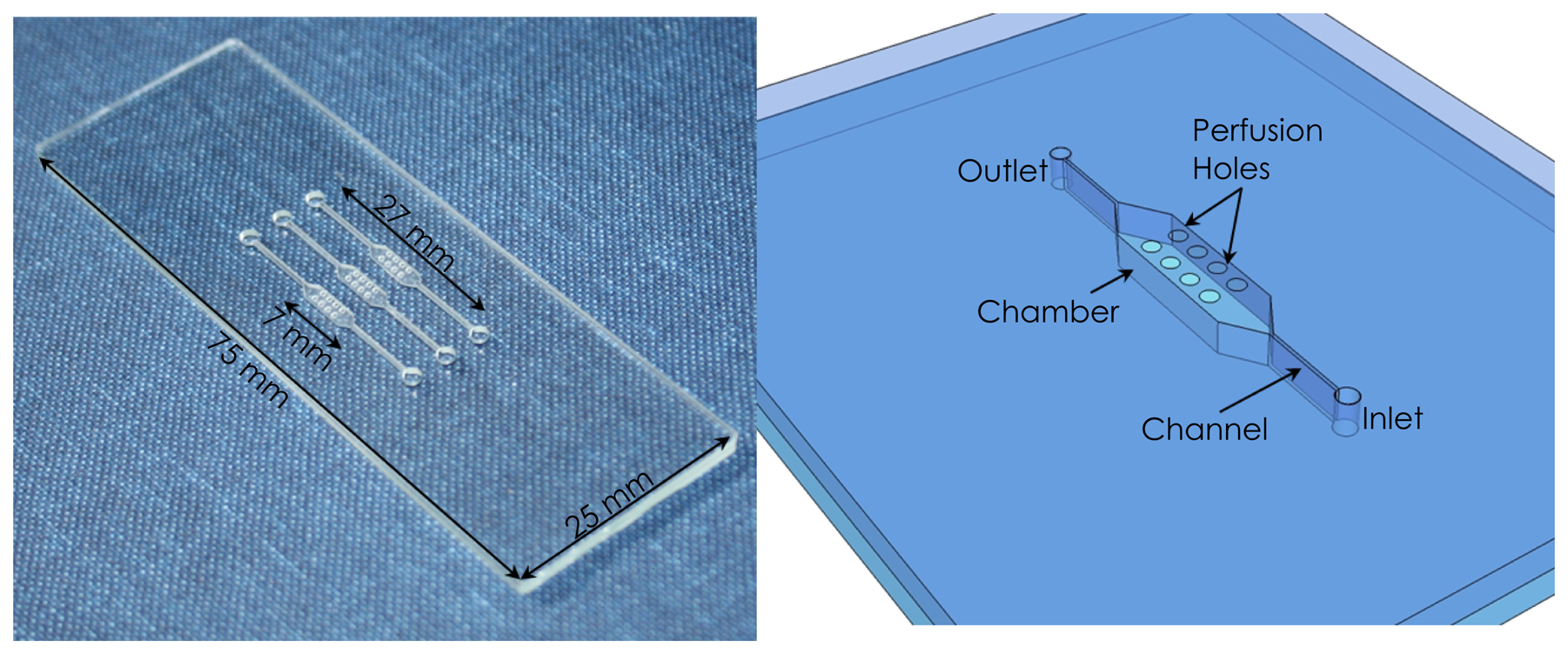
The device was designed in AutoCad2000 and fabricated by micromilling in Topas®. The final device and its outline are depicted in Figure 1. It consists of a bottom unstructured microscope slide size (25 × 75 mm) made of Topas® or glass slide. The top part of the device is made in Topas® with structured microfluidic channels and chambers with perfusion holes. In case of glass bottom device the Topas® top part with microfluidics was bonded using structured double sided adhesive tape. For the Topas® bottom device the Topas® top part was thermally bonded at 120 °C for 10 min at a force of 7 kN after 1 min exposure to UV. All the micromilled structures in the top part of the device are 200 (μm deep, channels are 500 (μm wide and 10 mm long, while the chambers are 2 mm wide and 7 mm long. The 8 perfusion holes (diameter of 600 μm) are placed over the chamber to allow for fast fixative evaporation and better visualization of chromosome spreads.

2.3. Topas® Modification
To improve surface hydrophilicity of Topas®, the slides were treated with oxygen plasma followed by immediate UV photografting of a hydrophilic 2-HEA. Prior to modification, the slides were cleaned with a mixture of isopropanol/acetone in an ultrasonic bath at 40 °C for 30 min and dried with N2. First, oxygen plasma under work pressure of 0.2–0.5 mbar was applied for 3 min. Afterwards, Topas® samples were soaked in an acetone solution of 3 M 2-HEA and 0.2 M benzophenone and placed in a UV-photoreactor at 50 °C. UV photografting was performed in a photoreactor with 360 nm wavelength lamps and 5 cm distance from the treated slides. Total irradiation was set to 7 mW/cm2 at 50 °C and the samples were treated for 4 min. After treatment, the slides were washed in isopropanol/acetone bath followed by 24 h washing in MilliQ water on a magnetic stirrer. The measurements of the water contact angle was performed on the prepared Topas® slides using Contact Angle System (OCA-15, DataPhysics Instruments GmbH, Filderstadt, Germany). At least 5 deionized 1 μL water drops were deposited on each tested surface and average contact angles have been recorded in a ‘sessile drop’ regime after 30 s. Detailed information and comprehensive statistical record regarding Topas® surface modification can be found in an additional investigation in [18].
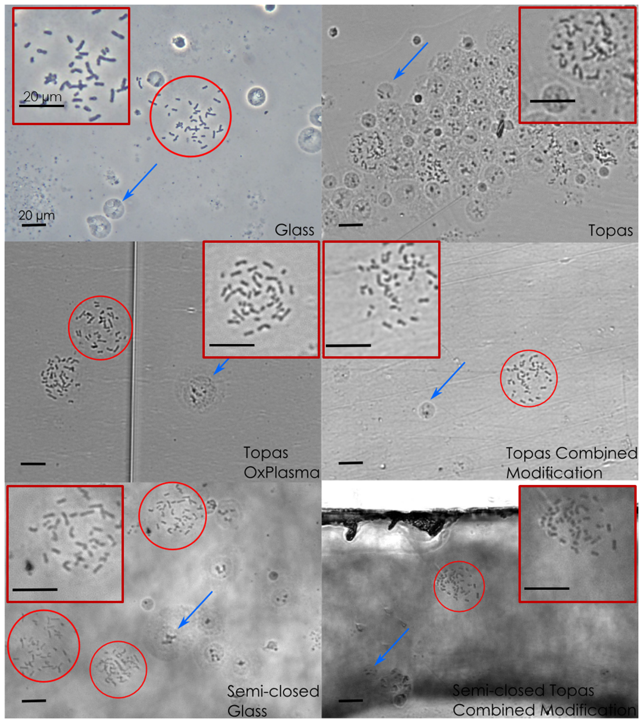
For comparison of the Topas® modification methods four different slides were used for chromosome spreading testing-conventional glass slide as a control, unmodified Topas® slide, Topas® slide treated with oxygen plasma and Topas® slide modified by oxygen plasma and photografting (Topas® modified with combined technique) (see Figure 2). Additionally, a glass and a Topas® modified with combined technique were assembled as a bottom substrate for a semi-closed device and tested for chromosome spreading. Prior to spreading all slides were immersed in water at 4 °C and stored in this way, which is a standard procedure for metaphase spreading on glass slides.

2.4. Chromosome Spreading
Before spreading, the cell suspensions were centrifuged and a fresh portion of fixative containing methanol and acetic acid in 3:1 ratio was slowly introduced drop by drop while mixing the solution to resuspend the cell pellet. As a control the standard spreading protocol was carried out on glass and Topas® slides kept in water at 4°C. The same was repeated for Topas® slides treated with oxygen plasma and Topas® modified with combined technique. The cell suspension was dropped on the control slides and air dried. To test the metaphase spread formation in the semi-closed device, 10 μL of the cell suspension was introduced to the channel filled with water at 4 °C. Subsequently, the semi-closed device was placed at 40 °C for 10–15 min to allow evaporation of the fixative. Between 10–20 spreads were analyzed using a bright-field optical microscope (Olympus) on each sample and the average spread area was calculated by measuring the area in ImageJ.
2.5. FISH Protocol
The complete FISH protocol was performed on both control glass slides and a Topas® modified with combined technique with assembled semi-closed device. The inlet was connected to the syringe by a fixed silicone tubing and the chamber perforation was closed with a tape typically used for polymerase chain reaction (PCR) to allow easy exchange of the FISH reagents and probes. The spreads denaturation was performed by heat treatment on a hot plate at 75 °C. Firstly RNAse (10 μg/μL) diluted 100 × in 2×SSC and 5 μL was injected through the inlet. The slides were incubated in a humidity chamber at 37 °C for 1 h. Following the RNAse treatment, the metaphase spreads were washed with 2×SSC solution at room temperature. Dehydration was carried out with increasing concentrations of ethanol (70%, 80% and 90%). Subsequently, the slides were heated at 75 °C for 5 min on a hot plate to denature the chromosomal DNA. DNA probe denaturation was performed simultaneously at 75 °C for 5 min in a water bath. After 5 μL of the probe solution was injected into the device, the inlet and outlet were closed to prevent drying of the probe solution. The device was incubated overnight at 37 °C in a humidity chamber to allow for the probe hybridization to chromosome spreads. Subsequently, 50% formamide was introduced as post hybridization wash at 42 °C. Final washing was performed with 0.1×SSC at 60 °C, 4×SSC and 1×PBS at room temperature. For the analysis of signals using Olympus fluorescent microscope the chromosomal DNA was stained with diamidino-2-phenylindole (DAPI).
3. Results and Discussion
3.1. Topas® Modification
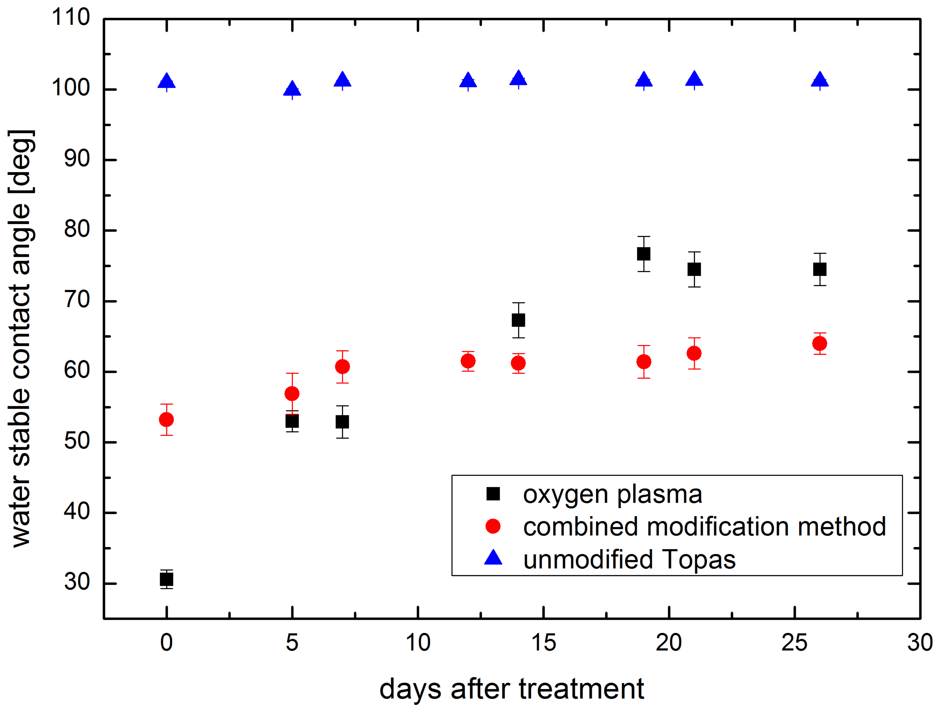
The initial water contact angle for Topas® is around 100°, which is not sufficient for efficient spreading of chromosomes. The Topas® modification applied in this paper was based on the reports by Jena and others regarding the photografting solution composition [19]. For a stable treatment, it was coupled with an oxygen plasma, which is a frequently used method for changing surface properties. Plasma surface treatment tunes the surface properties to promote adhesion, diminish the surface roughness and enhance wettability [20]. However, active radicals on plasma treated surface reduce the treatment stability and hydrophobicity recovers to the initial value during two weeks observation period in case of argon and oxygenated-argon plasma [21,22]. In the present investigation several of the Topas® slides were only treated with oxygen plasma to compare the recovery rates and long term stability with the proposed combined technique. The stability was investigated by measurements of the water contact angle repeated over time and the results are presented in Figure 3. As a control, changes in the measured contact angle over time were also performed on the untreated Topas®. The values of the contact angle were not varying significantly thus data shown in Figure 3 as blue triangles have small error bars. It can be seen that just after treatment the contact angle value for oxygen plasma treated sample is around 30°, which increases rapidly a few days after the treatment. Whereas for slides modified with combined technique the initial value of the water contact angle is higher, around 53°, but stable over the 3 weeks time. The highly reactive surface radicals formed during plasma treatment were further bound to the monomers applied during photografting, which prolongs the stability of the treatment. The photografting was conducted at elevated temperature for a better efficiency [23].

3.2. Chromosome Spreading
The chromosome spreading was performed on various slides with different surface properties. As a control, the cell suspension sample was dropped on a glass slide, untreated Topas® slide, Topas® slide treated with oxygen plasma and also on Topas® slide modified with combined technique. Untreated Topas® surface has a water contact angle of 100° in contrast to glass, which has a hydrophilic surface. The wettability of the surface is crucial during preparation of metaphase chromosome spreads as it allows for the cell suspension drop to spread quickly over the surface. Such a spread drop has a much bigger surface area and thus the fixative evaporation occurs faster resulting in well spread chromosomes with few overlaps (Figure 2 Glass). In contrast, the cell suspension placed on an untreated Topas® slide forms a standing drop, which prolongs the evaporation. Moreover, the chromosomes do not have enough space to properly spread. The visual inspection of the spreads on hydrophobic Topas® surface revealed the presence of cell clusters at the edges of the drop (Figure 2 Topas). The chromosomes spread well after treating Topas® with oxygen plasma and also after additional photografting. However, the slides after photografting have a foggy appearance, which can be reduced by optimizing the post-photografting washing procedure.
Furthermore, the spreading in a semi-closed device was done with evaporation enhancement through perforation and keeping the devices at elevated temperature. Both glass and Topas® modified with combined technique were used as a device bottom substrate to ensure that the spreading is not only affected by the surface properties. In both cases the spreads were formed in the chamber below the perforation. However, in case of Topas® slides modified with combined technique the area of each spread was smaller and their quality was worse. As the spreads were formed properly in a semi-closed device with a glass bottom we suspect that the surface treatment of Topas® might not have been sufficient to achieve good quality spreads. The evaporation in the device for spreads formation was successful although the performance of the device with a glass bottom was superior.
The average spread area on each sample was measured in ImageJ. The graph with the analyzed results is shown in Figure 4. The chromosome spreads have the largest area on a control glass sample, which is comparable to the average area of spreads in a semi-closed glass device. This shows that the evaporation of the fixative solution does not hinder the spreads formation. Furthermore, there were many spreads available for the analysis. On a control Topas® the chromosomes were spread on an untreated Topas® slide. Only few spreads were found on the surface and they were condensed as indicated by a small average area. The chromosome spreads had a bigger area on a Topas® slide treated with oxygen plasma, with the value of 878 μm2, which is close to the one for control glass slide. The Topas® modified with combined technique performed relatively well both as an open slide and as a bottom of semi-closed modified Topas® device. The spreads occupy a larger area in comparison to the untreated Topas® but the value is much smaller than the one for glass slide. There were only few spreads available for the analysis and their area varied greatly.

3.3. FISH Analysis
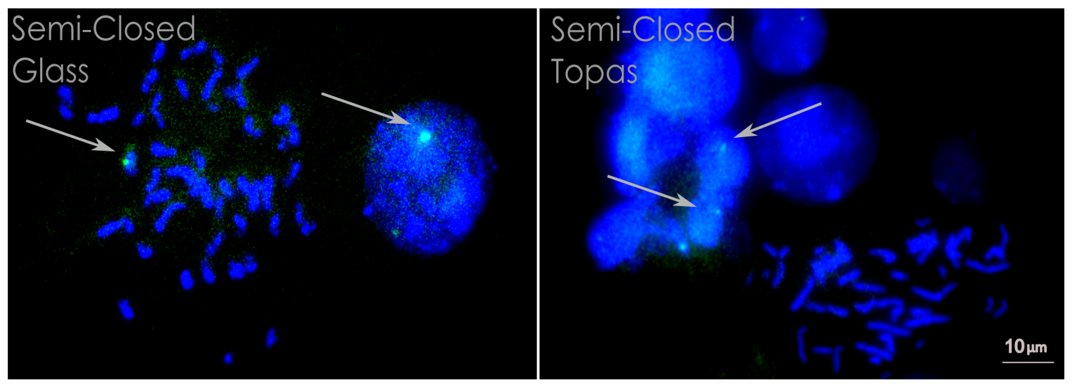
After preparation of the chromosome spreads we performed FISH analysis using an X chromosome centromeric probe to check usability of the prepared metaphase chromosome spreads. We used sample from a male patient with 46, XY karyotype and a single positive signal corresponding to the X chromosome was observed. The entire protocol was applied for both semi-closed devices with a glass and modified Topas® as a bottom. The probe was labelled with fluorescein isothiocyanate (FITC) thus it appears green in the picture. The chromosomal DNA was stained with DAPI, which intercalates between the two DNA strands and appears blue under a fluorescent microscope. The visual inspection of stained chromosomes revealed that only interphase FISH signals could be found on semi-closed Topas® device (Figure 5). None of the metaphase chromosome spreads obtained in the semi-closed Topas® device had FISH signals. In case of a semi-closed glass device some of the metaphase spreads showed positive FISH signals, which is an indication that the chromosomes were less condensed and spread better allowing the probe to bind the matching sequence on the chromosomes. Interphase signals were also obtained in the semi-closed glass device. In case of semi-closed Topas® device no hybridization signals from metaphase spreads were observed. The analysis was performed on all spreads found on the bottom of the device and showed only interphase signals, which indicates that the FISH protocol was conducted properly. The lack of metaphase signals might arise from insufficient spreading of the chromosome with their compact structure preventing the hybridization of FISH probes. Moreover, the applied FISH protocol is a standard method which is optimized for glass substrate that has a different thermal conductivity than polymers. Thus, the chromosome denaturation for 5 min at 75 °C might not be sufficient to loosen the chromosome structure required for hybridization.

4. Conclusions
We have developed a simple microfluidic device to facilitate fast evaporation of the solution from the device. The device top layer was fabricated in Topas®, with a bottom substrate made of glass or Topas® modified by oxygen plasma and photografting. This modification technique was applied to achieve long-term hydrophilic properties of the Topas® to resemble glass. We investigated that the treatment is stable over 3 weeks while oxygen plasma treatment gradually diminishes. The device design incorporated perfusion holes to allow for reliable chromosome spreading during evaporation of the fixative. The openings proved to be sufficient for solution evaporation after a short time at 40 °C, which was tested for both glass and Topas® semi-closed devices. It was possible to achieve proper spreading in both devices with an average spread area comparable to the respective substrates exposed to air. Further chromosome usability was assessed by FISH protocol completed in both semi-closed Topas® and semi-closed glass device. However, the positive metaphase FISH signals were observed only in case of a glass bottom device. Such a validation method was chosen to assess the metaphase spreads usability and showed that chromosomes on treated Topas® slide are not spread sufficiently well to be used in further cytogenetic analysis. The lack of FISH signals can be the consequence of insufficient wettability of the Topas® modified with combined technique. Moreover, the slide after modification resulted in foggy surface, which could form obstacles for the proper chromosome spreading. We concluded that the evaporation of the fixative in the semi-closed device is sufficient for preparation of reliable chromosome spreads. Although, the primary requirement for the spread usability is the surface wettability, which is superior for glass slides.
Acknowledgments
The authors would like to thank the Danish Council for Strategic Research (via the NanoKaryotyping project) and the Villum Kann Rasmussen Centre of Excellence “NAMEC” under Contract No.65286 for financial support.
Conflicts of Interest
The authors declare no conflicts of interest.
References
- Henegariu, O.; Heerema, N.A.; Wright, L.L.; Bray-Ward, P.; Ward, D.C.; Vance, G.H. Improvements in cytogenetic slide preparation: Controlled chromosome spreading, chemical aging and gradual denaturing. Cytometry 2001, 43, 101–109. [Google Scholar]
- Spurbeck, J.L.; Zinsmeister, A.R.; Meyer, K.J.; Jalal, S.M. Dynamics of chromosome spreading. Am. J. Med. Genet. 1996, 61, 387–393. [Google Scholar]
- Deng, W.; Tsao, S.W.; Lucas, J.N.; Leung, C.S.; Cheung, A.L.M. A new method for improving metaphase chromosome spreading. Cytometry 2003, 51A, 46–51. [Google Scholar]
- Claussen, U.; Michel, S.; Muhlig, P.; Westermann, M.; Grummt, U.W.; Kromeyer-Hauschild, K.; Liehr, T. Demystifying chromosome preparation and the implications for the concept of chromosome condensation during mitosis. Cytogenet. Genome Res. 2002, 98, 136–146. [Google Scholar]
- Qu, Y.Y.; Xing, L.Y.; Hughes, E.D.; Saunders, T.L. Chromosome dropper tool: Effect of slide angles on chromosome spread quality for murine embryonic stem cells. J. Histotechnol. 2008, 31, 75–79. [Google Scholar]
- Vedarethinam, I.; Shah, P.; Dimaki, M.; Tumer, Z.; Tommerup, N.; Svendsen, W.E. Metaphase FISH on a chip: Miniaturized microfluidic device for fluorescence in situ hybridization. Sensors 2010, 10, 9831–9846. [Google Scholar]
- Sieben, V.J.; Marun, C.S.D.; Pilarski, P.M.; Kaigala, G.V.; Pilarski, L.M.; Backhouse, C.J. FISH and chips: Chromosomal analysis on microfluidic platforms. IET Nanobiotechnol. 2007, 1, 27–35. [Google Scholar]
- Sieben, V.J.; Debes-Marun, C.S.; Pilarski, L.M.; Backhouse, C.J. An integrated microfluidic chip for chromosome enumeration using fluorescence in situ hybridization. Lab Chip. 2008, 8, 2151–2156. [Google Scholar]
- Shah, P.; Vedarethinam, I.; Kwasny, D.; Andresen, L.; Skov, S.; Silahtaroglu, A.; Tumer, Z.; Dimaki, M.; Svendsen, W.E. FISHprep: A novel integrated device for metaphase FISH sample preparation. Micromachines 2011, 2, 116–128. [Google Scholar]
- Shah, P.; Vedarethinam, I.; Kwasny, D.; Andresen, L.; Dimaki, M.; Skov, S.; Svendsen, W.E. Microfluidic bioreactors for culture of non-adherent cells. Sens. Actuators B Chem. 2011, 156, 1002–1008. [Google Scholar]
- Zanardi, A.; Bandiero, D.; Bertolini, F.; Corsini, C.A.; Gregato, G.; Milani, P.; Barborini, E.; Carbone, R. Miniaturized FISH for screening of onco-hematological malignancies. BioTechniques 2010, 49, 497–504. [Google Scholar]
- Tai, C.-H.; Ho, C.-L.; Chen, Y.-L.; Chen, W.L.; Lee, G.-B. A novel integrated microfluidic platform to perform fluorescence in situ hybridization for chromosomal analysis. Microfluid. Nanofluid 2013, 15, 745–752. [Google Scholar]
- Tai, C.-H.; Chen, Y.-L.; Ho, C.-L.; Lee, G.-B. An Integrated Microfluidic Platform for Chromosomal Analysis. Proceedings of the IEEE MEMS 25th International Conference, Paris, France, 29 January– 2 February 2012; pp. 918–921.
- Kao, K.-J.; Tai, C.-H.; Luo, W.-Y.; Yeh, T.-S.; Lee, G.-B. Fluorescence in situ Hybridization (FISH) Microfluidic Platform for Detection of HER-2 Over-expression in Cancer Cells. Proceedings of the 17th International Conference μTAS, Freiburg, Germany, 27–31 October 2013; pp. 509–511.
- Kurz, C.M.v.d.; Moosdijk, S.; Thielecke, H.; Velten, T. Towards a Cellular Multi-parameter Analysis Platform: Fluorescence in situ Hybridization (FISH) on Microhole-array Chips. Proceedings of the IEEE EMBS 33rd Annual International Conference, Boston, MA, USA, 30 August–3 September 2011; pp. 8408–8411.
- Yu, Y.; Wang, X.; Oberthur, D.; Meyer, A.; Perbandt, M.; Duan, L.; Kang, Q. Design and application of a microfluidic device for protein crystallization using an evaporation-based crystallization technique. J. Appl. Crystallogr 2012, 45, 53–60. [Google Scholar]
- Zhang, J.Y.; Mahalanabis, M.; Liu, L.; Chang, J.; Pollock, N.R.; Klapperich, C.M. A disposable microfluidic virus concentration device based on evaporation and interfacial tension. Diagnostics 2013, 3, 155–169. [Google Scholar]
- Mednova, O.; Kwasny, D.; Rozlosnik, N.; Svendsen, W.E.; Almdal, K. New approach of long-term modification of Topas® to acquire surface hydrophilicity for chromosome spreading. Appl. Surf. Sci. 2014, 292, 1045–1051. [Google Scholar]
- Jena, R.; Yue, C.; Anand, L. Improvement of thermal bond strength and surface properties of Cyclic Olefin Copolymer (COC) based microfluidic device using the photo-grafting technique. Sens. Actuators B Chem. 2011, 157, 518–526. [Google Scholar]
- Guimond, S.; Wertheimer, M.R. Surface degradation and hydrophobic recovery of polyolefins treated by air corona and nitrogen atmospheric pressure glow discharge. J. Appl. Polym. Sci. 2004, 94, 1291–1303. [Google Scholar]
- Sunanda, R.; Yue, C.E.; Lam, Y.C.; Wang, Z.Y.; Huifang, H. Surface analysis, hydrophilic enhancement, ageing behavior and flow in plasma modified cyclic olefin copolymer (COC)-based microfluidic devices. Sens. Actuators B Chem. 2010, 150, 537–549. [Google Scholar]
- Tsao, C.W.; Hromada, L.L.; Liu, J. Low temperature bonding of PMMA and COC microfluidic substrates using UV/ozone surface treatment. Lab Chip. 2007, 7, 499–505. [Google Scholar]
- De Smet, N.; Rymarczyk-Machal, M.; Schacht, E. Modification of poly-dimethylsiloxane surfaces using benzophenone. J. Biomater. Sci. 2009, 20, 2039–2053. [Google Scholar]
© 2014 by the authors; licensee MDPI, Basel, Switzerland. This article is an open access article distributed under the terms and conditions of the Creative Commons Attribution license (http://creativecommons.org/licenses/by/3.0/).
Share and Cite
Kwasny, D.; Mednova, O.; Vedarethinam, I.; Dimaki, M.; Silahtaroglu, A.; Tümer, Z.; Almdal, K.; Svendsen, W.E. A Semi-Closed Device for Chromosome Spreading for Cytogenetic Analysis. Micromachines 2014, 5, 158-170. https://doi.org/10.3390/mi5020158
Kwasny D, Mednova O, Vedarethinam I, Dimaki M, Silahtaroglu A, Tümer Z, Almdal K, Svendsen WE. A Semi-Closed Device for Chromosome Spreading for Cytogenetic Analysis. Micromachines. 2014; 5(2):158-170. https://doi.org/10.3390/mi5020158
Chicago/Turabian StyleKwasny, Dorota, Olga Mednova, Indumathi Vedarethinam, Maria Dimaki, Asli Silahtaroglu, Zeynep Tümer, Kristoffer Almdal, and Winnie E. Svendsen. 2014. "A Semi-Closed Device for Chromosome Spreading for Cytogenetic Analysis" Micromachines 5, no. 2: 158-170. https://doi.org/10.3390/mi5020158