EGFR Protein Expression in KRAS Wild-Type Metastatic Colorectal Cancer Is Another Negative Predictive Factor of the Cetuximab Therapy

 ,
,
Abstract
:1. Introduction
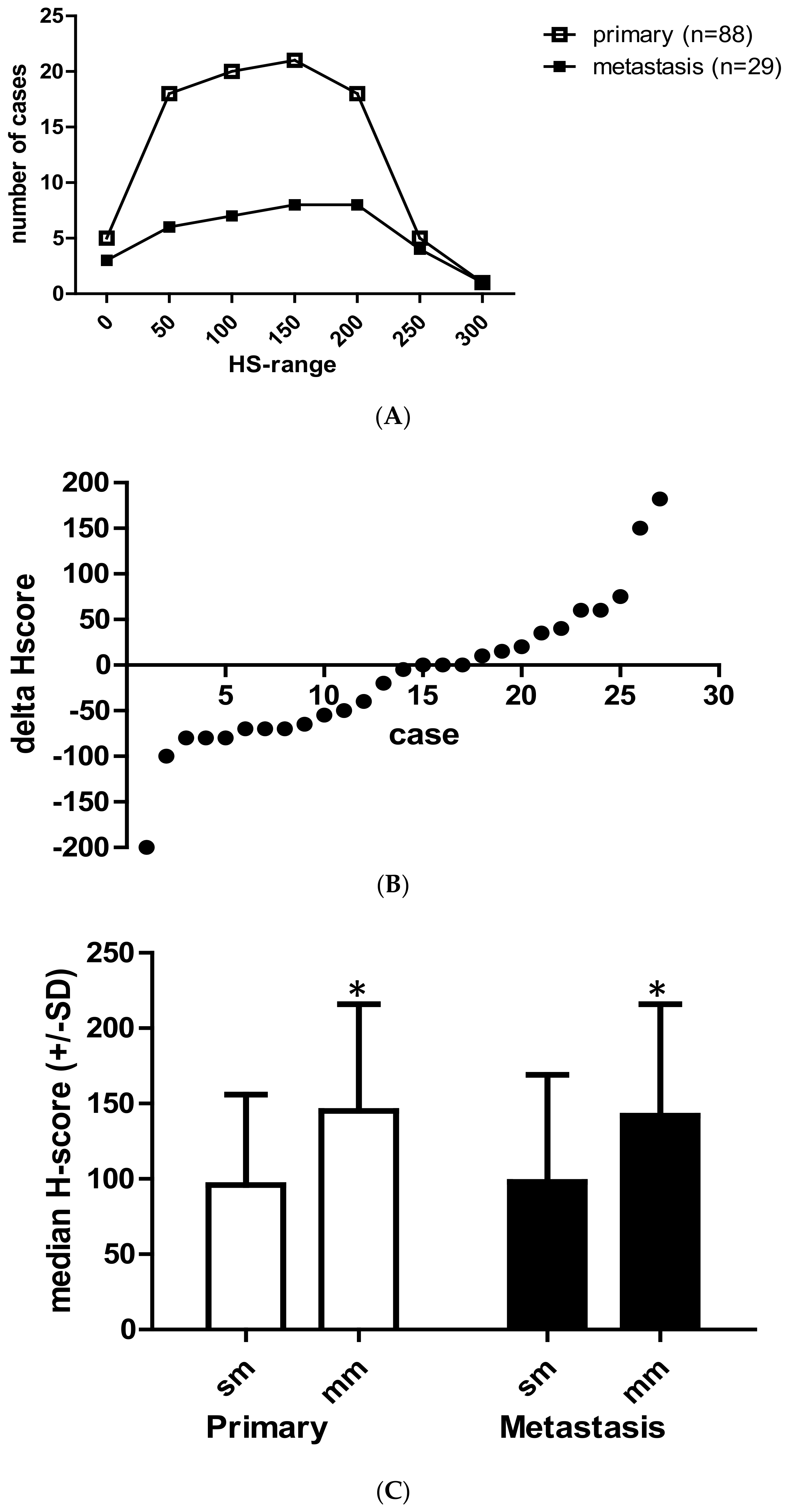
2. Results
3. Discussion
4. Matherials and Methods
4.1. Patients
4.2. RAS Testing
4.3. EGFR Protein Expression
4.4. Statistical Analysis
5. Conclusions
Supplementary Materials
Author Contributions
Funding
Conflicts of Interest
Abbreviations
| DAB | diamino benzidine |
| EGFR | epidermal growth factor receptor |
| FFPE | formalyn-fixed paraffin-embedded sample |
| FOLFIRI | folic acid+5-FU+irinotecan containing chemotherapeutic regimen |
| HS | H-score |
| OS | overall survival |
| PFS | progression-free survival |
References
- Van Cutsem, E.; Köhne, C.H.; Hitre, E.; Zaluski, J.; Chang Chien, C.R.; Markhson, A.; D’Haens, G.; Pinter, T.; Lim, R.; Bodoky, G.; et al. Cetuximab and chemotherapy as initial treatment for metastatic colorectal cancer. N. Engl. J. Med. 2009, 360, 1408–1417. [Google Scholar] [CrossRef] [PubMed] [Green Version]
- Summary of Product Characteristics. Available online: https://www.ema.europa.eu/en/documents/product-information/erbitux-epar-product-information_en.pdf (accessed on 10 December 2019).
- Hecht, J.R.; Mitchell, E.; Neubauer, M.A.; Burris, H.A.; Swanson, P.; Lopez, T.; Buchanan, G.; Reiner, M.; Gansert, J.; Berlin, J. Lack of correlation between epidermal growth factor receptor status and response to Panitumumab monotherapy in metastatic colorectal cancer. Clin. Cancer Res. 2010, 16, 2205–2213. [Google Scholar] [CrossRef] [PubMed] [Green Version]
- Licitra, L.; Störkel, S.; Kerr, K.M.; Van Cutsem, E.; Pirker, R.; Hirsch, F.R.; Vermorken, J.B.; von Hexdebreck, A.; Esser, R.; Celik, I.; et al. Predicitive value of epidermal growth factor receptor expression for first- line chenmotherapy plus cetuximab in patients with head and neck and colorectal cancer: Analysis of data from the EXTREME and CRYSTAL studies. Eur. J. Cancer 2013, 49, 1161–1168. [Google Scholar] [CrossRef] [PubMed]
- Chung, K.Y.; Shia, J.; Kemeny, N.E.; Shah, M.; Schwartz, G.K.; Tse, A.; Hamilton, A.; Pan, D.; Schrag, D.; Schwartz, L.; et al. Cetuximab shows activity in colorectal cancer patients with tumors that do not express the epidermal growth factor receptor by immunohistochemistry. J. Clin. Oncol. 2005, 23, 1803–1810. [Google Scholar] [CrossRef] [PubMed]
- Derecskei, K.; Moldvay, J.; Bogos, K.; Tímár, J. Protocol modifications influence the result of EGFR receptor immunodetection by EGFR pharmDX in paraffin-embedded cancer tissues. Pathol. Oncol. Res. 2006, 12, 243–245. [Google Scholar] [CrossRef]
- Fan, C.C.; Wang, T.Y.; Kung, C.M. Epidermal growth factor receptor inconsistency by immunohistochemistry method using different monoclonal antibodies in colorectal cancer patients. Clin. Lab. 2015, 61, 1635–1641. [Google Scholar] [CrossRef]
- Lievre, A.; Ouine, B.; Canet, J.; Cartier, A.; Amar, Y.; Cacheux, W.; Mariani, O.; Guimbaud, R.; Selves, J.; Lecomte, T.; et al. Protein biomarkers predictive for response to anti-EGFR treatment in RAS wild-type metastatic colorectal carcinoma. Br. J. Cancer 2017, 117, 1819–1827. [Google Scholar] [CrossRef] [Green Version]
- Tol, J.; Dijkstra, J.R.; Klomp, M.; Teerenstra, S.; Dommerholt, M.; Vink-Borger, M.E.; van Cleef, P.H.; van Krieken, J.H.; Punt, C.J.; Nastegaal, I.D. Markers for EGFR pathway activation as predictor of outcome in metastatic colorectal cancer patients treated with or without cetuximab. Eur. J. Cancer 2010, 46, 1997–2009. [Google Scholar] [CrossRef]
- Algars, A.; Sundstrom, J.; Lintunen, M.; Jokilehto, T.; Kytola, S.; Kaare, M.; Vainionpaa, R.; Orpana, A.; Osterlund, P.; Ristimaki, A.; et al. EGFR copy number predicts response to anti-EGFR treatment in RAS wild type and RAS/BRAF/PI3CA wild type metastatic colorectal cancer. Int. J. Cancer 2017, 40, 922–929. [Google Scholar] [CrossRef] [Green Version]
- Uhlyarik, A.; Piurko, V.; Vizkeleti, L.; Papai, Z.; Raso, E.; Lahm, E.; Kiss, E.; Sikter, M.; Vachaja, J.; Kenessey, I.; et al. EGFR protein expression of KRAS wild type colorectal cancer: Predictive value of the sidedness for efficacy of anti-EGFR therapy. Pathol. Oncol. Res. 2019. [Google Scholar] [CrossRef]
- Montagut, C.; Dalmases, A.; Bellosillo, B.; Crespo, M.; Pairet, S.; Iglesias, M.; Salido, M.; Gallen, M.; Marsters, S.; Tsai, S.P.; et al. Identification of mutation in the extracellular domain of the epidermal growth factor receptor conferring cetuximab resistance in colorectal cancer. Nat. Med. 2012, 18, 221–223. [Google Scholar] [CrossRef]
- Bertotti, A.; Papp, E.; Jones, S.; Adkeff, V.; Anagnostou, V.; Lupo, B.; Sausen, M.; Phallen, J.; Hruban, C.A.; Tokheim, C.; et al. The genomic landscape of response to EGFR blockade in colorectal cancer. Nature 2015, 526, 263–267. [Google Scholar] [CrossRef] [PubMed]
- Bokemeyer, C.; Van Cutsem, E.; Rougiuer, P.; Ciardiello, F.; Heeger, S.; Schlichting, M.; Celik, I.; Kohne, C.H. Addition of cetuximab to chemotherapy as first line treatment for KRAS wild-type metastatic colorectal cancer: Pooled analysis of the CRYSTAL and OPUS randomised clinical trials. Eur. J. Cancer 2012, 48, 1466–1475. [Google Scholar] [CrossRef] [PubMed]
- Bokemeyer, C.; Köhne, C.H.; Ciardiello, F.; Lenz, H.J.; Heinemann, V.; Klinkhardt, U.; Beier, F.; Duecker, K.; van Krieken, J.H.; Tejpar, S. FOLFOX4 plus cetuximab treatment and RAS mutations in colorectal cancer. Eur. J. Cancer 2015, 51, 1243–1252. [Google Scholar] [CrossRef]
- Sanz-Garcia, E.; Argiles, G.; Elez, E.; Tabernero, J. BRAF mutant colorectal cancer: Prognosis, treatment and new pwerspectives. Ann. Oncol. 2017, 28, 2648–2657. [Google Scholar] [CrossRef] [PubMed]
- Li, D.; Fu, Q.; Li, M.; Yin, C.; Zhao, J.; Li, F. Primary tumor site and anti-EGFR monoclonal antibody benefit in metastatic colorectal cancer: A meta-analysis. Future Oncol. 2017, 13, 1115–1127. [Google Scholar] [CrossRef] [PubMed]
- Cao, D.D.; Xu, H.L.; Xu, X.M.; Ge, W. The impact of primary tumor location on efficacy of cetuximab in metastatic colorectal cancer patients with different KRAS status: A systematic review and meta-analysis. Oncotarget 2017, 8, 53631–53641. [Google Scholar] [CrossRef] [PubMed] [Green Version]
- Salem, M.E.; Weinberg, B.A.; Xiu, J.; El-Eeiry, W.S.; Hwang, J.J.; Gatalica, Z.; Philip, P.A.; Shields, A.F.; Lenz, H.J.; Marshall, J.L. Comparative molecular analyses of left-sided colon, right-sided colon, and rectal cancers. Oncotarget 2017, 8, 86356–86368. [Google Scholar] [CrossRef] [Green Version]
- Van Krieken, J.H.J.M.; Jung, A.; Kirchner, T.; Cameiro, F.; Seruca, R.; Bosman, F.T.; Quirke, P.; Flejou, J.F.; Plato, H.T.; deHertogh, G.; et al. KRAS mutation testing for predicting response to anti-EGFR therapy for colorectal carcinoma: Proposal for an European quality assurance program. Virchows Arch. 2008, 453, 417–431. [Google Scholar] [CrossRef] [Green Version]
- Khelwatty, S.; Essapen, S.; Bagwan, I.; Green, M.; Seddon, A.; Modjtahedi, H. The impact of co-expression of wild-type EGFR and its ligands determined by immunohistochemistry for response to treatment with cetuximab in patients with metastatic colorectal cancer. Oncotarget 2017, 8, 7666–7677. [Google Scholar] [CrossRef] [Green Version]
- Hirsch, F.R.; Dziadziuszko, R.; Thatcher, N.; Mann, H.; Watkins, C.; Parums, D.V.; Speake, G.; Holloway, B.; Bunn, P.A.; Franklin, W.A. Epidermal growth factor immunohistochemistry: Comparison of antibodies and cutoff points to predict benefit from Gefitinib in a phase-III placebo-controlled study in advanced nonsmall-cell-lung cancer. Cancer 2008, 112, 1114–1121. [Google Scholar] [CrossRef] [PubMed] [Green Version]
- Gao, L.; Xu, J.; He, G.; Huang, J.; Xu, W.; Qin, J.; Zheng, P.; Ji, M.; Chang, W.; Ren, W.; et al. CCR7 high expression leads to cetuximab resistance by ccross-talking with EGFR pathway in PI3K/AKT signals in colorectal cancer. Am. J. Cancer Res. 2019, 9, 2531–2543. [Google Scholar] [PubMed]
- Martini, G.; Cardone, C.; Vitiello, P.P.; Belli, V.; Napolitano, S.; Troiani, T.; Ciardiello, D.; Della Corte, C.M.; Morgillo, F.; Matrone, N.; et al. EPHA2 is a predictive biomarker of resistance and potential therapeutic target for improving anti-epidermal growth factor receptor therapy of colorectal cancer. Mol. Cancer Ther. 2019, 18, 845–855. [Google Scholar] [CrossRef] [PubMed] [Green Version]
- Annadappa, G.; Lampis, A.; Cunningham, D.; Khan, K.H.; Kouvelakis, K.; Vlachiogiannis, G.; Hedayat, S.; Tunariu, N.; Rao, S.; Watkins, D.; et al. miR-31-3p expression and benefot from anti-EGFR inhibitors in metastatic colorectal cancer patients enrolled in the prospective phase II Prospect-C trial. Clin. Cancer Res. 2019, 25, 3830–3838. [Google Scholar] [CrossRef] [PubMed] [Green Version]
- Woolston, A.; Khan, K.; Spain, G.; Barber, L.J.; Griffiths, B.; Gonzalez-Exposito, R.; Hornstainer, L.; Punta, M.; Patil, Y.; Newey, A.; et al. Genomic and transcriptomic determinants of therapy resistance and immune landscape evolution during anti-EGFR treatment in colorectal cancer. Cancer Cell 2019, 36, 35–50. [Google Scholar] [CrossRef] [PubMed] [Green Version]
- Cserepes, M.; Ostoros, G.; Lohinai, Z.; Raso, E.; Barbai, T.; Tímár, J.; Rozsas, A.; Moldvay, J.; Kovalszky, I.; Fabian, K.; et al. Subtype-specific KRAS mutations in advanced lung adenocarcinoma: A retrospective study of patients treated with platinum-based chemotherapy. Eur. J. Cancer 2014, 50, 1819–1828. [Google Scholar] [CrossRef] [Green Version]


| Sex | [N] | [%] |
|---|---|---|
| Male | 63 | 70.0 |
| Female | 27 | 30.0 |
| Age (years) | [N] | [%] |
| Median | 64 | |
| Range | 24–79 | |
| Primary Tumor Location | [N] | [%] |
| Rectum | 26 | 28.9 |
| Rectosigmoid | 6 | 6.7 |
| Sigma | 28 | 31.1 |
| Descending colon | 8 | 8.9 |
| Lienal flexure | 3 | 3.3 |
| Transverse colon | 4 | 4.4 |
| Hepatic flexure | 1 | 1.1 |
| Ascending colon | 8 | 8.9 |
| Coecum | 6 | 6.7 |
| Primary Tumor T | [N] | [%] |
| NA | 13 | 14.4 |
| 1 | 0 | 0.0 |
| 2 | 6 | 6.7 |
| 3 | 54 | 60.0 |
| 4 | 17 | 18.9 |
| Primary Tumor N | [N] | [%] |
| NA | 13 | 14.4 |
| 0 | 21 | 23.3 |
| 1 | 26 | 28.9 |
| 2 | 29 | 32.2 |
| 3 | 1 | 1.1 |
| Resection of Primary | [N] | [%] |
| Yes | 77 | 85.6 |
| No | 13 | 14.4 |
| RAS Testing | [N] | [%] |
| KRAS exon2 | 75 | 83.3 |
| Extended RAS | 15 | 16.7 |
| Number of Evaluated Primary Tumors (N = 88) | [N] | [%] |
| Right | 19 | 21.6 |
| Left | 69 | 78.4 |
| Number of Metastases Evaluated by IHC (N = 29) | [N] | [%] |
| Liver | 17 | 58.6 |
| Lung | 2 | 6.9 |
| Lymphnode | 2 | 6.9 |
| Cerebellum | 1 | 3.4 |
| Skin | 1 | 3.4 |
| Ovarium | 1 | 3.4 |
| Peritoneum | 3 | 10.3 |
| Soft tissue | 1 | 3.4 |
| Mesocolon | 1 | 3.4 |
| Number of Metastases Evaluated by IHC (N = 29) | [N] | [%] |
| Right | 11 | 37.9 |
| Left | 18 | 62.1 |
| Abbreviation(s): immunohistochemistry (IHC) |
| Variables | p | RR (95% CI) |
|---|---|---|
| age | 0.001 | 0.967 (0.943–0.993) |
| sex | 0.715 | 1.099 (0.661–1.826) |
| EGFR-HS | 0.53 | 1.001 (0.997–1.006) |
| sidedness | 0.009 | 2.028 (1.193–3.448) |
| metastasis (S vs. M) | 0.1 | 0.643 (0.38–1.089) |
| Variables | p | RR (95% CI) |
|---|---|---|
| age | 0.001 | 0.916 (0.871–0.964) |
| sex | 0.427 | 1.468 (0.57–3.784) |
| EGFR-HS | 0.083 | 1.007 (0.999–1.016) |
| sidedness | 0.006 | 5.694 (1.641–19.757) |
| metastasis (S vs M) | 0.19 | 0.469 (0.151–1.455) |
© 2020 by the authors. Licensee MDPI, Basel, Switzerland. This article is an open access article distributed under the terms and conditions of the Creative Commons Attribution (CC BY) license (http://creativecommons.org/licenses/by/4.0/).
Share and Cite
Uhlyarik, A.; Piurko, V.; Papai, Z.; Raso, E.; Lahm, E.; Kiss, E.; Sikter, M.; Vachaja, J.; Kenessey, I.; Timar, J. EGFR Protein Expression in KRAS Wild-Type Metastatic Colorectal Cancer Is Another Negative Predictive Factor of the Cetuximab Therapy. Cancers 2020, 12, 614. https://doi.org/10.3390/cancers12030614
Uhlyarik A, Piurko V, Papai Z, Raso E, Lahm E, Kiss E, Sikter M, Vachaja J, Kenessey I, Timar J. EGFR Protein Expression in KRAS Wild-Type Metastatic Colorectal Cancer Is Another Negative Predictive Factor of the Cetuximab Therapy. Cancers. 2020; 12(3):614. https://doi.org/10.3390/cancers12030614
Chicago/Turabian StyleUhlyarik, Andrea, Violetta Piurko, Zsuzsanna Papai, Erzsebet Raso, Erika Lahm, Edina Kiss, Marta Sikter, Jozsef Vachaja, Istvan Kenessey, and Jozsef Timar. 2020. "EGFR Protein Expression in KRAS Wild-Type Metastatic Colorectal Cancer Is Another Negative Predictive Factor of the Cetuximab Therapy" Cancers 12, no. 3: 614. https://doi.org/10.3390/cancers12030614