
Regulation of PD-L1 Expression by YY1 in Cancer: Therapeutic Efficacy of Targeting YY1
by
 , and
, and
Ana Dillen
1,†,
Indy Bui
1,†,
Megan Jung
1,
Stephanie Agioti
2,
Apostolos Zaravinos
2,3 and
and
Benjamin Bonavida
1,* 1
Department of Microbiology, Immunology & Molecular Genetics, Jonsson Comprehensive Cancer, David Geffen School of Medicine, University of California, Los Angeles, CA 90095, USA
2
Cancer Genetics, Genomic and Systems Biology Group, Basic and Translational Cancer Research Center (BTCRC), 1516 Nicosia, Cyprus
3
Department of Life Sciences, School of Sciences, European University Cyprus, 2404 Nicosia, Cyprus
*
Author to whom correspondence should be addressed.
†
These authors contributed equally to this work.
Cancers 2024, 16(6), 1237; https://doi.org/10.3390/cancers16061237
Submission received: 9 January 2024
/
Revised: 23 February 2024
/
Accepted: 11 March 2024
/
Published: 21 March 2024
(This article belongs to the Section Molecular Cancer Biology)
Abstract
:Simple Summary
We have recently witnessed several milestones in the treatment of various cancers with immunotherapy through modulating the activity of the immune system to suppress and destroy cancer cells. One main factor involved in the anti-cancer immune system is cytotoxic immune cells known as T cells. T cells can recognize pathogenic cancer cells and launch targeted destruction attacks. However, these T cells may become non-functional due to their close association with cancer cells within the tumor microenvironment. Cancer cell activity can ultimately suppress T-cell function. New approaches that prevent the inactivation of T cells by cancer cells would result in recovery of the T-cell functions, cancer regression, and overall survival. Targeting cancer cells specifically to prevent T-cell inactivation would result in tumor disappearance, inhibition of metastasis, and the reversal of resistance to therapy. Therefore, this proposed alternative approach is promising as it is applicable to numerous cancers, including those that are resistant to treatment.
Abstract
During the last decade, we have witnessed several milestones in the treatment of various resistant cancers including immunotherapeutic strategies that have proven to be superior to conventional treatment options, such as chemotherapy and radiation. This approach utilizes the host’s immune response, which is triggered by cancer cells expressing tumor-associated antigens or neoantigens. The responsive immune cytotoxic CD8+ T cells specifically target and kill tumor cells, leading to tumor regression and prolongation of survival in some cancers; however, some cancers may exhibit resistance due to the inactivation of anti-tumor CD8+ T cells. One mechanism by which the anti-tumor CD8+ T cells become dysfunctional is through the activation of the inhibitory receptor programmed death-1 (PD-1) by the corresponding tumor cells (or other cells in the tumor microenvironment (TME)) that express the programmed death ligand-1 (PD-L1). Hence, blocking the PD-1/PD-L1 interaction via specific monoclonal antibodies (mAbs) restores the CD8+ T cells’ functions, leading to tumor regression. Accordingly, the Food and Drug Administration (FDA) has approved several checkpoint antibodies which act as immune checkpoint inhibitors. Their clinical use in various resistant cancers, such as metastatic melanoma and non-small-cell lung cancer (NSCLC), has shown significant clinical responses. We have investigated an alternative approach to prevent the expression of PD-L1 on tumor cells, through targeting the oncogenic transcription factor Yin Yang 1 (YY1), a known factor overexpressed in many cancers. We report the regulation of PD-L1 by YY1 at the transcriptional, post-transcriptional, and post-translational levels, resulting in the restoration of CD8+ T cells’ anti-tumor functions. We have performed bioinformatic analyses to further explore the relationship between both YY1 and PD-L1 in cancer and to corroborate these findings. In addition to its regulation of PD-L1, YY1 has several other anti-cancer activities, such as the regulation of proliferation and cell viability, invasion, epithelial–mesenchymal transition (EMT), metastasis, and chemo-immuno-resistance. Thus, targeting YY1 will have a multitude of anti-tumor activities resulting in a significant obliteration of cancer oncogenic activities. Various strategies are proposed to selectively target YY1 in human cancers and present a promising novel therapeutic approach for treating unresponsive cancer phenotypes. These findings underscore the distinct regulatory roles of YY1 and PD-L1 (CD274) in cancer progression and therapeutic response.
1. Introduction
As a major and complex disease, cancer consists of over a hundred different subtypes with various phenotypes, etiologies, and treatment strategies. The current common treatments for cancer consist of surgery, radiation, hormonal therapy, chemotherapy, and immunotherapy. Though these various conventional treatment strategies have resulted in significant clinical responses in a large subset of patients, cancer drug resistance remains a substantial complication. A significant subset of cancer patients is either initially unresponsive to therapies or acquires treatment resistance following initial therapy exposures [1,2]. Thus, unresponsive patients require novel therapies to overcome therapeutic resistance and suppress cancer activity. The evolution of these therapies must also align with the concurrent advancement of innovative targeting strategies. Such therapies should be designed to specifically target underlying factors accountable for therapy resistance and include small-molecule inhibitors/activators [3,4] and anti-tumor monoclonal antibodies (mAbs) [5] that activate the host anti-tumor immune response [6,7,8].
In many therapy-resistant cancer phenotypes, the anti-cancer immune response is suppressed or inactivated [9,10]. Mechanisms that are tied to cancer immune unresponsiveness and evasion involve the immunosuppressive environment of the tumor microenvironment (TME) [6,11,12]. The TME holds several immunosuppressive cells and factors including cancer-associated fibroblasts (CAFs) [13,14], myeloid-derived suppressor cells (MDSCs) [15,16], tumor-associated macrophages (TAMs) [17,18], T regulatory cells (Tregs) [19,20], and other additional suppressive factors such as transforming growth factor-β (TGF-β) [21,22], chemokines, cytokines [23,24], etc.
Anti-tumor CD8+ T cells express an inhibitory receptor, programmed cell death receptor 1 (PD-1). PD-1, a receptor found on immune T cells, B cells, and macrophages, plays a role in regulating the immune system to maintain immune tolerance through signaling and also prevents the negative effects of prolonged immune responses [25,26,27,28]. PD-1 on T cells interacts with programmed cell death ligands 1 (PD-L1) or 2 (PD-L2). PD-L1 and PD-L2 are typically expressed by several other immune cells during inflammatory responses; however, these ligands are also often expressed by cancer cells or tumor-associated cells. Activation of PD-1 by PD-L1 or PD-L2 results in the inactivation of the cytotoxic functions of anti-tumor CD8+ T cells, leading to tumor progression and cancer cell invasion [29,30]. Therefore, blocking the interaction between PD-1 and PD-L1/2 can restore the anti-tumor immune activity and allow for tumor regression [31,32]. Many pre-clinical and clinical findings have allowed for the development of Food and Drug Administration (FDA)-approved checkpoint inhibitor mAbs directed against PD-1 and PD-L1. This novel therapeutic approach has been beneficial in targeting chemoresistant cancers, including melanoma, lung cancers, and several other human cancers [31,33].
Another approach to blocking the PD-1/PD-L1/2 CD8+ T-cell inactivation focuses on inhibiting the induction of PD-1 on T cells or inhibiting the expression of PD-L1/2 in tumor cells. Of importance, the expression of PD-L1 on tumor cells has been demonstrated to be regulated by the overexpression of the transcription factor (TF) Yin Yang 1 (YY1) [34,35,36,37,38,39]. YY1 is known to be implicated in the pathogenesis of various cancer cells as an immunosuppressive factor [40]. In this report, we address the underlying mechanisms of YY1 in the regulation of PD-L1 expression on cancer cells and determine whether its inhibition will restore not only the anti-tumor activities of CD8+ T cells but also reverse other pro-tumorigenic activities associated with YY1, such as its role in tumor cell proliferation, cell viability, tumor invasion, epithelial–mesenchymal transition (EMT), metastasis, and chemo-immuno-resistance.
2. YY1 Overexpression in Cancer, Oncogenic and Immunosuppressive Properties
The transcription factor YY1 plays a critical role in regulating a number of cellular processes [41,42,43,44]. The name “Yin Yang” is due to its dual context-dependent abilities in both transcriptional activation and repression. YY1 belongs to the zinc finger transcription factor in the GLI-Kruppel class and is commonly found in enhancer and promoter regions of DNA [41,45,46,47]. The YY1 gene is 23 kb long and is highly conserved across cells; its translated protein consists of 414 amino acids [45,46,47]. Due to its roles in the cell cycle, gene expression, DNA repair, cellular metabolism, differentiation, survival, and apoptosis, YY1 is vital in early developmental processes as well as lifelong regulatory processes [48,49,50]. However, it has also been found that YY1 is overexpressed in many types of cancers, specifically in several metastatic cancers [51,52,53,54]. This overexpression of YY1 is associated with poor prognosis and resistance to both chemo- and immunotherapeutics. Though YY1 has been determined to be linked to cancer progression and treatment resistance, the exact downstream effects of YY1 overexpression in cancer are still being explored [55,56] (See Figure 1).
YY1, a ubiquitously expressed transcription factor, plays a crucial role in proliferation, cell survival, development, and differentiation through its interaction with several complexes [46,56,57]. The cell cycle is regulated by YY1 through its promotion or suppression of a number of genes [58]. Additionally, YY1 promotes cell survival by regulating anti-apoptotic factors and signaling pathways [59,60,61]. Cell development and differentiation are also influenced by YY1 due to its control of gene expression patterns [41,44,62,63]. Therefore, the overexpression of YY1 results in dysfunction of these regulatory loops, leading to poor health outcomes [56,64,65]. One relevant pathway is the nuclear factor kappa B (NF-κB)/Snail/YY1/Raf kinase inhibitory protein (RKIP) loop, which regulates the EMT [51,52,53,54]. NF-κB stimulates overexpression of YY1, leading to transcriptional activation of Snail and activation of downstream EMT-related molecules [51,66,67,68,69,70]. EMT dysregulation contributes to cancer metastasis through the disruption of cell–cell adhesion and the promotion of a migratory cellular state [51,66,71]. Another factor implicated with YY1 is the TF specificity protein 1 (Sp1), which acts as a coactivator to form a YY1-Sp1 complex [56,72]. This initiates the transcription of the mu opioid receptor (MOR) gene in human lymphocytes [56,72]. The activation of MOR causes the suppression of lymphocyte proliferation. Therefore, overexpression of YY1 may contribute to the suppression of lymphocyte proliferation through the upregulation of MOR expression [56,73].
Although YY1 may act as an activator, it also has many important inhibitory roles, one of which involves the protein p53 which is critical to the regulation of the cell cycle. YY1 can directly bind to the ACAT sequence of p53 and inhibit its function by displacing transcriptional activators [74]. The protein p53 is a checkpoint for cellular division and triggers autophagy in damaged cells [48]. Further, the loss of p53 function occurs in more than 50% of tumors and is a primary indicator of failure of clinical responses [75,76]. Overexpression of YY1 may cause dysfunction within the cell cycle if p53 is overly inhibited. Additionally, the overexpression of YY1 leads to the stimulation of p53 ubiquitination and degradation [74,77]. This inhibition is achieved via disruption of p53 and its coactivator p300 [77]. However, the interaction between p300 and YY1 is context-dependent [57,78,79]. Acetylation of YY1 through p300 results in transcriptional activation, while deacetylation through histone deacetylases (HDACs) results in transcriptional repression [57,78,79]. Additionally, YY1 has been found to directly bind to the p53-half-binding sites in the p21 and GADD45 genes, which regulate growth arrest and DNA repair [80]. This regulatory relationship between YY1 and p53 is important in understanding the oncogenic potential of YY1 in human cancers.
Furthermore, YY1 can inhibit apoptosis through several mechanisms, contributing to cancer cell survival and chemoresistance. One potential molecule that has been shown to be negatively regulated by YY1 is Fas, an inducer of apoptosis [81]. Transcriptional repression of the Fas gene causes resistance to Fas-induced apoptosis by cytotoxic CD8+ T cells [81,82]. A study conducted by Pothoulakis and colleagues [82] on colorectal cancer (CRC) patients revealed that upregulation of YY1 resulted in the repression of host immunosurveillance mechanisms through Fas downregulation on tumor cells, and that mRNA expression levels of YY1 and Fas were inversely correlated. Additionally, it was found that YY1 is negatively regulated by corticotropin-releasing hormone receptor-2 (CRHR2)/Urocortin-2 (Ucn2) signaling, which can result in transcriptional de-repression of Fas and can resensitize the tumor to anti-Fas antibody-mediated apoptosis [82]. CRHR2/Ucn2 was identified as a regulator of Fas transcriptional de-repression via microRNA (miRNA) mediated inhibition of YY1 [82]. Galloway et al. [83] reported that YY1 regulates the expression of survivin. YY1 also negatively regulates the transcription of death receptor 5 (DR5) in CD8+ T cells. YY1 suppression of DR5 can lead to resistance to tumor necrosis factor-related apoptosis-inducing ligand (TRAIL)-induced apoptosis [64]. YY1 interacts with the binding site on the DR5 promoter by recruiting histone-acetyl-transferase, histone-deacetylase, and histone-methyl-transferase enzymes, and may direct histone acetylation, deacetylation, and methylation [67,84]. Furthermore, upregulation of DR5 and resensitization to TRAIL-mediated apoptosis were observed following YY1 inhibition [67]. The consequences of YY1 overexpression in various cancers are still being explored and its many oncogenic and immune-suppressive properties make it a promising potential therapeutic target.
3. Role of the PD-1/PD-L1 Interaction in the Inactivation of Anti-Tumor CD8+ T Cells and Immune Escape
Cancer immune evasion presents a significant challenge in immunotherapy; thus, understanding these mechanisms of immune escape is vital in creating successful outcomes from treatment [85,86]. Additionally, the T-cell-mediated immune response plays an important role in fighting the body’s infected or cancerous cells [87]. Recent advances in T-cell therapies have focused on extracting and amplifying the number of T cells that recognize tumor cells in order to enhance the immune response [87,88,89]. Certain receptors on T cells act as immune checkpoints, regulating activity levels and limiting tissue damage [90,91]. One important regulatory immune checkpoint is PD-1. Stimulation of PD-1 by PD-L1 or PD-L2 induces inhibition of T-cell proliferation and T-cell activity [87,91]. Consequently, overexpression of PD-L1 in tumor cells can lead to PD-1 activation and suppression of T-cell immune response within the TME [87,91].
PD-1 is a transmembrane protein consisting of 288 amino acids and an extracellular N-terminal domain [29,92,93]. As a receptor on several immune cells, it serves as an inhibitor of both adaptive and innate immune responses [29,92]. It is prominently expressed on T cells but can also be found on natural killer (NK) cells, B lymphocytes, macrophages, dendritic cells (DCs), and monocytes [29,92]. Its ligand PD-L1 is a transmembrane glycoprotein made up of 290 amino acids that is commonly expressed by macrophages, some activated T and B cells, DCs, and some epithelial cells [29,94]. The expression of PD-1 is a marker of activated T cells and plays an important role in reducing the regulation of ineffective or harmful immune responses. However, in the context of cancer immune evasion [29,95], its expression by tumor cells can lead to the development of an adaptive immune mechanism that evades anti-tumor responses [29,96,97].
T-cell exhaustion can also contribute to the progression of tumorigenesis and failure of long-term responses to immunotherapy [98,99,100]. The effects of T-cell exhaustion manifest in decreased proliferation, increased expression of inhibitory checkpoint molecules such as PD-1, impaired production of type I cytokines, and reduced ability to kill antigenic targets [101,102,103]. The inactivation of PD-1-expressing CD8+ T cells by PD-L1 inhibits tumor-infiltrating CD4+/CD8+ T cells (CD4+/CD8+ TILs) and leads to a decrease in cytokines including tumor necrosis factor-alpha (TNF-α), interferon gamma (IFN-γ), and interleukin 2 (IL-2), allowing cancer cells to escape the immune reaction [104,105,106,107]. In a study by Baitsch et al. [101], melanoma samples were found to have a majority of their tumor-infiltrating lymphocytes (TILs) exhausted from PD-1 compared to normal T-cell function in non-cancerous skin cells [101]. It was concluded that upregulation of PD-L1 expression is a mechanism for tumor cells to defend themselves from cell death by cytotoxic T cells [104,105,106,107] (Figure 2).
Figure 2.
Effect of the PD-L1/PD-1 interaction on T-cell immune function and cytokines. The modulation of the immune T-cell system occurs when the immune checkpoint PD-L1, present on tumor cells, interacts with PD-1 on CD8 T cells. The interaction of PD-1 and PD-L1 blocks and suppresses T-cell function, including anti-tumor CD8 T cells. Additionally, PD-L1/PD-1 leads to a decrease in the cytokines TNF-α, IFN-γ, and IL-2, allowing for immune escape in tumor cells.

Given the PD-1/PD-L1 axis’ association with immune activity suppression and its downstream pro-cancer effects, both PD-1 and PD-L1 have been the focus of several targeted therapies. Checkpoint proteins such as PD-1 and PD-L1 act to dampen the immune system, blocking harmful immune responses. In cancer, overexpressed PD-L1 by tumor cells hijacks this immune regulatory system, preventing proper targeting and response against cancer cells. Therefore, by blocking these checkpoint proteins, the immune system can recognize and target cancer cells more effectively [66,108,109,110]. PD-1/PD-L1-targeted therapy is an approved and successful checkpoint inhibitor therapy used to treat cancer [111,112,113,114]. Zhou et al. [115] explored this implication in castration-resistant prostate cancer (CRPC) by finding a positive correlation between the expression of PD-L2 and the protein heterogeneous nuclear ribonucleoprotein L (HnRNP L). Thus, decreasing HnRNP L levels reduces PD-L1 expression in cancer cells, making them more vulnerable to T-cell attacks. As a potential therapeutic agent, HnRNP L knockdown as a PD-1/PD-L1 blockade strategy may enhance immune checkpoint inhibitor treatment for CRPC [115].
Several immune checkpoint inhibitors, such as those targeting cytotoxic T-lymphocyte-associated protein-4 (CTLA-4), PD-1, and PD-L1, have demonstrated significant promise in improving immunotherapies [8,31,116,117,118,119]. Ipilimumab is one of the drugs that target the immune checkpoint CTLA-4, while Nivolumab targets PD-1; in some trials, various immune checkpoint inhibitors are used in combination with one another or with other therapeutics [111,120,121]. Combination therapies such as Ipilimumab + Nivolumab have been demonstrated to be more effective than single-checkpoint inhibitor monotherapies alone [122,123]. Atezolizumab, Avelumab, and Durvalumab, on the other hand, are designed to solely target PD-L1 [124]. Inhibitors targeting PD-L1 or PD-1 have proven effective in blocking the PD-1/PD-L1 axis, leading to improved response rates in numerous cancer patients [108,125,126]. Despite success in certain cancers, this approach poses challenges, as immune checkpoint inhibitors targeting tumor-specific CD8+ T cells may inadvertently impact non-tumor-specific immune responses. This modulation of the immune system may risk autoimmunity and harm to healthy tissues [127,128,129]. Notably, immune-related adverse events most frequently manifest as dermatological conditions or gastrointestinal distress, with common outcomes including hepatotoxicity, endocrinopathies, pneumonitis, pancreatitis, and neurotoxicity [130,131]. Therefore, due to the toxicities associated with checkpoint inhibitors, it is important to consider specificity in immune checkpoint treatments.
4. Regulation of PD-L1 by YY1
4.1. Transcriptional
In a study by Zhang et al., YY1 was observed as a transcriptional regulator of PD-L1 in human trophoblasts [132]. They found that YY1 can directly bind to the CD274 gene and associate specifically with the promoter region. Additionally, it was also shown in vivo that decreased YY1 levels in trophoblasts resulted in lower PD-L1 levels [132]. YY1 may also indirectly regulate PD-L1 through control over several other factors. Specifically, the intracellular and extracellular factors TGF-β, MYC, and IFN-γ may modulate the expression of PD-L1 downstream of YY1 control [13,85,133]. Several studies have shown that YY1 acts as a suppressor to IFN-γ through direct interaction of the IFN-γ promoter at several DNA binding sites [3,134,135]. YY1 represses the factor TGF-β through the inhibition of its immediate early genes, including plasminogen activator inhibitor 1 gene (PAI-1) and the inhibitor of differentiation (Id1) gene [63]. Recently, TGF-β has been a novel target for PD-L1 through its signaling in the TME and its implications with the PD-1/PD-L1 signaling pathway [136,137]. Additionally, downstream of TGF-β, PD-L1 expression is induced through signal transducer and activator of transcription 3 (STAT3) signaling [34,138]. Thus, the regulation of TGF-β by YY1 in certain contexts may act as another mechanism of YY1 transcriptional regulation of PD-L1. While further research is necessary to confirm the relationship between YY1, TGF-β, and PD-L1, there is promising evidence that this connection could influence cancer immunotherapy. YY1 has been found to be an active component in the MYC transcription network and is also associated with inhibition of MYC function [139,140]. MYC, as a regulator of PD-L1, was found to bind directly to the PD-L1 promoter regions, upregulating PD-L1 in tumor cells and leading to a dampened immune response [35,141].
YY1 overexpression has been determined to activate several signaling pathways as observed in several cancers [142,143,144]. Further, activation of these signaling pathways may indirectly regulate PD-L1 transcription as well. YY1 has been determined to activate the phosphatidylinositol 3-kinase/protein kinase B (PI3K/Akt) pathway through several mechanisms including phosphatase and tensin homolog (PTEN) inhibition [145]. Similar data showed that loss of PTEN was significantly associated with the resistance phenotype in tumor samples following treatment with CTL4-A blockade [146]. Downstream of the PI3K/Akt, the mechanistic Target of Rapamycin (mTOR) is highly associated with PD-L1 expression in non-small-cell lung cancer models [147]. Therefore, there is a likely connection between YY1 and increased PD-L1 expression via stimulation of PTEN/PI3K/Akt/mTOR signaling as proposed by us previously [138,147,148]. Moreover, PI3K inhibitors have been shown to act synergistically with checkpoint inhibitor therapies [149]. In addition to PI3K/Akt signaling, YY1 may also indirectly regulate PD-L1 transcription via the regulation of interleukin 6 (IL-6) and STAT3. YY1 has been determined to positively regulate IL-6 through direct binding to its promoter region and activation of transcription [34,37]. Further, IL-6 positively regulates STAT3, and STAT3 activates PD-L1 transcription via direct binding [34,150]. Thus, IL-6 may be an important factor in the regulation of PD-L1 by YY1. Furthermore, YY1 plays a dual role in regulating IFN-γ both positively and negatively in different cancers [34,151,152]. Hays and Bonavida [34] and Bellucci et al. [153] previously proposed that YY1 indirectly modulates PD-L1 expression through IFN-γ-induced activation of janus kinase-1 (JAK1), JAK2, and STAT1. In a recent study on ovarian cancer, it was similarly shown that IFN-γ may induce PD-L1 expression through JAK1, interferon regulatory factor 1, and STAT1-dependent signaling [154]. However, further experiments directly connecting YY1 to the IFN-γ/JAK1/STAT1-mediated regulation of PD-L1 are required to truly illustrate this proposed relationship.
Cyclooxygenase 2 (COX-2), an enzyme associated with inflammatory responses, is another mediator in the regulation of PD-L1 by YY1. YY1 binds directly to the COX-2 promoter, positively regulating its expression in inflammatory contexts [155,156]. In vitro, inhibition of COX-2 was shown to downregulate PD-L1 expression in melanoma cells [157]. More recent evidence shows that COX-2 inhibitors can decrease the expression of PD-L1 in colon tumor cells [158]. COX-2 expression is also correlated with PD-L1 expression in breast cancer [159] and TAMs [160]. Its effect on PD-L1 may be traced to its known ability to activate STAT3 and NF-κB pathways, which are known regulators of PD-L1 [34,161,162,163]. STAT3 is a transcription factor that mediates cellular responses to various growth factors and cytokines [164,165,166,167]. In the context of cancer, its dysregulation can contribute to tumor progression due to its regulatory role in gene expression of cell differentiation, growth, and inflammation [168,169,170] (Figure 3).
Figure 3.
Upregulation and downregulation of PD-L1 by YY1 through various factors. YY1 indirectly regulates the expression of PD-L1 through several factors. YY1 plays a dual role in positively or negatively regulating IFN-γ in different cancers. The downregulation of PD-L1 is achieved when YY1 inhibits IFN-γ or COX-2. YY1 activation of IFN-γ leads to the activation of the JAK1 and STAT1 pathways, contributing to the upregulation of PD-L1 expression. Moreover, YY1 inhibition of PTEN activates the PI3K/Akt pathway, and ultimately, mTOR upregulation increases the expression of PD-L1. IL-6 and TGF-β are activated and inhibited, respectively, by YY1, leading to STAT3 upregulation and increased PD-L1 expression.

4.2. Epigenetics
The most common epigenetic regulations of PD-L1 are DNA methylation and histone acetylation and methylation [171,172,173]. The enhancer of zeste homolog (EZH2) has been reported to be involved in the chromatin remodeling of PD-L1 via histone methylation [171,174,175]. EZH2 has also been shown to interact with YY1 in various pathways, including silencing tumor-suppressive miRNAs and other pro-tumorigenic properties [56,65]. Increasing immunotherapy effects, the STAT family targets PD-L1 through epigenetic regulation. Particularly, the small molecule Stri-201 inhibits PD-L1 expression [176,177]. Further mediations of STAT3 activity led to a reduction in PD-L1 such as eukaryotic initiation factor 4F (EIF4F) and HDAC6 [178,179]. Verheul et al. revealed that YY1 represses factors within the STAT family, particularly mammary gland factor (MGF) and STAT5A in mammary epithelial cells [41,81].
Interestingly, several HDACs are implicated in PD-L1 regulation, and several HDAC inhibitor therapies have been shown to promote an anti-cancer effect, in part, due to modification of PD-L1 expression [180,181,182]. In B-cell lymphoma, inhibition of HDAC3 was shown to upregulate PD-L1 transcription [183], and in another study on lung cancer in mice, inhibition of HDAC6 was shown to decrease PD-L1 mRNA and protein levels [184]. Furthermore, YY1 has been confirmed to regulate and interact with several HDACs [72,185]. In acute myeloid leukemia (AML) cells, YY1 was revealed to bind and interact with HDAC1/3 [185]. In ovarian cancer, HDAC1, HDAC5, and HDAC6 were observed to be increased in cells overexpressing YY1. Additionally, YY1 was found to interact with both HDAC5 and HDAC6, though the interaction between YY1 and HDAC5 was found to be more robust [186]. While the ability of YY1 to epigenetically regulate PD-L1 warrants further investigation, YY1 may likely regulate PD-L1, in part, through acetylation via the regulation of HDACs (Figure 4).
Figure 4.
YY1 regulation of PD-L1: Epigenetic. Epigenetic regulation of PD-L1 through YY1 is achieved through various proposed mechanisms and relationships. YY1 directly activates EZH2 and, through histone methylation, inhibits PD-L1. Furthermore, YY1 and Stri-201, known inhibitors of STAT3, may indirectly downregulate PD-L1 expression. Next, YY1 has been found to interact with both HDAC5 and HDAC6 in many cancer cells. HDAC6 has a positive relationship with PD-L1, leading to the proposed mechanism between these factors.

4.3. Post-Transcriptional
YY1 is known to regulate the activity of several miRNAs upstream of PD-L1. YY1 upregulates PD-L1 expression through the inhibition of p53 [138,187]. Specifically, its transcriptional activity, acetylation, ubiquitination, and stability are regulated by YY1 [138,188,189]. In addition, p53 negatively regulates PD-L1 expression via interaction with miR-34a, and in non-small-cell lung cancer (NSCLC) cell models, miR-34a directly binds and downregulates PD-L1 [187]. The p53/miR-34/PD-L1 pathway is associated with tumor immune evasion, and YY1 regulation upstream of this pathway is likely a primary driver of this effect.
In addition, miR-200 contains a promoter sequence and binding site for YY1. YY1 expression is negatively correlated with miR-200, indicating downregulation upon binding [190,191]. A recent study by Zhang et al. [192] highlighted the use of miR-200 as an upstream target for PD-L1 in lung cancer prevention. This is due to the anti-tumor immunity effects of miR-200 and the reduction in PD-L1 expression [192]. Therefore, there is a potential correlation between YY1’s negative relationship with miR-200 which would ultimately modulate PD-L1 expression (Figure 5).
Figure 5.
Post-translational regulation of PD-L1 expression. YY1 regulates PD-L1 via a post-translational mechanism. Firstly, miR-200 is negatively correlated with YY1, suggesting that miR-200 may decrease YY1 expression. YY1 is a known factor that inhibits p53, and p53 upregulates miR-34a. Ultimately this relationship leads to PD-L1 inhibition, and it is important to further identify ways to target PD-L1.

4.4. Post-Translational
Glycosylation, ubiquitination, sumoylation, and acetylation are all important factors in the stability of the PD-L1 protein. Post-translational modifications (PTMs) of PD-L1 lead to immune resistance, which modulates immunosuppression in cancer cells [133,193,194]. Generally, phosphorylation of PD-L1 promotes its degradation via the proteasome [133,194]. YY1 may indirectly mediate PD-L1 degradation via post-translational modification by glycogen synthase kinase 3 beta (GSK3β), a molecule that has been observed to be downstream of YY1 [194,195]. Specifically, YY1 has been shown to indirectly regulate the phosphorylation of GSK3β via several pathways [196]. In colorectal cancer, YY1 was shown to transcriptionally regulate transmembrane protein 97 (TMEM97), leading to downstream modification of GSK3β via phosphorylation [196]. Additionally, YY1 has been demonstrated to negatively regulate poly (ADP-ribose) polymerase (PARP) [195]. Later studies revealed that treatment of tumors with PARP inhibitors upregulated PD-L1 through suppression of GSK3β [195]. GSK3β is able to phosphorylate PD-L1, leading to proteasomal degradation [195]. Further, glycosylation of PD-L1 inhibits GSK3β binding and phosphorylation-dependent degradation. Therefore, only unglycosylated PD-L1 may be degraded in the aforementioned manner.
Two other relevant molecules may also be linked to PD-L1 post-translational regulation. IL-6 and downstream-stimulated JAK1 are molecules downstream of YY1 also found to be implicated in PD-L1 phosphorylation. Differently from GSK3β, IL-6 and JAK1 are able to promote phosphorylation of PD-L1 at Tyr112, leading to overall PD-L1 stabilization [197]. Chan and colleagues determined that phosphorylation at this site leads to the recruitment of an N-glycosyltransferase, promoting glycosylation of PD-L1 and its stabilization. Overall, the various proposed mechanisms outlined above by which PD-L1 is regulated are summarized in Table 1.
5. Implications of YY1-Mediated Regulation of PD-1/PD-L1 Axis in Immune Escape
Impeding immune recognition is a crucial factor in tumor proliferation and cancer cell survival [198,199]. The activation of PD-1 by PD-L1 plays an important role in immunosuppression [200,201]. The PD-1/PD-L1 axis plays a large role in establishing an immune-inhibitory TME, shielding tumor cells from immune cell-mediated cell death [200,202,203,204]. In several solid cancers, the expression of PD-L1 on tumor cells is associated with heightened tumor aggressiveness and poor prognosis [203,205]. Upon activation of the PD-1/PD-L1 signaling pathway, several immune T-cell functions are inhibited, including antigen-specific T-cell activation, T-cell proliferation, and overall effector function [206,207]. Similarly, the transcription factor YY1 has been extensively studied and has been linked to pro-cancer activity in several cases [55,175]. As previously discussed, there are several mechanisms of PD-L1 regulation, with YY1 of specific interest due to its extensive oncogenic properties. As discussed above, we have outlined several mechanisms of regulation of PD-L1 by YY1. Overall, the upregulation of PD-L1 expression via YY1 through transcriptional, post-transcriptional, epigenetic, and post-translational regulation likely enhances the immune evasion of cancer cells.
In addition, YY1 is implicated with PD-1 upregulation in chronically stimulated T cells [196]. Balkhi and colleagues demonstrated that YY1-related signaling plays a significant role in upregulating checkpoint inhibitors such as PD-1, downregulating important cytokines, and contributing to T-cell exhaustion. Further, YY1 regulation of PD-1 is tied to tumor-infiltrating lymphocyte exhaustion [102,103]. T-cell exhaustion also plays a role in the progression of several cancer types and is a promising target for novel immunotherapies [101,201,208]. Furthermore, Balkhi et al. [102,107] found that in human melanoma cells, the activation of the p38/mitogen-activated protein kinase (MAPK)/jun N-terminal kinase (JNK) pathway leads to increased YY1 expression. YY1 then binds to the PD-1 promoter, ultimately leading to transcriptional upregulation. Thus, this pathway stimulates PD-1 upregulation via increased YY1 expression, leading to the exhaustion of tumor-infiltrating T lymphocytes and cancer progression (Figure 6).
Figure 6.
YY1 regulation of PD-L1: Transcriptional. YY1 regulates PD-L1 transcriptionally through binding to the promoter region found on the PD-L1 CD274 gene. It is also regulated via the p38/MAPK/JNK pathway which upregulates YY1. This causes increased YY1 binding to the PD-L1 promoter and an increase in PD-L1 expression. This interaction leads to cancer progression and T-cell exhaustion of tumor-infiltrating lymphocytes. The red and blue lines represent the DNA helix.

Overexpression of YY1 is also correlated with immune resistance through the regulation of EMT. In the NF-κB/Snail/YY1/RKIP loop that regulates EMT, YY1 upregulates Snail, and in turn, Snail represses the immune surveillance gene product RKIP [206,207]. Further, the anti-apoptosis properties of YY1 via Fas, TRAIL, and DR5 regulation, as previously discussed, present further mechanisms of immune resistance as a result of YY1 overexpression. Previous studies have also suggested a high correlation between immune evasion due to EMT dysfunction and high PD-L1 expression in multiple cancers [209,210]. A study conducted by Kim et al. found that Snail expression was positively correlated with PD-L1 levels [211]. This relationship between EMT dysregulation and increased PD-L1 expression for immune evasion supports the importance of YY1 as a therapeutic target due to its critical role in the regulation of both EMT and PD-L1 (Figure 7).
The heterogeneous nuclear ribonucleoprotein L (HnRNP L), a protein involved in the regulation of mRNA splicing, is reported to be linked to cancer resistance and immune escape in prostate cancer. HnRNP L’s pro-cancer effects have been determined to be primarily caused by the stimulation of the YY1/PD-L1 axis [115]. It was shown that HnRNP L increases the expression of PD-L1 through the stabilization of YY1 mRNA via a direct binding mechanism. This stimulation of the YY1/PD-L1 axis inhibited Jurkat T-cell-mediated ferroptosis [212], promoting immune escape in prostate cancer cells. Overall, the inhibition of HnRNP L amplifies anti-tumor immune activity and is important in sensitizing cancer cells to anti-PD-1 therapy.
Tang et al. [213] outlined the role of the YY1–fibrinogen-like protein 1 (FGL1)–MYH9 axis in immune regulation. As a promising potential therapeutic target for lung cancer, researchers found that FGL1 expression was a promoter of the immune cytokine IL-2 secretion and T-cell apoptosis. They also discovered that FGL1 expression was positively associated with PD-L1 in lung adenocarcinoma (LUAD). A novel immunosuppressive agent to enhance the signal axis of FGL1/PD-L1 (FP) was developed by displaying FP on small extracellular vesicles (sEVs). This enhances axis expression and, in turn, increases immune inhibition [214]. Thus, it is proposed that PD-L1 may be coexpressed with FGL1 in tumor immunity as regulators [213].
In the context of NSCLC and LUAD, researchers Liu et al. [215] explored the resistance to the common treatment of epidermal growth factor receptor tyrosine kinase inhibitors (EGFR-TKIs) and their implications with PD-L1 and YY1. They found that NADPH oxidase 4 (NOX4) and YY1 expressions were positively linked to acquired resistance to EGFR-TKIs, while PD-L1, a downstream NOX4 target, ultimately contributed to immune escape in cancer cells. They further found that NOX4 induced YY1 expression, leading to YY1-mediated transcriptional activation of the factor interleukin 8 (IL-8). In vivo, NOX4, YY1, and IL-8 levels were shown to be heavily correlated with PD-L1 expression. It was determined that the NOX4/YY1/IL-8/PD-L1 signaling pathway is significantly implicated in NOX4-mediated therapy resistance and LUAD immune escape [215].
In summary, the various mechanisms by which PD-L1 is upregulated via YY1 contribute to heightened immune evasion. While PD-L1 is regulated by other transcription factors and mechanisms beyond YY1, the overexpression of YY1 does putatively lead to augmented immune evasion through multiple avenues, making it a promising therapeutic target via the several methods outlined in this section [211,212].
6. Bioinformatics Analysis of the Pathway Activity Scores (PASs) between High and Low YY1 and PD-L1-Expressing Tumors
To highlight the critical importance of targeting both YY1 and the PD-L1/PD-1 axis in several human cancers, we explored the differences in YY1 and CD274 gene expressions between pathway activity groups (activation and inhibition), as defined by their median pathway scores (Supplementary Table S1 and Figure 8).
To this end, we used reverse-phase protein array (RPPA) data from the cancer proteome atlas (TCPA, https://tcpaportal.org/tcpa/, accessed on 10 October 2023) to calculate the pathway activity scores (PASs) of 10 cancer-related pathways from 32 cancer types found in the cancer genome atlas (TCGA) project. RPPA is a high-throughput antibody-based technique during which proteins are extracted from tumor tissue or cultured cells, denatured by sodium dodecyl-sulfate (SDS), and printed on nitrocellulose-coated slides followed by an antibody probe.
We paid attention to the following 10 cancer-related pathways: TSC/mTOR, receptor tyrosine kinase (RTK), RAS/MAPK, PI3K/AKT, Hormone ER, Hormone androgen receptor (AR), EMT, DNA Damage Response, Cell Cycle, and Apoptosis.
RPPA data were median-centered and normalized by the standard deviation across all samples for each component to obtain the relative protein level. The pathway score was calculated as the sum of the relative protein level of all positive regulatory components minus that of negative regulatory components in a particular pathway, as previously described.
Samples were then divided into two groups (high- and low-expressing tumors) by the median expression of each gene (CD274 or YY1), and the difference in pathway activity score between the two groups was defined by the Student t test. p values were adjusted by false discovery rate (FDR), and FDR ≤ 0.05 was considered significant. The activating or inhibitory effect of YY1 or CD274 on each pathway was applied as described in Ye et al. [63].
Our analysis showed that both genes are involved in the activation of apoptosis in pan-cancer, CD274 at a much higher rate (16% of tumors for YY1 and 31% for CD274, respectively). YY1 was also shown to potentially activate the Cell Cycle pathway in 31% of all tumors and to inhibit the EMT, RTK, and RASMAPK pathways (in 12%, 12%, and 19% of all tumors, respectively). On the other hand, CD274 mRNA expression was shown to have an inhibitory effect on the DNA Damage Response and Hormone AR pathways (28% and 19% of all tumors, respectively) and to activate the EMT, Hormone ER, RASMAPK, and RTK pathways (in 22%, 12%, 16% and 12% of all cancer types, respectively).
We also examined the potential effect of each gene in specific tumor types. For example, in breast cancer, our analysis showed that tumors expressing high levels of YY1 had higher Cell Cycle PASs compared to those expressing low YY1 mRNA levels. Similarly, breast tumors expressing high levels of CD274 had higher Apoptosis PASs compared to those expressing low CD274 levels (Figure 8).
Figure 8.
Bioinformatic analysis of PAS between high- and low-YY1 (or CD274)-expressing tumors (A) The heat map shows the percentage of cancers in which the YY1 and CD274 (PD-L1) genes have an activating (red) or inhibitory (blue) effect (FDR ≤ 0.05) on 10 cancer-related pathways, across 32 TCGA cancer types. The number in each cell indicates the corresponding percentage. (B) Two examples of the activity of the Cell Cycle pathway between high- and low-YY1-expressing breast cancers, as well as of the activity of the Apoptosis pathway between high- and low-CD274 (PD-L1)-expressing breast cancers (TCGA-BRCA).

7. Therapeutic Targeting of YY1 for Immune Checkpoint Blockade
Due to YY1’s role in tumor progression and development, its inhibition may promote apoptosis and chemo-response, enhance immune CD8+ T-cell activity, and block tumor growth. Specific YY1-targeted therapies include the use of RNA interference, gene editing, and small-molecule inhibitors [141]. As outlined previously, inhibition of YY1 can indirectly downregulate PD-L1 through several mechanisms. The dual inhibition of YY1 and downstream PD-L1 may be an effective means of therapeutic targeting. It has been found that in melanoma patients, inhibiting YY1 restores downstream Fas- and TRAIL-mediated apoptosis [60,67,107]. Moreover, utilizing miRNAs to downregulate YY1 expression led to beneficial outcomes in melanoma patients due to the inhibition of the EMT [107,216]. More recently, YY1 regulation has been linked to the overexpression of several anti-apoptotic factors including Bcl-2, Bcl-xL, Mcl-1, and survivin [59,217,218,219,220]. Within the NF-κB/Snail/YY1/RKIP loop regulating the EMT, an inverse relationship between YY1 and RKIP exists, suggesting an additional potential source for indirect YY1 inhibition [74,221].
Nitric oxide (NO) has also been identified as an inhibitor of YY1, leading to suppressed EMT. This inhibition also sensitizes tumor cells to immunotherapies, including Fas-, TRAIL-, and anti-PD-L1-dependent treatments [67,107,222,223]. YY1-binding activity is inhibited via redox regulation by NO and occurs through thiol modification and disruption of YY1 activity [83]. Thus, targeting NO and blocking YY1 would allow for the downregulation of PD-L1 and the reversal of immune resistance. For example, in malignant melanoma, it was found that YY1 plays a critical role in apoptosis resistance, metastasis promotion, and the regulation of immune response [107,224,225,226]. This is achieved through its regulation of PD-1 and lymphocyte-activation gene 3. Inhibition of YY1 through NO allows melanoma cells to be more responsive to immunotherapies including anti-PD-L1-dependent treatment, further allowing anti-PD-L1 therapy to attack the cancer. NO is also known to play a role in the NF-κB/SNAIL/YY1/RKIP/PTEN loop in cancer cells through the repression of SNAIL. NO indirectly blocks YY1, which leads to chemotherapy sensitization. Additionally, in a study by Wang et al. [227] on skeletal myogenesis, the researchers discovered that YY1 serves as a transcriptional target of NF-κB, with NF-κB found to exert positive regulation of YY1 [227]. This is achieved through transcriptional regulation with NF-κB directly binding to the YY1 promoter, and it is not specific to cell types. Therefore, this suggests that inhibiting NF-κB could serve as another potential target by inhibiting YY1 expression on a transcriptional level.
The current development of small-molecule inhibitors of YY1 presents another promising method of YY1 inhibition [142,228,229]. Small-molecule inhibitors are chemical compounds that target specific proteins such as YY1 and modify their function [145]. The FDA has approved 89 small-molecule-targeted drugs for anti-tumor therapies and many more are currently undergoing clinical trials [75,142]. Examples include NO donors, small interfering RNA (siRNA) YY1, proteasome inhibitors, and inhibitors of activated survival pathways [75,81]. By working to directly or indirectly inhibit YY1 activity, small-molecule inhibitors are promising in achieving PD-1 inhibition [141,230].
A recent study by Gu et al. outlined a new potential immunotherapy via the blockage of PD-L1 [231]. The small-molecule antagonist to PD-L1, pentamidine, may be involved in inhibiting PD-1-to-PD-L1 interaction. Mechanistically, this is achieved through direct binding to the PD-L1 protein, promoting T-cell activity. The mediated cytotoxicity is permeated through the promotion of the factors IFN-γ and TNF-α [231]. YY1 has known implications with these factors as it is responsible for the inhibition of IFN-γ [134,135]. Additionally, the axis of TNF-α/NF-κB/YY1 highlights the relationship of TNF-α as an enhancer of YY1 expression [232,233,234,235]. Therefore, further research on the synergistic effects of YY1 blockers along with the PD-L1 blocker pentamidine could shed light on a more robust immune attack against cancer cells. The specific implications between YY1 and pentamidine would need to further be evaluated.
The development of a small synthetic small-molecule-specific YY1 inhibitor (Inh-YY1) can inhibit the YY1-DNA binding activity due to repressor activity on the Fas gene activation of the MDR1 gene [236]. The findings with drug-resistant pediatric lymphoblastoid cell lines demonstrated that treatment with Inh-YY1 reverted resistance when used in combination with chemotherapeutic drugs. However, further research is needed to develop tumor-cell-specific Inh-YY1, possibly through the use of Inh-YY1–antibody conjugates and nanotechnologies, for clinical applications [236].
Additionally, the compound isorhamnetin (ISO) was found to target YY1 in hepatocellular carcinoma (HCC) in a study conducted by Liu et al. [237]. In slowing the progression of HCC, anti-PD-L1 antibodies are normally used as immunotherapy; however, the EMT of these tumor cells may create an immunosuppressive environment and promote tumor migration. YY1 is an important regulator of the EMT and migration of HCC cells, and, thus, can be targeted to inhibit HCC tumor growth through ISO. The development of a combined nanoparticle-based therapy (HMSN-ISO@ProA-PD-L1 Ab) has revealed promising results, by both releasing ISO in a controlled manner and reducing the immunosuppressive properties of PD-L1 in the TME [237].
Antisense oligonucleotides (ASOs) are chemically synthesized oligonucleotides that bind to a target RNA strand and modulate its function through various mechanisms [238,239]. There has been increasing interest in the therapeutic applications of ASOs with the approval of several ASO drugs in late-stage clinical studies [238,240]. ASOs are generally 12–30 nucleotides long and promote RNA cleavage and degradation or block protein translation [238,239]. Occupancy-only mechanisms involve the ASOs binding to RNA to block translation and replication. The more common type of ASOs involves RNA degradation mechanisms, where ASOs are designed to promote RNA cleavage via RNase H1 or argonaute 2 [238,240]. The limitations of the therapeutic use of ASOs are tied to the rapid degradation of unmodified RNA or DNA oligonucleotides in biological matrices. Therefore, chemical modifications are necessary to ensure their role as a viable therapeutic agent [238,241,242,243]. Overall, the ability to target specific RNA sequences suggests a potential method of inhibiting YY1 expression at the RNA level but requires further research and development.
Similar to the specificity achieved through the use of ASOs, another possible mechanism of YY1 inhibition is via CRISPR/Cas9 technologies [60,244]. With the use of guide RNAs (gRNAs), it is possible to directly target the YY1 gene for CRISPR/Cas9-mediated knockout. Once YY1 is targeted, Cas9 can cleave the DNA sequence and disrupt gene function. This inhibition of YY1 expression would inhibit the downstream effects of immune evasion by PD-L1 expression and other oncogenic properties associated with the overexpression of YY1 [60,102].
Epigenetic modifications have been known to play a role in the development of cancer; however, the development of novel drugs aims to reverse these modulations [245]. Thus, YY1 inhibition via targeting epigenetic changes is an attractive approach to consider. An approach aimed specifically at modifying epigenetic marks, called epidrugs, may be an effective novel therapeutic strategy. The use of epidrugs to target, reverse, or inhibit DNA methylation, acetylation, HDAC activation, transcription, and post-transcriptional activities can reduce chemoresistance in certain cancers [245]. MiRNAs are one form of epigenetic modulation, consisting of short strands of non-coding RNA that target and modulate mRNA expression post-transcriptionally. Several miRNAs have been found to have an association with YY1 expression in cancer [75,246]. In CRC, the overexpression of miR-7 is correlated with the inhibition of proliferation, induction of apoptosis, and induced cell arrest in G1 [75,247]. MiR-7 directly binds to YY1 and downregulates YY1 expression in CRC, ultimately inducing apoptosis [59,75,198]. Furthermore, studies focused on cervical cancer cases found that miR-181 negatively regulates YY1 expression and additionally inhibits tumor cell proliferation [248]. In esophageal squamous cell carcinoma, an association with miR-34A was found, and in cases of pancreatic cancer, miR-489 associations were noted [249,250]. Additionally, Yang et al. [246] reported that miRNAs can modulate YY1 gene expression through the association between miRNA-193a-5P and YY1. Thus, miRNAs offer another promising solution for the inhibition of YY1 and other tumorigenic effects.
In a study by Liu and colleagues [251] on glioblastoma patients, siRNA was tested as a possible approach for therapeutic interventions. A T7 peptide-decorated exosome (T7-exo) was used to efficiently target and deliver the siRNA to tumor cells both in vitro and in vivo [251]. The in vitro experiments found that the T7-siYY1-exo complex was able to enhance chemoradiotherapy sensitivity and reverse therapeutic resistance while also significantly improving the survival time of the mice with glioblastoma [251]. YY1-siRNA in combination with a tumor-specific delivery system such as a peptide-decorated exosome is a possible mechanism of YY1 inhibition and requires further exploration.
The development of YY1-targeted nanoparticles poses another potential mechanism for delivering targeted treatment. The clinical uses of targeted nanoparticles are still being investigated, but many nanoparticles are designed to deliver immunotherapeutic agents directly to T cells [252,253]. A study by Riley et al. [252,253] targeted TGF-βR1 inhibitors to T cells via nanoparticles and extended the survival in a mouse model of CRC when compared to the use of free drugs. The use of a nanoparticle drug delivery system increased the proportion of tumor-infiltrating CD8+ T cells and sensitized tumors to anti-PD-1 therapy [252,253]. In combination with the previously discussed methods of YY1 inhibition, YY1-targeted nanoparticle delivery is a potential method of selective inhibition of YY1 to reverse immune evasion and resistance (Figure 9).
The above-discussed methods and potential routes for YY1 inhibition are promising therapeutic targets for tumor cells. However, blocking PD-L1 via YY1 inhibition poses the need for a therapy that is specific and selective to tumor cells to ensure no adverse effects on healthy cells. Achieving this will require a combination of strategies, technologies, methods, and further investigations. The increased immune evasion associated with the overexpression of YY1 and its regulation of PD-L1 among other cellular pathways strongly supports targeting YY1 for inhibition. Doing so may reverse the oncogenic phenotype of tumor cells. Each of these proposed methods of selectively targeting YY1 for inhibition requires further research and exploration for efficacy.
8. Overall Perspectives and Future Directions
The findings as described above have illustrated several likely mechanisms for the upregulation of PD-L1 in cancer by YY1. We have also specifically highlighted several putative and previously observed means of transcriptional, post-transcriptional, epigenetic, and post-translational regulations of PD-L1 by YY1. YY1 has been shown to regulate PD-L1 transcription both directly and indirectly through stimulation of several factors including MYC, TGF-β, IFN-γ, and IL-6 [34,35,36,37,38,39]. Furthermore, YY1 regulates PD-L1 through activation of the transcriptional pathways STAT, JAK, PI3K/Akt, and NF-κB. Another inflammatory enzyme, COX-2, has also been identified as a potential downstream target of YY1 and a positive regulator of PD-L1 [155,157,158,222]. Additionally, YY1 may indirectly control PD-L1 epigenetically through the regulation of HDACs and STAT downstream small molecules [176,180,184]. Some miRNAs have also been found to be involved in YY1 regulation of PD-L1, including miR-34 and miR-200. Finally, YY1 is implicated in PD-L1 degradation or stabilization through post-translational modification. YY1 regulates PD-L1 through indirect control of both GSK3β and JAK1, leading to its phosphorylation and either stabilization or degradation [194,195,196,197].
While immune checkpoint inhibitors that target PD-L1 have been promising in treating cancer, many patients are not responsive to these therapies. Thus, it is important to further understand its implications and associated pathways that play a role in therapy resistance [111]. There is significant current evidence regarding the impact of YY1 regulation of PD-L1 in several cancers. Multiple studies have implicated the YY1/PD-L1 axis in immune escape in melanoma, NSCLC, liver cancer, and lymphoma [47,156]. It has been determined that YY1 is an important regulator of PD-L1 during T-cell exhaustion and promotes immune escape in prostate cancer [115,208,212]. YY1 is also implicated in the resistance of immune checkpoint therapies, and it can disrupt immunotherapy treatments through disruption of the PD-1/PD-L1 axis [156,213]. Additionally, YY1 is linked to tumor immune evasion through the upregulation of PD-L1 through regulation of the p53/miR-34/PD-L1 pathway [187]. Therefore, we have proposed that YY1 targeting is a beneficial therapeutic strategy for circumventing resistance to conventional immunotherapies. Further, we have illustrated several novel ways of targeting both YY1 and PD-L1 in hopes of reversing resistance and sensitizing tumor cells to current treatments [59,142,231,237]. Though findings indicate a strong connection between YY1 and PD-L1, the complete landscape of YY1 regulation of PD-L1 requires further investigation. Future studies on other shared pathways are needed to strengthen our understanding of the YY1/PD-L1 axis as well as the various proposed mechanisms of YY1 inhibition.
Despite the various potential methods of YY1 inhibition, there remain challenges in the development of a viable therapeutic agent. The most significant being the necessity of selectivity and specificity in inhibition. In order to reduce the negative consequences of a potential therapeutic, a YY1 inhibitor must selectively target YY1 expression in tumor cells, leaving healthy cells untouched. To combat this, it is likely that a selective nanoparticle, antibody, or other technology will be needed in combination with the YY1 inhibitor to target YY1 expression solely in tumor cells. This specificity remains a crucial factor for the various methods of cancer treatment and will require further investigation of its possible avenues.
This review highlights the regulation of PD-L1 expression via YY1 or other crosstalk pathways in cancer. Targeting YY1 will not only be an effective strategy for reducing PD-L1 expression in certain cancers but also for promoting various positive effects through interactions with other crosstalk pathways and downstream inhibition of several other known cancer-promoting gene products. Further research focused on identifying the connections between these crosstalk pathways, YY1, and PD-L1 is likely to yield promising results for the development of future therapies.
In conclusion, our study underscores the significance of YY1 as a master regulator in cancer immune evasion, intricately controlling PD-L1 expression through various transcriptional, post-transcriptional, epigenetic, and post-translational mechanisms. Targeting YY1 offers a multifaceted approach to not only disrupt the PD-1/PD-L1 axis but also attenuate other pathways contributing to tumor aggressiveness and therapy resistance. However, the development of specific YY1 inhibitors that selectively target tumor cells while sparing healthy tissues poses a substantial challenge and necessitates further research. This strategy, integrating selective YY1 inhibition with current immunotherapies, could potentially revolutionize the treatment paradigm for resistant cancers, ultimately improving patient outcomes.
Supplementary Materials
The following supporting information can be downloaded at https://www.mdpi.com/article/10.3390/cancers16061237/s1, Supplementary Table S1. Reverse phase protein array data as shown in Figure 8. We investigated variations in the expression of the YY1 and CD274 (PD-L1) genes across pathway activity groups, as defined by their median pathway scores. The bioinformatics analysis spanned 32 diverse cancer types, with a particular focus on the following 10 cancer-related pathways: TSC/mTOR, receptor tyrosine kinase (RTK), RAS/MAPK, PI3K/AKT, Hormone ER, Hormone androgen receptor (AR), EMT, DNA Damage Response, Cell Cycle, and Apoptosis.
Author Contributions
Conceptualization, B.B.; research investigations, A.D., M.J. and I.B.; writing drafts: A.D. and B.B.; supervision: S.A. and A.Z.; bioinformatics, review, and editing: A.D., B.B., M.J., I.B. and A.Z. All authors have read and agreed to the published version of the manuscript.
Funding
This research received no external or internal fundings.
Acknowledgments
We acknowledge the assistance of the Department of Microbiology, Immunology & Molecular Genetics for its valuable assistance. We also acknowledge the Jonsson Comprehensive Cancer Center at UCLA for their support.
Conflicts of Interest
The authors declare no conflicts of interests.
Abbreviations
| AML | acute myeloid leukemia |
| AR | androgen receptor |
| ASO | antisense oligonucleotide |
| CAFS | cancer-associated fibroblasts |
| CD4+/CD8+ TILs | tumor-infiltrating CD4+/CD8+ T cells |
| COX-2 | cyclooxygenase 2 |
| CRC | colorectal cancer |
| CRHR2 | corticotropin-releasing hormone receptor 2 |
| CRPC | castration-resistant prostate cancer |
| CTLA-4 | cytotoxic T-lymphocyte-associated protein-4 |
| DC | dendritic cell |
| DR5 | death receptor 5 |
| EGFR-TKI | epidermal growth factor receptor tyrosine kinase inhibitor |
| EIF4F | eukaryotic initiation factor 4F |
| EMT | epithelial–mesenchymal transition |
| EZH2 | enhancer of zeste homolog |
| FDA | Food and Drug Administration |
| FDR | false discovery rate |
| FGL1 | fibrinogen-like protein 1 |
| FP | FGL1/PD-L1 |
| GSK3β | glycogen synthase kinase 3 beta |
| GRNA | guide RNA |
| HCC | hepatocellular carcinoma |
| HDAC | histone deacetylase |
| HnRNP L | heterogeneous nuclear ribonucleoprotein L |
| Id1 | inhibitor of differentiation |
| IFN-γ | interferon gamma |
| IL-2 | interleukin 2 |
| IL-6 | interleukin 6 |
| IL-8 | interleukin 8 |
| Inh-YY1 | YY1 inhibitor |
| ISO | isorhamnetin |
| JAK | janus kinase |
| JNK | jun N-terminal |
| LUAD | MYH9 lung adenocarcinoma |
| mAbs | monoclonal antibodies |
| MDSC | myeloid-derived suppressor cell |
| MGF | mammary gland factor |
| miRNA | microRNA |
| MOR | mu opioid receptor |
| mTOR | mechanistic Target of Rapamycin |
| NF-κB | nuclear factor kappa B |
| NK | natural killer |
| NO | nitric oxide |
| NOX4 | NADPH oxidase 4 |
| NSCLC | non-small-cell lung cancer |
| PAI-1 | plasminogen activator inhibitor 1 gene |
| PARP | poly (ADP-ribose) polymerase |
| PAS | pathway activity scores |
| PD-1 | programmed death-1 |
| PD-L1 | programmed death ligand-1 |
| PD-L2 | programmed death ligand-2 |
| PI3k/AKT | phosphatidylinositol 3-kinase/protein kinase B |
| PTEN | phosphatase and tensin homolog |
| PTM | post-translational modification |
| RAS/MAPK | Ras/mitogen-activated protein kinase |
| RKIP | Raf kinase inhibitory protein |
| RPPA | reverse-phase protein array |
| RTK | receptor tyrosine kinase |
| sEVs | small extracellular vesicles |
| SDS | sodium dodecyl-sulfate |
| siRNA | small interfering RNA |
| Sp1 | specificity protein 1 |
| STAT | signal transducer and activator of transcription |
| T7-exo | T7 peptide-decorated exosome |
| TAM | tumor-associated macrophage |
| TCGA | the cancer genome atlas |
| TCPA | the cancer proteome atlas |
| TF | transcription factor |
| TGF-β | transforming growth factor-β |
| TIL | tumor-infiltrating lymphocyte |
| TME | tumor microenvironment |
| TMEM97 | transmembrane protein 97 |
| TNF-α | tumor necrosis factor-alpha |
| TRAIL | tumor necrosis factor-related apoptosis-inducing ligand |
| Treg | regulatory T cell |
| Ucn2 | Urocortin-2 |
| YY1 | Yin Yang 1 |
References
- Longley, D.; Johnston, P. Molecular mechanisms of drug resistance. J. Pathol. 2005, 205, 275–292. [Google Scholar] [CrossRef] [PubMed]
- Mansoori, B.; Mohammadi, A.; Davudian, S.; Shirjang, S.; Baradaran, B. The Different Mechanisms of Cancer Drug Resistance: A Brief Review. Adv. Pharm. Bull. 2017, 7, 339–348. [Google Scholar] [CrossRef] [PubMed]
- Zhong, L.; Li, Y.; Xiong, L.; Wang, W.; Wu, M.; Yuan, T.; Yang, W.; Tian, C.; Miao, Z.; Wang, T.; et al. Small molecules in targeted cancer therapy: Advances, challenges, and future perspectives. Signal Transduct. Target. Ther. 2021, 6, 201. [Google Scholar] [CrossRef]
- Burgess, B.T.; Anderson, A.M.; McCorkle, J.R.; Wu, J.; Ueland, F.R.; Kolesar, J.M. Olaparib Combined with an ATR or Chk1 Inhibitor as a Treatment Strategy for Acquired Olaparib-Resistant BRCA1 Mutant Ovarian Cells. Diagnostics 2020, 10, 121. [Google Scholar] [CrossRef] [PubMed]
- Deligne, C.; Gros, L. Anti-tumor monoclonal antibodies: New insights to elicit a long-term immune response. Med. Sci. 2019, 35, 982–989. [Google Scholar] [CrossRef]
- Zhang, Y.; Zhang, Z. The history and advances in cancer immunotherapy: Understanding the characteristics of tumor-infiltrating immune cells and their therapeutic implications. Cell Mol. Immunol. 2020, 17, 807–821. [Google Scholar] [CrossRef]
- Gandhi, L.; Rodríguez-Abreu, D.; Gadgeel, S.; Esteban, E.; Felip, E.; De Angelis, F.; Domine, M.; Clingan, P.; Hochmair, M.J.; Powell, S.F.; et al. Pembrolizumab plus Chemotherapy in Metastatic Non–Small-Cell Lung Cancer. N. Engl. J. Med. 2018, 378, 2078–2092. [Google Scholar] [CrossRef]
- Hodi, F.S.; O’Day, S.J.; McDermott, D.F.; Weber, R.W.; Sosman, J.A.; Haanen, J.B.; Gonzalez, R.; Robert, C.; Schadendorf, D.; Hassel, J.C.; et al. Improved Survival with Ipilimumab in Patients with Metastatic Melanoma. N. Engl. J. Med. 2010, 363, 711–723. [Google Scholar] [CrossRef]
- Monteleone, G.; Franzè, E.; Maresca, C.; Colella, M.; Pacifico, T.; Stolfi, C. Targeted Therapy of Interleukin-34 as a Promising Approach to Overcome Cancer Therapy Resistance. Cancers 2023, 15, 971. [Google Scholar] [CrossRef]
- Baghdadi, M.; Wada, H.; Nakanishi, S.; Abe, H.; Han, N.; Putra, W.E.; Endo, D.; Watari, H.; Sakuragi, N.; Hida, Y.; et al. Chemotherapy-Induced IL34 Enhances Immunosuppression by Tumor-Associated Macrophages and Mediates Survival of Chemoresistant Lung Cancer Cells. Cancer Res. 2016, 76, 6030–6042. [Google Scholar] [CrossRef]
- Tirosh, I.; Izar, B.; Prakadan, S.M.; Wadsworth, M.H.; Treacy, D.; Trombetta, J.J.; Rotem, A.; Rodman, C.; Lian, C.; Murphy, G.; et al. Dissecting the Multicellular Ecosystem of Metastatic Melanoma by Single-Cell RNA-Seq. Science 2016, 352, 189–196. [Google Scholar] [CrossRef]
- Zheng, C.; Zheng, L.; Yoo, J.-K.; Guo, H.; Zhang, Y.; Guo, X.; Kang, B.; Hu, R.; Huang, J.Y.; Zhang, Q.; et al. Landscape of Infiltrating T Cells in Liver Cancer Revealed by Single-Cell Sequencing. Cell 2017, 169, 1342–1356.e16. [Google Scholar] [CrossRef]
- Chen, Y.; McAndrews, K.M.; Kalluri, R. Clinical and therapeutic relevance of cancer-associated fibroblasts. Nat. Rev. Clin. Oncol. 2021, 18, 792–804. [Google Scholar] [CrossRef]
- Kalluri, R. The biology and function of fibroblasts in cancer. Nat. Rev. Cancer 2016, 16, 582–598. [Google Scholar] [CrossRef]
- Gabrilovich, D.I. Myeloid-Derived Suppressor Cells. Cancer Immunol. Res. 2017, 5, 3–8. [Google Scholar] [CrossRef]
- Talmadge, J.E.; Gabrilovich, D.I. History of myeloid-derived suppressor cells. Nat. Rev. Cancer 2013, 13, 739–752. [Google Scholar] [CrossRef]
- Pan, Y.; Yu, Y.; Wang, X.; Zhang, T. Tumor-Associated Macrophages in Tumor Immunity. Front. Immunol. 2020, 11, 583084. [Google Scholar] [CrossRef] [PubMed]
- Yin, M.; Li, X.; Tan, S.; Zhou, H.J.; Ji, W.; Bellone, S.; Xu, X.; Zhang, H.; Santin, A.D.; Lou, G.; et al. Tumor-Associated Macrophages Drive Spheroid Formation during Early Transcoelomic Metastasis of Ovarian Cancer. J. Clin. Investig. 2016, 126, 4157–4173. [Google Scholar] [CrossRef] [PubMed]
- Sakaguchi, S.; Yamaguchi, T.; Nomura, T.; Ono, M. Regulatory T Cells and Immune Tolerance. Cell 2008, 133, 775–787. [Google Scholar] [CrossRef] [PubMed]
- Tanaka, A.; Sakaguchi, S. Regulatory T cells in cancer immunotherapy. Cell Res. 2017, 27, 109–118. [Google Scholar] [CrossRef] [PubMed]
- MaruYama, T.; Chen, W.; Shibata, H. TGF-β and Cancer Immunotherapy. Biol. Pharm. Bull. 2022, 45, 155–161. [Google Scholar] [CrossRef]
- Fan, Q.-M.; Jing, Y.-Y.; Yu, G.-F.; Kou, X.-R.; Ye, F.; Gao, L.; Li, R.; Zhao, Q.-D.; Yang, Y.; Lu, Z.-H.; et al. Tumor-Associated Macrophages Promote Cancer Stem Cell-like Properties via Transforming Growth Factor-Beta1-Induced Epithelial–Mesenchymal Transition in Hepatocellular Carcinoma. Cancer Lett. 2014, 352, 160–168. [Google Scholar] [CrossRef]
- Propper, D.J.; Balkwill, F.R. Harnessing cytokines and chemokines for cancer therapy. Nat. Rev. Clin. Oncol. 2022, 19, 237–253. [Google Scholar] [CrossRef]
- Sodir, N.M.; Kortlever, R.M.; Barthet VJ, A.; Campos, T.; Pellegrinet, L.; Kupczak, S.; Anastasiou, P.; Swigart, L.B.; Soucek, L.; Arends, M.J.; et al. MYC Instructs and Maintains Pancreatic Adenocarcinoma Phenotype. Cancer Discov. 2020, 10, 588–607. [Google Scholar] [CrossRef] [PubMed]
- Chen, R.Y.; Zhu, Y.; Shen, Y.Y.; Xu, Q.Y.; Tang, H.Y.; Cui, N.X.; Jiang, L.; Dai, X.M.; Chen, W.Q.; Lin, Q.; et al. The role of PD-1 signaling in health and immune-related diseases. Front. Immunol. 2023, 14, 1163633. [Google Scholar] [CrossRef] [PubMed]
- Tang, Q.; Chen, Y.; Li, X.; Long, S.; Shi, Y.; Yu, Y.; Wu, W.; Han, L.; Wang, S. The Role of PD-1/PD-L1 and Application of Immune-Checkpoint Inhibitors in Human Cancers. Front. Immunol. 2022, 13, 964442. [Google Scholar] [CrossRef] [PubMed]
- Daud, A.I.; Wolchok, J.D.; Robert, C.; Hwu, W.-J.; Weber, J.S.; Ribas, A.; Hodi, F.S.; Joshua, A.M.; Kefford, R.; Hersey, P.; et al. Programmed Death-Ligand 1 Expression and Response to the Anti-Programmed Death 1 Antibody Pembrolizumab in Melanoma. J. Clin. Oncol. 2016, 34, 4102–4109. [Google Scholar] [CrossRef]
- Agata, Y.; Kawasaki, A.; Nishimura, H.; Ishida, Y.; Tsubata, T.; Yagita, H.; Honjo, T. Expression of the PD-1 Antigen on the Surface of Stimulated Mouse T and B Lymphocytes. Int. Immunol. 1996, 8, 765–772. [Google Scholar] [CrossRef]
- Han, Y.; Liu, D.; Li, L. PD-1/PD-L1 pathway: Current researches in cancer. Am. J. Cancer Res. 2020, 10, 727–742. [Google Scholar]
- Ljunggren, H.-G.; Jonsson, R.; Höglund, P. Seminal Immunologic Discoveries with Direct Clinical Implications: The 2018 Nobel Prize in Physiology or Medicine Honours Discoveries in Cancer Immunotherapy. Scand. J. Immunol. 2018, 88, e12731. [Google Scholar] [CrossRef]
- Hargadon, K.M.; Johnson, C.E.; Williams, C.J. Immune checkpoint blockade therapy for cancer: An overview of FDA-approved immune checkpoint inhibitors. Int. Immunopharmacol. 2018, 62, 29–39. [Google Scholar] [CrossRef]
- Freeman, G.J.; Long, A.J.; Iwai, Y.; Bourque, K.; Chernova, T.; Nishimura, H.; Fitz, L.J.; Malenkovich, N.; Okazaki, T.; Byrne, M.C.; et al. Engagement of the Pd-1 Immunoinhibitory Receptor by a Novel B7 Family Member Leads to Negative Regulation of Lymphocyte Activation. J. Exp. Med. 2000, 192, 1027–1034. [Google Scholar] [CrossRef]
- Weber, J.S.; D’Angelo, S.P.; Minor, D.; Hodi, F.S.; Gutzmer, R.; Neyns, B.; Hoeller, C.; Khushalani, N.I.; Miller, W.H., Jr.; Lao, C.D.; et al. Nivolumab versus Chemotherapy in Patients with Advanced Melanoma Who Progressed after Anti-CTLA-4 Treatment (CheckMate 037): A Randomised, Controlled, Open-Label, Phase 3 Trial. Lancet Oncol. 2015, 16, 375–384. [Google Scholar] [CrossRef] [PubMed]
- Hays, E.; Bonavida, B. YY1 regulates cancer cell immune resistance by modulating PD-L1 expression. Drug Resist. 2019, 43, 10–28. [Google Scholar] [CrossRef]
- Casey, S.C.; Tong, L.; Li, Y.; Do, R.; Walz, S.; Fitzgerald, K.N.; Gouw, A.M.; Baylot, V.; Gütgemann, I.; Eilers, M.; et al. MYC Regulates the Antitumor Immune Response through CD47 and PD-L1. Science 2016, 352, 227–231. [Google Scholar] [CrossRef] [PubMed]
- Baritaki, S.; Chatzinikola, A.M.; Vakis, A.F.; Soulitzis, N.; Karabetsos, D.A.; Neonakis, I.; Bonavida, B.; Spandidos, D.A. YY1 Over-Expression in Human Brain Gliomas and Meningiomas Correlates with TGF-B1, IGF-1 and FGF-2 mRNA Levels. Cancer Investig. 2009, 27, 184–192. [Google Scholar] [CrossRef] [PubMed]
- Lin, J.; He, Y.; Chen, J.; Zeng, Z.; Yang, B.; Ou, Q. A critical role of transcription factor YY1 in rheumatoid arthritis by regulation of interleukin-6. J. Autoimmun. 2017, 77, 67–75. [Google Scholar] [CrossRef] [PubMed]
- Siednienko, J.; Maratha, A.; Yang, S.; Mitkiewicz, M.; Miggin, S.M.; Moynagh, P.N. Nuclear Factor κB Subunits RelB and cRel Negatively Regulate Toll-like Receptor 3-Mediated β-Interferon Production via Induction of Transcriptional Repressor Protein YY1*. J. Biol. Chem. 2011, 286, 44750–44763. [Google Scholar] [CrossRef] [PubMed]
- Vo, B.T.; Morton, D., Jr.; Komaragiri, S.; Millena, A.C.; Leath, C.; Khan, S.A. TGF-β Effects on Prostate Cancer Cell Migration and Invasion Are Mediated by PGE2 through Activation of PI3K/AKT/mTOR Pathway. Endocrinology 2013, 154, 1768–1779. [Google Scholar] [CrossRef]
- Hwang, S.S.; Jang, S.W.; Kim, M.K.; Kim, L.K.; Kim, B.S.; Kim, H.S.; Kim, K.; Lee, W.; Flavell, R.A.; Lee, G.R. YY1 inhibits differentiation and function of regulatory T cells by blocking Foxp3 expression and activity. Nat. Commun. 2016, 7, 10789. [Google Scholar] [CrossRef]
- Verheul, T.C.J.; van Hijfte, L.; Perenthaler, E.; Barakat, T.S. The Why of YY1: Mechanisms of Transcriptional Regulation by Yin Yang 1. Front. Cell Dev. Biol. 2020, 8, 592164. [Google Scholar] [CrossRef]
- Varum, S.; Baggiolini, A.; Zurkirchen, L.; Atak, Z.K.; Cantù, C.; Marzorati, E.; Bossart, R.; Wouters, J.; Häusel, J.; Tuncer, E.; et al. Yin Yang 1 Orchestrates a Metabolic Program Required for Both Neural Crest Development and Melanoma Formation. Cell Stem Cell 2019, 24, 637–653.e9. [Google Scholar] [CrossRef] [PubMed]
- Chen, Z.S.; Li, L.; Peng, S.; Chen, F.M.; Zhang, Q.; An, Y.; Lin, X.; Li, W.; Koon, A.C.; Chan, T.; et al. Planar Cell Polarity Gene Fuz Triggers Apoptosis in Neurodegenerative Disease Models. EMBO Rep. 2018, 19, e45409. [Google Scholar] [CrossRef]
- Zurkirchen, L.; Varum, S.; Giger, S.; Klug, A.; Häusel, J.; Bossart, R.; Zemke, M.; Cantù, C.; Atak, Z.K.; Zamboni, N.; et al. Yin Yang 1 Sustains Biosynthetic Demands during Brain Development in a Stage-Specific Manner. Nat. Commun. 2019, 10, 2192. [Google Scholar] [CrossRef] [PubMed]
- Kaufhold, S.; Aziz, N.; Bonavida, B. The Forgotten YY2 in Reported YY1 Expression Levels in Human Cancers. Crit. Rev. Oncog. 2017, 22, 63–73. [Google Scholar] [CrossRef]
- Shi, Y.; Lee, J.S.; Galvin, K.M. Everything you have ever wanted to know about Yin Yang 1……. Biochim. Biophys. Acta BBA-Rev. Cancer 1997, 1332, F49–F66. [Google Scholar] [CrossRef]
- Shrivastava, A.; Calame, K. An analysis of genes regulated by the multi-functional transcriptional regulator Yin Yang-1. Nucleic Acids Res. 1994, 22, 5151–5155. [Google Scholar] [CrossRef] [PubMed]
- Bonavida, B. Chapter 2—Crosstalks between Yin-Yang 1 (YY1) and autophagy in cancer. In Autophagy in Immune Response: Impact on Cancer Immunotherapy; Chouaib, S., Ed.; Breaking Tolerance to Anti-Cancer Cell-Mediated Immunotherapy; Academic Press: Cambridge, MA, USA, 2020; Volume 1, pp. 9–27. [Google Scholar] [CrossRef]
- Meliala, I.T.S.; Hosea, R.; Kasim, V.; Wu, S. The biological implications of Yin Yang 1 in the hallmarks of cancer. Theranostics 2020, 10, 4183–4200. [Google Scholar] [CrossRef]
- Vivarelli, S.; Falzone, L.; Ligresti, G.; Candido, S.; Garozzo, A.; Magro, G.G.; Bonavida, B.; Libra, M. Role of the Transcription Factor Yin Yang 1 and Its Selectively Identified Target Survivin in High-Grade B-Cells Non-Hodgkin Lymphomas: Potential Diagnostic and Therapeutic Targets. Int. J. Mol. Sci. 2020, 21, 6446. [Google Scholar] [CrossRef]
- Cho, A.A.; Bonavida, B. Targeting the Overexpressed YY1 in Cancer Inhibits EMT and Metastasis. Crit. Rev. Oncog. 2017, 22, 49–61. [Google Scholar] [CrossRef]
- Seligson, D.; Horvath, S.; Huerta-Yepez, S.; Hanna, S.; Garban, H.; Roberts, A.; Shi, T.; Liu, X.; Chia, D.; Goodglick, L.; et al. Expression of Transcription Factor Yin Yang 1 in Prostate Cancer. Int. J. Oncol. 2005, 27, 131–141. [Google Scholar] [CrossRef]
- Chinnappan, D.; Xiao, D.; Ratnasari, A.; Andry, C.; King, T.C.; Weber, H.C. Transcription Factor YY1 Expression in Human Gastrointestinal Cancer Cells. Int. J. Oncol. 2009, 34, 1417–1423. [Google Scholar] [CrossRef]
- Thomassen, M.; Tan, Q.; Kruse, T.A. Gene Expression Meta-Analysis Identifies Metastatic Pathways and Transcription Factors in Breast Cancer. BMC Cancer 2008, 8, 394. [Google Scholar] [CrossRef] [PubMed]
- de Nigris, F.; Zanella, L.; Cacciatore, F.; De Chiara, A.; Fazioli, F.; Chiappetta, G.; Apice, G.; Infante, T.; Monaco, M.; Rossiello, R.; et al. YY1 overexpression is associated with poor prognosis and metastasis-free survival in patients suffering osteosarcoma. BMC Cancer 2011, 11, 472. [Google Scholar] [CrossRef] [PubMed]
- Sarvagalla, S.; Kolapalli, S.P.; Vallabhapurapu, S. The Two Sides of YY1 in Cancer: A Friend and a Foe. Front. Oncol. 2019, 9, 1230. [Google Scholar] [CrossRef]
- Lee, T.C.; Shi, Y.; Schwartz, R.J. Displacement of BrdUrd-Induced YY1 by Serum Response Factor Activates Skeletal Alpha-Actin Transcription in Embryonic Myoblasts. Proc. Natl. Acad. Sci. USA 1992, 89, 9814–9818. [Google Scholar] [CrossRef]
- Rizkallah, R.; Hurt, M.M. Regulation of the Transcription Factor YY1 in Mitosis through Phosphorylation of Its DNA-binding Domain. Mol. Biol. Cell 2009, 20, 4766–4776. [Google Scholar] [CrossRef] [PubMed]
- Jung, M.; Bui, I.; Bonavida, B. Role of YY1 in the Regulation of Anti-Apoptotic Gene Products in Drug-Resistant Cancer Cells. Cancers 2023, 15, 4267. [Google Scholar] [CrossRef]
- Vega, M.I.; Jazirehi, A.R.; Huerta-Yepez, S.; Bonavida, B. Rituximab-Induced Inhibition of YY1 and Bcl-xL Expression in Ramos Non-Hodgkin’s Lymphoma Cell Line via Inhibition of NF-Kappa B Activity: Role of YY1 and Bcl-xL in Fas Resistance and Chemoresistance, Respectively. J. Immunol. 2005, 175, 2174–2183. [Google Scholar] [CrossRef]
- Wu, K.; Bonavida, B. The Activated NF-κB-Snail-RKIP Circuitry in Cancer Regulates Both the Metastatic Cascade and Resistance to Apoptosis by Cytotoxic Drugs. Crit. Rev. Immunol. 2009, 29, 241–254. [Google Scholar] [CrossRef]
- Katsushima, K.; Natsume, A.; Ohka, F.; Shinjo, K.; Hatanaka, A.; Ichimura, N.; Sato, S.; Takahashi, S.; Kimura, H.; Totoki, Y.; et al. Targeting the Notch-Regulated Non-Coding RNA TUG1 for Glioma Treatment. Nat. Commun. 2016, 7, 13616. [Google Scholar] [CrossRef] [PubMed]
- Kurisaki, K.; Kurisaki, A.; Valcourt, U.; Terentiev, A.A.; Pardali, K.; Dijke, P.T.; Heldin, C.-H.; Ericsson, J.; Moustakas, A. Nuclear Factor YY1 Inhibits Transforming Growth Factor Beta- and Bone Morphogenetic Protein-Induced Cell Differentiation. Mol. Cell. Biol. 2003, 23, 4494–4510. [Google Scholar] [CrossRef] [PubMed]
- Bonavida, B.; Huerta-Yepez, S.; Baritaki, S.; Vega, M.; Liu, H.; Chen, H.; Berenson, J. Overexpression of Yin Yang 1 in the pathogenesis of human hematopoietic malignancies. Crit. Rev. Oncog. 2011, 16, 261–267. [Google Scholar] [CrossRef] [PubMed]
- Tsang, D.P.; Wu, W.K.; Kang, W.; Lee, Y.; Wu, F.; Yu, Z.; Xiong, L.; Chan, A.W.; Tong, J.H.; Yang, W.; et al. Yin Yang 1-mediated epigenetic silencing of tumour-suppressive microRNAs activates nuclear factor-κB in hepatocellular carcinoma. J. Pathol. 2016, 238, 651–664. [Google Scholar] [CrossRef] [PubMed]
- Stanisavljevic, J.; Porta-de-la-Riva, M.; Batlle, R.; de Herreros, A.G.; Baulida, J. The P65 Subunit of NF-κB and PARP1 Assist Snail1 in Activating Fibronectin Transcription. J. Cell Sci. 2011, 124 Pt 24, 4161–4171. [Google Scholar] [CrossRef] [PubMed]
- Huerta-Yepez, S.; Vega, M.; Escoto-Chavez, S.E.; Murdock, B.; Sakai, T.; Baritaki, S.; Bonavida, B. Nitric oxide sensitizes tumor cells to TRAIL-induced apoptosis via inhibition of the DR5 transcription repressor Yin Yang 1. Nitric Oxide 2009, 20, 39–52. [Google Scholar] [CrossRef] [PubMed]
- Huerta-Yepez, S.; Vega, M.; Jazirehi, A.; Garban, H.; Hongo, F.; Cheng, G.; Bonavida, B. Nitric Oxide x Cell Lines to TRAIL-Mediated Apoptosis via Inactivation of NF-Kappa B and Inhibition of Bcl-Xl Expression. Oncogene 2004, 23, 4993–5003. [Google Scholar] [CrossRef] [PubMed]
- Baritaki, S.; Chapman, A.; Yeung, K.; Spandidos, D.A.; Palladino, M.; Bonavida, B. Inhibition of Epithelial to Mesenchymal Transition in Metastatic Prostate Cancer Cells by the Novel Proteasome Inhibitor, NPI-0052: Pivotal Roles of Snail Repression and RKIP Induction. Oncogene 2009, 28, 3573–3585. [Google Scholar] [CrossRef]
- Baritaki, S.; Yeung, K.; Palladino, M.; Berenson, J.; Bonavida, B. Pivotal Roles of Snail Inhibition and RKIP Induction by the Proteasome Inhibitor NPI-0052 in Tumor Cell Chemoimmunosensitization. Cancer Res. 2009, 69, 8376–8385. [Google Scholar] [CrossRef]
- Burute, M.; Prioux, M.; Blin, G.; Truchet, S.; Letort, G.; Tseng, Q.; Bessy, T.; Lowell, S.; Young, J.; Filhol, O.; et al. Polarity Reversal by Centrosome Repositioning Primes Cell Scattering during Epithelial-to-Mesenchymal Transition. Dev. Cell 2017, 40, 168–184. [Google Scholar] [CrossRef]
- Li, M.; Li, M.; Xia, Y.; Li, G.; Su, X.; Wang, D.; Ye, J.; Lu, F.; Sun, T.; Ji, C. HDAC1/3-dependent moderate liquid–liquid phase separation of YY1 promotes METTL3 expression and AML cell proliferation. Cell Death Dis. 2022, 13, 992. [Google Scholar] [CrossRef] [PubMed]
- Li, H.; Liu, H.; Wang, Z.; Liu, X.; Guo, L.; Huang, L.; Gao, L.; McNutt, M.A.; Li, G. The role of transcription factors Sp1 and YY1 in proximal promoter region in initiation of transcription of the mu opioid receptor gene in human lymphocytes. J. Cell. Biochem. 2008, 104, 237–250. [Google Scholar] [CrossRef] [PubMed]
- Wottrich, S.; Kaufhold, S.; Chrysos, E.; Zoras, O.; Baritaki, S.; Bonavida, B. Inverse correlation between the metastasis suppressor RKIP and the metastasis inducer YY1: Contrasting roles in the regulation of chemo/immuno-resistance in cancer. Drug Resist. Updates 2017, 30, 28–38. [Google Scholar] [CrossRef] [PubMed]
- Bonavida, B. Therapeutic YY1 Inhibitors in Cancer: ALL in ONE. Crit. Rev. Oncog. 2017, 22, 37–47. [Google Scholar] [CrossRef] [PubMed]
- Levine, A.J.; Oren, M. The first 30 years of p53: Growing ever more complex. Nat. Rev. Cancer 2009, 9, 749–758. [Google Scholar] [CrossRef] [PubMed]
- Deng, Z.; Wan, M.; Cao, P.; Rao, A.; Cramer, S.D.; Sui, G. Yin Yang 1 regulates the transcriptional activity of androgen receptor. Oncogene 2009, 28, 3746–3757. [Google Scholar] [CrossRef]
- Yao, Y.L.; Yang, W.M.; Seto, E. Regulation of Transcription Factor YY1 by Acetylation and Deacetylation. Mol. Cell. Biol. 2001, 21, 5979–5991. [Google Scholar] [CrossRef]
- Lee, J.S.; Galvin, K.M.; See, R.H.; Eckner, R.; Livingston, D.; Moran, E.; Shi, Y. Relief of YY1 Transcriptional Repression by Adenovirus E1A Is Mediated by E1A-Associated Protein P300. Genes Dev. 1995, 9, 1188–1198. [Google Scholar] [CrossRef]
- Yakovleva, T.; Kolesnikova, L.; Vukojević, V.; Gileva, I.; Tan-No, K.; Austen, M.; Lüscher, B.; Ekström, T.J.; Terenius, L.; Bakalkin, G. YY1 binding to a subset of p53 DNA-target sites regulates p53-dependent transcription. Biochem. Biophys. Res. Commun. 2004, 318, 615–624. [Google Scholar] [CrossRef]
- Garbán, H.J.; Bonavida, B. Nitric Oxide Inhibits the Transcription Repressor Yin-Yang 1 Binding Activity at the Silencer Region of the Fas Promoter: A Pivotal Role for Nitric Oxide in the Up-Regulation of Fas Gene Expression in Human Tumor Cells. J. Immunol. 2001, 167, 75–81. [Google Scholar] [CrossRef]
- Pothoulakis, C.; Torre-Rojas, M.; Duran-Padilla, M.A.; Gevorkian, J.; Zoras, O.; Chrysos, E.; Chalkiadakis, G.; Baritaki, S. CRHR2/Ucn2 signaling is a novel regulator of miR-7/YY1/Fas circuitry contributing to reversal of colorectal cancer cell resistance to Fas-mediated apoptosis. Int. J. Cancer 2018, 142, 334–346. [Google Scholar] [CrossRef]
- Galloway, N.R.; Ball, K.F.; Stiff, T.; Wall, N.R. Yin Yang 1 (YY1): Regulation of Survivin and Its Role in Invasion and Metastasis. Crit. Rev. Oncog. 2017, 22, 23–36. [Google Scholar] [CrossRef]
- Coull, J.J.; Romerio, F.; Sun, J.M.; Volker, J.L.; Galvin, K.M.; Davie, J.R.; Shi, Y.; Hansen, U.; Margolis, D.M. The human factors YY1 and LSF repress the human immunodeficiency virus type 1 long terminal repeat via recruitment of histone deacetylase 1. J. Virol. 2000, 74, 6790–6799. [Google Scholar] [CrossRef] [PubMed]
- Chen, J.; Jiang, C.C.; Jin, L.; Zhang, X.D. Regulation of PD-L1: A novel role of pro-survival signalling in cancer. Ann. Oncol. 2016, 27, 409–416. [Google Scholar] [CrossRef] [PubMed]
- Sumimoto, H.; Imabayashi, F.; Iwata, T.; Kawakami, Y. The BRAF-MAPK Signaling Pathway Is Essential for Cancer-Immune Evasion in Human Melanoma Cells. J. Exp. Med. 2006, 203, 1651–1656. [Google Scholar] [CrossRef] [PubMed]
- Met, Ö.; Jensen, K.M.; Chamberlain, C.A.; Donia, M.; Svane, I.M. Principles of adoptive T cell therapy in cancer. Semin. Immunopathol. 2019, 41, 49–58. [Google Scholar] [CrossRef] [PubMed]
- Tran, K.Q.; Zhou, J.; Durflinger, K.H.; Langhan, M.M.; Shelton, T.E.; Wunderlich, J.R.; Robbins, P.F.; Rosenberg, S.A.; Dudley, M.E. Minimally Cultured Tumor-Infiltrating Lymphocytes Display Optimal Characteristics for Adoptive Cell Therapy. J. Immunother. 2008, 31, 742–751. [Google Scholar] [CrossRef] [PubMed]
- Dudley, M.E.; Wunderlich, J.R.; Shelton, T.E.; Even, J.; Rosenberg, S.A. Generation of Tumor-Infiltrating Lymphocyte Cultures for Use in Adoptive Transfer Therapy for Melanoma Patients. J. Immunother. 2003, 26, 332–342. [Google Scholar] [CrossRef] [PubMed]
- Sun, Q.; Melino, G.; Amelio, I.; Jiang, J.; Wang, Y.; Shi, Y. Recent advances in cancer immunotherapy. Discov. Oncol. 2021, 12, 27. [Google Scholar] [CrossRef] [PubMed]
- Ishida, Y.; Agata, Y.; Shibahara, K.; Honjo, T. Induced Expression of PD-1, a Novel Member of the Immunoglobulin Gene Superfamily, upon Programmed Cell Death. EMBO J. 1992, 11, 3887–3895. [Google Scholar] [CrossRef]
- Ahmadzadeh, M.; Johnson, L.A.; Heemskerk, B.; Wunderlich, J.R.; Dudley, M.E.; White, D.E.; Rosenberg, S.A. Tumor antigen–specific CD8 T cells infiltrating the tumor express high levels of PD-1 and are functionally impaired. Blood 2009, 114, 1537–1544. [Google Scholar] [CrossRef]
- Neel, B.G.; Gu, H.; Pao, L. The ‘Shp’ing news: SH2 domain-containing tyrosine phosphatases in cell signaling. Trends Biochem. Sci. 2003, 28, 284–293. [Google Scholar] [CrossRef]
- Sharpe, A.H.; Wherry, E.J.; Ahmed, R.; Freeman, G.J. The function of programmed cell death 1 and its ligands in regulating autoimmunity and infection. Nat. Immunol. 2007, 8, 239–245. [Google Scholar] [CrossRef] [PubMed]
- Salmaninejad, A.; Khoramshahi, V.; Azani, A.; Soltaninejad, E.; Aslani, S.; Zamani, M.R.; Zal, M.; Nesaei, A.; Hosseini, S.M. PD-1 and cancer: Molecular mechanisms and polymorphisms. Immunogenetics 2018, 70, 73–86. [Google Scholar] [CrossRef] [PubMed]
- Ohaegbulam, K.C.; Assal, A.; Lazar-Molnar, E.; Yao, Y.; Zang, X. Human cancer immunotherapy with antibodies to the PD-1 and PD-L1 pathway. Trends Mol. Med. 2015, 21, 24–33. [Google Scholar] [CrossRef] [PubMed]
- Hofmeyer, K.A.; Jeon, H.; Zang, X. The PD-1/PD-L1 (B7-H1) Pathway in Chronic Infection-Induced Cytotoxic T Lymphocyte Exhaustion. J. Biomed. Biotechnol. 2011, 2011, 451694. [Google Scholar] [CrossRef]
- Philip, M.; Schietinger, A. CD8+ T cell differentiation and dysfunction in cancer. Nat. Rev. Immunol. 2022, 22, 209–223. [Google Scholar] [CrossRef] [PubMed]
- Wherry, E.J.; Blattman, J.N.; Murali-Krishna, K.; van der Most, R.; Ahmed, R. Viral Persistence Alters CD8 T-Cell Immunodominance and Tissue Distribution and Results in Distinct Stages of Functional Impairment. J. Virol. 2003, 77, 4911–4927. [Google Scholar] [CrossRef] [PubMed]
- Wherry, E.J.; Ha, S.-J.; Kaech, S.M.; Haining, W.N.; Sarkar, S.; Kalia, V.; Subramaniam, S.; Blattman, J.N.; Barber, D.L.; Ahmed, R. Molecular Signature of CD8+ T Cell Exhaustion during Chronic Viral Infection. Immunity 2007, 27, 670–684. [Google Scholar] [CrossRef]
- Baitsch, L.; Baumgaertner, P.; Devêvre, E.; Raghav, S.K.; Legat, A.; Barba, L.; Wieckowski, S.; Bouzourene, H.; Deplancke, B.; Romero, P.; et al. Exhaustion of tumor-specific CD8+ T cells in metastases from melanoma patients. J. Clin. Investig. 2011, 121, 2350–2360. [Google Scholar] [CrossRef]
- Balkhi, M.Y.; Wittmann, G.; Xiong, F.; Junghans, R.P. YY1 Upregulates Checkpoint Receptors and Downregulates Type I Cytokines in Exhausted, Chronically Stimulated Human T Cells. iScience 2018, 2, 105–122. [Google Scholar] [CrossRef] [PubMed]
- Balkhi, M.Y.; Ma, Q.; Ahmad, S.; Junghans, R.P. T cell exhaustion and Interleukin 2 downregulation. Cytokine 2015, 71, 339–347. [Google Scholar] [CrossRef] [PubMed]
- Daassi, D.; Mahoney, K.M.; Freeman, G.J. The importance of exosomal PDL1 in tumour immune evasion. Nat. Rev. Immunol. 2020, 20, 209–215. [Google Scholar] [CrossRef] [PubMed]
- Francisco, L.M.; Sage, P.T.; Sharpe, A.H. The PD-1 Pathway in Tolerance and Autoimmunity. Immunol. Rev. 2010, 236, 219–242. [Google Scholar] [CrossRef] [PubMed]
- Liu, J.; Chen, Z.; Li, Y.; Zhao, W.; Wu, J.; Zhang, Z. PD-1/PD-L1 Checkpoint Inhibitors in Tumor Immunotherapy. Front. Pharmacol. 2021, 12, 731798. [Google Scholar] [CrossRef]
- Kwiatkowska, D.; Mazur, E.; Reich, A. YY1 Is a Key Player in Melanoma Immunotherapy/Targeted Treatment Resistance. Front. Oncol. 2022, 12, 856963. [Google Scholar] [CrossRef]
- Ai, L.; Chen, J.; Yan, H.; He, Q.; Luo, P.; Xu, Z.; Yang, X. Research Status and Outlook of PD-1/PD-L1 Inhibitors for Cancer Therapy. Drug Des. Dev. Ther. 2020, 14, 3625–3649. [Google Scholar] [CrossRef]
- Goepfert, K.; Dinsart, C.; Rommelaere, J.; Foerster, F.; Moehler, M. Rational Combination of Parvovirus H1 With CTLA-4 and PD-1 Checkpoint Inhibitors Dampens the Tumor Induced Immune Silencing. Front. Oncol. 2019, 9, 425. [Google Scholar] [CrossRef]
- Sun, X.; Roudi, R.; Dai, T.; Chen, S.; Fan, B.; Li, H.; Zhou, Y.; Zhou, M.; Zhu, B.; Yin, C.; et al. Immune-Related Adverse Events Associated with Programmed Cell Death Protein-1 and Programmed Cell Death Ligand 1 Inhibitors for Non-Small Cell Lung Cancer: A PRISMA Systematic Review and Meta-Analysis. BMC Cancer 2019, 19, 558. [Google Scholar] [CrossRef] [PubMed]
- Yuan, Y.; Adam, A.; Zhao, C.; Chen, H. Recent Advancements in the Mechanisms Underlying Resistance to PD-1/PD-L1 Blockade Immunotherapy. Cancers 2021, 13, 663. [Google Scholar] [CrossRef] [PubMed]
- American Association for Cancer Research. Tiragolumab Impresses in Multiple Trials. Cancer Discov. 2020, 10, 1086–1087. [Google Scholar] [CrossRef]
- Chen, C.; Liu, Y.; Cui, B. Effect of Radiotherapy on T Cell and PD-1/PD-L1 Blocking Therapy in Tumor Microenvironment. Hum. Vaccin. Immunother. 2021, 17, 1555–1567. [Google Scholar] [CrossRef]
- Muth, S.T.; Saung, M.T.; Blair, A.B.; Henderson, M.G.; Thomas, D.L.; Zheng, L. CD137 Agonist-Based Combination Immunotherapy Enhances Activated, Effector Memory T Cells and Prolongs Survival in Pancreatic Adenocarcinoma. Cancer Lett. 2021, 499, 99–108. [Google Scholar] [CrossRef]
- Zhou, X.; Zou, L.; Liao, H.; Luo, J.; Yang, T.; Wu, J.; Chen, W.; Wu, K.; Cen, S.; Lv, D.; et al. Abrogation of HnRNP L enhances anti-PD-1 therapy efficacy via diminishing PD-L1 and promoting CD8+ T cell-mediated ferroptosis in castration-resistant prostate cancer. Acta Pharm. Sin. B 2022, 12, 692–707. [Google Scholar] [CrossRef]
- Antonia, S.J.; Villegas, A.; Daniel, D.; Vicente, D.; Murakami, S.; Hui, R.; Yokoi, T.; Chiappori, A.; Lee, K.H.; De Wit, M.; et al. Durvalumab after Chemoradiotherapy in Stage III Non–Small-Cell Lung Cancer. N. Engl. J. Med. 2017, 377, 1919–1929. [Google Scholar] [CrossRef]
- Robert, C.; Long, G.V.; Brady, B.; Dutriaux, C.; Maio, M.; Mortier, L.; Hassel, J.C.; Rutkowski, P.; McNeil, C.; Kalinka-Warzocha, E.; et al. Nivolumab in Previously Untreated Melanoma without BRAF Mutation. N. Engl. J. Med. 2015, 372, 320–330. [Google Scholar] [CrossRef]
- Wei, S.C.; Levine, J.H.; Cogdill, A.P.; Zhao, Y.; Anang, N.-A.A.S.; Andrews, M.C.; Sharma, P.; Wang, J.; Wargo, J.A.; Pe’Er, D.; et al. Distinct Cellular Mechanisms Underlie Anti-CTLA-4 and Anti-PD-1 Checkpoint Blockade. Cell 2017, 170, 1120–1133.e17. [Google Scholar] [CrossRef] [PubMed]
- Peggs, K.S.; Quezada, S.A.; Chambers, C.A.; Korman, A.J.; Allison, J.P. Blockade of CTLA-4 on Both Effector and Regulatory T Cell Compartments Contributes to the Antitumor Activity of Anti–CTLA-4 Antibodies. J. Exp. Med. 2009, 206, 1717–1725. [Google Scholar] [CrossRef] [PubMed]
- Motzer, R.J.; Penkov, K.; Haanen, J.; Rini, B.; Albiges, L.; Campbell, M.T.; Venugopal, B.; Kollmannsberger, C.; Negrier, S.; Uemura, M.; et al. Avelumab plus Axitinib versus Sunitinib for Advanced Renal-Cell Carcinoma. N. Engl. J. Med. 2019, 380, 1103–1115. [Google Scholar] [CrossRef] [PubMed]
- Motzer, R.J.; Tannir, N.M.; McDermott, D.F.; Aren Frontera, O.; Melichar, B.; Choueiri, T.K.; Plimack, E.R.; Barthélémy, P.; Porta, C.; George, S.; et al. Nivolumab plus Ipilimumab versus Sunitinib in Advanced Renal-Cell Carcinoma. N. Engl. J. Med. 2018, 378, 1277–1290. [Google Scholar] [CrossRef] [PubMed]
- Chen, J.; Li, S.; Yao, Q.; Du, N.; Fu, X.; Lou, Y.; Wang, M.; Mao, F.; Mao, D.; Khadaroo, P.A.; et al. The efficacy and safety of combined immune checkpoint inhibitors (nivolumab plus ipilimumab): A systematic review and meta-analysis. World J. Surg. Oncol. 2020, 18, 150. [Google Scholar] [CrossRef] [PubMed]
- Morse, M.A.; Hochster, H.; Benson, A. Perspectives on Treatment of Metastatic Colorectal Cancer with Immune Checkpoint Inhibitor Therapy. Oncologist 2020, 25, 33–45. [Google Scholar] [CrossRef] [PubMed]
- Lee, H.T.; Lee, J.Y.; Lim, H.; Lee, S.H.; Moon, Y.J.; Pyo, H.J.; Ryu, S.E.; Shin, W.; Heo, Y.S. Molecular mechanism of PD-1/PD-L1 blockade via anti-PD-L1 antibodies atezolizumab and durvalumab. Sci. Rep. 2017, 7, 5532. [Google Scholar] [CrossRef] [PubMed]
- Reck, M.; Rodríguez-Abreu, D.; Robinson, A.G.; Hui, R.; Csőszi, T.; Fülöp, A.; Gottfried, M.; Peled, N.; Tafreshi, A.; Cuffe, S.; et al. Pembrolizumab versus Chemotherapy for PD-L1-Positive Non-Small-Cell Lung Cancer. N. Engl. J. Med. 2016, 375, 1823–1833. [Google Scholar] [CrossRef] [PubMed]
- Robert, C.; Ribas, A.; Wolchok, J.D.; Hodi, F.S.; Hamid, O.; Kefford, R.; Weber, J.S.; Joshua, A.M.; Hwu, W.-J.; Gangadhar, T.C.; et al. Anti-Programmed-Death-Receptor-1 Treatment with Pembrolizumab in Ipilimumab-Refractory Advanced Melanoma: A Randomised Dose-Comparison Cohort of a Phase 1 Trial. Lancet 2014, 384, 1109–1117. [Google Scholar] [CrossRef] [PubMed]
- Pardoll, D.M. The blockade of immune checkpoints in cancer immunotherapy. Nat. Rev. Cancer 2012, 12, 252–264. [Google Scholar] [CrossRef] [PubMed]
- Phan, G.Q.; Yang, J.C.; Sherry, R.M.; Hwu, P.; Topalian, S.L.; Schwartzentruber, D.J.; Restifo, N.P.; Haworth, L.R.; Seipp, C.A.; Freezer, L.J.; et al. Cancer Regression and Autoimmunity Induced by Cytotoxic T Lymphocyte-Associated Antigen 4 Blockade in Patients with Metastatic Melanoma. Proc. Natl. Acad. Sci. USA 2003, 100, 8372–8377. [Google Scholar] [CrossRef] [PubMed]
- Wing, K.; Onishi, Y.; Prieto-Martin, P.; Yamaguchi, T.; Miyara, M.; Fehervari, Z.; Nomura, T.; Sakaguchi, S. CTLA-4 Control over Foxp3+ Regulatory T Cell Function. Science 2008, 322, 271–275. [Google Scholar] [CrossRef]
- Apalla, Z.; Rapoport, B.; Sibaud, V. Dermatologic immune-related adverse events: The toxicity spectrum and recommendations for management. Int. J. Women’s Dermatol. 2021, 7, 625–635. [Google Scholar] [CrossRef]
- Martins, F.; Sofiya, L.; Sykiotis, G.P.; Lamine, F.; Maillard, M.; Fraga, M.; Shabafrouz, K.; Ribi, C.; Cairoli, A.; Guex-Crosier, Y.; et al. Adverse effects of immune-checkpoint inhibitors: Epidemiology, management and surveillance. Nat. Rev. Clin. Oncol. 2019, 16, 563–580. [Google Scholar] [CrossRef]
- Zhang, Y.; Wang, H.; Qiu, P.; Jiang, J.; Wu, X.; Mei, J.; Sun, H. Decidual macrophages derived NO downregulates PD-L1 in trophoblasts leading to decreased Treg cells in recurrent miscarriage. Front. Immunol. 2023, 14, 1180154. [Google Scholar] [CrossRef]
- Hsu, J.M.; Li, C.W.; Lai, Y.J.; Hung, M.C. Posttranslational Modifications of PD-L1 and Their Applications in Cancer Therapy. Cancer Res. 2018, 78, 6349–6353. [Google Scholar] [CrossRef]
- Wang, B.; Jiang, X.; Li, Y.; Xu, H.; Ying, B.; Qiu, J.; Huang, Z.; Shao, X. YY1 alleviates lupus nephritis-induced renal injury by reducing the Th17/Treg cell ratio via the IFN-γ/Fra2 axis. Lab. Investig. 2022, 102, 872–884. [Google Scholar] [CrossRef]
- Ye, J.; Cippitelli, M.; Dorman, L.; Ortaldo, J.R.; Young, H.A. The nuclear factor YY1 suppresses the human gamma interferon promoter through two mechanisms: Inhibition of AP1 binding and activation of a silencer element. Mol. Cell. Biol. 1996, 16, 4744–4753. [Google Scholar] [CrossRef] [PubMed]
- Yi, M.; Niu, M.; Xu, L.; Luo, S.; Wu, K. Regulation of PD-L1 expression in the tumor microenvironment. J. Hematol. Oncol. 2021, 14, 10. [Google Scholar] [CrossRef] [PubMed]
- David, J.M.; Dominguez, C.; McCampbell, K.K.; Gulley, J.L.; Schlom, J.; Palena, C. A Novel Bifunctional Anti-PD-L1/TGF-β Trap Fusion Protein (M7824) Efficiently Reverts Mesenchymalization of Human Lung Cancer Cells. Oncoimmunology 2017, 6, e1349589. [Google Scholar] [CrossRef] [PubMed]
- Song, M.; Chen, D.; Lu, B.; Wang, C.; Zhang, J.; Huang, L.; Wang, X.; Timmons, C.L.; Hu, J.; Liu, B.; et al. PTEN Loss Increases PD-L1 Protein Expression and Affects the Correlation between PD-L1 Expression and Clinical Parameters in Colorectal Cancer. PLoS ONE 2013, 8, e65821. [Google Scholar] [CrossRef] [PubMed]
- Austen, M.; Cerni, C.; Lüscher-Firzlaff, J.M.; Lüscher, B. YY1 can inhibit c-Myc function through a mechanism requiring DNA binding of YY1 but neither its transactivation domain nor direct interaction with c-Myc. Oncogene 1998, 17, 511–520. [Google Scholar] [CrossRef]
- Vella, P.; Barozzi, I.; Cuomo, A.; Bonaldi, T.; Pasini, D. Yin Yang 1 extends the Myc-related transcription factors network in embryonic stem cells. Nucleic Acids Res. 2012, 40, 3403–3418. [Google Scholar] [CrossRef] [PubMed]
- Lee, J.V.; Housley, F.; Yau, C.; Nakagawa, R.; Winkler, J.; Anttila, J.M.; Munne, P.M.; Savelius, M.; Houlahan, K.E.; Van de Mark, D.; et al. Combinatorial immunotherapies overcome MYC-driven immune evasion in triple negative breast cancer. Nat. Commun. 2022, 13, 3671. [Google Scholar] [CrossRef]
- Hosea, R.; Hillary, S.; Wu, S.; Kasim, V. Targeting Transcription Factor YY1 for Cancer Treatment: Current Strategies and Future Directions. Cancers 2023, 15, 3506. [Google Scholar] [CrossRef]
- You, J.; Tao, B.; Peng, L.; Peng, T.; He, H.; Zeng, S.; Han, J.; Chen, L.; Xia, X.; Yang, X.; et al. Transcription Factor YY1 Mediates Self-Renewal of Glioblastoma Stem Cells through Regulation of the SENP1/METTL3/MYC Axis. Cancer Gene Ther. 2023, 30, 683–693. [Google Scholar] [CrossRef]
- Guo, Q.; Wang, T.; Yang, Y.; Gao, L.; Zhao, Q.; Zhang, W.; Xi, T.; Zheng, L. Transcriptional Factor Yin Yang 1 Promotes the Stemness of Breast Cancer Cells by Suppressing miR-873-5p Transcriptional Activity. Mol. Ther. Nucleic Acids 2020, 21, 527–541. [Google Scholar] [CrossRef]
- Zhao, L.; Li, R.; Qiu, J.Z.; Yu, J.B.; Cao, Y.; Yuan, R.T. YY1-mediated PTEN dephosphorylation antagonizes IR-induced DNA repair contributing to tongue squamous cell carcinoma radiosensitization. Mol. Cell. Probes 2020, 53, 101577. [Google Scholar] [CrossRef]
- Roh, W.; Chen, P.-L.; Reuben, A.; Spencer, C.N.; Prieto, P.A.; Miller, J.P.; Gopalakrishnan, V.; Wang, F.; Cooper, Z.A.; Reddy, S.M.; et al. Integrated molecular analysis of tumor biopsies on sequential CTLA-4 and PD-1 blockade reveals markers of response and resistance. Sci. Transl. Med. 2017, 9, eaah3560. [Google Scholar] [CrossRef]
- Lastwika, K.J.; Wilson, W., III; Li, Q.K.; Norris, J.; Xu, H.; Ghazarian, S.R.; Kitagawa, H.; Kawabata, S.; Taube, J.M.; Yao, S.; et al. Control of PD-L1 Expression by Oncogenic Activation of the AKT–mTOR Pathway in Non–Small Cell Lung Cancer. Cancer Res. 2016, 76, 227–238. [Google Scholar] [CrossRef]
- Lin, J.; He, Y.; Wang, B.; Xun, Z.; Chen, S.; Zeng, Z.; Ou, Q. Blocking of YY1 Reduce Neutrophil Infiltration by Inhibiting IL-8 Production via the PI3K-Akt-mTOR Signaling Pathway in Rheumatoid Arthritis. Clin. Exp. Immunol. 2019, 195, 226–236. [Google Scholar] [CrossRef] [PubMed]
- De Henau, O.; Rausch, M.; Winkler, D.; Campesato, L.F.; Liu, C.; Cymerman, D.H.; Budhu, S.; Ghosh, A.; Pink, M.; Tchaicha, J.; et al. Overcoming resistance to checkpoint blockade therapy by targeting PI3Kγ in myeloid cells. Nature 2016, 539, 443–447. [Google Scholar] [CrossRef] [PubMed]
- Wölfle, S.J.; Strebovsky, J.; Bartz, H.; Sähr, A.; Arnold, C.; Kaiser, C.; Dalpke, A.H.; Heeg, K. PD-L1 expression on tolerogenic APCs is controlled by STAT-3. Eur. J. Immunol. 2011, 41, 413–424. [Google Scholar] [CrossRef] [PubMed]
- Sun, W.; Wu, H.Y.; Chen, S. Influence of TBX21 T-1993C variant on autoimmune hepatitis development by Yin-Yang 1 binding. World J. Gastroenterol. 2017, 23, 8500–8511. [Google Scholar] [CrossRef]
- Weill, L.; Shestakova, E.; Bonnefoy, E. Transcription Factor YY1 Binds to the Murine Beta Interferon Promoter and Regulates Its Transcriptional Capacity with a Dual Activator/Repressor Role. J. Virol. 2003, 77, 2903–2914. [Google Scholar] [CrossRef]
- Bellucci, R.; Martin, A.; Bommarito, D.; Wang, K.; Hansen, S.H.; Freeman, G.J.; Ritz, J. Interferon-γ-induced activation of JAK1 and JAK2 suppresses tumor cell susceptibility to NK cells through upregulation of PD-L1 expression. Oncoimmunology 2015, 4, e1008824. [Google Scholar] [CrossRef] [PubMed]
- Padmanabhan, S.; Gaire, B.; Zou, Y.; Uddin, M.M.; Vancurova, I. IFNγ-induced PD-L1 expression in ovarian cancer cells is regulated by JAK1, STAT1 and IRF1 signaling. Cell. Signal. 2022, 97, 110400. [Google Scholar] [CrossRef] [PubMed]
- Joo, M.; Wright, J.G.; Hu, N.N.; Sadikot, R.T.; Park, G.Y.; Blackwell, T.S.; Christman, J.W. Yin Yang 1 enhances cyclooxygenase-2 gene expression in macrophages. Am. J. Physiol. Lung Cell. Mol. Physiol. 2007, 292, L1219–L1226. [Google Scholar] [CrossRef] [PubMed]
- Li, M.; Wei, J.; Xue, C.; Zhou, X.; Chen, S.; Zheng, L.; Duan, Y.; Deng, H.; Xiong, W.; Tang, F.; et al. Dissecting the roles and clinical potential of YY1 in the tumor microenvironment. Front. Oncol. 2023, 13, 1122110. [Google Scholar] [CrossRef] [PubMed]
- Botti, G.; Fratangelo, F.; Cerrone, M.; Liguori, G.; Cantile, M.; Anniciello, A.M.; Scala, S.; D’Alterio, C.; Trimarco, C.; Ianaro, A.; et al. COX-2 expression positively correlates with PD-L1 expression in human melanoma cells. J. Transl. Med. 2017, 15, 46. [Google Scholar] [CrossRef] [PubMed]
- Cecil, D.L.; Gad, E.A.; Corulli, L.R.; Drovetto, N.; Lubet, R.A.; Disis, M.L. COX-2 Inhibitors Decrease Expression of PD-L1 in Colon Tumors and Increase the Influx of Type I Tumor-infiltrating Lymphocytes. Cancer Prev. Res. 2022, 15, 225–231. [Google Scholar] [CrossRef] [PubMed]
- Markosyan, N.; Chen, E.P.; Evans, R.A.; Ndong, V.; Vonderheide, R.H.; Smyth, E.M. Mammary carcinoma cell derived cyclooxygenase 2 suppresses tumor immune surveillance by enhancing intratumoral immune checkpoint activity. Breast Cancer Res. 2013, 15, R75. [Google Scholar] [CrossRef] [PubMed]
- Prima, V.; Kaliberova, L.N.; Kaliberov, S.; Curiel, D.T.; Kusmartsev, S. COX2/mPGES1/PGE2 pathway regulates PD-L1 expression in tumor-associated macrophages and myeloid-derived suppressor cells. Proc. Natl. Acad. Sci. USA 2017, 114, 1117–1122. [Google Scholar] [CrossRef]
- Antonangeli, F.; Natalini, A.; Garassino, M.C.; Sica, A.; Santoni, A.; Di Rosa, F. Regulation of PD-L1 Expression by NF-κB in Cancer. Front. Immunol. 2020, 11, 58426. [Google Scholar] [CrossRef]
- De Cicco, P.; Panza, E.; Ercolano, G.; Armogida, C.; Sessa, G.; Pirozzi, G.; Cirino, G.; Wallace, J.L.; Ianaro, A. ATB-346, a novel hydrogen sulfide-releasing anti-inflammatory drug, induces apoptosis of human melanoma cells and inhibits melanoma development in vivo. Pharmacol. Res. 2016, 114, 67–73. [Google Scholar] [CrossRef]
- Yang, M.Y.; Lee, H.T.; Chen, C.M.; Shen, C.C.; Ma, H.I. Celecoxib Suppresses the Phosphorylation of STAT3 Protein and Can Enhance the Radiosensitivity of Medulloblastoma-Derived Cancer Stem-Like Cells. Int. J. Mol. Sci. 2014, 15, 11013–11029. [Google Scholar] [CrossRef]
- Levy, D.E.; Lee, C.-K. What does Stat3 do? J. Clin. Investig. 2002, 109, 1143–1148. [Google Scholar] [CrossRef]
- Takeda, K.; Kaisho, T.; Yoshida, N.; Takeda, J.; Kishimoto, T.; Akira, S. Stat3 Activation Is Responsible for IL-6-Dependent T Cell Proliferation through Preventing Apoptosis: Generation and Characterization of T Cell-Specific Stat3-Deficient Mice. J. Immunol. 1998, 161, 4652–4660. [Google Scholar] [CrossRef]
- Raz, R.; Lee, C.K.; Cannizzaro, L.A.; d’Eustachio, P.; Levy, D.E. Essential Role of STAT3 for Embryonic Stem Cell Pluripotency. Proc. Natl. Acad. Sci. USA 1999, 96, 2846–2851. [Google Scholar] [CrossRef] [PubMed]
- Niwa, H.; Burdon, T.; Chambers, I.; Smith, A. Self-Renewal of Pluripotent Embryonic Stem Cells Is Mediated via Activation of STAT3. Genes Dev. 1998, 12, 2048–2060. [Google Scholar] [CrossRef]
- Zou, S.; Tong, Q.; Liu, B.; Huang, W.; Tian, Y.; Fu, X. Targeting STAT3 in Cancer Immunotherapy. Mol. Cancer 2020, 19, 145. [Google Scholar] [CrossRef] [PubMed]
- Bromberg, J.F.; Wrzeszczynska, M.H.; Devgan, G.; Zhao, Y.; Pestell, R.G.; Albanese, C.; Darnell, J.E. Stat3 as an Oncogene. Cell 1999, 98, 295–303. [Google Scholar] [CrossRef] [PubMed]
- Johnson, D.E.; O’Keefe, R.A.; Grandis, J.R. Targeting the IL-6/JAK/STAT3 Signalling Axis in Cancer. Nat. Rev. Clin. Oncol. 2018, 15, 234–248. [Google Scholar] [CrossRef] [PubMed]
- Glorieux, C.; Xia, X.; Huang, P. The Role of Oncogenes and Redox Signaling in the Regulation of PD-L1 in Cancer. Cancers 2021, 13, 4426. [Google Scholar] [CrossRef]
- Elashi, A.A.; Sasidharan Nair, V.; Taha, R.Z.; Shaath, H.; Elkord, E. DNA Methylation of Immune Checkpoints in the Peripheral Blood of Breast and Colorectal Cancer Patients. OncoImmunology 2019, 8, e1542918. [Google Scholar] [CrossRef]
- Chatterjee, A.; Rodger, E.J.; Ahn, A.; Stockwell, P.A.; Parry, M.; Motwani, J.; Gallagher, S.J.; Shklovskaya, E.; Tiffen, J.; Eccles, M.R.; et al. Marked Global DNA Hypomethylation Is Associated with Constitutive PD-L1 Expression in Melanoma. iScience 2018, 4, 312–325. [Google Scholar] [CrossRef]
- Zhao, Y.; Wang, X.; Wu, W.; Long, H.; Huang, J.; Wang, Z.; Li, T.; Tang, S.; Zhu, B.; Chen, D. EZH2 Regulates PD-L1 Expression via HIF-1α in Non-Small Cell Lung Cancer Cells. Biochem. Biophys. Res. Commun. 2019, 517, 201–209. [Google Scholar] [CrossRef]
- Xiao, G.; Jin, L.-L.; Liu, C.-Q.; Wang, Y.-C.; Meng, Y.-M.; Zhou, Z.-G.; Chen, J.; Yu, X.-J.; Zhang, Y.-J.; Xu, J.; et al. EZH2 Negatively Regulates PD-L1 Expression in Hepatocellular Carcinoma. J. Immunother. Cancer 2019, 7, 300. [Google Scholar] [CrossRef]
- Liu, Z.; Yu, X.; Xu, L.; Li, Y.; Zeng, C. Current insight into the regulation of PD-L1 in cancer. Exp. Hematol. Oncol. 2022, 11, 44. [Google Scholar] [CrossRef]
- Bu, L.L.; Yu, G.T.; Wu, L.; Mao, L.; Deng, W.W.; Liu, J.F.; Kulkarni, A.B.; Zhang, W.F.; Zhang, L.; Sun, Z.J. STAT3 Induces Immunosuppression by Upregulating PD-1/PD-L1 in HNSCC. J. Dent. Res. 2017, 96, 1027–1034. [Google Scholar] [CrossRef]
- Cerezo, M.; Guemiri, R.; Druillennec, S.; Girault, I.; Malka-Mahieu, H.; Shen, S.; Allard, D.; Martineau, S.; Welsch, C.; Agoussi, S.; et al. Translational control of tumor immune escape via the eIF4F-STAT1-PD-L1 axis in melanoma. Nat. Med. 2018, 24, 1877–1886. [Google Scholar] [CrossRef] [PubMed]
- Lienlaf, M.; Perez-Villarroel, P.; Knox, T.; Pabon, M.; Sahakian, E.; Powers, J.; Woan, K.; Lee, C.; Cheng, F.; Deng, S.; et al. Essential role of HDAC6 in the regulation of PD-L1 in melanoma. Mol. Oncol. 2016, 10, 735–750. [Google Scholar] [CrossRef] [PubMed]
- Nanamori, H.; Sawada, Y. Epigenetic Modification of PD-1/PD-L1-Mediated Cancer Immunotherapy against Melanoma. Int. J. Mol. Sci. 2022, 23, 1119. [Google Scholar] [CrossRef] [PubMed]
- Ny, L.; Jespersen, H.; Karlsson, J.; Alsén, S.; Filges, S.; All-Eriksson, C.; Andersson, B.; Carneiro, A.; Helgadottir, H.; Levin, M.; et al. The PEMDAC Phase 2 Study of Pembrolizumab and Entinostat in Patients with Metastatic Uveal Melanoma. Nat. Commun. 2021, 12, 5155. [Google Scholar] [CrossRef] [PubMed]
- Sah, V.R.; Karlsson, J.; Jespersen, H.; Lindberg, M.F.; Nilsson, L.M.; Ny, L.; Nilsson, J.A. Epigenetic Therapy to Enhance Therapeutic Effects of PD-1 Inhibition in Therapy-Resistant Melanoma. Melanoma Res. 2022, 32, 241–248. [Google Scholar] [CrossRef]
- Deng, S.; Hu, Q.; Zhang, H.; Yang, F.; Peng, C.; Huang, C. HDAC3 Inhibition Upregulates PD-L1 Expression in B-Cell Lymphomas and Augments the Efficacy of Anti-PD-L1 Therapy. Mol. Cancer Ther. 2019, 18, 900–908. [Google Scholar] [CrossRef]
- Shin, H.S.; Choi, J.; Lee, J.; Lee, S.Y. Histone Deacetylase as a Valuable Predictive Biomarker and Therapeutic Target in Immunotherapy for Non–Small Cell Lung Cancer. Cancer Res. Treat. 2022, 54, 458–468. [Google Scholar] [CrossRef]
- Tang, W.; Zhou, W.; Xiang, L.; Wu, X.; Zhang, P.; Wang, J.; Liu, G.; Zhang, W.; Peng, Y.; Huang, X.; et al. The p300/YY1/miR-500a-5p/HDAC2 signalling axis regulates cell proliferation in human colorectal cancer. Nat. Commun. 2019, 10, 663. [Google Scholar] [CrossRef]
- Zhang, Y.; Qiu, J.G.; Jia, X.Y.; Ke, Y.; Zhang, M.K.; Stieg, D.; Liu, W.J.; Liu, L.Z.; Wang, L.; Jiang, B.H. METTL3-mediated N6-methyladenosine modification and HDAC5/YY1 promote IFFO1 downregulation in tumor development and chemo-resistance. Cancer Lett. 2023, 553, 215971. [Google Scholar] [CrossRef]
- Cortez, M.A.; Ivan, C.; Valdecanas, D.; Wang, X.; Peltier, H.J.; Ye, Y.; Araujo, L.; Carbone, D.P.; Shilo, K.; Giri, D.K.; et al. PDL1 Regulation by p53 via miR-34. JNCI J. Natl. Cancer Inst. 2016, 108, djv303. [Google Scholar] [CrossRef]
- Grönroos, E.; Terentiev, A.A.; Punga, T.; Ericsson, J. YY1 inhibits the activation of the p53 tumor suppressor in response to genotoxic stress. Proc. Natl. Acad. Sci. USA 2004, 101, 12165–12170. [Google Scholar] [CrossRef]
- Khachigian, L.M. The Yin and Yang of YY1 in tumor growth and suppression. Int. J. Cancer 2018, 143, 460–465. [Google Scholar] [CrossRef]
- Jo, H.; Shim, K.; Jeoung, D. Potential of the miR-200 Family as a Target for Developing Anti-Cancer Therapeutics. Int. J. Mol. Sci. 2022, 23, 5881. [Google Scholar] [CrossRef]
- Hafsi, S.; Candido, S.; Maestro, R.; Falzone, L.; Soua, Z.; Bonavida, B.; Spandidos, D.A.; Libra, M. Correlation between the Overexpression of Yin Yang 1 and the Expression Levels of miRNAs in Burkitt’s Lymphoma: A Computational Study. Oncol. Lett. 2016, 11, 1021–1025. [Google Scholar] [CrossRef]
- Zhang, Q.; Pan, J.; Xiong, D.; Zheng, J.; McPherson, K.N.; Lee, S.; Huang, M.; Xu, Y.; Chen, S.-H.; Wang, Y.; et al. Aerosolized miR-138-5p and miR-200c targets PD-L1 for lung cancer prevention. Front. Immunol. 2023, 14, 1166951. [Google Scholar] [CrossRef]
- Li, C.-W.; Lim, S.-O.; Chung, E.M.; Kim, Y.-S.; Park, A.H.; Yao, J.; Cha, J.-H.; Xia, W.; Chan, L.-C.; Kim, T.; et al. Eradication of Triple-Negative Breast Cancer Cells by Targeting Glycosylated PD-L1. Cancer Cell 2018, 33, 187–201.e10. [Google Scholar] [CrossRef]
- Li, C.-W.; Lim, S.-O.; Xia, W.; Lee, H.-H.; Chan, L.-C.; Kuo, C.-W.; Khoo, K.-H.; Chang, S.-S.; Cha, J.-H.; Kim, T.; et al. Glycosylation and stabilization of programmed death ligand-1 suppresses T-cell activity. Nat. Commun. 2016, 7, 12632. [Google Scholar] [CrossRef]
- Jiao, S.; Xia, W.; Yamaguchi, H.; Wei, Y.; Chen, M.K.; Hsu, J.M.; Hsu, J.L.; Yu, W.H.; Du, Y.; Lee, H.H.; et al. PARP inhibitor upregulates PD-L1 expression and enhances cancer-associated immunosuppression. Clin. Cancer Res. 2017, 23, 3711–3720. [Google Scholar] [CrossRef]
- Mao, D.; Zhang, X.; Wang, Z.; Xu, G.; Zhang, Y. TMEM97 is transcriptionally activated by YY1 and promotes colorectal cancer progression via the GSK-3β/β-catenin signaling pathway. Hum. Cell 2022, 35, 1535–1546. [Google Scholar] [CrossRef]
- Chan, L.-C.; Li, C.-W.; Xia, W.; Hsu, J.-M.; Lee, H.-H.; Cha, J.-H.; Wang, H.-L.; Yang, W.-H.; Yen, E.-Y.; Chang, W.-C.; et al. IL-6/JAK1 pathway drives PD-L1 Y112 phosphorylation to promote cancer immune evasion. J. Clin. Investig. 2019, 129, 3324–3338. [Google Scholar] [CrossRef]
- Jiang, X.; Wang, J.; Deng, X.; Xiong, F.; Ge, J.; Xiang, B.; Wu, X.; Ma, J.; Zhou, M.; Li, X.; et al. Role of the tumor microenvironment in PD-L1/PD-1-mediated tumor immune escape. Mol. Cancer 2019, 18, 10. [Google Scholar] [CrossRef]
- Yu, J.; Du, W.; Yan, F.; Wang, Y.; Li, H.; Cao, S.; Yu, W.; Shen, C.; Liu, J.; Ren, X. Myeloid-Derived Suppressor Cells Suppress Antitumor Immune Responses through IDO Expression and Correlate with Lymph Node Metastasis in Patients with Breast Cancer. J. Immunol. 2013, 190, 3783–3797. [Google Scholar] [CrossRef]
- Blank, C.; Mackensen, A. Contribution of the PD-L1/PD-1 pathway to T-cell exhaustion: An update on implications for chronic infections and tumor evasion. Cancer Immunol. Immunother. 2007, 56, 739–745. [Google Scholar] [CrossRef]
- Barber, D.L.; Wherry, E.J.; Masopust, D.; Zhu, B.; Allison, J.P.; Sharpe, A.H.; Freeman, G.J.; Ahmed, R. Restoring Function in Exhausted CD8 T Cells during Chronic Viral Infection. Nature 2006, 439, 682–687. [Google Scholar] [CrossRef]
- Ahmad, S.M.; Borch, T.H.; Hansen, M.; Andersen, M.H. PD-L1-specific T cells. Cancer Immunol. Immunother. 2016, 65, 797–804. [Google Scholar] [CrossRef]
- Durgan, K.; Ali, M.; Warner, P.; Latchman, Y.E. Targeting NKT cells and PD-L1 pathway results in augmented anti-tumor responses in a melanoma model. Cancer Immunol. Immunother. 2011, 60, 547–558. [Google Scholar] [CrossRef]
- Ostrand-Rosenberg, S.; Horn, L.A.; Alvarez, J.A. Novel strategies for inhibiting PD-1 pathway-mediated immune suppression while simultaneously delivering activating signals to tumor-reactive T cells. Cancer Immunol. Immunother. 2015, 64, 1287–1293. [Google Scholar] [CrossRef]
- Hamanishi, J.; Mandai, M.; Iwasaki, M.; Okazaki, T.; Tanaka, Y.; Yamaguchi, K.; Higuchi, T.; Yagi, H.; Takakura, K.; Minato, N.; et al. Programmed cell death 1 ligand 1 and tumor-infiltrating CD8+ T lymphocytes are prognostic factors of human ovarian cancer. Proc. Natl. Acad. Sci. USA 2007, 104, 3360–3365. [Google Scholar] [CrossRef]
- Bonavida, B.; Kaufhold, S. Prognostic significance of YY1 protein expression and mRNA levels by bioinformatics analysis in human cancers: A therapeutic target. Pharmacol. Ther. 2015, 150, 149–168. [Google Scholar] [CrossRef]
- Palmer, M.B.; Majumder, P.; Cooper, J.C.; Yoon, H.; Wade, P.A.; Boss, J.M. Yin Yang 1 Regulates the Expression of Snail through a Distal Enhancer. Mol. Cancer Res. 2009, 7, 221–229. [Google Scholar] [CrossRef]
- Chow, A.; Perica, K.; Klebanoff, C.A.; Wolchok, J.D. Clinical implications of T cell exhaustion for cancer immunotherapy. Nat. Rev. Clin. Oncol. 2022, 19, 775–790. [Google Scholar] [CrossRef]
- Jiang, Y.; Zhan, H. Communication between EMT and PD-L1 signaling: New insights into tumor immune evasion. Cancer Lett. 2020, 468, 72–81. [Google Scholar] [CrossRef]
- Celià-Terrassa, T.; Meca-Cortés, O.; Mateo, F.; de Paz, A.M.; Rubio, N.; Arnal-Estapé, A.; Ell, B.J.; Bermudo, R.; Díaz, A.; Guerra-Rebollo, M.; et al. Epithelial-Mesenchymal Transition Can Suppress Major Attributes of Human Epithelial Tumor-Initiating Cells. J. Clin. Investig. 2012, 122, 1849–1868. [Google Scholar] [CrossRef]
- Kim, S.; Koh, J.; Kim, M.Y.; Kwon, D.; Go, H.; Kim, Y.A.; Jeon, Y.K.; Chung, D.H. PD-L1 expression is associated with epithelial-to-mesenchymal transition in adenocarcinoma of the lung. Hum. Pathol. 2016, 58, 7–14. [Google Scholar] [CrossRef]
- Wang, W.; Green, M.; Choi, J.E.; Gijón, M.; Kennedy, P.D.; Johnson, J.K.; Liao, P.; Lang, X.; Kryczek, I.; Sell, A.; et al. CD8+ T cells regulate tumour ferroptosis during cancer immunotherapy. Nature 2019, 569, 270–274. [Google Scholar] [CrossRef]
- Tang, X.-Y.; Xiong, Y.-L.; Zhao, Y.-B.; Yang, J.; Shi, A.-P.; Zheng, K.-F.; Liu, Y.-J.; Shu, C.; Jiang, T.; Ma, N.; et al. Dual immunological and proliferative regulation of immune checkpoint FGL1 in lung adenocarcinoma: The pivotal role of the YY1–FGL1–MYH9 axis. Front. Immunol. 2022, 13, 1014053. [Google Scholar] [CrossRef]
- Tsai, H.; Wu, Y.; Liu, X.; Xu, Z.; Liu, L.; Wang, C.; Zhang, H.; Huang, Y.; Wang, L.; Zhang, W.; et al. Engineered Small Extracellular Vesicles as a FGL1/PD-L1 Dual-Targeting Delivery System for Alleviating Immune Rejection. Adv. Sci. 2021, 9, 2102634. [Google Scholar] [CrossRef] [PubMed]
- Liu, W.-J.; Wang, L.; Zhou, F.-M.; Liu, S.-W.; Wang, W.; Zhao, E.-J.; Yao, Q.-J.; Li, W.; Zhao, Y.-Q.; Shi, Z.; et al. Elevated NOX4 promotes tumorigenesis and acquired EGFR-TKIs resistance via enhancing IL-8/PD-L1 signaling in NSCLC. Drug Resist. Updates 2023, 70, 100987. [Google Scholar] [CrossRef]
- Hosseinahli, N.; Aghapour, M.; Duijf, P.H.G.; Baradaran, B. Treating cancer with microRNA replacement therapy: A literature review. J. Cell. Physiol. 2018, 233, 5574–5588. [Google Scholar] [CrossRef] [PubMed]
- Guan, H.; Zhang, W.; Liu, H.; Jiang, Y.; Li, F.; Wu, M.; Waterhouse, G.I.N.; Sun-Waterhouse, D.; Li, D. Quercetin Induces Apoptosis in HepG2 Cells via Directly Interacting with YY1 to Disrupt YY1-P53 Interaction. Metabolites 2023, 13, 229. [Google Scholar] [CrossRef]
- Shao, Z.; Yang, W.; Meng, X.; Li, M.; Hou, P.; Li, Z.; Chu, S.; Zheng, J.; Bai, J. The Role of Transcription Factor Yin Yang-1 in Colorectal Cancer. Cancer Med. 2023, 12, 11177–11190. [Google Scholar] [CrossRef] [PubMed]
- Galloway, N.R.; Osterman CJ, D.; Reiber, K.; Jutzy JM, S.; Li, F.; Sui, G.; Soto, U.; Wall, N.R. Yin Yang 1 Regulates the Transcriptional Repression of Survivin. Biochem. Biophys. Res. Commun. 2014, 445, 208–213. [Google Scholar] [CrossRef] [PubMed]
- Martinez-Paniagua, M.; Vega, M.I.; Huerta-Yepez, S.; Hariharan, H.; Chen, H.; Berenson, J.R.; Bonavida, B. The Pivotal Role of Yin Yang 1 (YY1) Inhibition (and Downstream Bcl-2/Bclxl) by Galiximab (Anti-CD80 mAb) In the Reversal of Resistance of B-NHL Cells to Chemotherapy. Blood 2010, 116, 2887. [Google Scholar] [CrossRef]
- Notarbartolo, M.; Giannitrapani, L.; Vivona, N.; Poma, P.; Labbozzetta, M.; Florena, A.M.; Porcasi, R.; Muggeo, V.M.R.; Sandonato, L.; Cervello, M.; et al. Frequent Alteration of the Yin Yang 1/Raf-1 Kinase Inhibitory Protein Ratio in Hepatocellular Carcinoma. OMICS J. Integr. Biol. 2011, 15, 267–272. [Google Scholar] [CrossRef]
- Hays, E.; Bonavida, B. Nitric Oxide-Mediated Enhancement and Reversal of Resistance of Anticancer Therapies. Antioxidants 2019, 8, 407. [Google Scholar] [CrossRef]
- Hongo, F.; Garban, H.; Huerta-Yepez, S.; Vega, M.; Jazirehi, A.R.; Mizutani, Y.; Miki, T.; Bonavida, B. Inhibition of the transcription factor Yin Yang 1 activity by S-nitrosation. Biochem. Biophys. Res. Commun. 2005, 336, 692–701. [Google Scholar] [CrossRef]
- Donohoe, M.E.; Zhang, X.; McGinnis, L.; Biggers, J.; Li, E.; Shi, Y. Targeted Disruption of Mouse Yin Yang 1 Transcription Factor Results in Peri-Implantation Lethality. Mol. Cell. Biol. 1999, 19, 7237–7244. [Google Scholar] [CrossRef]
- Thomas, W.D.; Hersey, P. TNF-Related Apoptosis-Inducing Ligand (TRAIL) Induces Apoptosis in Fas Ligand-Resistant Melanoma Cells and Mediates CD4 T Cell Killing of Target Cells. J. Immunol. 1998, 161, 2195–2200. [Google Scholar] [CrossRef]
- Huerta-Yepez, S.; Vega, M.; Garban, H.; Bonavida, B. Involvement of the TNF-Alpha Autocrine-Paracrine Loop, via NF-kappaB and YY1, in the Regulation of Tumor Cell Resistance to Fas-Induced Apoptosis. Clin. Immunol. 2006, 120, 297–309. [Google Scholar] [CrossRef]
- Wang, H.; Hertlein, E.; Bakkar, N.; Sun, H.; Acharyya, S.; Wang, J.; Carathers, M.; Davuluri, R.; Guttridge, D.C. NF-κB Regulation of YY1 Inhibits Skeletal Myogenesis through Transcriptional Silencing of Myofibrillar Genes. Mol. Cell. Biol. 2007, 27, 4374–4387. [Google Scholar] [CrossRef]
- Huerta-Yepez, S.; Baritaki, S.; Baay-Guzman, G.; Hernandez-Luna, M.A.; Hernandez-Cueto, A.; Vega, M.I.; Bonavida, B. Contribution of Either YY1 or BclXL-Induced Inhibition by the NO-Donor DETANONOate in the Reversal of Drug Resistance, Both in Vitro and in Vivo. YY1 and BclXL Are Overexpressed in Prostate Cancer. Nitric Oxide 2013, 29, 17–24. [Google Scholar] [CrossRef]
- Liu, X.; Jutooru, I.; Lei, P.; Kim, K.; Lee, S.-O.; Brents, L.K.; Prather, P.L.; Safe, S. Betulinic Acid Targets YY1 and ErbB2 through Cannabinoid Receptor-Dependent Disruption of microRNA-27a:ZBTB10 in Breast Cancer. Mol. Cancer Ther. 2012, 11, 1421–1431. [Google Scholar] [CrossRef] [PubMed]
- Henley, M.J.; Koehler, A.N. Advances in targeting “undruggable” transcription factors with small molecules. Nat. Rev. Drug Discov. 2021, 20, 669–688. [Google Scholar] [CrossRef] [PubMed]
- Gu, T.; Tian, X.; Wang, Y.; Yang, W.; Li, W.; Song, M.; Zhao, R.; Wang, M.; Gao, Q.; Li, T.; et al. Repurposing pentamidine for cancer immunotherapy by targeting the PD1/PD-L1 immune checkpoint. Front. Immunol. 2023, 14, 1145028. [Google Scholar] [CrossRef] [PubMed]
- Pabian-Jewuła, S.; Bragiel-Pieczonka, A.; Rylski, M. Ying Yang 1 engagement in brain pathology. J. Neurochem. 2022, 161, 236–253. [Google Scholar] [CrossRef]
- Karki, P.; Johnson, J.; Son, D.-S.; Aschner, M.; Lee, E. Transcriptional Regulation of Human Transforming Growth Factor-α in Astrocytes. Mol. Neurobiol. 2017, 54, 964–976. [Google Scholar] [CrossRef]
- Karki, P.; Kim, C.; Smith, K.; Son, D.-S.; Aschner, M.; Lee, E. Transcriptional Regulation of the Astrocytic Excitatory Amino Acid Transporter 1 (EAAT1) via NF-κB and Yin Yang 1 (YY1). J. Biol. Chem. 2015, 290, 23725–23737. [Google Scholar] [CrossRef]
- Karki, P.; Webb, A.; Smith, K.; Johnson, J.; Lee, K.; Son, D.-S.; Aschner, M.; Lee, E. Yin Yang 1 Is a Repressor of Glutamate Transporter EAAT2, and It Mediates Manganese-Induced Decrease of EAAT2 Expression in Astrocytes. Mol. Cell. Biol. 2014, 34, 1280–1289. [Google Scholar] [CrossRef] [PubMed]
- Tania, L.-P.; Mariana, H.-C.; Mario, M.-M.; Johannes, T.; Till, O.; Thomas, E.; Benjamin, B.; Sara, H.-Y. A newly synthesized small chemical inhibitor of Ying Yang (YY1): Inhibition of its DNA-binding activity and sensitized drug-resistant human ALL cell lines to cell death and apoptosis by chemotherapeutic drugs. submitted for publication.
- Liu, H.; Han, J.; Lv, Y.; Zhao, Z.; Zheng, S.; Sun, Y.; Sun, T. Isorhamnetin and anti-PD-L1 antibody dual-functional mesoporous silica nanoparticles improve tumor immune microenvironment and inhibit YY1-mediated tumor progression. J. Nanobiotechnology 2023, 21, 208. [Google Scholar] [CrossRef]
- Bennett, C.F. Therapeutic Antisense Oligonucleotides Are Coming of Age. Annu. Rev. Med. 2019, 70, 307–321. [Google Scholar] [CrossRef]
- Zamecnik, P.C.; Stephenson, M.L. Inhibition of Rous Sarcoma Virus Replication and Cell Transformation by a Specific Oligodeoxynucleotide. Proc. Natl. Acad. Sci. USA 1978, 75, 280–284. [Google Scholar] [CrossRef] [PubMed]
- Crooke, S.T.; Witztum, J.L.; Bennett, C.F.; Baker, B.F. RNA-Targeted Therapeutics. Cell Metab. 2018, 27, 714–739. [Google Scholar] [CrossRef]
- Bennett, C.F.; Baker, B.F.; Pham, N.; Swayze, E.; Geary, R.S. Pharmacology of Antisense Drugs. Annu. Rev. Pharmacol. Toxicol. 2017, 57, 81–105. [Google Scholar] [CrossRef] [PubMed]
- Goel, S.; Desai, K.; Macapinlac, M.; Wadler, S.; Goldberg, G.; Fields, A.; Einstein, M.; Volterra, F.; Wong, B.; Martin, R.; et al. A Phase I Safety and Dose Escalation Trial of Docetaxel Combined with GEM®231, a Second Generation Antisense Oligonucleotide Targeting Protein Kinase A R1α in Patients with Advanced Solid Cancers. Investig. New Drugs 2006, 24, 125–134. [Google Scholar] [CrossRef]
- Voit, T.; Topaloglu, H.; Straub, V.; Muntoni, F.; Deconinck, N.; Campion, G.; De Kimpe, S.J.; Eagle, M.; Guglieri, M.; Hood, S.; et al. Safety and Efficacy of Drisapersen for the Treatment of Duchenne Muscular Dystrophy (DEMAND II): An Exploratory, Randomised, Placebo-Controlled Phase 2 Study. Lancet Neurol. 2014, 13, 987–996. [Google Scholar] [CrossRef]
- Chen, S.; Lu, K.; Hou, Y.; You, Z.; Shu, C.; Wei, X.; Wu, T.; Shi, N.; Zhang, G.; Wu, J.; et al. YY1 Complex in M2 Macrophage Promotes Prostate Cancer Progression by Upregulating IL-6. J. Immunother. Cancer 2023, 11, e006020. [Google Scholar] [CrossRef]
- Wójcik, P.; Berlicki, Ł. Peptide-based inhibitors of protein–protein interactions. Bioorg. Med. Chem. Lett. 2016, 26, 707–713. [Google Scholar] [CrossRef] [PubMed]
- Yang, Y.; Zhou, L.; Lu, L.; Wang, L.; Li, X.; Jiang, P.; Chan, L.K.Y.; Zhang, T.; Yu, J.; Kwong, J.; et al. A novel miR-193a-5p-YY1-APC regulatory axis in human endometrioid endometrial adenocarcinoma. Oncogene 2013, 32, 3432–3442. [Google Scholar] [CrossRef] [PubMed]
- Zhang, N.; Li, X.; Wu, C.W.; Dong, Y.; Cai, M.; Mok, M.T.S.; Wang, H.; Chen, J.; Ng, S.S.M.; Chen, M.; et al. microRNA-7 is a novel inhibitor of YY1 contributing to colorectal tumorigenesis. Oncogene 2013, 32, 5078–5088. [Google Scholar] [CrossRef] [PubMed]
- Zhou, W.Y.; Chen, J.C.; Jiao, T.T.; Hui, N.; Qi, X. MicroRNA-181 targets Yin Yang 1 expression and inhibits cervical cancer progression. Mol. Med. Rep. 2015, 11, 4541–4546. [Google Scholar] [CrossRef] [PubMed]
- Nie, J.; Ge, X.; Geng, Y.; Cao, H.; Zhu, W.; Jiao, Y.; Wu, J.; Zhou, J.; Cao, J. miR-34a inhibits the migration and invasion of esophageal squamous cell carcinoma by targeting Yin Yang-1. Oncol. Rep. 2015, 34, 311–317. [Google Scholar] [CrossRef]
- Yuan, P.; He, X.-H.; Rong, Y.-F.; Cao, J.; Li, Y.; Hu, Y.-P.; Liu, Y.; Li, D.; Lou, W.; Liu, M.-F. KRAS/NF-κB/YY1/miR-489 Signaling Axis Controls Pancreatic Cancer Metastasis. Cancer Res. 2017, 77, 100–111. [Google Scholar] [CrossRef]
- Liu, X.; Cao, Z.; Liu, N.; Gao, G.; Du, M.; Wang, Y.; Cheng, B.; Zhu, M.; Jia, B.; Pan, L.; et al. Kill two birds with one stone: Engineered exosome-mediated delivery of cholesterol modified YY1-siRNA enhances chemoradiotherapy sensitivity of glioblastoma. Front. Pharmacol. 2022, 13, 975291. [Google Scholar] [CrossRef]
- Riley, R.S.; June, C.H.; Langer, R.; Mitchell, M.J. Delivery technologies for cancer immunotherapy. Nat. Rev. Drug Discov. 2019, 18, 175–196. [Google Scholar] [CrossRef]
- Schmid, D.; Park, C.G.; Hartl, C.A.; Subedi, N.; Cartwright, A.N.; Puerto, R.B.; Zheng, Y.; Maiarana, J.; Freeman, G.J.; Wucherpfennig, K.W.; et al. T cell-targeting nanoparticles focus delivery of immunotherapy to improve antitumor immunity. Nat. Commun. 2017, 8, 1747. [Google Scholar] [CrossRef] [PubMed]
Figure 1.
YY1 regulation of cellular processes and its role in cancer cells. YY1 is an activator or repressor in several cellular processes including gene expression, apoptosis, DNA repair, cellular metabolism, cell survival, cellular differentiation, and cell cycle regulation. Disruption of YY1 signaling and overexpression of YY1 in cancer is associated with metastasis, resistance to chemotherapy and immunotherapy in cancer patients, and poor prognosis in patients [55,56]. Figure 1, Figure 2, Figure 3, Figure 4, Figure 5 and Figure 6 were generated using https://www.biorender.com/ accessed on 12 January 2023.

Figure 7.
YY1-mediated regulation of PD-L1 and EMT. YY1 is an important therapeutic target due to its role in the regulation of the EMT and PD-L1. Through the NF-κB/Snail/YY1/RKIP loop, YY1 can be targeted to modulate the EMT, an important factor for immune escape in cancer cells. Additionally, YY1 upregulates PD-L1, leading to an increase in the PD-L1/PD-1 interaction. This leads to T-cell exhaustion, immune tolerance of tumor cells, and cancer progression.

Figure 9.
Various targets that lead to the inhibition of YY1. Through the inhibition of YY1 and PD-L1 downregulation, oncogenic effects may decrease. Several targets are identified to achieve negative regulation of YY1. RKIP is a target that has a known inverse relationship with YY1 through the NF-κB/Snail/YY1/RKIP loop. NO is also implicated with the inhibition of YY1 through the repression of SNAIL, while NF-κB is also a promising factor to consider in the context of the NF-κB/SNAIL/YY1/RKIP/PTEN loop. ASOs also provide specificity in inhibiting YY1 through their ability to bind to specific RNA target sequences. Next, the use of miR-7, miR-34a, miR-489, or miR-193a-5p may have the ability to regulate YY1 gene expression negatively. The use of gRNAs through CRISPR/Cas9 may block YY1 gene function through disruption of the DNA sequence. The use of a novel synthetic small-molecule inhibitor, Inh-YY1, may directly inhibit the DNA-binding activity of YY1. Finally, YY1-targeted nanoparticle delivery is a promising selective method of YY1 inhibition. It is proposed to be used in combination with other methods of YY1 inhibition as previously mentioned.


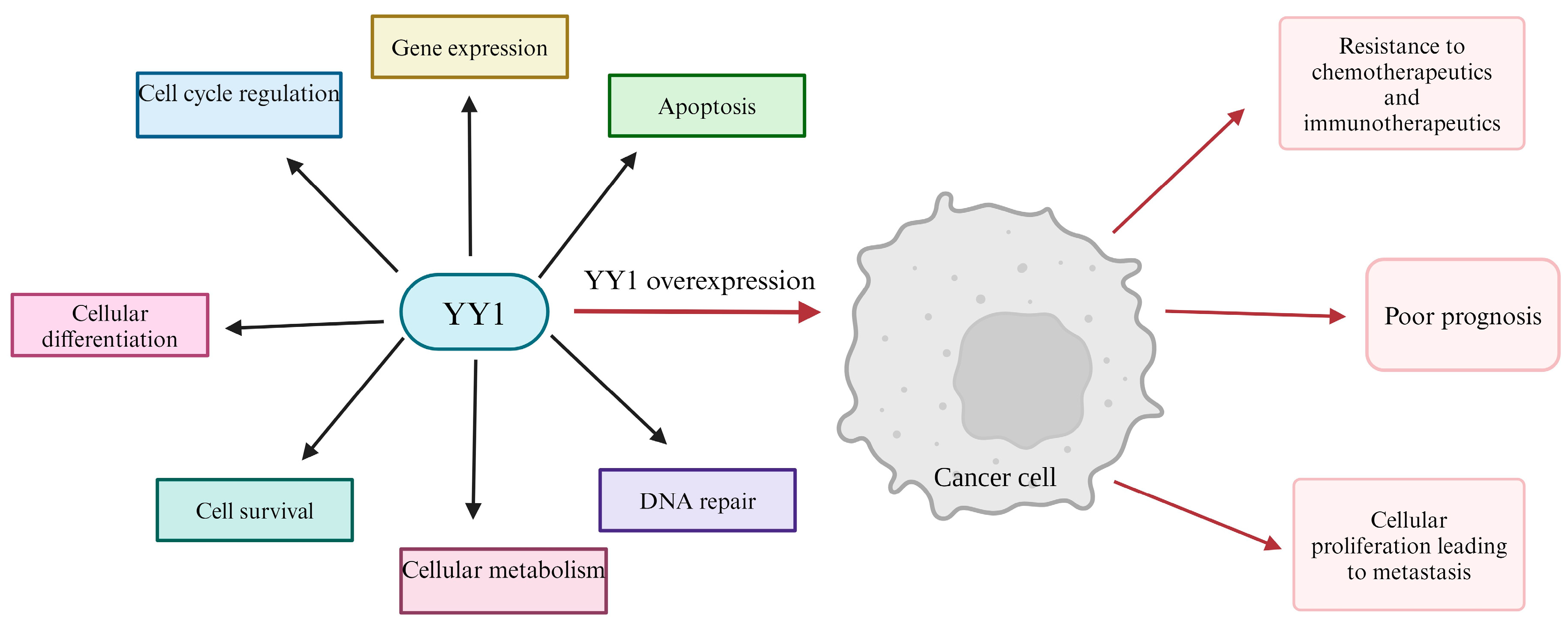
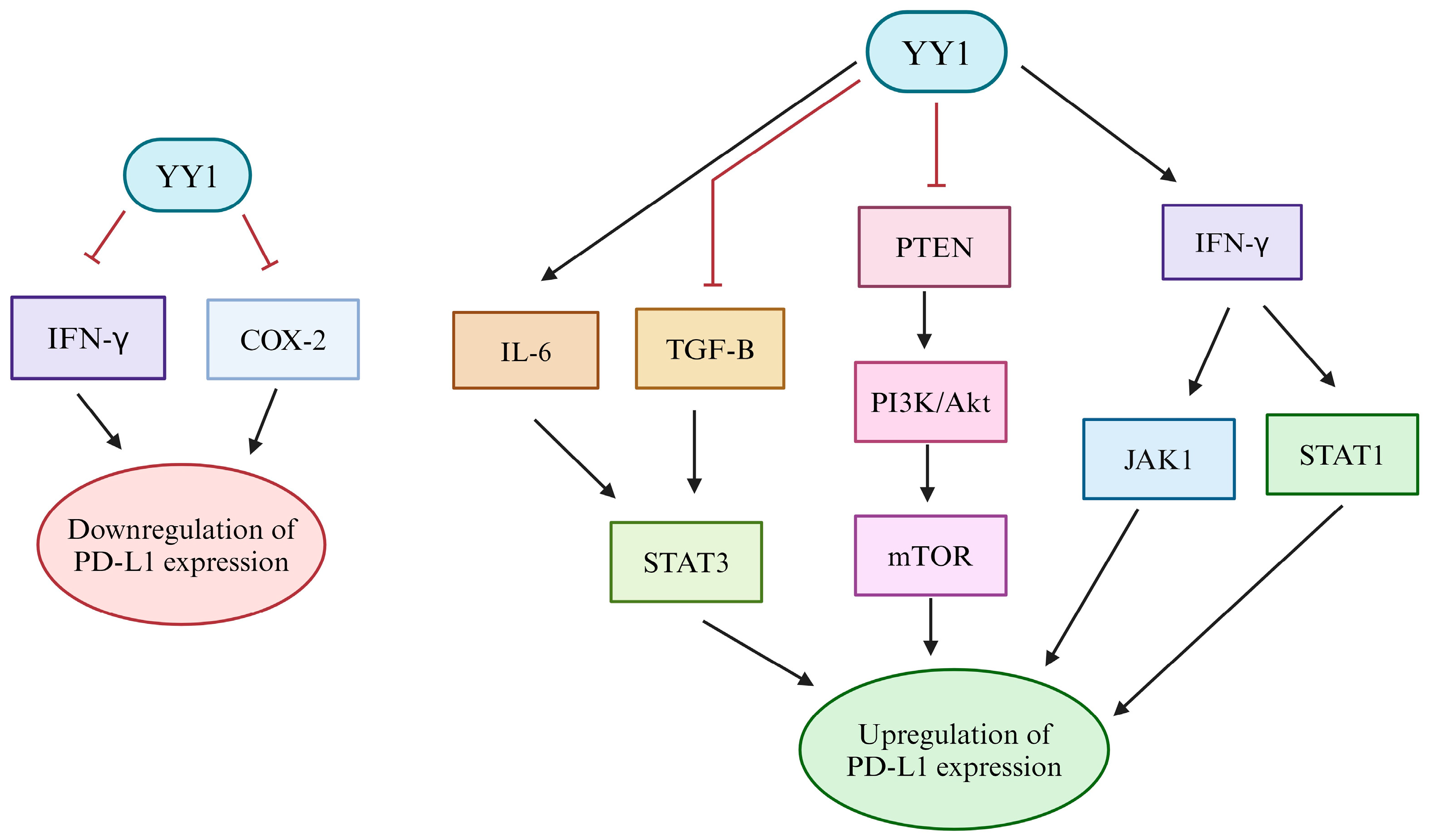
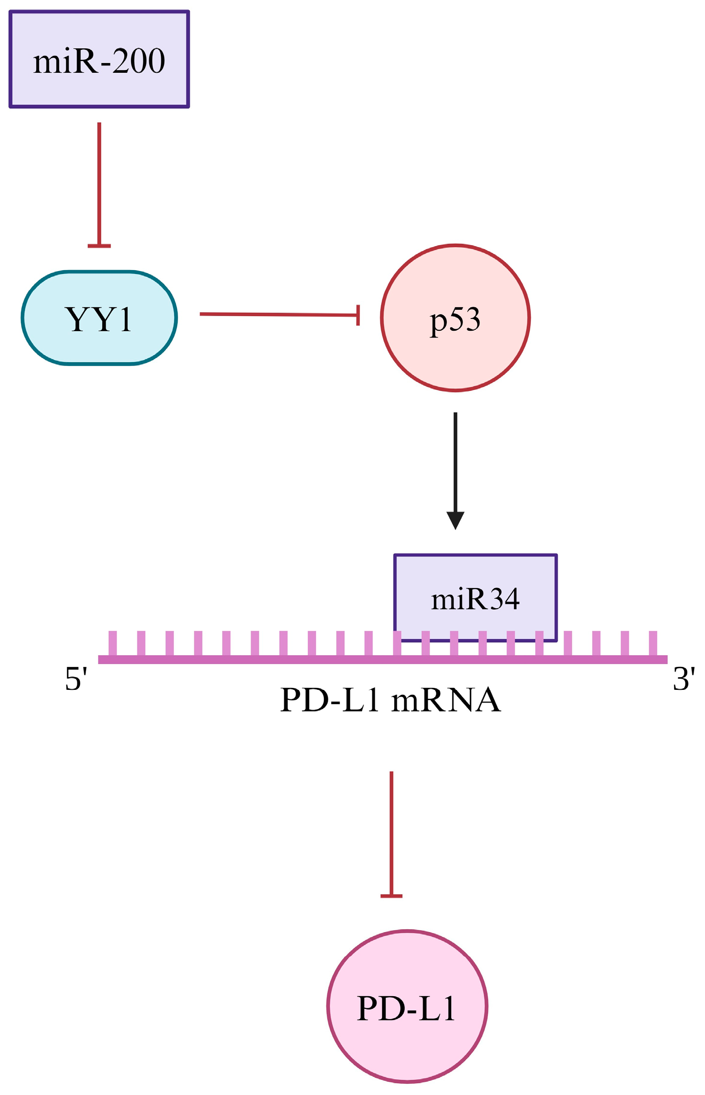
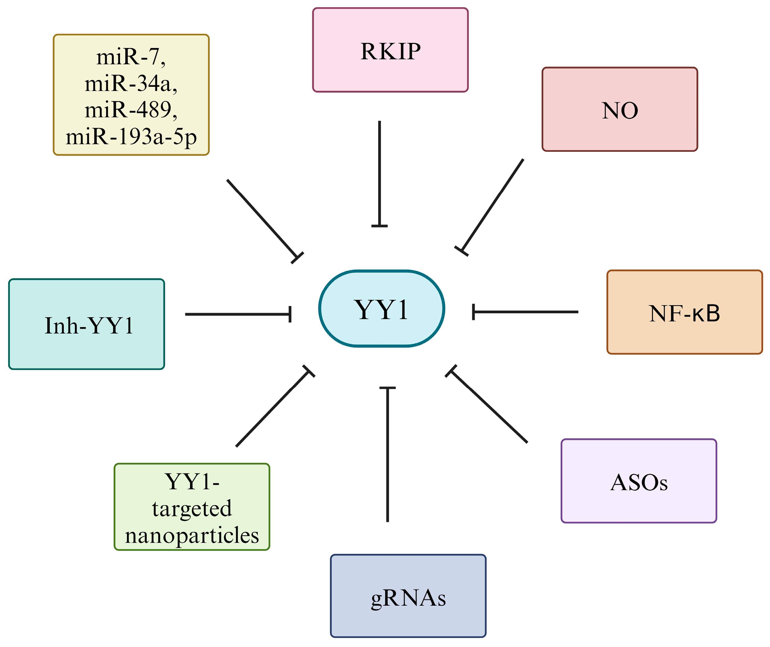
Table 1.
Summarized molecular regulation of PD-L1 expression by YY1.
| Type of Regulation | Modifier or Pathway Downstream of YY1 | Effect on PD-L1 | Reference |
|---|---|---|---|
| Transcriptional | YY1 direct binding to the PD-L1 promoter | Promotes Transcription | [132] |
| Repression of TGF-β | Decreased expression | [63,136] | |
| Extension of MYC-related transcription factor networks | Increased expression | [80,81,82] | |
| Inhibition of MYC function | Decreased expression | [35,140,141] | |
| PI3K/Akt/mTOR activation via PTEN inhibition | Increased expression | [34,145,146,147] | |
| Positive regulation of IL-6/STAT3 | Increased expression | [37,150,161,162,163] | |
| IFN-γ-induced activation of JAK1 and STAT1 | Increased expression | [34,153,154] | |
| Increased COX-2 expression | Increased expression | [155,156,157,158,159,160] | |
| Epigenetic | Increased HDAC6 | Upregulation | [184,186] |
| Interaction with EZH2 | Upregulation | [56,65,171] | |
| Repression of STAT family proteins | Upregulation | [141,142] | |
| Post-transcriptional | Inhibition of p53 and miR-34 | Upregulation | [187,188,189] |
| Downregulation of miR-200 | Downregulation | [190,192] | |
| Post-translational | Suppression of PARP/GSK3β | Degradation | [194,195,196] |
| Stimulation of IL-6/JAK1 | Stabilization | [37,197] |
COX-2: cyclooxygenase 2; EZH2: enhancer of zeste homolog; GSK3β: glycogen synthase kinase 3 beta; HDAC: histone deacetylase; IFN-γ: interferon gamma; IL-6: interleukin 6; JAK: janus kinase; mTOR: mechanistic Target of Rapamycin; miR-200: microRNA-200; miR-34: microRNA-34; MYC: myelocytomatosis oncogene; p53: tumor protein p53; PARP: poly (ADP-ribose) polymerase; PD-L1: programmed death ligand-1; PI3k/AKT: phosphatidylinositol 3-kinase/protein kinase B; PTEN: phosphatase and tensin homolog; STAT: signal transducer and activator of transcription; STAT1: signal transducer and activator of transcription 1; STAT3: signal transducer and activator of transcription 3; TGF-β: transforming growth factor-β; YY1: Yin Yang 1.
Disclaimer/Publisher’s Note: The statements, opinions and data contained in all publications are solely those of the individual author(s) and contributor(s) and not of MDPI and/or the editor(s). MDPI and/or the editor(s) disclaim responsibility for any injury to people or property resulting from any ideas, methods, instructions or products referred to in the content. |
© 2024 by the authors. Licensee MDPI, Basel, Switzerland. This article is an open access article distributed under the terms and conditions of the Creative Commons Attribution (CC BY) license (https://creativecommons.org/licenses/by/4.0/).
Share and Cite
MDPI and ACS Style
Dillen, A.; Bui, I.; Jung, M.; Agioti, S.; Zaravinos, A.; Bonavida, B. Regulation of PD-L1 Expression by YY1 in Cancer: Therapeutic Efficacy of Targeting YY1. Cancers 2024, 16, 1237. https://doi.org/10.3390/cancers16061237
AMA Style
Dillen A, Bui I, Jung M, Agioti S, Zaravinos A, Bonavida B. Regulation of PD-L1 Expression by YY1 in Cancer: Therapeutic Efficacy of Targeting YY1. Cancers. 2024; 16(6):1237. https://doi.org/10.3390/cancers16061237
Chicago/Turabian StyleDillen, Ana, Indy Bui, Megan Jung, Stephanie Agioti, Apostolos Zaravinos, and Benjamin Bonavida. 2024. "Regulation of PD-L1 Expression by YY1 in Cancer: Therapeutic Efficacy of Targeting YY1" Cancers 16, no. 6: 1237. https://doi.org/10.3390/cancers16061237
Note that from the first issue of 2016, this journal uses article numbers instead of page numbers. See further details here.