Next Issue
Volume 9, November
Previous Issue
Volume 9, September


Cancers, Volume 9, Issue 10 (October 2017) – 17 articles

Cover Story (view full-size image): 
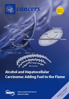
Alcohol consumption is associated with a large number of chronic diseases and deaths worldwide. About 10% of all cancer cases in men and 3% in women are estimated to be attributable to alcohol abuse, with a high impact on the liver where it seems to be responsible for 20–30% of hepatic cancers in both genders. Although it is known that most of the cytotoxic effects of alcohol depend on metabolites generating from its hepatic catabolism, new evidence emerged for the characterization of genetic, epigenetic and environmental factors that synergistically influence liver injury enabling tumor development. View the paper
  • Issues are regarded as officially published after their release is announced to the table of contents alert mailing list.
  • You may sign up for e-mail alerts to receive table of contents of newly released issues.
  • PDF is the official format for papers published in both, html and pdf forms. To view the papers in pdf format, click on the "PDF Full-text" link, and use the free Adobe Reader to open them.
Order results
Result details
Section
Select all
Export citation of selected articles as:
824 KiB  
Article
Association of Vitamin D3 Level with Breast Cancer Risk and Prognosis in African-American and Hispanic Women
by Yanyuan Wu, Marianna Sarkissyan, Sheilah Clayton, Rowan Chlebowski and Jaydutt V. Vadgama
Cancers 2017, 9(10), 144; https://doi.org/10.3390/cancers9100144 - 24 Oct 2017
Cited by 24 | Viewed by 7885
Abstract
Background: This study investigated the association of vitamin D3 levels with breast cancer risk and progression in African-Americans and Hispanics. Methods: A total of 237 African-American (Cases = 119, Control = 118) and 423 Hispanic women (Cases = 124, Control = [...] Read more.
Background: This study investigated the association of vitamin D3 levels with breast cancer risk and progression in African-Americans and Hispanics. Methods: A total of 237 African-American (Cases = 119, Control = 118) and 423 Hispanic women (Cases = 124, Control = 299) were recruited in the study. Blood samples were collected at the time of breast cancer screening and prior to cancer treatment for 4 weeks on average for the cases. The serum 25-hydroxyvitamin D (25(OH)D3) was measured at a Quest-Diagnostics facility. Results: The results showed that 69.2% of African-Americans and 37.8% of Hispanics had 25(OH)D3 levels below 20 ng/mL. The 25(OH)D3 level below 20 ng/mL was significantly associated with breast cancer in both African-Americans (OR = 2.5, 95% CI = 1.3–4.8) and Hispanics (OR = 1.9, 95% CI = 1.1–3.0). However, the predicted probabilities of breast cancer in African-Americans were significantly higher than in Hispanics (p < 0.001). The 25(OH)D3 below 20 ng/mL was significantly associated with triple negative breast cancer (TNBC) in African-Americans (OR = 5.4, p = 0.02, 95% CI = 1.4–15), but not in Hispanics in our cohort of participants. Levels of 25(OH)D3 below 26 ng/mL predicts a decrease in disease-free survival, but it was not an independent predictor. Conclusions: Our data shows an association between 25(OH)D3 levels and the risk of breast cancer. Further studies on the relationship between 25(OH)D3 level and breast cancer risk are warranted. Full article
Show Figures

Figure 1

753 KiB  
Review
Roles of microRNAs and RNA-Binding Proteins in the Regulation of Colorectal Cancer Stem Cells
by Junko Mukohyama, Yohei Shimono, Hironobu Minami, Yoshihiro Kakeji and Akira Suzuki
Cancers 2017, 9(10), 143; https://doi.org/10.3390/cancers9100143 - 24 Oct 2017
Cited by 28 | Viewed by 7199
Abstract
Colorectal cancer stem cells (CSCs) are responsible for the initiation, progression and metastasis of human colorectal cancers, and have been characterized by the expression of cell surface markers, such as CD44, CD133, CD166 and LGR5. MicroRNAs (miRNAs) are differentially expressed between CSCs and [...] Read more.
Colorectal cancer stem cells (CSCs) are responsible for the initiation, progression and metastasis of human colorectal cancers, and have been characterized by the expression of cell surface markers, such as CD44, CD133, CD166 and LGR5. MicroRNAs (miRNAs) are differentially expressed between CSCs and non-tumorigenic cancer cells, and play important roles in the maintenance and regulation of stem cell properties of CSCs. RNA binding proteins (RBPs) are emerging epigenetic regulators of various RNA processing events, such as splicing, localization, stabilization and translation, and can regulate various types of stem cells. In this review, we summarize current evidences on the roles of miRNA and RBPs in the regulation of colorectal CSCs. Understanding the epigenetic regulation of human colorectal CSCs will help to develop biomarkers for colorectal cancers and to identify targets for CSC-targeting therapies. Full article
Show Figures

Figure 1

2401 KiB  
Article
The Role of Platelet-Derived ADP and ATP in Promoting Pancreatic Cancer Cell Survival and Gemcitabine Resistance
by Omar Elaskalani, Marco Falasca, Niamh Moran, Michael C. Berndt and Pat Metharom
Cancers 2017, 9(10), 142; https://doi.org/10.3390/cancers9100142 - 24 Oct 2017
Cited by 35 | Viewed by 7629
Abstract
Platelets have been demonstrated to be vital in cancer epithelial-mesenchymal transition (EMT), an important step in metastasis. Markers of EMT are associated with chemotherapy resistance. However, the association between the development of chemoresistance, EMT, and the contribution of platelets to the process, is [...] Read more.
Platelets have been demonstrated to be vital in cancer epithelial-mesenchymal transition (EMT), an important step in metastasis. Markers of EMT are associated with chemotherapy resistance. However, the association between the development of chemoresistance, EMT, and the contribution of platelets to the process, is still unclear. Here we report that platelets regulate the expression of (1) human equilibrative nucleoside transporter 1 (hENT1) and (2) cytidine deaminase (CDD), markers of gemcitabine resistance in pancreatic cancer. Human ENT1 (hENT1) is known to enable cellular uptake of gemcitabine while CDD deactivates gemcitabine. Knockdown experiments demonstrate that Slug, a mesenchymal transcriptional factor known to be upregulated during EMT, regulates the expression of hENT1 and CDD. Furthermore, we demonstrate that platelet-derived ADP and ATP regulate Slug and CDD expression in pancreatic cancer cells. Finally, we demonstrate that pancreatic cancer cells express the purinergic receptor P2Y12, an ADP receptor found mainly on platelets. Thus ticagrelor, a P2Y12 inhibitor, was used to examine the potential therapeutic effect of an ADP receptor antagonist on cancer cells. Our data indicate that ticagrelor negated the survival signals initiated in cancer cells by platelet-derived ADP and ATP. In conclusion, our results demonstrate a novel role of platelets in modulating chemoresistance in pancreatic cancer. Moreover, we propose ADP/ATP receptors as additional potential drug targets for treatment of pancreatic cancer. Full article
Show Figures

Figure 1

520 KiB  
Perspective
An Exploration into the Origins and Pathogenesis of Anaplastic Large Cell Lymphoma, Anaplastic Lymphoma Kinase (ALK)-Positive
by Suzanne D. Turner
Cancers 2017, 9(10), 141; https://doi.org/10.3390/cancers9100141 - 24 Oct 2017
Cited by 4 | Viewed by 6224
Abstract
T-cell non-Hodgkin lymphoma is a heterogeneous disease ranging from malignancies arising from thymic T cells halted in development, through to mature, circulating peripheral T cells. The latter cases are diagnostically problematic with many entering the category of peripheral T-cell lymphoma, not otherwise specified [...] Read more.
T-cell non-Hodgkin lymphoma is a heterogeneous disease ranging from malignancies arising from thymic T cells halted in development, through to mature, circulating peripheral T cells. The latter cases are diagnostically problematic with many entering the category of peripheral T-cell lymphoma, not otherwise specified (PTCL, NOS). Anaplastic large cell lymphoma (ALCL) is one of the exceptions to this whereby aberrant expression of anaplastic lymphoma kinase (ALK) and the distinctive presence of cell surface CD30 places this entity in its own class. Besides the expression of a well-studied oncogenic translocation, ALCL, ALK+ may also have a unique pathogenesis with a thymic origin like T lymphoblastic lymphoma but a peripheral presentation akin to PTCL. This perspective discusses evidence towards the potential origin of ALCL, ALK+, and mechanisms that may give rise to its unique phenotype. Full article
(This article belongs to the Special Issue Targeting ALK in Cancer)
Show Figures

Figure 1

2019 KiB  
Review
Targeting PDK1 for Chemosensitization of Cancer Cells
by Aikaterini Emmanouilidi and Marco Falasca
Cancers 2017, 9(10), 140; https://doi.org/10.3390/cancers9100140 - 24 Oct 2017
Cited by 46 | Viewed by 11447
Abstract
Despite the rapid development in the field of oncology, cancer remains the second cause of mortality worldwide, with the number of new cases expected to more than double in the coming years. Chemotherapy is widely used to decelerate or stop tumour development in [...] Read more.
Despite the rapid development in the field of oncology, cancer remains the second cause of mortality worldwide, with the number of new cases expected to more than double in the coming years. Chemotherapy is widely used to decelerate or stop tumour development in combination with surgery or radiation therapy when appropriate, and in many cases this improves the symptomatology of the disease. Unfortunately though, chemotherapy is not applicable to all patients and even when it is, there are many cases where a successful initial treatment period is followed by chemotherapeutic drug resistance. This is caused by a number of reasons, ranging from the genetic background of the patient (innate resistance) to the formation of tumour-initiating cells (acquired resistance). In this review, we discuss the potential role of PDK1 in the development of chemoresistance in different types of malignancy, and the design and application of potent inhibitors which can promote chemosensitization. Full article
(This article belongs to the Special Issue PI3K/PDK1/Akt Pathways in Cancer)
Show Figures

Figure 1

4376 KiB  
Article
CD47-CAR-T Cells Effectively Kill Target Cancer Cells and Block Pancreatic Tumor Growth
by Vita Golubovskaya, Robert Berahovich, Hua Zhou, Shirley Xu, Hizkia Harto, Le Li, Cheng-Chi Chao, Mike Ming Mao and Lijun Wu
Cancers 2017, 9(10), 139; https://doi.org/10.3390/cancers9100139 - 21 Oct 2017
Cited by 66 | Viewed by 13711
Abstract
CD47 is a glycoprotein of the immunoglobulin superfamily that is often overexpressed in different types of hematological and solid cancer tumors and plays important role in blocking phagocytosis, increased tumor survival, metastasis and angiogenesis. In the present report, we designed CAR (chimeric antigen [...] Read more.
CD47 is a glycoprotein of the immunoglobulin superfamily that is often overexpressed in different types of hematological and solid cancer tumors and plays important role in blocking phagocytosis, increased tumor survival, metastasis and angiogenesis. In the present report, we designed CAR (chimeric antigen receptor)-T cells that bind CD47 antigen. We used ScFv (single chain variable fragment) from mouse CD47 antibody to generate CD47-CAR-T cells for targeting different cancer cell lines. CD47-CAR-T cells effectively killed ovarian, pancreatic and other cancer cells and produced high level of cytokines that correlated with expression of CD47 antigen. In addition, CD47-CAR-T cells significantly blocked BxPC3 pancreatic xenograft tumor growth after intratumoral injection into NSG mice. Moreover, we humanized mouse CD47 ScFv and showed that it effectively bound CD47 antigen. The humanized CD47-CAR-T cells also specifically killed ovarian, pancreatic, and cervical cancer cell lines and produced IL-2 that correlated with expression of CD47. Thus, CD47-CAR-T cells can be used as a novel cellular therapeutic agent for treating different types of cancer. Full article
(This article belongs to the Special Issue CAR-T Cell Therapy against Different Types of Cancer)
Show Figures

Figure 1

644 KiB  
Review
From Pathology to Precision Medicine in Anaplastic Large Cell Lymphoma Expressing Anaplastic Lymphoma Kinase (ALK+ ALCL)
by Michael T. Werner, Qian Zhang and Mariusz A. Wasik
Cancers 2017, 9(10), 138; https://doi.org/10.3390/cancers9100138 - 16 Oct 2017
Cited by 8 | Viewed by 5252
Abstract
Anaplastic large cell lymphoma expressing anaplastic lymphoma kinase (ALK+ ALCL) is a distinct subtype of non-Hodgkin lymphoma. In this review, we discuss the historical findings that led to its classification as a unique disease, despite its varied clinical presentation and histology. We discuss [...] Read more.
Anaplastic large cell lymphoma expressing anaplastic lymphoma kinase (ALK+ ALCL) is a distinct subtype of non-Hodgkin lymphoma. In this review, we discuss the historical findings that led to its classification as a unique disease, despite its varied clinical presentation and histology. We discuss the molecular mechanisms underlying ALK+ ALCL pathology and the questions that remain in the field. Finally, we visit how decades of ALK+ ALCL research has yielded more precise drugs that hold promise for the future. Full article
(This article belongs to the Special Issue Targeting ALK in Cancer)
Show Figures

Figure 1

2440 KiB  
Article
STAT3 but Not HIF-1α Is Important in Mediating Hypoxia-Induced Chemoresistance in MDA-MB-231, a Triple Negative Breast Cancer Cell Line
by Hoda Soleymani Abyaneh, Nidhi Gupta, Aneta Radziwon-Balicka, Paul Jurasz, John Seubert, Raymond Lai and Afsaneh Lavasanifar
Cancers 2017, 9(10), 137; https://doi.org/10.3390/cancers9100137 - 14 Oct 2017
Cited by 27 | Viewed by 7949
Abstract
Hypoxia-induced chemoresistance (HICR) is a well-recognized phenomenon, and in many experimental models, hypoxia inducible factor-1α (HIF-1α) is believed to be a key player. We aimed to better understand the mechanism underlying HICR in a triple negative breast cancer cell line, MDA-MB-231, with a [...] Read more.
Hypoxia-induced chemoresistance (HICR) is a well-recognized phenomenon, and in many experimental models, hypoxia inducible factor-1α (HIF-1α) is believed to be a key player. We aimed to better understand the mechanism underlying HICR in a triple negative breast cancer cell line, MDA-MB-231, with a focus on the role of HIF-1α. In this context, the effect of hypoxia on the sensitivity of MDA-MB-231 cells to cisplatin and their stem-like features was evaluated and the role of HIF-1α in both phenomena was assessed. Our results showed that hypoxia significantly increased MDA-MB-231 resistance to cisplatin. Correlating with this, intracellular uptake of cisplatin was significantly reduced under hypoxia. Furthermore, the stem-like features of MDA-MB-231 cells increased as evidenced by the significant increases in the expression of ATP-binding cassette (ABC) drug transporters, the proportion of CD44+/CD24 cells, clonogenic survival and cisplatin chemoresistance. Under hypoxia, both the protein level and DNA binding of HIF-1α was dramatically increased. Surprisingly, siRNA knockdown of HIF-1α did not result in an appreciable change to HICR. Instead, signal transducer and activator of transcription 3 (STAT3) activation was found to be important. STAT3 activation may confer HICR by upregulating ABC transporters, particularly ABCC2 and ABCC6. This study has demonstrated that, in MDA-MB-231 cells, STAT3 rather than HIF-1α is important in mediating HICR to cisplatin. Full article
(This article belongs to the Special Issue Drug Resistance in Cancers)
Show Figures

Figure 1

2412 KiB  
Article
Radiation-Induced Changes of microRNA Expression Profiles in Radiosensitive and Radioresistant Leukemia Cell Lines with Different Levels of Chromosome Abnormalities
by Daria Liamina, Wladimir Sibirnyj, Anna Khokhlova, Viacheslav Saenko, Eugenia Rastorgueva, Aleksandr Fomin and Yury Saenko
Cancers 2017, 9(10), 136; https://doi.org/10.3390/cancers9100136 - 13 Oct 2017
Cited by 6 | Viewed by 4860
Abstract
In our study, we estimate an effect from chromosome aberrations and genome mutations on changes in microRNA expression profiles in cancer cell lines demonstrating different radiosensitivity. Here, cell viability and microRNA spectrum have been estimated 1, 4, and 24 h after irradiation. MiSeq [...] Read more.
In our study, we estimate an effect from chromosome aberrations and genome mutations on changes in microRNA expression profiles in cancer cell lines demonstrating different radiosensitivity. Here, cell viability and microRNA spectrum have been estimated 1, 4, and 24 h after irradiation. MiSeq high-throughput sequencing system (Illumina, San Diego, CA, USA) is employed to perform microRNA spectrum estimation. In the K562 cell line, the number of expressed microRNAs in chromosomes demonstrates a more pronounced variation. An analysis of microRNA effects on signaling pathway activity demonstrates differences in post-transcriptional regulation of the expression of genes included into 40 signaling pathways. In the K562 cell line, microRNA dynamics analyzed for their dependence on chromosome localization show a wider scattering of microRNA expression values for a pair of chromosomes compared to the HL-60 cell line. An analysis of microRNAs expression in the K562 and HL-60 cell lines after irradiation has shown that chromosome abnormalities can affect microRNA expression changes. A study of radiation-induced changes of microRNA expression profiles in the K562 and HL-60 cell lines has revealed a dependence of microRNA expression changes on the number of chromosome aberrations and genome mutations. Full article
(This article belongs to the Special Issue Radiation-Induced Carcinogenesis)
Show Figures

Graphical abstract

731 KiB  
Review
Can Intensity-Modulated-Radiotherapy Reduce Toxicity in Head and Neck Squamous Cell Carcinoma?
by Julie Van der Veen and Sandra Nuyts
Cancers 2017, 9(10), 135; https://doi.org/10.3390/cancers9100135 - 06 Oct 2017
Cited by 45 | Viewed by 9012
Abstract
Intensity modulated radiotherapy (IMRT) is a modern radiotherapy technique that was implemented in the mid-1990s. It allows closer shaping of dose, to target volumes, thereby sparing organs at risk (OARs). Before the IMRT-era, two-dimensional radiotherapy (2DRT) and later three-dimensional conformal radiotherapy (3DCRT) were [...] Read more.
Intensity modulated radiotherapy (IMRT) is a modern radiotherapy technique that was implemented in the mid-1990s. It allows closer shaping of dose, to target volumes, thereby sparing organs at risk (OARs). Before the IMRT-era, two-dimensional radiotherapy (2DRT) and later three-dimensional conformal radiotherapy (3DCRT) were the techniques of choice, but this robust way of irradiating caused more normal tissue to receive a higher dose. Radiation of cancers in the head and neck region is complex because of close proximity to critical normal tissue and the large target volumes that need to be treated at high doses. IMRT offers an elegant solution compared with 3DCRT and surgery because it allows organ preservation and improved function preservation. In this manuscript, we review the rationales for IMRT, with an emphasis on toxicity outcomes compared with 3DCRT. We performed a review of the literature and looked at the most important randomised controlled trials comparing IMRT with 3DCRT. We conclude that IMRT is safe in regard to disease outcome, and that it allows better sparing of normal tissue, thereby causing less toxicity, resulting in a smaller impact on quality of life compared with conventional radiotherapy in the treatment of head and neck cancer. Full article
(This article belongs to the Special Issue Intensity Modulated Radiation Therapy)
Show Figures

Figure 1

817 KiB  
Review
The Epithelial-to-Mesenchymal Transition in Breast Cancer: Focus on Basal-Like Carcinomas
by Monica Fedele, Laura Cerchia and Gennaro Chiappetta
Cancers 2017, 9(10), 134; https://doi.org/10.3390/cancers9100134 - 30 Sep 2017
Cited by 98 | Viewed by 11213
Abstract
Breast cancer is a heterogeneous disease that is characterized by a high grade of cell plasticity arising from the contribution of a diverse range of factors. When combined, these factors allow a cancer cell to transition from an epithelial to a mesenchymal state [...] Read more.
Breast cancer is a heterogeneous disease that is characterized by a high grade of cell plasticity arising from the contribution of a diverse range of factors. When combined, these factors allow a cancer cell to transition from an epithelial to a mesenchymal state through a process of dedifferentiation that confers stem-like features, including chemoresistance, as well as the capacity to migrate and invade. Understanding the complex events that lead to the acquisition of a mesenchymal phenotype will therefore help to design new therapies against metastatic breast cancer. Here, we recapitulate the main endogenous molecular signals involved in this process, and their cross-talk with paracrine factors. These signals and cross-talk include the extracellular matrix; the secretome of cancer-associated fibroblasts, macrophages, cancer stem cells, and cancer cells; and exosomes with their cargo of miRNAs. Finally, we highlight some of the more promising therapeutic perspectives based on counteracting the epithelial-to-mesenchymal transition in breast cancer cells. Full article
(This article belongs to the Special Issue The Epithelial-to-Mesenchymal Transition (EMT) in Cancer)
Show Figures

Figure 1

849 KiB  
Review
Platelet Integrins in Tumor Metastasis: Do They Represent a Therapeutic Target?
by Marion Lavergne, Emily Janus-Bell, Mathieu Schaff, Christian Gachet and Pierre H. Mangin
Cancers 2017, 9(10), 133; https://doi.org/10.3390/cancers9100133 - 28 Sep 2017
Cited by 61 | Viewed by 7935
Abstract
Platelets are small anucleated cell fragments that ensure the arrest of bleeding after a vessel wall injury. They are also involved in non-hemostatic function such as development, immunity, inflammation, and in the hematogeneous phase of metastasis. While the role of platelets in tumor [...] Read more.
Platelets are small anucleated cell fragments that ensure the arrest of bleeding after a vessel wall injury. They are also involved in non-hemostatic function such as development, immunity, inflammation, and in the hematogeneous phase of metastasis. While the role of platelets in tumor metastasis has been recognized for 60 years, the molecular mechanism underlying this process remains largely unclear. Platelets physically and functionally interact with various tumor cells through surface receptors including integrins. Platelets express five integrins at their surface, namely α2β1, α5β1, α6β1, αvβ3, and αIIbβ3, which bind preferentially to collagen, fibronectin, laminin, vitronectin, and fibrinogen, respectively. The main role of platelet integrins is to ensure platelet adhesion and aggregation at sites of vascular injury. Two of these, α6β1 and αIIbβ3, were proposed to participate in platelet–tumor cell interaction and in tumor metastasis. It has also been reported that pharmacological agents targeting both integrins efficiently reduce experimental metastasis, suggesting that platelet integrins may represent new anti-metastatic targets. This review focuses on the role of platelet integrins in tumor metastasis and discusses whether these receptors may represent new potential targets for novel anti-metastatic approaches. Full article
(This article belongs to the Special Issue Integrins in Cancer)
Show Figures

Figure 1

417 KiB  
Review
Regulation of mTOR, Metabolic Fitness, and Effector Functions by Cytokines in Natural Killer Cells
by Sébastien Viel, Laurie Besson, Marie Marotel, Thierry Walzer and Antoine Marçais
Cancers 2017, 9(10), 132; https://doi.org/10.3390/cancers9100132 - 28 Sep 2017
Cited by 22 | Viewed by 7533
Abstract
The control of cellular metabolism is now recognized as key to regulate functional properties of immune effectors such as T or Natural Killer (NK) cells. During persistent infections or in the tumor microenvironment, multiple metabolic changes have been highlighted in T cells that [...] Read more.
The control of cellular metabolism is now recognized as key to regulate functional properties of immune effectors such as T or Natural Killer (NK) cells. During persistent infections or in the tumor microenvironment, multiple metabolic changes have been highlighted in T cells that contribute to their dysfunctional state or exhaustion. NK cells may also undergo major phenotypic and functional modifications when infiltrating tumors that could be linked to metabolic alterations. The mammalian target of rapamycin (mTOR) kinase is a central regulator of cellular metabolism. mTOR integrates various extrinsic growth or immune signals and modulates metabolic pathways to fulfill cellular bioenergetics needs. mTOR also regulates transcription and translation thereby adapting cellular pathways to the growth or activation signals that are received. Here, we review the role and regulation of mTOR in NK cells, with a special focus on cytokines that target mTOR such as IL-15 and TGF-β. We also discuss how NK cell metabolic activity could be enhanced or modulated to improve their effector anti-tumor functions in clinical settings. Full article
(This article belongs to the Special Issue mTOR Pathway in Cancer)
Show Figures

Figure 1

1141 KiB  
Review
The Emerging Role of Polo-Like Kinase 1 in Epithelial-Mesenchymal Transition and Tumor Metastasis
by Zheng Fu and Donghua Wen
Cancers 2017, 9(10), 131; https://doi.org/10.3390/cancers9100131 - 27 Sep 2017
Cited by 45 | Viewed by 8084
Abstract
Polo-like kinase 1 (PLK1) is a serine/threonine kinase that plays a key role in the regulation of the cell cycle. PLK1 is overexpressed in a variety of human tumors, and its expression level often correlates with increased cellular proliferation and poor prognosis in [...] Read more.
Polo-like kinase 1 (PLK1) is a serine/threonine kinase that plays a key role in the regulation of the cell cycle. PLK1 is overexpressed in a variety of human tumors, and its expression level often correlates with increased cellular proliferation and poor prognosis in cancer patients. It has been suggested that PLK1 controls cancer development through multiple mechanisms that include canonical regulation of mitosis and cytokinesis, modulation of DNA replication, and cell survival. However, emerging evidence suggests novel and previously unanticipated roles for PLK1 during tumor development. In this review, we will summarize the recent advancements in our understanding of the oncogenic functions of PLK1, with a focus on its role in epithelial-mesenchymal transition and tumor invasion. We will further discuss the therapeutic potential of these functions. Full article
(This article belongs to the Special Issue The Epithelial-to-Mesenchymal Transition (EMT) in Cancer)
Show Figures

Figure 1

1416 KiB  
Review
Alcohol and Hepatocellular Carcinoma: Adding Fuel to the Flame
by Pierluigi Ramadori, Francisco Javier Cubero, Christian Liedtke, Christian Trautwein and Yulia A. Nevzorova
Cancers 2017, 9(10), 130; https://doi.org/10.3390/cancers9100130 - 25 Sep 2017
Cited by 45 | Viewed by 9787
Abstract
Primary tumors of the liver represent the fifth most common type of cancer in the world and the third leading cause of cancer-related death. Case-control studies from different countries report that chronic ethanol consumption is associated with an approximately 2-fold increased odds ratio [...] Read more.
Primary tumors of the liver represent the fifth most common type of cancer in the world and the third leading cause of cancer-related death. Case-control studies from different countries report that chronic ethanol consumption is associated with an approximately 2-fold increased odds ratio for hepatocellular carcinoma (HCC). Despite the substantial epidemiologic data in humans demonstrating that chronic alcohol consumption is a major risk factor for HCC development, the pathways causing alcohol-induced liver cancer are poorly understood. In this overview, we summarize the epidemiological evidence for the association between alcohol and liver cancer, review the genetic, oncogenic, and epigenetic factors that drive HCC development synergistically with ethanol intake and discuss the essential molecular and metabolic pathways involved in alcohol-induced liver tumorigenesis. Full article
(This article belongs to the Special Issue Alcohol and Cancer)
Show Figures

Figure 1

470 KiB  
Case Report
Alcohol Misuse Link to POEMS Syndrome in a Patient
by John Neary, Susan E. Goodwin, Lawrence B. Cohen and Manuela G. Neuman
Cancers 2017, 9(10), 129; https://doi.org/10.3390/cancers9100129 - 23 Sep 2017
Cited by 3 | Viewed by 4802
Abstract
Previously called Crow–Fukase syndrome, POEMS syndrome is characterized by poly-neuropathy, osteo-sclerotic myeloma, organomegaly, endocrinopathy, monoclonal plasma cell disorder, and skin changes. Extremely elevated levels of serum vascular endothelial growth factor (VEGF) are characteristic of the syndrome. Chronic hepatitis B (HBV) and C (HCV) [...] Read more.
Previously called Crow–Fukase syndrome, POEMS syndrome is characterized by poly-neuropathy, osteo-sclerotic myeloma, organomegaly, endocrinopathy, monoclonal plasma cell disorder, and skin changes. Extremely elevated levels of serum vascular endothelial growth factor (VEGF) are characteristic of the syndrome. Chronic hepatitis B (HBV) and C (HCV) infections can also be present in POEMS. The pathogenesis of the syndrome is not well understood. The link between chronic alcohol consumption and this malignant condition has not been reported until now. In addition, no previous study has evaluated the influence of cytokine and chemokines or viruses in the severity and evolution of POEMS. Objectives: (1) to describe a heavy-alcohol user, who was diagnosed with POEMS; (2) to demonstrate the utility of quantitative measurement of serum levels of VEGF in the diagnosis of POEMS and the monitoring of therapeutic interventions; (3) to demonstrate that overproduction of pro-inflammatory cytokines is a characteristic of POEMS. Methods: We describe a case of a POEMS patient presenting HCV and who is a heavy drinker; we compare the serum levels of cytokines and chemokines between the POEMS patient with 80 patients with HCV, 12 healthy controls, and 80 individuals with alcoholic liver disease (ALD). We quantified (ELISA pg/mL) the levels of VEGF, Interferon gamma (IFN-γ), Tumor Necrosis Factor alpha (TNF-α), Regulated-upon-Activation Normal-T-cell-Expressed and presumably-Secreted (RANTES), and Nuclear Factor kappa-B (NFκB). Results: In POEMS patients, VEGF levels were elevated versus control or other diseases, TNFα levels were higher versus control, but lower when compared with HCV or ALD patients. VEGF levels in POEMS patients decreased with therapeutic intervention. Conclusions: Chronic alcohol misuse can be a strong risk factor to rare malignancies such as POEMS syndrome. Extreme elevation of VEGF levels is diagnostic for POEMS syndrome, and should be followed to assess response to therapy. In addition, other comorbidities should be considered individually to ensure personalized therapeutic intervention. Full article
(This article belongs to the Special Issue Alcohol and Cancer)
Show Figures

Figure 1

1356 KiB  
Article
Investigating the Interaction of Cyclic RGD Peptidomimetics with αVβ6 Integrin by Biochemical and Molecular Docking Studies
by Monica Civera, Daniela Arosio, Francesca Bonato, Leonardo Manzoni, Luca Pignataro, Simone Zanella, Cesare Gennari, Umberto Piarulli and Laura Belvisi
Cancers 2017, 9(10), 128; https://doi.org/10.3390/cancers9100128 - 21 Sep 2017
Cited by 17 | Viewed by 6469
Abstract
The interaction of a small library of cyclic RGD (Arg-Gly-Asp) peptidomimetics with αVβ6 integrin has been investigated by means of competitive solid phase binding assays to the isolated receptor and docking calculations in the crystal structure of the αV [...] Read more.
The interaction of a small library of cyclic RGD (Arg-Gly-Asp) peptidomimetics with αVβ6 integrin has been investigated by means of competitive solid phase binding assays to the isolated receptor and docking calculations in the crystal structure of the αVβ6 binding site. To this aim, a rigid receptor-flexible ligand docking protocol has been set up and then applied to predict the binding mode of the cyclic RGD peptidomimetics to αVβ6 integrin. Although the RGD interaction with αVβ6 recapitulates the RGD binding mode observed in αVβ3, differences between the integrin binding pockets can strongly affect the ligand binding ability. In general, the peptidomimetics exhibited IC50 values for integrin αVβ6 (i.e., the concentration of compound required for 50% inhibition of biotinylated fibronectin binding to isolated αVβ6 integrin) in the nanomolar range (77–345 nM), about 10–100 times higher than those for the related αVβ3 receptor, with a single notable ligand displaying a low nanomolar IC50 value (2.3 nM). Insights from the properties of the binding pocket combined with the analysis of the docking poses provided a rationale for ligand recognition and selectivity. Full article
(This article belongs to the Special Issue Integrins in Cancer)
Show Figures

Figure 1

Previous Issue
Next Issue
Back to TopTop