Next Issue
Volume 9, August
Previous Issue
Volume 9, June


Cancers, Volume 9, Issue 7 (July 2017) – 25 articles

Cover Story (view full-size image): 
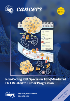
Metastasis represents a critical step in tumor progression that is responsible for more than 90% of cancer-induced mortality. During cancer progression of epithelial tumors, cells become significantly more invasive after completing EMT as they display newly-acquired mesenchymal features. TGF-β is a powerful regulator of EMT through both transcriptional and post-transcriptional mechanisms, and recent advances underline the critical roles of non-coding RNAs in these processes. Our review discusses the current literature including the most recent reports emphasizing the regulatory functions of non-coding RNA in TGF-β-mediated EMT, provides original experimental evidence, and advocates in general for a broader approach in the quest of new regulatory RNAs. View this paper
  • Issues are regarded as officially published after their release is announced to the table of contents alert mailing list.
  • You may sign up for e-mail alerts to receive table of contents of newly released issues.
  • PDF is the official format for papers published in both, html and pdf forms. To view the papers in pdf format, click on the "PDF Full-text" link, and use the free Adobe Reader to open them.
Order results
Result details
Section
Select all
Export citation of selected articles as:
2000 KiB  
Review
Targeting Platelets for the Treatment of Cancer
by Omar Elaskalani, Michael C. Berndt, Marco Falasca and Pat Metharom
Cancers 2017, 9(7), 94; https://doi.org/10.3390/cancers9070094 - 22 Jul 2017
Cited by 53 | Viewed by 9467
Abstract
The majority of cancer-associated mortality results from the ability of tumour cells to metastasise leading to multifunctional organ failure and death. Disseminated tumour cells in the blood circulation are faced with major challenges such as rheological shear stresses and cell-mediated cytotoxicity mediated by [...] Read more.
The majority of cancer-associated mortality results from the ability of tumour cells to metastasise leading to multifunctional organ failure and death. Disseminated tumour cells in the blood circulation are faced with major challenges such as rheological shear stresses and cell-mediated cytotoxicity mediated by natural killer cells. Nevertheless, circulating tumour cells with metastatic ability appear equipped to exploit host cells to aid their survival. Despite the long interest in targeting tumour-associated host cells such as platelets for cancer treatment, the clinical benefit of this strategy is still under question. In this review, we provide a summary of the latest mechanistic and clinical evidence to evaluate the validity of targeting platelets in cancer. Full article
Show Figures

Figure 1

478 KiB  
Review
Seed-in-Soil: Pancreatic Cancer Influenced by Tumor Microenvironment
by Huey-Jen Lin and Jiayuh Lin
Cancers 2017, 9(7), 93; https://doi.org/10.3390/cancers9070093 - 21 Jul 2017
Cited by 24 | Viewed by 5217
Abstract
Pancreatic ductal adenocarcinoma is a fatal malignancy with a five-year survival rate lower than 7%, and most patients dying within six months of diagnosis. The factors that contribute to the aggressiveness of the disease include, but are not limited to: late diagnosis, prompt [...] Read more.
Pancreatic ductal adenocarcinoma is a fatal malignancy with a five-year survival rate lower than 7%, and most patients dying within six months of diagnosis. The factors that contribute to the aggressiveness of the disease include, but are not limited to: late diagnosis, prompt metastasis to adjacent vital organs, poor response, and resistance to anticancer treatments. This malignancy is uniquely associated with desmoplastic stroma that accounts for 80% of tumor mass. Understanding the biology of stroma can aid the discovery of innovative strategies for eradicating this lethal cancer in the future. This review highlights the critical components in the stroma and how they interact with the cancer cells to convey the devastating tumor progression. Full article
(This article belongs to the Special Issue Cancer-Associated Fibroblasts)
Show Figures

Figure 1

3408 KiB  
Review
Regional Delivery of Chimeric Antigen Receptor (CAR) T-Cells for Cancer Therapy
by Praveen Sridhar and Fabio Petrocca
Cancers 2017, 9(7), 92; https://doi.org/10.3390/cancers9070092 - 18 Jul 2017
Cited by 67 | Viewed by 9546
Abstract
Chimeric Antigen Receptor (CAR) T-cells are T-cells with recombinant receptors targeted to tumor antigens. CAR-T cell therapy has emerged as a mode of immunotherapy and is now being extensively explored in hematologic cancer. In contrast, CAR-T cell use in solid tumors has been [...] Read more.
Chimeric Antigen Receptor (CAR) T-cells are T-cells with recombinant receptors targeted to tumor antigens. CAR-T cell therapy has emerged as a mode of immunotherapy and is now being extensively explored in hematologic cancer. In contrast, CAR-T cell use in solid tumors has been hampered by multiple obstacles. Several approaches have been taken to circumvent these obstacles, including the regional delivery of CAR-T cells. Regional CAR-T cell delivery can theoretically compensate for poor T-cell trafficking and tumor antigen specificity while avoiding systemic toxicity associated with intravenous delivery. We reviewed completed clinical trials for the treatment of glioblastoma and metastatic colorectal cancer and examined the data in these studies for safety, efficacy, and potential advantages that regional delivery may confer over systemic delivery. Our appraisal of the available literature revealed that regional delivery of CAR-T cells in both glioblastoma and hepatic colorectal metastases was generally well tolerated and efficacious in select instances. We propose that the regional delivery of CAR-T cells is an area of potential growth in the solid tumor immunotherapy, and look towards future clinical trials in head and neck cancer, mesothelioma, and peritoneal carcinomatosis as the use of this technique expands. Full article
(This article belongs to the Special Issue CAR-T Cell Therapy against Different Types of Cancer)
Show Figures

Figure 1

1537 KiB  
Review
Complex DNA Damage: A Route to Radiation-Induced Genomic Instability and Carcinogenesis
by Ifigeneia V. Mavragani, Zacharenia Nikitaki, Maria P. Souli, Asef Aziz, Somaira Nowsheen, Khaled Aziz, Emmy Rogakou and Alexandros G. Georgakilas
Cancers 2017, 9(7), 91; https://doi.org/10.3390/cancers9070091 - 18 Jul 2017
Cited by 119 | Viewed by 10955
Abstract
Cellular effects of ionizing radiation (IR) are of great variety and level, but they are mainly damaging since radiation can perturb all important components of the cell, from the membrane to the nucleus, due to alteration of different biological molecules ranging from lipids [...] Read more.
Cellular effects of ionizing radiation (IR) are of great variety and level, but they are mainly damaging since radiation can perturb all important components of the cell, from the membrane to the nucleus, due to alteration of different biological molecules ranging from lipids to proteins or DNA. Regarding DNA damage, which is the main focus of this review, as well as its repair, all current knowledge indicates that IR-induced DNA damage is always more complex than the corresponding endogenous damage resulting from endogenous oxidative stress. Specifically, it is expected that IR will create clusters of damage comprised of a diversity of DNA lesions like double strand breaks (DSBs), single strand breaks (SSBs) and base lesions within a short DNA region of up to 15–20 bp. Recent data from our groups and others support two main notions, that these damaged clusters are: (1) repair resistant, increasing genomic instability (GI) and malignant transformation and (2) can be considered as persistent “danger” signals promoting chronic inflammation and immune response, causing detrimental effects to the organism (like radiation toxicity). Last but not least, the paradigm shift for the role of radiation-induced systemic effects is also incorporated in this picture of IR-effects and consequences of complex DNA damage induction and its erroneous repair. Full article
(This article belongs to the Special Issue Radiation-Induced Carcinogenesis)
Show Figures

Figure 1

885 KiB  
Review
SIRT3: Oncogene and Tumor Suppressor in Cancer
by Margalida Torrens-Mas, Jordi Oliver, Pilar Roca and Jorge Sastre-Serra
Cancers 2017, 9(7), 90; https://doi.org/10.3390/cancers9070090 - 12 Jul 2017
Cited by 92 | Viewed by 8513
Abstract
Sirtuin 3 (SIRT3), the major deacetylase in mitochondria, plays a crucial role in modulating oxygen reactive species (ROS) and limiting the oxidative damage in cellular components. SIRT3 targets different enzymes which regulate mitochondrial metabolism and participate in ROS detoxification, such as the complexes [...] Read more.
Sirtuin 3 (SIRT3), the major deacetylase in mitochondria, plays a crucial role in modulating oxygen reactive species (ROS) and limiting the oxidative damage in cellular components. SIRT3 targets different enzymes which regulate mitochondrial metabolism and participate in ROS detoxification, such as the complexes of the respiratory chain, the isocitrate dehydrogenase, or the manganese superoxide dismutase. Thus, SIRT3 activity is essential in maintaining mitochondria homeostasis and has recently received great attention, as it is considered a fidelity protein for mitochondrial function. In some types of cancer, SIRT3 functions as a tumoral promoter, since it keeps ROS levels under a certain threshold compatible with cell viability and proliferation. On the contrary, other studies describe SIRT3 as a tumoral suppressor, as SIRT3 could trigger cell death under stress conditions. Thus, SIRT3 could have a dual role in cancer. In this regard, modulation of SIRT3 activity could be a new target to develop more personalized therapies against cancer. Full article
Show Figures

Figure 1

908 KiB  
Article
The Role of Radiation Induced Injury on Lung Cancer
by Stephanie Puukila, Christopher Thome, Antone L. Brooks, Gayle Woloschak and Douglas R. Boreham
Cancers 2017, 9(7), 89; https://doi.org/10.3390/cancers9070089 - 12 Jul 2017
Cited by 14 | Viewed by 4556
Abstract
This manuscript evaluates the role of cell killing, tissue disorganization, and tissue damage on the induction of lung cancer following low dose rate radiation exposures from internally deposited radioactive materials. Beagle dogs were exposed by inhalation to 90Y, 91Y, 144Ce, [...] Read more.
This manuscript evaluates the role of cell killing, tissue disorganization, and tissue damage on the induction of lung cancer following low dose rate radiation exposures from internally deposited radioactive materials. Beagle dogs were exposed by inhalation to 90Y, 91Y, 144Ce, or 90Sr in fused clay particles. Dogs lived out their life span with complete pathology conducted at the time of death. The radiation dose per cell turnover was characterized and related to the cause of death for each animal. Large doses per cell turnover resulted in acute death from lung damage with extensive cell killing, tissue disorganization, chronic inflammatory disease, fibrosis, and pneumonitis. Dogs with lower doses per cell turnover developed a very high frequency of lung cancer. As the dose per cell turnover was further decreased, no marked tissue damage and no significant change in either life span or lung cancer frequency was observed. Radiation induced tissue damage and chronic inflammatory disease results in high cancer frequencies in the lung. At doses where a high frequency of chromosome damage and mutations would be predicted to occur there was no decrease in life span or increase in lung cancer. Such research suggests that cell killing and tissue damage and the physiological responses to that damage are important mechanisms in radiation induced lung cancer. Full article
(This article belongs to the Special Issue Radiation-Induced Carcinogenesis)
Show Figures

Figure 1

217 KiB  
Opinion
Cost Saving Opportunities in NSCLC Therapy by Optimized Diagnostics
by Ilija Nenadić, Janine Staber, Susanne Dreier, Guus Simons, Verena Schildgen, Michael Brockmann and Oliver Schildgen
Cancers 2017, 9(7), 88; https://doi.org/10.3390/cancers9070088 - 11 Jul 2017
Cited by 6 | Viewed by 3828
Abstract
With an incidence of 68 new cases per 100,000 people per year, an estimated total number of up to 350,000 new non-small-cell lung cancer (NSCLC) cases are diagnosed each year in the European Union. Up to 10% of NSCLC patients are eligible for [...] Read more.
With an incidence of 68 new cases per 100,000 people per year, an estimated total number of up to 350,000 new non-small-cell lung cancer (NSCLC) cases are diagnosed each year in the European Union. Up to 10% of NSCLC patients are eligible for therapy with novel ALK (anaplastic lymphoma kinase) inhibitors, as they have been diagnosed with a mutation in the gene coding for ALK. The ALK inhibitor therapy costs add up to approx. 9,000 € per patient per month, with treatment durations of up to one year. Recent studies have shown that up to 10% of ALK cases are misdiagnosed by nearly 40% of pathologic investigations. The current state-of-the-art ALK diagnostic procedure comprises a Fluorescent in situ Hybridization (FISH) assay accompanied by ALK inhibitor therapy (Crizotinib). The therapy success ranges between a full therapy failure and the complete remission of the tumor (i.e., healing), but the biomedical and systemic reasons for this range remain unknown so far. It appears that the variety of different ALK mutations and variants contributes to the discrepancy in therapy results. Although the major known fusion partner for ALK in NSCLC is the Echinoderm microtubule-associated protein-like 4 (EML4), of which a minimum of 15 variants have been described, an additional 20 further ALK fusion variants with other genes are known, of which three have already been found in NSCLC. We hypothesize that the wide variety of known (and unknown) ALK mutations is associated with a variable therapy success, thus rendering current companion diagnostic procedures (FISH) and therapy (Crizotinib) only partly applicable in ALK-related NSCLC treatment. In cell culture, differing sensitivity to Crizotinib has been shown for some fusion variants, but it is as yet unknown which of them are really biologically active in cancer patients, and how the respective variants affect the response to Crizotinib treatment. Moreover, it has been demonstrated that translocated ALK genes can also be observed in healthy tissues and are not compulsorily associated with tumors. Therefore, it is important to keep in mind that even for the known variants of ALK fusion genes, the biological function is not known for all variants, and that no information is available on the homogeneity of ALK fusion variants within a single tumor. These facts, in concert with data for ALK mutation prevalence and therapy outcomes of a German cohort of NSCLC patients, support the hypothesis that, by using novel companion diagnostic tools in combination with therapy outcome predictions, massive cost savings could be possible in European Health Care systems without a loss of patient care. Full article
(This article belongs to the Special Issue Targeting ALK in Cancer)
1438 KiB  
Review
Role and Therapeutic Targeting of the HGF/MET Pathway in Glioblastoma
by Nichola Cruickshanks, Ying Zhang, Fang Yuan, Mary Pahuski, Myron Gibert and Roger Abounader
Cancers 2017, 9(7), 87; https://doi.org/10.3390/cancers9070087 - 11 Jul 2017
Cited by 48 | Viewed by 7078
Abstract
Glioblastoma (GBM) is a lethal brain tumor with dismal prognosis. Current therapeutic options, consisting of surgery, chemotherapy and radiation, have only served to marginally increase patient survival. Receptor tyrosine kinases (RTKs) are dysregulated in approximately 90% of GBM; attributed to this, research has [...] Read more.
Glioblastoma (GBM) is a lethal brain tumor with dismal prognosis. Current therapeutic options, consisting of surgery, chemotherapy and radiation, have only served to marginally increase patient survival. Receptor tyrosine kinases (RTKs) are dysregulated in approximately 90% of GBM; attributed to this, research has focused on inhibiting RTKs as a novel and effective therapy for GBM. Overexpression of RTK mesenchymal epithelial transition (MET), and its ligand, hepatocyte growth factor (HGF), in GBM highlights a promising new therapeutic target. This review will discuss the role of MET in cell cycle regulation, cell proliferation, evasion of apoptosis, cell migration and invasion, angiogenesis and therapeutic resistance in GBM. It will also discuss the modes of deregulation of HGF/MET and their regulation by microRNAs. As the HGF/MET pathway is a vital regulator of multiple pro-survival pathways, efforts and strategies for its exploitation for GBM therapy are also described. Full article
(This article belongs to the Special Issue Hepatocyte Growth Factor Pathway in Cancer)
Show Figures

Figure 1

1081 KiB  
Review
Phosphoinositide 3-Kinase-Dependent Signalling Pathways in Cutaneous Squamous Cell Carcinomas
by Joanna M. Janus, Ryan F. L. O’Shaughnessy, Catherine A. Harwood and Tania Maffucci
Cancers 2017, 9(7), 86; https://doi.org/10.3390/cancers9070086 - 11 Jul 2017
Cited by 32 | Viewed by 6283
Abstract
Cutaneous squamous cell carcinoma (cSCC) derives from keratinocytes in the epidermis and accounts for 15–20% of all cutaneous malignancies. Although it is usually curable by surgery, 5% of these tumours metastasise leading to poor prognosis mostly because of a lack of therapies and [...] Read more.
Cutaneous squamous cell carcinoma (cSCC) derives from keratinocytes in the epidermis and accounts for 15–20% of all cutaneous malignancies. Although it is usually curable by surgery, 5% of these tumours metastasise leading to poor prognosis mostly because of a lack of therapies and validated biomarkers. As the incidence rate is rising worldwide it has become increasingly important to better understand the mechanisms involved in cSCC development and progression in order to develop therapeutic strategies. Here we discuss some of the evidence indicating that activation of phosphoinositide 3-kinases (PI3Ks)-dependent signalling pathways (in particular the PI3Ks targets Akt and mTOR) has a key role in cSCC. We further discuss available data suggesting that inhibition of these pathways can be beneficial to counteract the disease. With the growing number of different inhibitors currently available, it would be important to further investigate the specific contribution of distinct components of the PI3Ks/Akt/mTOR pathways in order to identify the most promising molecular targets and the best strategy to inhibit cSCC. Full article
(This article belongs to the Special Issue PI3K/PDK1/Akt Pathways in Cancer)
Show Figures

Figure 1

1045 KiB  
Review
The Role of miRNAs in Angiogenesis, Invasion and Metabolism and Their Therapeutic Implications in Gliomas
by Sasha Beyer, Jessica Fleming, Wei Meng, Rajbir Singh, S. Jaharul Haque and Arnab Chakravarti
Cancers 2017, 9(7), 85; https://doi.org/10.3390/cancers9070085 - 10 Jul 2017
Cited by 48 | Viewed by 7027
Abstract
MicroRNAs (miRNAs) are small, non-coding, endogenous RNA molecules that function in gene silencing by post-transcriptional regulation of gene expression. The dysregulation of miRNA plays a pivotal role in cancer tumorigenesis, including the development and progression of gliomas. Their small size, stability and ability [...] Read more.
MicroRNAs (miRNAs) are small, non-coding, endogenous RNA molecules that function in gene silencing by post-transcriptional regulation of gene expression. The dysregulation of miRNA plays a pivotal role in cancer tumorigenesis, including the development and progression of gliomas. Their small size, stability and ability to target multiple oncogenes have simultaneously distinguished miRNAs as attractive candidates for biomarkers and novel therapeutic targets for glioma patients. In this review, we summarize the most frequently cited miRNAs known to contribute to gliomagenesis and progression by regulating the defining hallmarks of gliomas, including angiogenesis, invasion, and cell metabolism. We also discuss their promising potential as prognostic and predictive biomarkers and novel therapeutic targets, in addition to the challenges that must be overcome before their translation from bench to bedside. Full article
Show Figures

Figure 1

603 KiB  
Review
Integrin αvβ3 Signaling in Tumor-Induced Bone Disease
by Kristin A. Kwakwa and Julie A. Sterling
Cancers 2017, 9(7), 84; https://doi.org/10.3390/cancers9070084 - 08 Jul 2017
Cited by 44 | Viewed by 8720
Abstract
Tumor-induced bone disease is common among patients with advanced solid cancers, especially those with breast, prostate, and lung malignancies. The tendency of these cancers to metastasize to bone and induce bone destruction is, in part, due to alterations in integrin expression and signaling. [...] Read more.
Tumor-induced bone disease is common among patients with advanced solid cancers, especially those with breast, prostate, and lung malignancies. The tendency of these cancers to metastasize to bone and induce bone destruction is, in part, due to alterations in integrin expression and signaling. Substantial evidence from preclinical studies shows that increased expression of integrin αvβ3 in tumor cells promotes the metastatic and bone-invasive phenotype. Integrin αvβ3 mediates cell adhesion to several extracellular matrix proteins in the bone microenvironment which is necessary for tumor cell colonization as well as the transmission of mechanical signals for tumor progression. This review will discuss the αvβ3 integrin receptor in the context of tumor-induced bone disease. Specifically, the focus will be the role of αvβ3 in modulating cancer metastasis to bone and tumor cell response to the bone microenvironment, including downstream signaling pathways that contribute to tumor-induced osteolysis. A better understanding of integrin dysregulation in cancer is critical to developing new therapeutics for the prevention and treatment of bone metastases. Full article
(This article belongs to the Special Issue Integrins in Cancer)
Show Figures

Figure 1

897 KiB  
Review
Potential of Integrin Inhibitors for Treating Ovarian Cancer: A Literature Review
by Masaki Kobayashi, Kenjiro Sawada and Tadashi Kimura
Cancers 2017, 9(7), 83; https://doi.org/10.3390/cancers9070083 - 08 Jul 2017
Cited by 38 | Viewed by 7212
Abstract
Epithelial ovarian cancer is a fatal disease, with a cure rate of only 30%. Several recent studies have targeted integrins for cancer treatment. Preclinical studies have shown the effectiveness of several integrin inhibitors for blocking cancer progression, especially by blocking angiogenesis. Because the [...] Read more.
Epithelial ovarian cancer is a fatal disease, with a cure rate of only 30%. Several recent studies have targeted integrins for cancer treatment. Preclinical studies have shown the effectiveness of several integrin inhibitors for blocking cancer progression, especially by blocking angiogenesis. Because the initial critical step in ovarian cancer metastasis is the attachment of cancer cells to the peritoneum or omentum and because clinical trials have provided positive results for anti-angiogenic therapy, therapies targeting integrins may be the most feasible approach for treating cancer. This review summarizes the current understanding of integrin biology in ovarian cancer metastasis and various therapeutic approaches involving integrin inhibitors. However, no integrin inhibitor has shown favorable results thus far. However, conjugates of cytotoxic agents with the triplet sequence arginine-glycine-aspartate (RGD) peptides targeting α5β1-, αvβ3-, and αvβ6-integrins may be promising integrin-targeting therapies for further clinical investigation. Full article
(This article belongs to the Special Issue Integrins in Cancer)
Show Figures

Figure 1

820 KiB  
Review
Inside the Cell: Integrins as New Governors of Nuclear Alterations?
by Elena Madrazo, Andrea Cordero Conde and Javier Redondo-Muñoz
Cancers 2017, 9(7), 82; https://doi.org/10.3390/cancers9070082 - 06 Jul 2017
Cited by 23 | Viewed by 6500
Abstract
Cancer cell migration is a complex process that requires coordinated structural changes and signals in multiple cellular compartments. The nucleus is the biggest and stiffest organelle of the cell and might alter its physical properties to allow cancer cell movement. Integrins are transmembrane [...] Read more.
Cancer cell migration is a complex process that requires coordinated structural changes and signals in multiple cellular compartments. The nucleus is the biggest and stiffest organelle of the cell and might alter its physical properties to allow cancer cell movement. Integrins are transmembrane receptors that mediate cell-cell and cell-extracellular matrix interactions, which regulate numerous intracellular signals and biological functions under physiological conditions. Moreover, integrins orchestrate changes in tumor cells and their microenvironment that lead to cancer growth, survival and invasiveness. Most of the research efforts have focused on targeting integrin-mediated adhesion and signaling. Recent exciting data suggest the crucial role of integrins in controlling internal cellular structures and nuclear alterations during cancer cell migration. Here we review the emerging role of integrins in nuclear biology. We highlight increasing evidence that integrins are critical for changes in multiple nuclear components, the positioning of the nucleus and its mechanical properties during cancer cell migration. Finally, we discuss how integrins are integral proteins linking the plasma membrane and the nucleus, and how they control cell migration to enable cancer invasion and infiltration. The functional connections between these cell receptors and the nucleus will serve to define new attractive therapeutic targets. Full article
(This article belongs to the Special Issue Integrins in Cancer)
Show Figures

Figure 1

897 KiB  
Review
The Role of the Core Non-Homologous End Joining Factors in Carcinogenesis and Cancer
by Brock J. Sishc and Anthony J. Davis
Cancers 2017, 9(7), 81; https://doi.org/10.3390/cancers9070081 - 06 Jul 2017
Cited by 107 | Viewed by 12189
Abstract
DNA double-strand breaks (DSBs) are deleterious DNA lesions that if left unrepaired or are misrepaired, potentially result in chromosomal aberrations, known drivers of carcinogenesis. Pathways that direct the repair of DSBs are traditionally believed to be guardians of the genome as they protect [...] Read more.
DNA double-strand breaks (DSBs) are deleterious DNA lesions that if left unrepaired or are misrepaired, potentially result in chromosomal aberrations, known drivers of carcinogenesis. Pathways that direct the repair of DSBs are traditionally believed to be guardians of the genome as they protect cells from genomic instability. The prominent DSB repair pathway in human cells is the non-homologous end joining (NHEJ) pathway, which mediates template-independent re-ligation of the broken DNA molecule and is active in all phases of the cell cycle. Its role as a guardian of the genome is supported by the fact that defects in NHEJ lead to increased sensitivity to agents that induce DSBs and an increased frequency of chromosomal aberrations. Conversely, evidence from tumors and tumor cell lines has emerged that NHEJ also promotes chromosomal aberrations and genomic instability, particularly in cells that have a defect in one of the other DSB repair pathways. Collectively, the data present a conundrum: how can a single pathway both suppress and promote carcinogenesis? In this review, we will examine NHEJ’s role as both a guardian and a disruptor of the genome and explain how underlying genetic context not only dictates whether NHEJ promotes or suppresses carcinogenesis, but also how it alters the response of tumors to conventional therapeutics. Full article
(This article belongs to the Special Issue DNA Repair Pathways in Cancer)
Show Figures

Figure 1

145 KiB  
Erratum
Erratum: Tanaka, T. et al. Cimetidine and Clobenpropit Attenuate Inflammation-Associated Colorectal Carcinogenesis in Male ICR Mice. Cancers, 2016, 8, 25
by Takuji Tanaka, Takahiro Kochi, Yohei Shirakami, Takayuki Mori, Ayumi Niwa, Naoki Watanabe, Hisataka Moriwaki and Masahito Shimizu
Cancers 2017, 9(7), 80; https://doi.org/10.3390/cancers9070080 - 05 Jul 2017
Cited by 1 | Viewed by 2886
Abstract
The authors wish to make the following correction to their paper [1]. The author Ayumi Kurata’s name should be changed to Ayumi Niwa. [...] Full article
1296 KiB  
Review
Roles of Integrin α6β4 Glycosylation in Cancer
by Yoshinobu Kariya, Yukiko Kariya and Jianguo Gu
Cancers 2017, 9(7), 79; https://doi.org/10.3390/cancers9070079 - 05 Jul 2017
Cited by 30 | Viewed by 7150
Abstract
Malignant transformation is accompanied with aberrant glycosylation of proteins. Such changes in glycan structure also occur in the integrins, which are a large family of cell surface receptors for the extracellular matrix and play key roles in tumor progression. There is now increasing [...] Read more.
Malignant transformation is accompanied with aberrant glycosylation of proteins. Such changes in glycan structure also occur in the integrins, which are a large family of cell surface receptors for the extracellular matrix and play key roles in tumor progression. There is now increasing evidence that glycosylation of integrins affects cellular signaling and interaction with the extracellular matrix, receptor tyrosine kinases, and galectins, thereby regulating cell adhesion, motility, growth, and survival. Integrin α6β4 is a receptor for laminin-332 and the increased expression level is correlated with malignant progression and poor survival in various types of cancers. Recent studies have revealed that integrin α6β4 plays central roles in tumorigenesis and the metastatic process. In this review, we summarize our current understanding of the molecular mechanisms of tumor progression driven by integrin α6β4 and also discuss the modification of glycans on integrin β4 subunit to address the important roles of glycan in integrin-mediated tumor progression. Full article
(This article belongs to the Special Issue Integrins in Cancer)
Show Figures

Figure 1

2144 KiB  
Review
Can Integrin Agonists Have Cards to Play against Cancer? A Literature Survey of Small Molecules Integrin Activators
by Alessandra Tolomelli, Paola Galletti, Monica Baiula and Daria Giacomini
Cancers 2017, 9(7), 78; https://doi.org/10.3390/cancers9070078 - 05 Jul 2017
Cited by 32 | Viewed by 8644
Abstract
The ability of integrins to activate and integrate intracellular communication illustrates the potential of these receptors to serve as functional distribution hubs in a bi-directional signal transfer outside-in and inside-out of the cells. Tight regulation of the integrin signaling is paramount for normal [...] Read more.
The ability of integrins to activate and integrate intracellular communication illustrates the potential of these receptors to serve as functional distribution hubs in a bi-directional signal transfer outside-in and inside-out of the cells. Tight regulation of the integrin signaling is paramount for normal physiological functions such as migration, proliferation, and differentiation, and misregulated integrin activity could be associated with several pathological conditions. Because of the important roles of integrins and their ligands in biological development, immune responses, leukocyte traffic, haemostasis, and cancer, their potential as therapeutic tools is now widely recognized. Nowadays extensive efforts have been made to discover and develop small molecule ligands as integrin antagonists, whereas less attention has been payed to agonists. In recent years, it has been recognized that integrin agonists could open up novel opportunities for therapeutics, which gain benefits to increase rather than decrease integrin-dependent adhesion and transductional events. For instance, a significant factor in chemo-resistance in melanoma is a loss of integrin-mediated adhesion; in this case, stimulation of integrin signaling by agonists significantly improved the response to chemotherapy. In this review, we overview results about small molecules which revealed an activating action on some integrins, especially those involved in cancer, and examine from a medicinal chemistry point of view, their structure and behavior. Full article
(This article belongs to the Special Issue Integrins in Cancer)
Show Figures

Figure 1

5955 KiB  
Review
Epithelial-to-Pericyte Transition in Cancer
by Jianrong Lu and Anitha K. Shenoy
Cancers 2017, 9(7), 77; https://doi.org/10.3390/cancers9070077 - 04 Jul 2017
Cited by 15 | Viewed by 5400
Abstract
During epithelial-to-mesenchymal transition (EMT), cells lose epithelial characteristics and acquire mesenchymal properties. These two processes are genetically separable and governed by distinct transcriptional programs, rendering the EMT outputs highly heterogeneous. Our recent study shows that the mesenchymal products generated by EMT often express [...] Read more.
During epithelial-to-mesenchymal transition (EMT), cells lose epithelial characteristics and acquire mesenchymal properties. These two processes are genetically separable and governed by distinct transcriptional programs, rendering the EMT outputs highly heterogeneous. Our recent study shows that the mesenchymal products generated by EMT often express multiple pericyte markers, associate with and stabilize blood vessels to fuel tumor growth, thus phenotypically and functionally resembling pericytes. Therefore, some EMT events represent epithelial-to-pericyte transition (EPT). The serum response factor (SRF) plays key roles in both EMT and differentiation of pericytes, and may inherently confer the pericyte attributes on EMT cancer cells. By impacting their intratumoral location and cell surface receptor expression, EPT may enable cancer cells to receive and respond to angiocrine factors produced by the vascular niche, and develop therapy resistance. Full article
(This article belongs to the Special Issue The Epithelial-to-Mesenchymal Transition (EMT) in Cancer)
Show Figures

Figure 1

947 KiB  
Review
Epithelial-to-Mesenchymal Transition in the Pathogenesis and Therapy of Head and Neck Cancer
by Julia Thierauf, Johannes Adrian Veit and Jochen Hess
Cancers 2017, 9(7), 76; https://doi.org/10.3390/cancers9070076 - 03 Jul 2017
Cited by 32 | Viewed by 6299
Abstract
Head and neck cancer (HNC) is one of the most prevalent human malignancies worldwide, with a high morbidity and mortality. Implementation of interdisciplinary treatment modalities has improved the quality of life, but only minor changes in overall survival have been achieved over the [...] Read more.
Head and neck cancer (HNC) is one of the most prevalent human malignancies worldwide, with a high morbidity and mortality. Implementation of interdisciplinary treatment modalities has improved the quality of life, but only minor changes in overall survival have been achieved over the past decades. Main causes for treatment failure are an aggressive and invasive tumor growth in combination with a high degree of intrinsic or acquired treatment resistance. A subset of tumor cells gain these properties during malignant progression by reactivating a complex program of epithelia-to-mesenchymal transition (EMT), which is integral in embryonic development, wound healing, and stem cell behavior. EMT is mediated by a core set of key transcription factors, which are under the control of a large range of developmental signals and extracellular cues. Unraveling molecular principles that drive EMT provides new concepts to better understand tumor cell plasticity and response to established as well as new treatment modalities, and has the potential to identify new drug targets for a more effective, less toxic, and individualized therapy of HNC patients. Here, we review the most recent findings on the clinical relevance of a mesenchymal-like phenotype for HNC patients, including more rare cases of mucosal melanoma and adenoid cystic carcinoma. Full article
(This article belongs to the Special Issue The Epithelial-to-Mesenchymal Transition (EMT) in Cancer)
Show Figures

Figure 1

1326 KiB  
Review
Pleiotropic Roles of Non-Coding RNAs in TGF-β-Mediated Epithelial-Mesenchymal Transition and Their Functions in Tumor Progression
by Simon Grelet, Ariel McShane, Renaud Geslain and Philip H. Howe
Cancers 2017, 9(7), 75; https://doi.org/10.3390/cancers9070075 - 01 Jul 2017
Cited by 20 | Viewed by 5970
Abstract
Epithelial-mesenchymal transition (EMT) is a spatially- and temporally-regulated process involved in physiological and pathological transformations, such as embryonic development and tumor progression. While the role of TGF-β as an EMT-inducer has been extensively documented, the molecular mechanisms regulating this transition and their implications [...] Read more.
Epithelial-mesenchymal transition (EMT) is a spatially- and temporally-regulated process involved in physiological and pathological transformations, such as embryonic development and tumor progression. While the role of TGF-β as an EMT-inducer has been extensively documented, the molecular mechanisms regulating this transition and their implications in tumor metastasis are still subjects of intensive debates and investigations. TGF-β regulates EMT through both transcriptional and post-transcriptional mechanisms, and recent advances underline the critical roles of non-coding RNAs in these processes. Although microRNAs and lncRNAs have been clearly identified as effectors of TGF-β-mediated EMT, the contributions of other atypical non-coding RNA species, such as piRNAs, snRNAs, snoRNAs, circRNAs, and even housekeeping tRNAs, have only been suggested and remain largely elusive. This review discusses the current literature including the most recent reports emphasizing the regulatory functions of non-coding RNA in TGF-β-mediated EMT, provides original experimental evidence, and advocates in general for a broader approach in the quest of new regulatory RNAs. Full article
(This article belongs to the Special Issue The Epithelial-to-Mesenchymal Transition (EMT) in Cancer)
Show Figures

Figure 1

1944 KiB  
Review
Specific Depletion of Leukemic Stem Cells: Can MicroRNAs Make the Difference?
by Tania Martiáñez Canales, David C. De Leeuw, Eline Vermue, Gert J. Ossenkoppele and Linda Smit
Cancers 2017, 9(7), 74; https://doi.org/10.3390/cancers9070074 - 30 Jun 2017
Cited by 6 | Viewed by 8432
Abstract
For over 40 years the standard treatment for acute myeloid leukemia (AML) patients has been a combination of chemotherapy consisting of cytarabine and an anthracycline such as daunorubicin. This standard treatment results in complete remission (CR) in the majority of AML patients. However, [...] Read more.
For over 40 years the standard treatment for acute myeloid leukemia (AML) patients has been a combination of chemotherapy consisting of cytarabine and an anthracycline such as daunorubicin. This standard treatment results in complete remission (CR) in the majority of AML patients. However, despite these high CR rates, only 30–40% (<60 years) and 10–20% (>60 years) of patients survive five years after diagnosis. The main cause of this treatment failure is insufficient eradication of a subpopulation of chemotherapy resistant leukemic cells with stem cell-like properties, often referred to as “leukemic stem cells” (LSCs). LSCs co-exist in the bone marrow of the AML patient with residual healthy hematopoietic stem cells (HSCs), which are needed to reconstitute the blood after therapy. To prevent relapse, development of additional therapies targeting LSCs, while sparing HSCs, is essential. As LSCs are rare, heterogeneous and dynamic, these cells are extremely difficult to target by single gene therapies. Modulation of miRNAs and consequently the regulation of hundreds of their targets may be the key to successful elimination of resistant LSCs, either by inducing apoptosis or by sensitizing them for chemotherapy. To address the need for specific targeting of LSCs, miRNA expression patterns in highly enriched HSCs, LSCs, and leukemic progenitors, all derived from the same patients’ bone marrow, were determined and differentially expressed miRNAs between LSCs and HSCs and between LSCs and leukemic progenitors were identified. Several of these miRNAs are specifically expressed in LSCs and/or HSCs and associated with AML prognosis and treatment outcome. In this review, we will focus on the expression and function of miRNAs expressed in normal and leukemic stem cells that are residing within the AML bone marrow. Moreover, we will review their possible prospective as specific targets for anti-LSC therapy. Full article
Show Figures

Figure 1

1110 KiB  
Article
Quality-of-Life (QOL) during Screening for Phase 1 Trial Studies in Patients with Advanced Solid Tumors and Its Impact on Risk for Serious Adverse Events
by Sidra Anwar, Wei Tan, Chi-Chen Hong, Sonal Admane, Askia Dozier, Francine Siedlecki, Amy Whitworth, Ann Marie DiRaddo, Dawn DePaolo, Sandra M. Jacob, Wen Wee Ma, Austin Miller, Alex A. Adjei and Grace K. Dy
Cancers 2017, 9(7), 73; https://doi.org/10.3390/cancers9070073 - 26 Jun 2017
Cited by 7 | Viewed by 4671
Abstract
Background: Serious adverse events (SAEs) and subject replacements occur frequently in phase 1 oncology clinical trials. Whether baseline quality-of-life (QOL) or social support can predict risk for SAEs or subject replacement among these patients is not known. Methods: Between 2011–2013, 92 [...] Read more.
Background: Serious adverse events (SAEs) and subject replacements occur frequently in phase 1 oncology clinical trials. Whether baseline quality-of-life (QOL) or social support can predict risk for SAEs or subject replacement among these patients is not known. Methods: Between 2011–2013, 92 patients undergoing screening for enrollment into one of 22 phase 1 solid tumor clinical trials at Roswell Park Cancer Institute were included in this study. QOL Questionnaires (EORTC QLQ-C30 and FACT-G), Medical Outcomes Study Social Support Survey (MOSSSS), Charlson comorbidity scores (CCS) and Royal Marsden scores (RMS) were obtained at baseline. Frequency of dose limiting toxicities (DLTs), subject replacement and SAEs that occurred within the first 4 cycles of treatment were recorded. Fisher’s exact test and Mann-Whitney-Wilcoxon test were used to study the association between categorical and continuous variables, respectively. A linear transformation was used to standardize QOL scores. p-value ≤ 0.05 was considered statistically significant. Results: Baseline QOL, MOSSSS, CCS and RMS were not associated with subject replacement nor DLTs. Baseline EORTC QLQ-C30 scores were significantly lower among patients who encountered SAEs within the first 4 cycles (p = 0.04). Conclusions: Lower (worse) EORTC QLQ-C30 score at baseline is associated with SAE occurrence during phase 1 oncology trials. Full article
(This article belongs to the Special Issue CAR-T Cell Therapy against Different Types of Cancer)
Show Figures

Figure 1

981 KiB  
Review
Epigenetic Regulation of the Epithelial to Mesenchymal Transition in Lung Cancer
by Joëlle Roche, Robert M. Gemmill and Harry A. Drabkin
Cancers 2017, 9(7), 72; https://doi.org/10.3390/cancers9070072 - 24 Jun 2017
Cited by 22 | Viewed by 8372
Abstract
Lung cancer is the leading cause of cancer deaths worldwide. It is an aggressive and devastating cancer because of metastasis triggered by enhanced migration and invasion, and resistance to cytotoxic chemotherapy. The epithelial to mesenchymal transition (EMT) is a fundamental developmental process that [...] Read more.
Lung cancer is the leading cause of cancer deaths worldwide. It is an aggressive and devastating cancer because of metastasis triggered by enhanced migration and invasion, and resistance to cytotoxic chemotherapy. The epithelial to mesenchymal transition (EMT) is a fundamental developmental process that is reactivated in wound healing and a variety of diseases including cancer where it promotes migration/invasion and metastasis, resistance to treatment, and generation and maintenance of cancer stem cells. The induction of EMT is associated with reprogramming of the epigenome. This review focuses on major mechanisms of epigenetic regulation mainly in lung cancer with recent data on EZH2 (enhancer of zeste 2 polycomb repressive complex 2 subunit ), the catalytic subunit of the PRC2 (Polycomb Group PcG), that behaves as an oncogene in lung cancer associated with gene repression, non-coding RNAs and the epitranscriptome. Full article
(This article belongs to the Special Issue The Epithelial-to-Mesenchymal Transition (EMT) in Cancer)
Show Figures

Figure 1

223 KiB  
Review
Exosomal MicroRNAs in Breast Cancer towards Diagnostic and Therapeutic Applications
by Lorenzo F. Sempere, Jessica Keto and Muller Fabbri
Cancers 2017, 9(7), 71; https://doi.org/10.3390/cancers9070071 - 24 Jun 2017
Cited by 77 | Viewed by 8155
Abstract
Soon after the discovery of microRNAs over 15 years ago, a myriad of research groups around the world sought to develop clinical applications in breast cancer for these short, noncoding, regulatory RNAs. While little of this knowledge has translated into the clinic, the [...] Read more.
Soon after the discovery of microRNAs over 15 years ago, a myriad of research groups around the world sought to develop clinical applications in breast cancer for these short, noncoding, regulatory RNAs. While little of this knowledge has translated into the clinic, the recent research explosion on cell-to-cell communication via exosomes and other extracellular vesicles has rekindled interest in microRNA-based clinical applications. microRNAs appear to be a preferential and important cargo of exosomes in mediating biological effects in recipient cells. This review highlights recent studies on the biology of exosomal microRNAs (exo-miRNAs) and discusses potential clinical applications. From a diagnostic perspective, circulating exo-miRNAs may represent breast cancer cell content and/or tumor microenvironmental reactions to cancer cell growth. Thus, serum or plasma analysis of exo-miRNAs could be useful for early disease detection or for monitoring treatment response and disease progression. From a therapeutic perspective, exo-miRNAs derived from different cell types have been implicated in supporting or restraining tumor growth, conferring drug resistance, and preparing the metastatic niche. Strategies to interfere with the loading or delivery of tumor-promoting exo-miRNAs or to replenish tumor-suppressive miRNAs via exosomal delivery are under investigation. These recent studies provide new hope and opportunities, but study design limitations and technical challenges will need to be overcome before seriously considering clinical application of exo-miRNAs. Full article
1714 KiB  
Review
Phenotypic Plasticity and Cell Fate Decisions in Cancer: Insights from Dynamical Systems Theory
by Dongya Jia, Mohit Kumar Jolly, Prakash Kulkarni and Herbert Levine
Cancers 2017, 9(7), 70; https://doi.org/10.3390/cancers9070070 - 22 Jun 2017
Cited by 64 | Viewed by 10232
Abstract
Waddington’s epigenetic landscape, a famous metaphor in developmental biology, depicts how a stem cell progresses from an undifferentiated phenotype to a differentiated one. The concept of “landscape” in the context of dynamical systems theory represents a high-dimensional space, in which each cell phenotype [...] Read more.
Waddington’s epigenetic landscape, a famous metaphor in developmental biology, depicts how a stem cell progresses from an undifferentiated phenotype to a differentiated one. The concept of “landscape” in the context of dynamical systems theory represents a high-dimensional space, in which each cell phenotype is considered as an “attractor” that is determined by interactions between multiple molecular players, and is buffered against environmental fluctuations. In addition, biological noise is thought to play an important role during these cell-fate decisions and in fact controls transitions between different phenotypes. Here, we discuss the phenotypic transitions in cancer from a dynamical systems perspective and invoke the concept of “cancer attractors”—hidden stable states of the underlying regulatory network that are not occupied by normal cells. Phenotypic transitions in cancer occur at varying levels depending on the context. Using epithelial-to-mesenchymal transition (EMT), cancer stem-like properties, metabolic reprogramming and the emergence of therapy resistance as examples, we illustrate how phenotypic plasticity in cancer cells enables them to acquire hybrid phenotypes (such as hybrid epithelial/mesenchymal and hybrid metabolic phenotypes) that tend to be more aggressive and notoriously resilient to therapies such as chemotherapy and androgen-deprivation therapy. Furthermore, we highlight multiple factors that may give rise to phenotypic plasticity in cancer cells, such as (a) multi-stability or oscillatory behaviors governed by underlying regulatory networks involved in cell-fate decisions in cancer cells, and (b) network rewiring due to conformational dynamics of intrinsically disordered proteins (IDPs) that are highly enriched in cancer cells. We conclude by discussing why a therapeutic approach that promotes “recanalization”, i.e., the exit from “cancer attractors” and re-entry into “normal attractors”, is more likely to succeed rather than a conventional approach that targets individual molecules/pathways. Full article
(This article belongs to the Special Issue The Epithelial-to-Mesenchymal Transition (EMT) in Cancer)
Show Figures

Figure 1

Previous Issue
Next Issue
Back to TopTop