
Crystal Indentation Hardness


1
Department of Mechanical Engineering, University of Maryland, College Park, MD 20742, USA
2
Cavendish Laboratory, Madingley Road, Cambridge CB3 0HE, UK
3
Department of Engineering, Loyola University Maryland, Baltimore, MD 21210, USA
*
Author to whom correspondence should be addressed.
†
These authors contributed equally to this work.
Crystals 2017, 7(1), 21; https://doi.org/10.3390/cryst7010021
Submission received: 5 January 2017
/
Accepted: 6 January 2017
/
Published: 12 January 2017
(This article belongs to the Special Issue Crystal Indentation Hardness)
Abstract
:There is expanded interest in the long-standing subject of the hardness properties of materials. A major part of such interest is due to the advent of nanoindentation hardness testing systems which have made available orders of magnitude increases in load and displacement measuring capabilities achieved in a continuously recorded test procedure. The new results have been smoothly merged with other advances in conventional hardness testing and with parallel developments in improved model descriptions of both elastic contact mechanics and dislocation mechanisms operative in the understanding of crystal plasticity and fracturing behaviors. No crystal is either too soft or too hard to prevent the determination of its elastic, plastic and cracking properties under a suitable probing indenter. A sampling of the wealth of measurements and reported analyses associated with the topic on a wide variety of materials are presented in the current Special Issue.
1. Introduction
The concept of material hardness has been tracked historically by Walley starting from biblical time and proceeding until the 1950s, with pictorial emphasis given to the earliest 19th century design of testing machines and accumulated measurements [1]. Walley’s review leads up to David Tabor setting a new course via science connection with his seminal 1951 book The Hardness of Metals [2] and further leading, for example, to a later 1973 conference proceedings on The Science of Hardness Testing and Its Research Applications [3]. The subject has gained increased importance with the relatively recent advent of orders of magnitude greater force and displacement measuring capabilities available with modern nano-indentation test systems. A review was presented in 2013 of the complete elastic, plastic and, when appropriate, cracking behaviors that can be monitored for crystals, polycrystals, composites and amorphous materials under suitable probing indentation [4]. A substantial reference list was included in the review of conferences and books produced until that time. As will be seen in the current updated collection of research and review articles, no crystal can be too soft or too hard within its environment to escape measurement with a suitable probing indenter applied using appropriate test conditions.
2. Continuous Indentation Testing
The original application of the hardness test corresponded to the obtainment of a single reference point for measurement of an applied load and resultant plastic deformation. Much has been made, and continues to be made, of correlating the measurement with some aspect of the same material unidirectional stress–strain behavior beginning from historical association with the ultimate tensile stress. In like manner, current achievements of continuous indentation load–deformation measurements, particularly obtained with nanoindentation test instruments, are being developed to describe the full material indentation-based stress–strain behavior. Figure 1 provides an example in which a number of measurements are compared. The hardness stress is load divided by contact area; and, the hardness strain is based on a spherical-tipped indenter and evaluated in terms of the surface-projected indentation diameter, d, divided by the actual or effective ball diameter, D [5]. The terminal open circle points on the dashed and solid elastic loading lines are computed for indicated D values on the basis of an indentation fracture mechanics description, as will be described. Pathak and Kalidindi have given an updated description of such nanoindentation stress–strain curves [6]. Here, a brief preview is given of information currently available from point-by-point measurements made conventionally or from continuous load/penetration measurements.
2.1. Continuous Load–Deformation Measurements
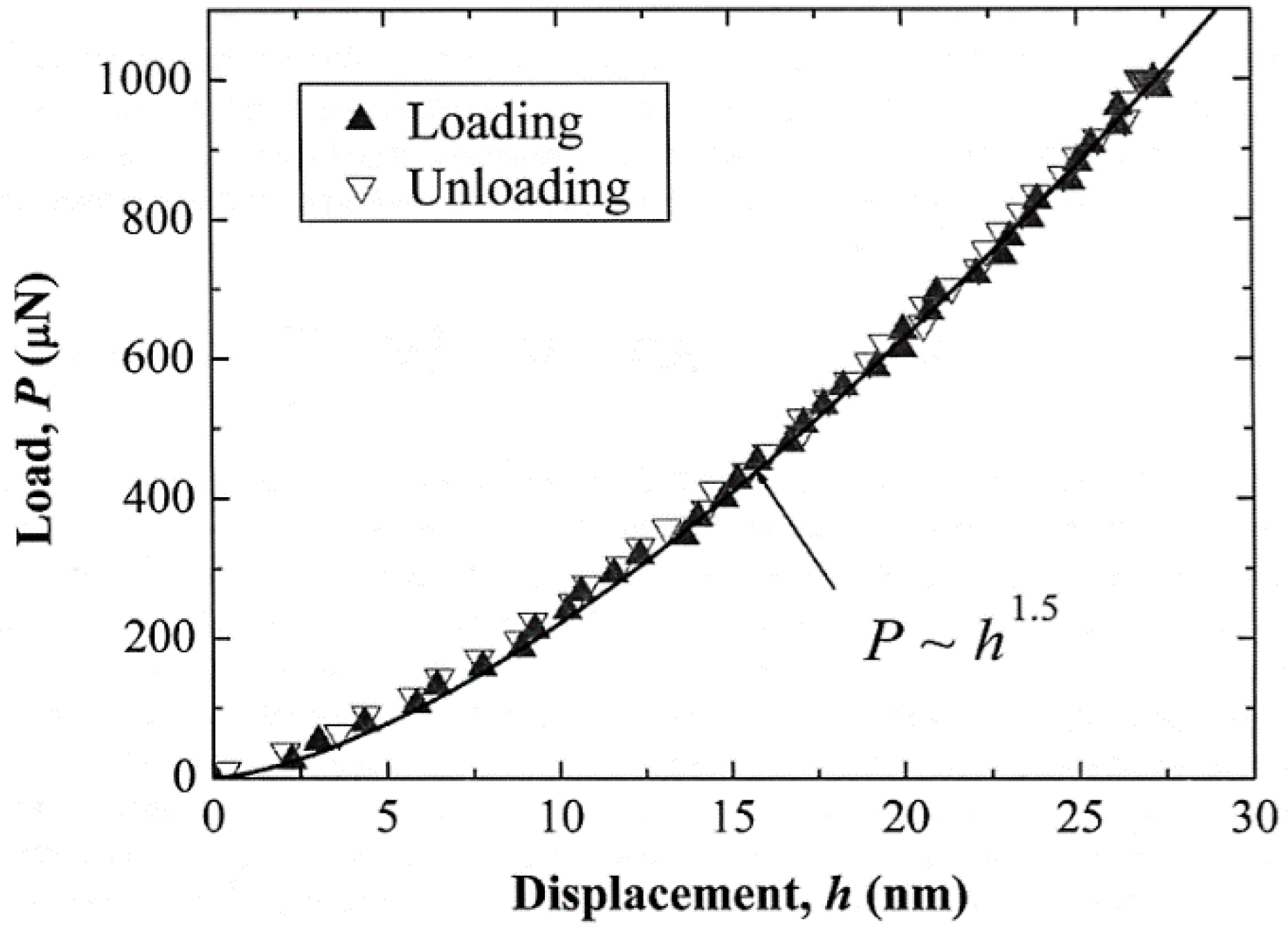
The NaCl crystal (solid curve) hardness stress–strain measurement shown in Figure 1 was obtained on indenting an {001} crystal surface with a 6.35 mm ball in a standard compression test. Such measurments are made much easier with the naturally-rounded tips of nanometer- or micrometer-scale indenter tips in nano-test indentation systems. Figure 2 shows an example elastic loading/unloading behavior recorded for nanoindentation of a {0001} sapphire crystal surface [7]. The nonlinear dependence on penetration depth, h, may be employed to determine the effective elastic modulus for a known tip diameter in accordance with the Hertzian relationship:
E* = [{(1 − νB)/EB} + {(1 − νS)/ES}]−1 = (3√2/4)(P/D1/2)h−3/2
In Equation (1), νB, EB and νS, ES are Poisson’s ratio and Young’s modulus for ball and specimen, respectively, P is load, and again, D is (effective) ball diameter. As indicated in Figure 2, the value of E* can be determined from fit to the indentation loading curve if D for the rounded indenter tip is known. Dub, Brazhkin, Novikov, Tolmachova et al. have reported similar measurements on sapphire and stishovite single crystals [8]. Elastic modulus determinations for aluminum have been reported for both micro- and nano-scale test systems [9,10]. Solhjoo and Vakis have provided a molecular dynamics assessment of surface roughness on E* determinations [11]. The initial loading method compares with an alternative method of determining E* from an unloading curve after small plastic deformation [12].
2.2. Crystal Plasticity
The omnipresent elastic loading is eventually interrupted moreso sooner than later by the so-called “pop-in” initiation of crystal plasticity. An example is shown in Figure 3 for the ambient temperature nanoindentation of several different body-centered cubic (bcc) tantalum crystal surfaces [13]. Beyond the higher hardness of the (001) crystal surface, one might note the rapid hardening at the end of the pop-in displacement and the indication of greater strain hardening within the hardened (001) indentation region.
Such measurements as shown in Figure 3 have provided valuable information for dislocation mechanics modeling of the crystal deformation behavior, particularly in connection with determining the theoretical shear stress required for dislocation nucleation within a zone below the indentation. Ruestes, Stukowski, Tang, Tramontina et al. have reported on atomic simulation of dislocation creation at nanoindentations in tantalum crystals, including the occurrence of deformation twinning [14]. Alhafez, Ruestes, Gao and Urbassek have investigated nanoindentations made in hexagonal close-packed (hcp) crystal surfaces [15]. A standard (elongated) Knoop indenter system is often employed to evaluate the plastic anisotropy of both ductile and brittle crystal materials [16].
2.3. Crystal Cracking
There are two main aspects of indentation-induced (cleavage) cracking determinations that are of interest: (1) investigation of dislocation mechanism(s) for crack formation; and (2) specification of the indentation fracture mechanics stress intensity for crack propagation.
2.3.1. Crack Formation
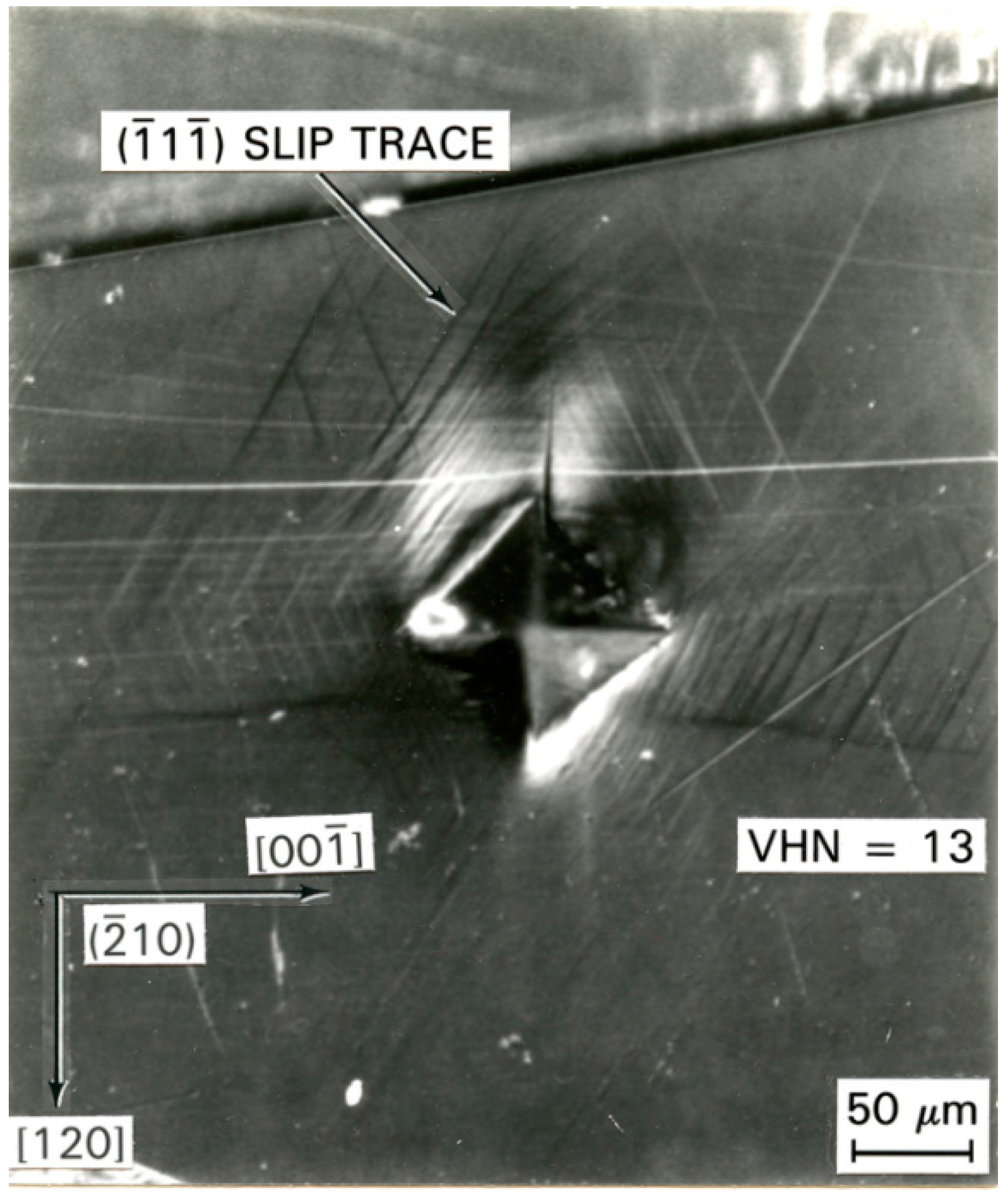
Figure 4 provides an example of crack formation at a crystallographically-aligned diamond pyramid indentation impressed into an ammonium perchlorate (AP) {210} crystal surface [17]. The top-side cleavage crack has been generated by sessile dislocations reacted at the intersections of the indicated juxtaposed {111} slip planes, in the same manner reported for cracking at similar indentations put into {001} MgO crystal surfaces and originally proposed for cleavage crack formation in α-iron and other bcc metals [4]. Close examination of the reflected optical image gives an indication that the crack has formed in the valley between otherwise raised surface regions on either side of the indentation. Such “piling up” results from secondary slip occurring to accommodate the primary indentation-forming displacement [4].
2.3.2. Indentation Fracture Mechanics
An example of elastic, plastic and cracking hardness measurements spanning load and size characterizations for nano- to micro-scale indentations and cracking is shown in Figure 5 [18]. The left-side linear solid and dashed line is the computed Hertzian dependence for elastic behavior, following a P ~ d3 dependence. The next broad band containing filled circle, triangle and square points applies for the determination of the hardness dependence, with di being the indentation diagonal length and is shown to approximate, at higher P value, to a P ~ di2 dependence. The open symbols correspond to the shift of the load and diagonal measurements to an effective ball diameter result. The furthest right-side measurements are for an indentation fracture mechanics assessment applied to crack extensions following a theoretical P ~ dC3/2 dependence. The figure indicates that plasticity precedes cracking, in line with other results shown in Figure 1 that also show that cracking requires a higher load at smaller effective ball sizes. Wan, H.; Shen, Y.; Chen, Q. and Chen, Y. have elaborated on the plastic ‘damage’ preceding such cracking produced in silicon crystals with different type indenters [19]. Vodenitcharova, Borrero-López and Hofffman described the related cracking associated with scratching along different directions on {100} silicon crystal wafers [20]. The hardness literature shows such Griffith-based crack analyses to be widely employed for characterizing the cracking behavior of brittle ceramic and glass materials.
3. Applications
Particular attention was given to example hardness aspects of surface films and coatings in the previous review [4]. Here, we mention, first, an important hardness connection with material compaction behaviors that arise for softer organic crystals relating to pharmaceutical tableting [21] and formulated energetic material processing [5]. Research effort on the former topic has been carried on to investigating temperature [22] and strain rate [23] influences on molecular crystal hardness. In the latter energetic crystal case, a recent report on hardness and molecular dynamics aspects of RDX (cyclotrimethylenetrinitramine) crystals has dealt with concern for their mechanical and tribological properties [24].
Beyond the referenced-above consideration of hardness-determined elastic deformation behavior of sapphire, its hardness properties have been investigated to relate with abrasive wear resistance [25]. Tribological concern has been with chemomechanical aspects of sapphire crystal, polycrystal ZnO coating and other ceramic material hardness properties [26]. In addition, there are interesting optical (cathodoluminescence) properties associated with dislocation zones at nanoindentations in ZnO crystals [27].
3.1. Polycrystals, Polyphases and Amorphous Phases
Particular attention was also directed in [4] to relating hardness properties between individual crystals and their polycrystalline counterparts. The connection was reasonably shown to be well-established in the so-called Hall–Petch (H–P) relationship for hardness:
H = H0 + kHℓ−1/2
In Equation (2), H is Meyers hardness, P/(πd2/4); H0 is the single crystal hardness; kH is a microstructural stress intensity; and ℓ is average (crystal) grain diameter, generally measured on a line intercept basis. Other hardness values are easily related to the Meyer hardness.
Equation (2) is related to the same type equation for the true compressive (or tensile) stress (σε)–true strain (ε) behavior for which kH ≈ 3kε. Recent estimations have been reported for H–P k values determined for steel materials through nanoindentation measurements [28]. In line with the H–P model description, the obstacle presented to slip penetrations by grain boundaries has been investigated in the bcc metals case by Soer, Aifantis and De Hosson [29] and more recently in (hcp) α-titanium via high resolution X-ray and electron microscope methods [30]. Related hardness and grain size dependent scratch results have been reported for polycrystalline copper [31]. The influence of H–P strengthening of polycrystalline diamond has been described [32] and also for cubic boron nitride via nanotwinned (boundary) strengthening [33].
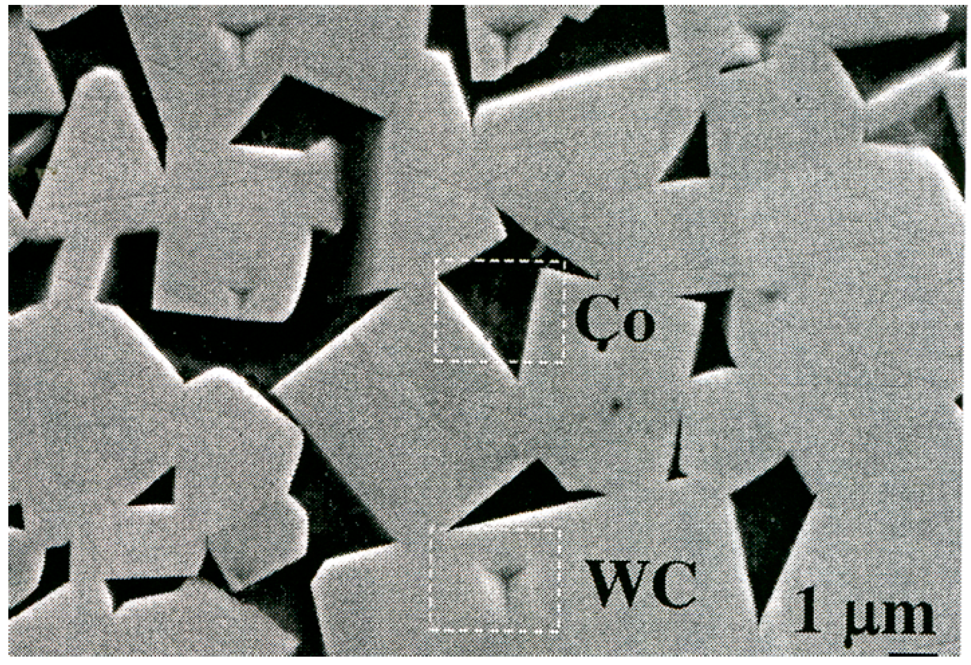
An important crystal size-dependent connection has been made with composite WC–Co cermet material for which indentations made on the individual components were known to follow separate H–P dependencies [34]. A recent report on the system has been made by Roa, Jimenez–Pique, Verge, Tarragό et al. [35]; see Figure 6. In related work, Roa et al. have determined an intermediate kH for the combined deformation of phases [36]. Zhang, Wang and Dai have employed nanoindentation testing to investigate the rate dependence of plastic flow in metallic glass materials [37].
3.2. Hardness as a Test Probe
In this concluding section, we return to reference [3] in which Gilman pointed to the important use of local hardness testing as a strength microprobe [38] and was able to correlate hardness with a number of material properties such as yield strength, elastic modulus, glide activation energy and energy gap density for a variety of crystals. The advent of nanoindentation testing has given greater meaning to such probe capability. Emphasis was given previously in [4] to connection with probe aspects of atomic force microscopy for which a new tip fabrication procedure has recently been reported [39]. Another recent application has been to investigate the strain hardening surrounding larger Rockwell indentations made in electrodeposited nanocrystalline nickel material with different grain sizes [40]. Additional examples include (1) investigating shear banding in metallic glass materials [41]; and, nano-probing of diffusion-controlled deformation on a copper crystal surface [42] and on different surfaces of ZnO crystals [43].
4. Summary
An editorial introduction to the Special Issue on crystal indentation hardness has been presented by the authors while building upon their previous review entitled Elastic, Plastic and Cracking Aspects of the Hardness of Materials [4]. The wide variation in currently referenced journals gives an indication of the broad interest in the subject. Hopefully, the more recent referenced reports will be viewed as indicating an expanded interest in the topic of crystal hardness testing, particularly as provided in this sampling of important measurements and analyses stemming from current attention being given to all aspects of nanoindentation hardness testing.
Conflicts of Interest
The authors declare no conflict of interest.
References
- Walley, S.M. Review: Historical origins of indentation hardness testing. Mater. Sci. Technol. 2012, 28, 1028–1044. [Google Scholar] [CrossRef]
- Tabor, D. The Hardness of Metals; Clarendon Press: Oxford, UK, 1951. [Google Scholar]
- Westbrook, J.H.; Conrad, H. (Eds.) The Science of Hardness Testing and Its Research Applications; American Society for Metals: Metals Park, OH, USA, 1973.
- Armstrong, R.W.; Elban, W.L.; Walley, S.M. Elastic, plastic and cracking aspects of the hardness of materials. Int. J. Mod. Phys. B 2013, 28, 1330004. [Google Scholar] [CrossRef]
- Armstrong, R.W.; Elban, W.L. Review: Hardness properties across multi-scales of applied loads and material structures. Mater. Sci. Technol. 2012, 28, 1060–1071. [Google Scholar] [CrossRef]
- Pathak, S.; Kalidindi, S.R. Spherical nanoindentation stress-strain curves. Mater. Sci. Eng. R 2015, 91, 1–36. [Google Scholar] [CrossRef]
- Lu, C.; Mai, Y.W.; Tam, P.L.; Shen, Y.G. Nanoindentation-induced elastic-plastic transition and size effect in α-Al2O3 (0001). Philos. Mag. Letts. 2007, 87, 409–415. [Google Scholar] [CrossRef]
- Dub, S.N.; Braxhkin, V.V.; Novikov, N.V.; Tolmachova, G.N.; Litvin, P.M.; Litagina, L.M.; Dyuzheva, T.I. Comparative Studies of Mechanical Properties of Stishovite and Sapphire Single Crystals by Nanoindentation. (RU) J. Superhard Mater. 2010, 32, 406–414. [Google Scholar] [CrossRef]
- Ferrante, L., Jr.; Armstrong, R.W.; Thadhani, N.N. Elastic/plastic deformation behavior in a continuous ball indentation test. Mater. Sci. Eng. A 2004, 371, 251–255. [Google Scholar] [CrossRef]
- Yoshida, M.; Sumomogi, T.; Endo, T.; Maeta, H.; Kino, T. Nanoscale Evaluation of Strength and Deformation Properties of Ultrahigh-Purity Aluminum. (JPN) Mater. Trans. 2007, 48, 1–5. [Google Scholar] [CrossRef]
- Solhjoo, S.; Vakis, A.I. Continuum mechanics at the atomic scale: Insights into non-adhesive contacts using molecular dynamics simulations. J. Appl. Phys. 2016, 120, 215102. [Google Scholar] [CrossRef]
- Oliver, W.C.; Pharr, G.M. An improved technique for determining hardness and elastic modulus using load and displacement sensing indentation experiments. J. Mater. Res. 1992, 7, 1564–1583. [Google Scholar] [CrossRef]
- Alcala, J.; Dalman, R.; Franke, O.; Biener, M.; Biener, J.; Hodge, A. Planar defect nucleation and annihilation mechanisms in nanocontact plasticity of metal surfaces. Phys. Rev. Lett. 2010, 109, 075502. [Google Scholar] [CrossRef] [PubMed]
- Ruestes, C.J.; Sukowski, A.; Tang, Y.; Tramontina, D.R.; Erhart, P.; Remington, B.A.; Urbassek, H.M.; Meyers, M.A.; Bringa, E.M. Atomistic simulation of tantalum nanoindentation: Effects of indenter diameter, penetration velocity, and interatomic potentials on defect mechanisms and evolution. Mater. Sci. Eng. A 2014, 613, 390–403. [Google Scholar] [CrossRef]
- Alhafez, I.A.; Ruestes, C.J.; Gao, Y.; Urbassek, H.M. Nanoindentation of hcp metals: A comparative simulation study of the evolution of dislocation networks. Nanotechnology 2016, 27, 045706. [Google Scholar] [CrossRef] [PubMed]
- Gao, F. Theoretical model of hardness anisotropy in brittle materials. J. Appl. Phys. 2012, 112, 023506. [Google Scholar] [CrossRef]
- Elban, W.L.; Armstrong, R.W. Plastic anisotropy and cracking at hardness impressions in single crystal ammonium perchlorate. Acta Mater. 1998, 46, 6041–6052. [Google Scholar] [CrossRef]
- Armstrong, R.W.; Ruff, A.W.; Shin, H. Elastic, plastic and cracking indentation behavior of silicon crystals. Mater. Sci. Eng. A 1996, 209, 91–96. [Google Scholar] [CrossRef]
- Wan, H.; Shen, Y.; Chen, Q.; Chen, Y. A plastic damage model for finite element analysis of cracking of silicon under indentation. J. Mater. Res. 2010, 25, 2224–2237. [Google Scholar] [CrossRef]
- Vodenitcharaova, T.; Borrero-López, O.; Hoffman, M. Mechanics prediction of the fracture pattern on scratching wafers of single crystal silicon. Acta Mater. 2012, 60, 4448–4460. [Google Scholar] [CrossRef]
- Egart, M.; Janković, B.; Srčič, S. Application of instrumented nanoindentation in preformulation studies of pharmaceutical active ingredients and excipients. Acta Pharm. 2016, 66, 303–330. [Google Scholar] [CrossRef] [PubMed]
- Mohamed, R.M.; Mishra, M.K.; Al-Harbi, L.M.; Al-Ghamdi, M.S.; Asiri, A.M.; Reddy, C.M.; Ramamurty, U. Temperature Dependence of Mechanical Properties in Molecular Crystals. Cryst. Growth Des. 2015, 15, 2474–2479. [Google Scholar] [CrossRef]
- Raut, D.; Kiran, M.S.R.N.; Mishra, M.K.; Asiri, A.M.; Ramamurty, U. On the loading rate sensitivity of plastic deformation in molecular crystals. CrystEngComm 2016, 18, 3551–3555. [Google Scholar] [CrossRef]
- Weingarten, N.S.; Sausa, R.C. Nanomechanics of RDX Single Crystals by Force-Displacement Measurements and Molecular Dynamics Simulations. J. Phys. Chem. A 2015, 119, 9338–9351. [Google Scholar] [CrossRef] [PubMed]
- Voloshin, A.V.; Dolzhenkova, E.F.; Litvinov, L.A. Anisotropy of Deformation and Fracture Processes in Sapphire Surface. (RU) J. Superhard Mater. 2015, 37, 341–345. [Google Scholar] [CrossRef]
- Bull, S.J.; Moharrami, N.; Hainsworth, S.V.; Page, T.F. The origins of chemomechanical effects in the low-load indentation hardness and tribology of ceramic materials. J. Mater. Sci. 2016, 51, 107–125. [Google Scholar] [CrossRef]
- Juday, R.; Silva, E.M.; Huang, J.Y.; Caldas, P.G.; Prioli, R.; Ponce, F.A. Strain-related optical properties of ZnO crystals due to nanoindentation on various surface orientations. J. Appl. Phys. 2013, 113, 183511. [Google Scholar] [CrossRef]
- Seok, M.-Y.; Choi, I.-C.; Moon, J.; Kim, S.; Ramamurty, U.; Jang, J.-I. Estimation of the Hall–Petch strengthening coefficient of steels through nanoindentation. Scr. Mater. 2014, 87, 49–52. [Google Scholar] [CrossRef]
- Soer, W.A.; Aifantis, K.E.; de Hosson, J.T.M. Incipient plasticity during nanoindentation at grain boundaries in body-centered cubic metals. Acta Mater. 2005, 53, 4665–4676. [Google Scholar] [CrossRef]
- Guo, Y.; Collins, D.M.; Tarleton, E.; Hofmann, F.; Tischler, J.; Liu, W.; Xu, R.; Wilkinson, A.J.; Britton, T.B. Measurements of stress fields near a grain boundary: Exploring blocked arrays of dislocations in 3D. Acta Mater. 2015, 96, 229–236. [Google Scholar] [CrossRef]
- Kareer, A.; Hou, X.D.; Jennett, N.M.; Hainsworth, S.V. The interaction between lateral size effect and grain size when scratching polycrystalline copper using a Berkovich indenter. Philos. Mag. 2016, 96, 3414–3429. [Google Scholar] [CrossRef]
- Xu, B.; Tian, Y. Ultrahardness: Measurement and Enhancement. J. Phys. Chem. 2015, 119, 5633–5638. [Google Scholar] [CrossRef]
- Li, B.; Sun, H.; Chen, C. Large indentation strain stiffening in nanotwinned cubic boron nitride. Nat. Commun. 2014, 5, 4965–4971. [Google Scholar] [CrossRef] [PubMed]
- Lee, H.C.; Gurland, J. Hardness and deformation of cemented tungsten carbide. Mater. Sci. Eng. 1978, 33, 125–133. [Google Scholar] [CrossRef]
- Roa, J.J.; Jiménez-Pique, E.; Verge, C.; Tarragό, J.M.; Mateo, A.; Llanes, L. Intrinsic hardness of constitutive phase in WC–Co composites: Nanoindentation testing, statistical analysis, WC crystal orientation effects and flow stress for the constrained metallic binder. J. Eur. Ceram. Soc. 2015, 35, 3419–3425. [Google Scholar] [CrossRef]
- Roa, J.J.; Jiménez-Pique, E.; Tarragό, J.M.; Sandoval, D.A.; Mateo, A.; Fair, J.; Llanes, L. Hall–Petch strengthening of the constrained metallic binder in WC–Co cemented carbides: Experimental assessment by means of massive nanoindentation and statistical analysis. Mater. Sci. Eng. A 2016, 676, 487–491. [Google Scholar] [CrossRef]
- Zhang, M.; Wang, Y.J.; Dai, L.H. Correlation between strain rate sensitivity and α relaxation of metallic glasses. AIP Adv. 2016, 6, 075022. [Google Scholar] [CrossRef]
- Gilman, J.J. Hardness—A Strength Microprobe. In The Science of Hardness Testing and Its Research Applications; Westbrook, J.H., Conrad, H., Eds.; American Society for Metals: Metals Park, OH, USA, 1973; pp. 51–74. [Google Scholar]
- Göring, G.; Dietrich, P.-I.; Blaicher, M.; Sharma, S.; Korvink, J.G.; Schimmel, T.; Koos, C.; Hölscher, H. Tailored probes for atomic force microscopy fabricated by two-photon polymerization. Appl. Phys. Lett. 2016, 109, 063101. [Google Scholar] [CrossRef]
- Tang, B.T.F.; Zhou, Y.; Zabev, T. Reduced hardening of nanocrystalline nickel under multiaxial indentation loading. J. Mater. Res. 2015, 30, 3528–3541. [Google Scholar] [CrossRef]
- Wang, J.Q.; Perepezko, J.H. Focus: Nucleation kinetics of shear bands in metallic glass. J. Chem. Phys. 2016, 145, 211803. [Google Scholar] [CrossRef]
- Samanta, A.; Weinan, E. Interfacial diffusion aided deformation during nanoindentation. AIP Adv. 2016, 6, 075002. [Google Scholar] [CrossRef]
- Lin, P.H.; Du, X.H.; Chen, Y.H.; Chen, H.C.; Huang, J.C. Nano-scaled diffusional or dislocation creep analysis of single-crystal ZnO. AIP Adv. 2016, 6, 095125. [Google Scholar] [CrossRef]
Figure 1.
Hardness-based stress–strain description of indentation test measurements. The hardness stress is load divided by the projected area of contact; and, the hardness strain is the projected contact diameter divided by the actual or effective ball diameter of the indenter tip [5]. The solid experimental (ball test) and dashed linear dependencies are computed in accordance with a Hertzian description; and, the Vickers hardness numbers are plotted on the abscissa scale at a position corresponding to the indenter diagonal, d = 0.375D.
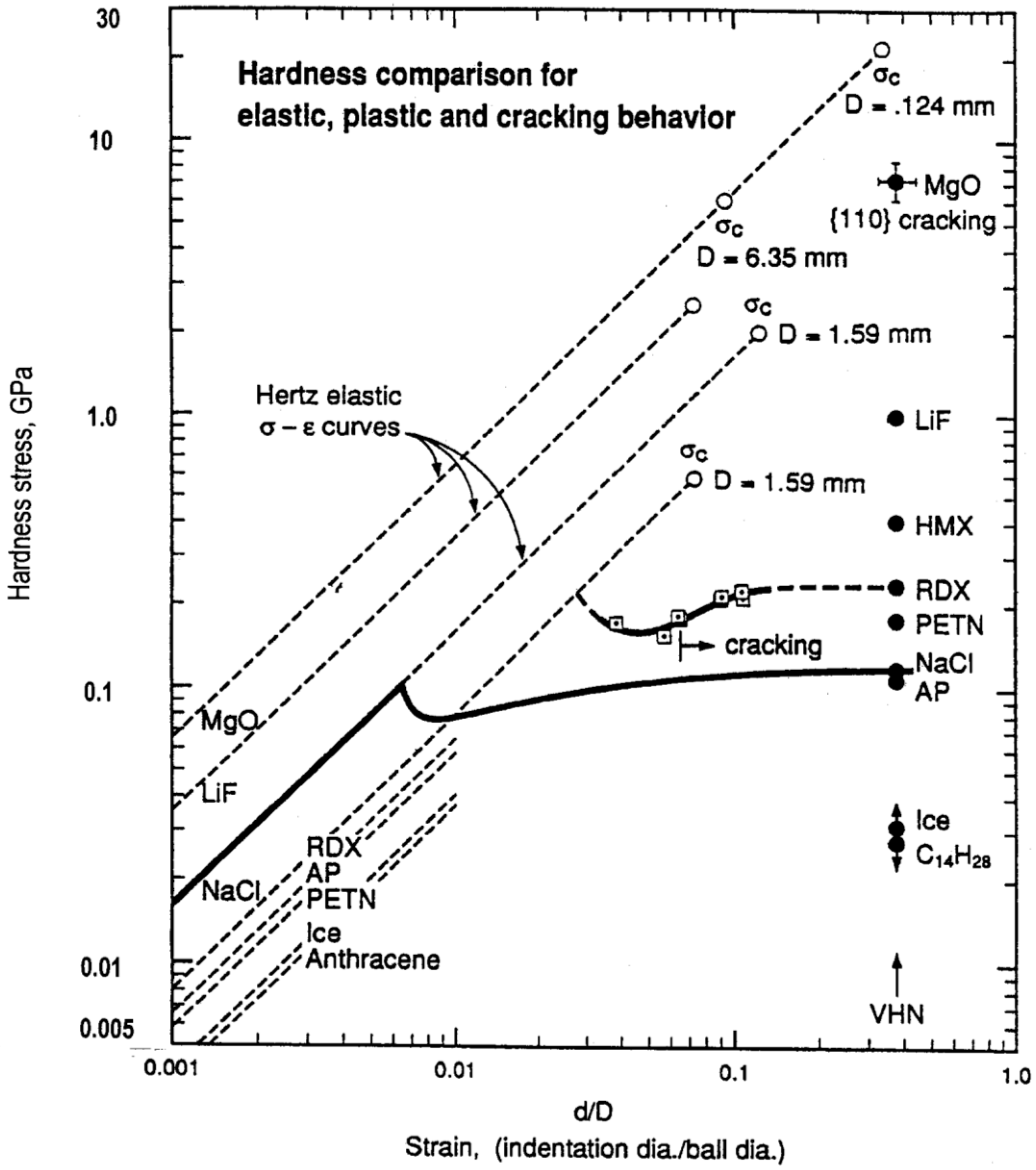
Figure 2.
Continuous load/unload curve for nanoindentation of a (0001) sapphire crystal surface [7].
Figure 2.
Continuous load/unload curve for nanoindentation of a (0001) sapphire crystal surface [7].

Figure 3.
Nanoindentation load–penetration curves obtained with impressing a (rounded point) Berkovich tri-pyramidal indenter into various tantalum (111), (011) and (001) crystal surfaces [13].
Figure 3.
Nanoindentation load–penetration curves obtained with impressing a (rounded point) Berkovich tri-pyramidal indenter into various tantalum (111), (011) and (001) crystal surfaces [13].
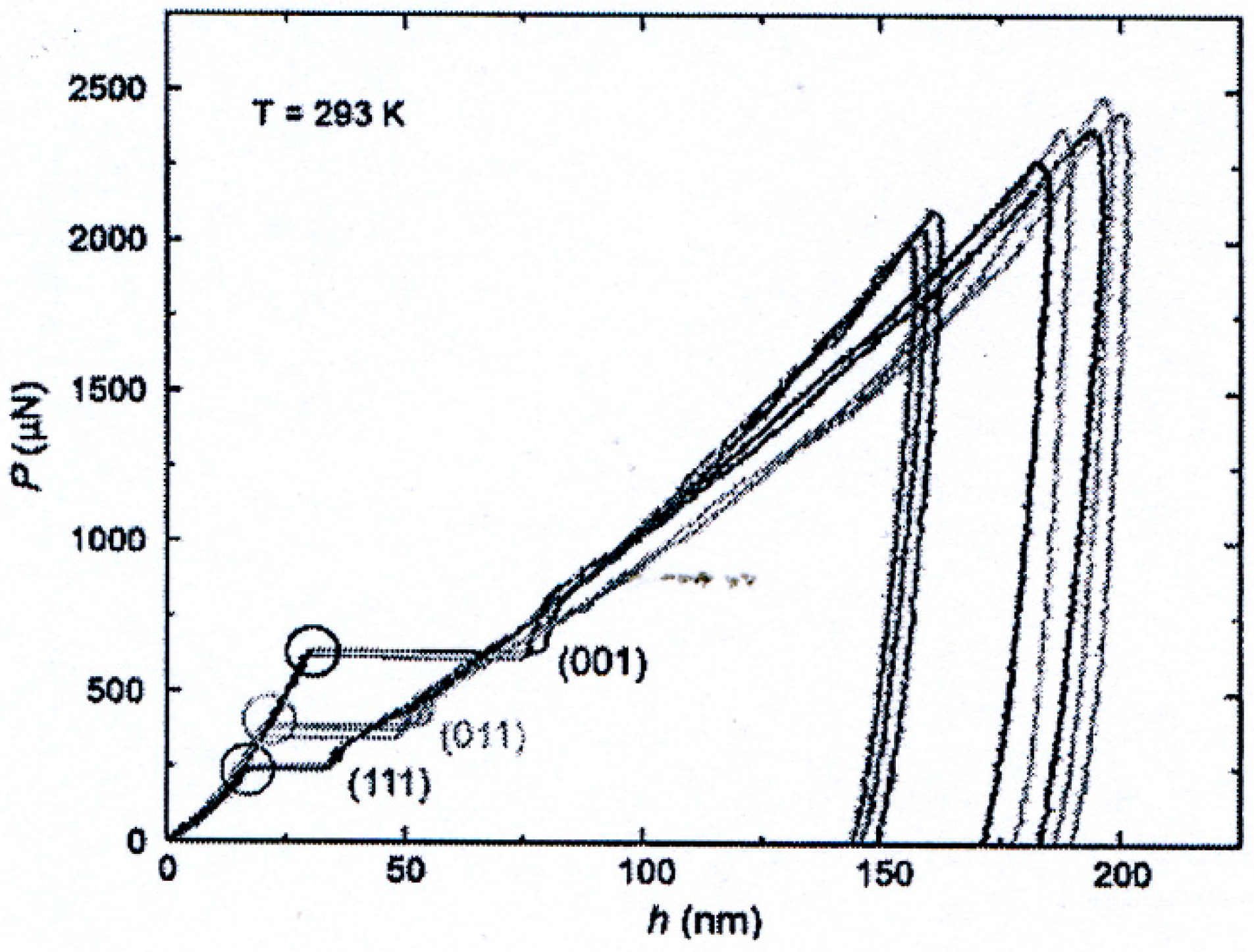
Figure 4.
{001} cleavage crack produced by Cottrell-type dislocation reaction at a diamond pyramid indentation impressed into a {210} ammonium perchlorate (AP), NH4ClO4, crystal surface [17].
Figure 4.
{001} cleavage crack produced by Cottrell-type dislocation reaction at a diamond pyramid indentation impressed into a {210} ammonium perchlorate (AP), NH4ClO4, crystal surface [17].

Figure 5.
The load, P, versus elastic, d, plastic, di, or crack extent, dc, dependence for cracking of silicon crystal surfaces indented with various sharp indenters [18].
Figure 5.
The load, P, versus elastic, d, plastic, di, or crack extent, dc, dependence for cracking of silicon crystal surfaces indented with various sharp indenters [18].
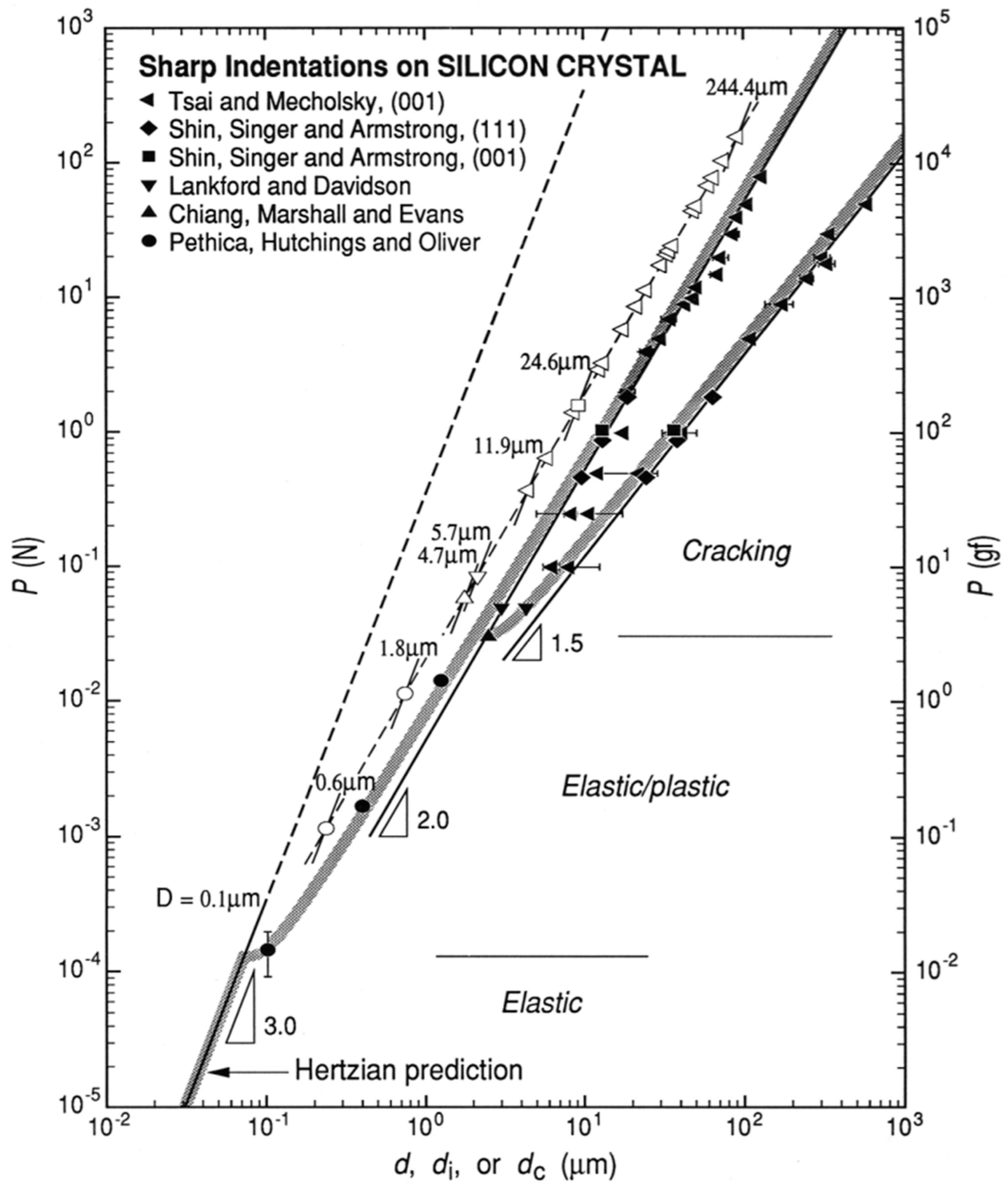
Figure 6.
Berkovich nanoindentations made within the WC particle and Co binder phases of the composite cermet material [35].
Figure 6.
Berkovich nanoindentations made within the WC particle and Co binder phases of the composite cermet material [35].

© 2017 by the authors; licensee MDPI, Basel, Switzerland. This article is an open access article distributed under the terms and conditions of the Creative Commons Attribution (CC-BY) license (http://creativecommons.org/licenses/by/4.0/).
Share and Cite
MDPI and ACS Style
Armstrong, R.W.; Walley, S.M.; Elban, W.L. Crystal Indentation Hardness. Crystals 2017, 7, 21. https://doi.org/10.3390/cryst7010021
AMA Style
Armstrong RW, Walley SM, Elban WL. Crystal Indentation Hardness. Crystals. 2017; 7(1):21. https://doi.org/10.3390/cryst7010021
Chicago/Turabian StyleArmstrong, Ronald W., Stephen M. Walley, and Wayne L. Elban. 2017. "Crystal Indentation Hardness" Crystals 7, no. 1: 21. https://doi.org/10.3390/cryst7010021
Note that from the first issue of 2016, this journal uses article numbers instead of page numbers. See further details here.