Article

Synthesis, Crystal Structure and Nonlinear Optical Property of RbHgI3

1
Department of Chemistry, Wuhan University of Science and Technology, Wuhan 430081, China
2
Department of Chemistry, Wuhan University, Wuhan 430072, China
3
College of Chemistry, Central China Normal University, Wuhan 430079, China
*
Authors to whom correspondence should be addressed.
Crystals 2017, 7(5), 148; https://doi.org/10.3390/cryst7050148
Submission received: 15 January 2017 / Revised: 15 May 2017 / Accepted: 16 May 2017 / Published: 22 May 2017

Abstract

:
Searching for new nonlinear optical (NLO) crystals to be used in the infrared (IR) region is still a challenge. This paper presents the synthesis, crystal structure and properties of a new halide, RbHgI3. Its non-centrosymmetric single crystal can be grown in solution. In its crystal structure, all the polar [HgI4]2− groups align in such a way that brings a favorable net polarization. The measurement by Kurtz–Perry powder technique indicates that RbHgI3 shows a phase-matchable second harmonic generation (SHG) property seven times stronger than that of KH2PO4 (KDP). RbHgI3 displays excellent transparency in the range of 0.48–25 μm with relatively good thermal stability. The UV absorption implies that this yellow compound’s band gap is about 2.56 eV, close to that of AgGaS2. A preliminary measurement indicates that the laser-induced damage threshold of the crystal is about 28.3 MW/cm2. These preliminary experimental data reveal that RbHgI3 is a new candidate as nonlinear optical material in the infrared region.

Graphical Abstract

1. Introduction

Second-order nonlinear optical (NLO) crystals are very important high-tech materials due to their application in laser frequency convention, optical parameter oscillator (OPO), and signal communication [1,2]. According to wavelength range, second-order NLO materials can be classified into three categories, namely ultraviolet NLO crystals, visible NLO crystals and infrared NLO crystals. Great progress has been made in ultraviolet NLO crystals in the last several decades. For instance, some excellent new crystals such as β-BaB2O4 (BBO) [3], LiB3O5 (LBO) [4], KBe2BO3F2 (KBBF) [5,6,7] have been developed by Chuangtian Chen and his colleagues. In the visible region, there are some excellent NLO crystals such as KH2PO4 (KDP) [8], KTiOPO4 (KTP) [9] and so forth. Nevertheless, numerous NLO crystals in IR regions are not fully satisfied. Some common infrared NLO crystals, such as AgGaS2 [10], AgGaSe2 [11] and ZnGeP2 [12] possess two drawbacks: one is their low laser damage thresholds (LDT), and the other is not easy to grow high-quality crystals. Therefore, searching for new infrared NLO crystals is still a challenge in this field.
However, general knowledge indicates that high LDT of the materials originates from the large gap. Guided by this purpose, thousands of IR NLO crystals have been discovered and many materials exhibit excellent performance such as halides, chalcogenides, iodates and phosphides etc., Two review articles [13,14] about IR NLO materials have been recently published. It has been accepted that the band gap which causes thermal and electronic effects is in accord with laser damage threshold [15,16]. As we know, halides usually exhibit large band gaps, and there are four halogen atoms to choose from. Moreover, halides can be easily dissolved in solvents so that it is easier to grow single crystals. On the other hand, mercury atom is a heavy metal element. This is beneficial for the wide transparence in the IR region. Furthermore, the mercury (II) cation (Hg2+) possesses d10 electronic configuration and does not emerge d-d transition. This will lead to relatively wide band gaps. Therefore, with Chuangtian Chen’s long-term help and collaboration, we have pursued research on exploring new second-order NLO crystals to be used in the mid-IR region from halides, and some new potential mid-IR NLO crystals such as Cs2HgCl2I2 [17], Cs2Hg3I8 [18], HgBrI [19], HgBr2 [20], Hg2BrI3 [21], CsHgBr3 [22], β-HgBrCl [23], NaSb3F10 [24] and Rb2CdBr2I2 [25] have been developed by this idea on our group.
In this paper, we report the synthesis, crystal structure, and NLO properties of a new compound RbHgI3. The compound is pure RbHgI3 without crystalline water and it differs from the compound of RbHgI3·H2O reported in the literature [26]. The powder second harmonic generation (SHG) effect of RbHgI3·H2O is very weak and not phase-matchable. The existence of the crystalline water is harmful for thermal stability and transparent region in the IR region. Differently, RbHgI3 shows a powder second harmonic generation (SHG) property seven times as high as that of KDP. It also exhibits a wide transparent window in the infrared region. It also shows reasonable band gap and thermal stability. To conclude, RbHgI3 is a potential candidate as new IR NLO material.

2. Results and Discussion

2.1. Synthesis and Analysis

RbHgI3 was synthesized with HgI2 and RbI by conventional solution reaction. The phase purity was checked by powder XRD. No impurities were observed, and the diffraction patterns are not consistent with those of HgI2 [27] or RbI [28] phase, and also not the sum of HgI2 and RbI phases by the XRD contrast analyses. The measured powder X-ray diffraction patterns are in agreement with the simulated patterns based on the single crystal structure of RbHgI3 obtained from the solution reaction (See Figure 1).

2.2. Crystal Structure

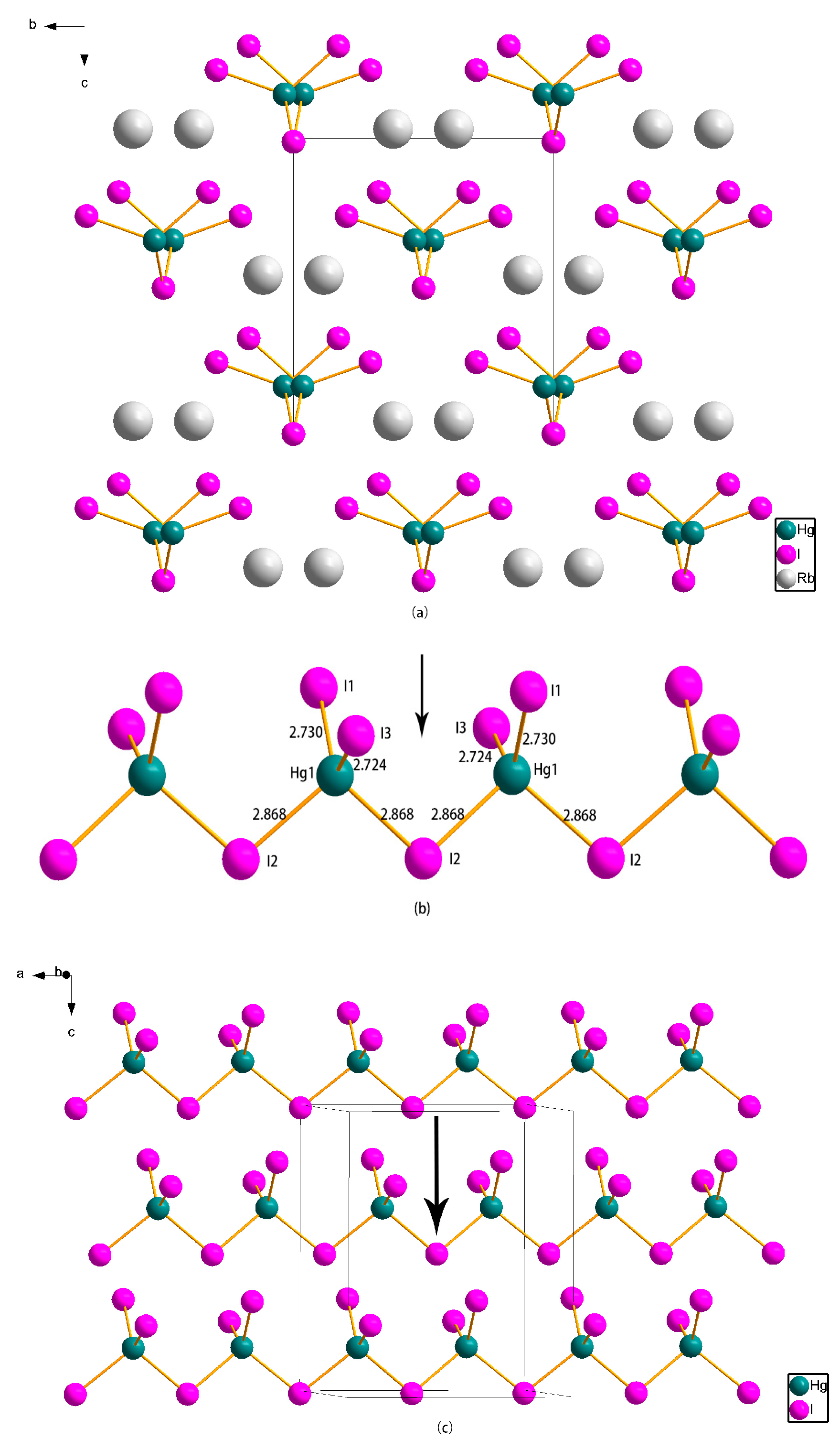
RbHgI3 crystallizes in the noncentrosymmetric orthorhombic space group Ama2 (No. 40). Figure 2a shows the packing diagram of RbHgI3 along bc plane. Each [HgI4]2− group forms a distorted tetrahedron (Figure 2b), which is connected with the neighbors by sharing an iodine atom to form a one-dimensional (1D) zigzag chain along the a-axis (Figure 2c). The chains are then to form layers along the ab plane, and the planes are further connected into a three-dimensional (3D) framework by the Rb atoms that occupy the empty spaces surrounded by iodine atoms (Figure 2). In the crystal structure, it also exhibits disorder of Rb atom in the symmetry unit with 50% occupancies (Figure 2a). As shown in Figure 2b, in each [HgI4]2− group there are two types of Hg-I bond lengths, namely, two shorter Hg-I bond length (2.724(3) Å and 2.730(3) Å, respectively), and two longer bond lengths (both 2.868(16) Å). Interestingly, all the shorter Hg-I bonds are always located above the Hg atoms in each [HgI4] tetrahedron (Figure 2c). This packing style gives rise to a net polarization parallel to the direction of the c axis (indicated by the black arrow in Figure 2b,c). Due to the favorable alignment and high density of the [HgI4]2− anionic group, the compound can show a relatively strong SHG response.

2.3. Infrared Spectrum and UV-Vis Diffuse Reflectance Spectrum

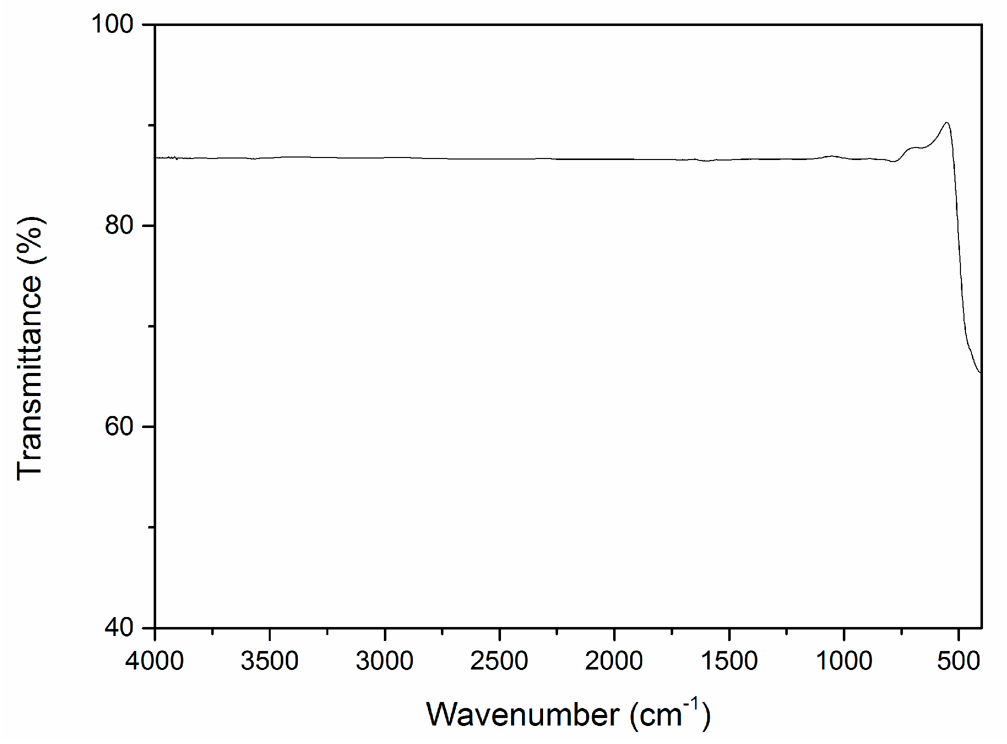
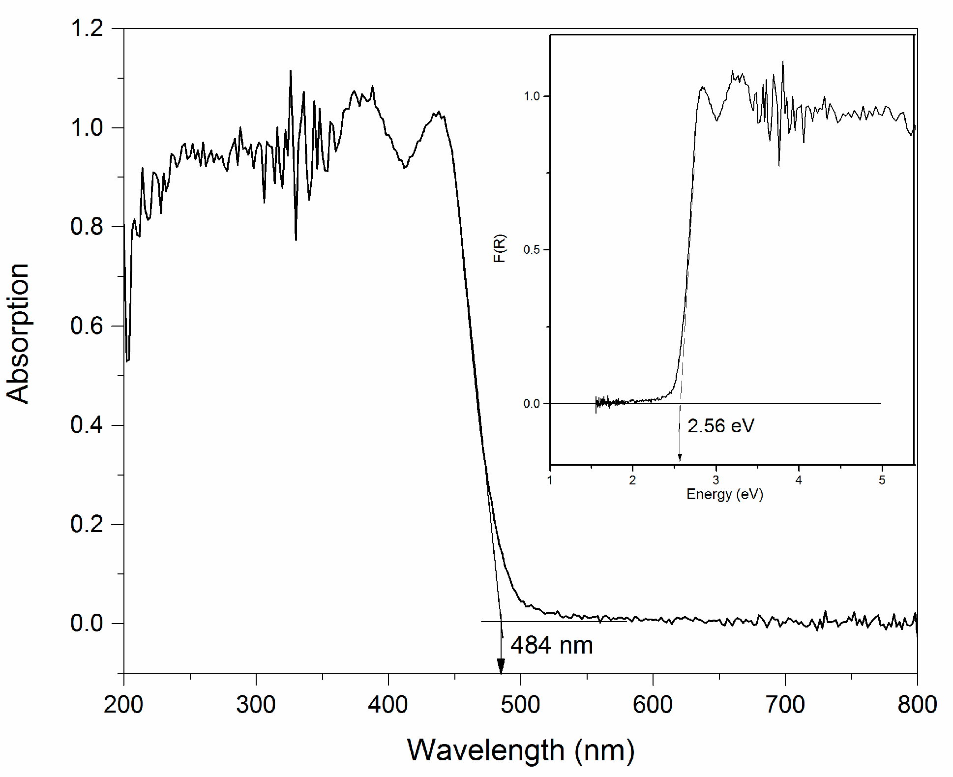
The attenuated total reflection Fourier-transformed infrared (ATR-FTIR) spectrum of RbHgI3 crystalline samples is shown in Figure 3. The ATR-FTIR spectrum indicates no absorption in the middle IR region from 4000 to 400 cm−1 (2.5–25 μm). This agrees quite well with the reported compounds such as Tl4HgI6 [29] and Cs2Hg3I8 [18,30], and they are transparent between 4000 and 400 cm−1. According to the references, Tl4HgI6 exhibits a broad range of transparency from 1.4 to 40 μm, while Cs2Hg3I8 is reported to be transparent between 0.5 and 25 μm. The UV-Vis diffuse reflectance spectrum of RbHgI3 is shown in Figure 4. The compound is yellow and the spectrum shows that the absorption edge near the UV side is about 484 nm, and this indicates that the optical band gap of the compound is approximately 2.56 eV. Based on these data, the transparent range of RbHgI3 is 0.48–25 μm.

2.4. NLO Property and LDT Measurement

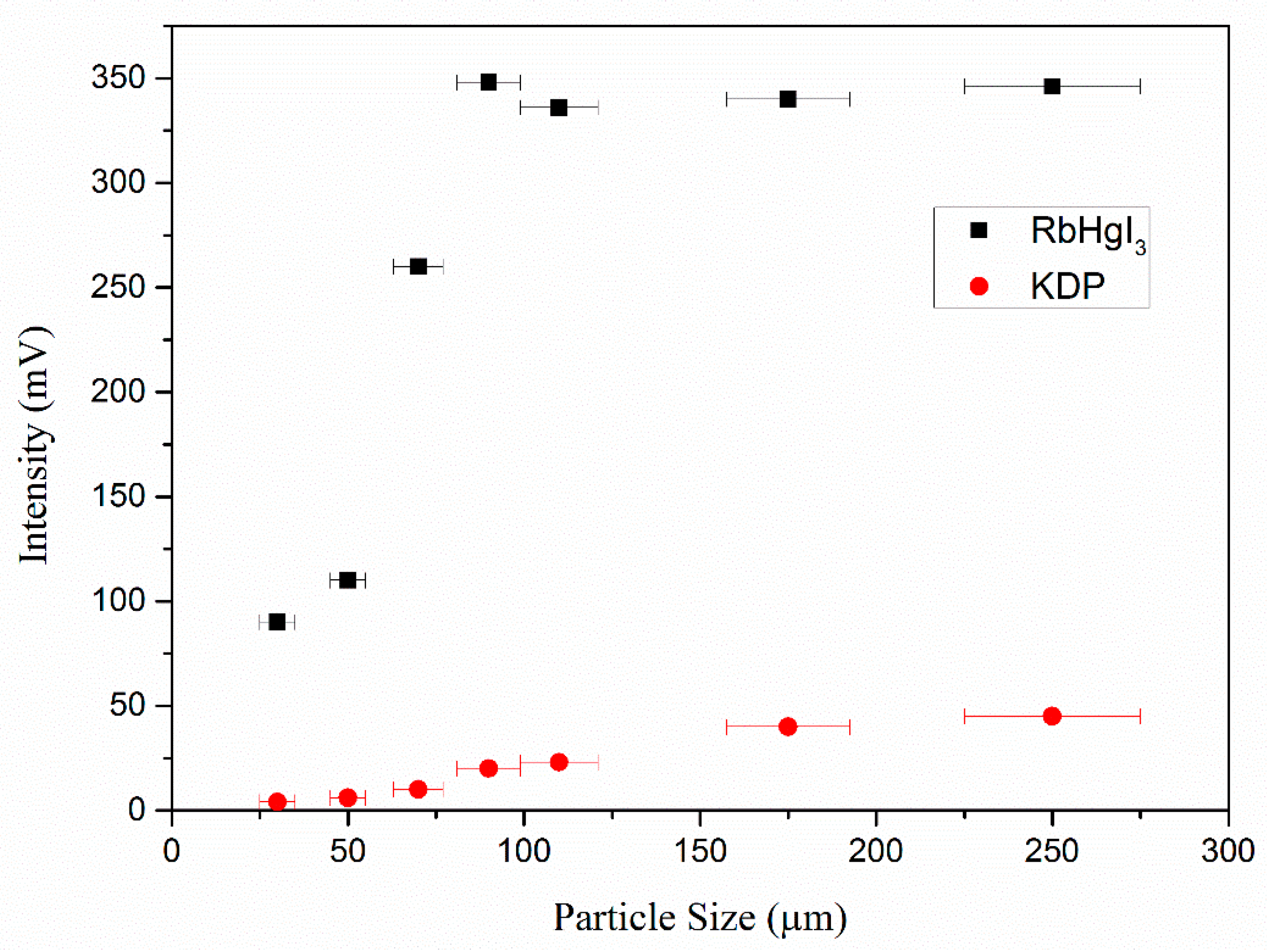
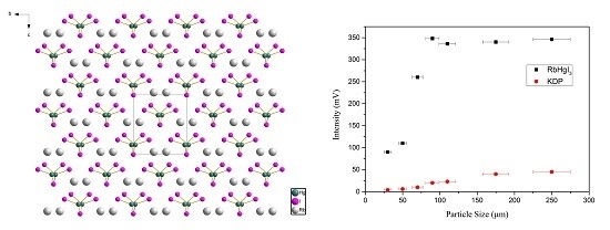
Powder SHG measurements using 1064 nm laser radiation revealed that RbHgI3 showed powder SHG efficiencies seven times as strong as that of KDP. The study result of the SHG intensity as a function of particle size (from 20 to 300 μm) is shown in Figure 5. The intensity of SHG signals at first increases gradually with the increase of the sample size and then reaches saturation when the sample size increases further. It is a typical curve for indicating that the SHG effect of RbHgI3 is phase-matchable [31].
A preliminary examination of the LDT measurement has been carried out on the crystalline sample using a Q-switched laser source. The samples showed a damage threshold of about 28.3 MW/cm2 (1064 nm, 10 ns), which is approximately equal to that of AgGaS2 (30 MW/cm2, 1064 nm) [32].

2.5. Thermogravimetric Analysis

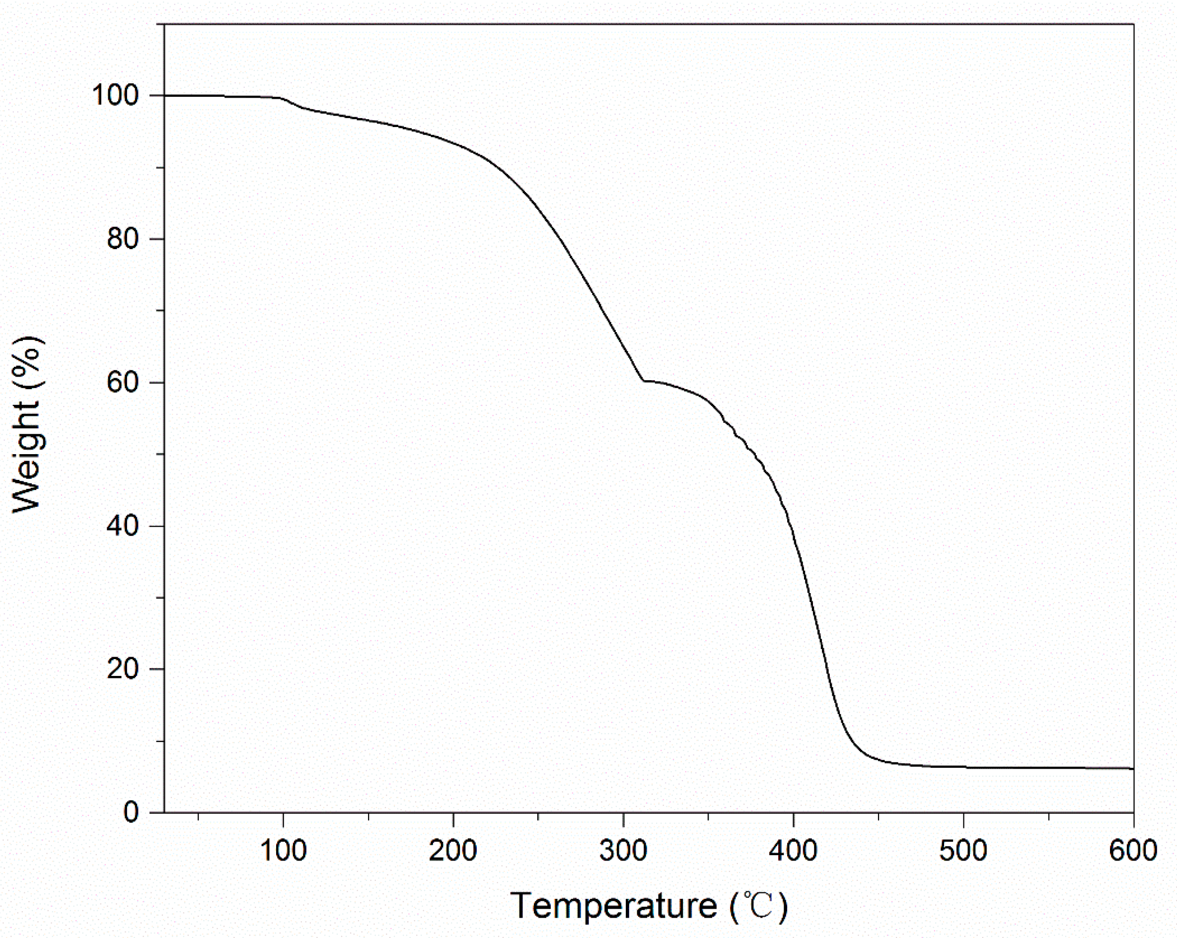
The yellow powder crystal of RbHgI3 is not hygroscopic and does not change its color after being exposed to air for about six months. The thermal behavior of RbHgI3 was investigated using thermogravimetric analysis (TGA). The TG curve reveals that the compound starts losing its weight when it is heated to above 120 °C (See Figure 6), and then it continues to lose weight and reach a platform at about 450 °C. The thermal stability of RbHgI3 is not very high and may be due to the existence of the iodine atom.

2.6. Electronic Structure and Optical Properties Calculation

To better understand the relationship of structure and the optical properties of the RbHgI3, the first-principles density functional theory (DFT) calculations were performed. The calculated band gap is 1.95 eV. The deviations from the experimental values should be attributed to the limitation of DFT [33]. Hence, scissors of 0.61 eV has been added to shift up the conduction band (CB) levels in order to agree with the measured value of the band gap for the following calculations and analyses.
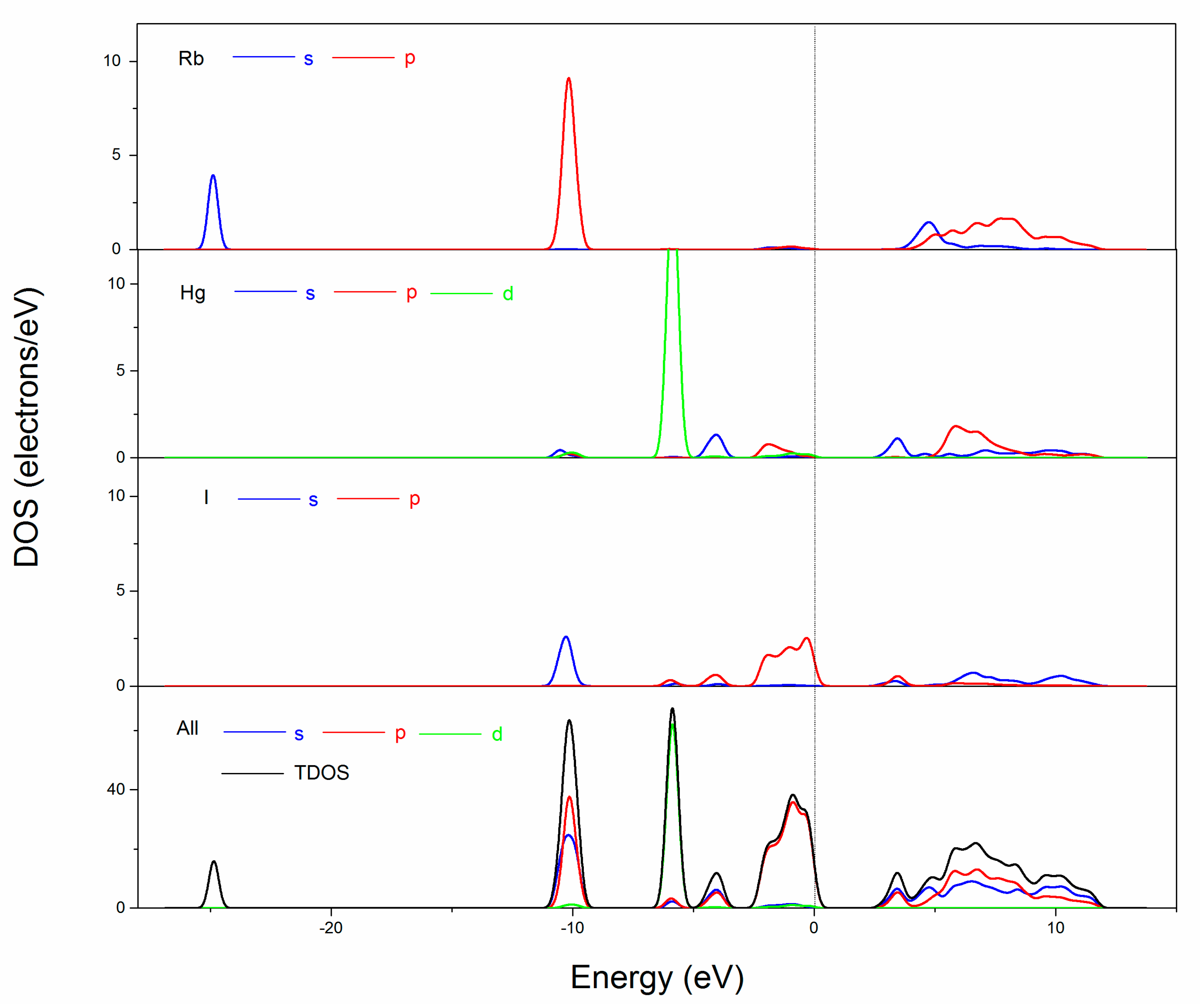
The partial density of states (PDOS) projected on the constitutional atoms in RbHgI3 are shown in Figure 7. The Rb s, Rb 4p, I 5s and Hg 5d orbitals are strongly localized in the valence band (VB) about −25 eV, −10 eV and −5.8 eV, respectively. The upper of the valence states from −2.5 eV show a large hybridization between Hg 5p (and 5d) and I 5p orbitals, indicating very strong chemical bonds between the Hg and I atoms, but the VB maximum is dominated by I 5p orbitals which are higher than Hg 5d orbitals in the VB. The bottom of CB is mainly composed of the 5s and 5p orbitals of I atom and 6s orbitals of Hg atom, but I 5s orbitals are lower than Hg 5s orbitals in the CB. This means that the iodide anion directly determines the energy band gap of RbHgI3. This is the reason why the laser damage threshold is not high and it agrees with the experimental value.
On the basis of the above electronic band structure, the refractive indices and second harmonic generation (SHG) coefficients of RbHgI3 were obtained. The refractive indices are shown in Figure S2 (see the supporting information) and the calculated refractive indices and birefringence at several radiation wavelengths are listed in Table 1. It is shown that the birefringence Δn is larger than 0.08 as the wavelength is longer than 1000 nm, so RbHgI3 is phase-matchable for the SHG in the IR region. Furthermore, we theoretically determined the SHG coefficients of RbHgI3. For the orthorhombic mm2 point group and considering the restriction of Kleinman’s symmetry, the title compound (Ama2) has three (d15, d24 and d33) independent SHG tensors and the static SHG coefficients are as follows: d15 = 28.99 pm/V, d24 = −27.19 pm/V and d33 = 2.95 pm/V. The calculated results agree with the experimental observation that RbHgI3 exhibits a SHG response seven times that of KDP (d36 = 0.39 pm/V). Therefore, we are confident that RbHgI3 possess strong SHG effects and is a promising candidate for the nonlinear optical applications.

3. Materials and Methods

3.1. Synthesis and Crystal Growth

All the starting materials are analytically pure from commercial sources and used without further purification. RbHgI3 was synthesized by conventional solution reaction. HgI2 and RbI (mole ratio 1:1) are carefully dissolved in ethanol. The mixture was stirred at 80 °C for 5 h. The yellow solution was filtered and slowly cooled, then kept in a flask at room temperature. After a few days, some yellow crystals appeared in the bottom of the flask. The yellow crystals were filtered and carefully washed with cool ethanol. A single phase of RbHgI3 crystals was obtained with the yield of 65% (based on HgI2). The RbHgI3 crystal can be grown from the ethanol solution and a photograph of the RbHgI3 crystal is presented in Figure S1 (see the supporting information).

3.2. Structure Determination

A single crystal of RbHgI3 with dimensions of ca. 0.10 × 0.10 × 0.08 mm3 was selected and used for single-crystal diffraction experiment. Data sets were collected using a Bruker SMART APEX diffractometer (Bruker, Karlsruhe, Germany) equipped with a CCD detector (graphite-monochromated Mo-Kα radiation λ = 0.71073 Å) at 298(2) K. Data sets reduction and integration were performed using the software package SAINT PLUS [34]. The crystal structure is solved by direct methods and refined using the SHELXTL 97 software package [35,36]. Single crystal data collection, cell parameters and basic information for RbHgI3 are summarized in Table 2. Further details of the crystal structure studies may be obtained from the Fachinformationszentrum Karlsruhe, 76344 Eggenstein-Leopoldshafen, Germany (Fax: (49) 7247808666; e-mail: [email protected]), on quoting registry number CSD-432180 for RbHgI3.

3.3. Powder XRD Measurement

X-ray powder diffraction (XRD) patterns of the polycrystalline material were collected using a Bruker D8 Advanced diffractometer (Bruker, Karlsruhe, Germany) with Cu kα1 radiation (λ = 154186 Å) in the range of 10–80° (2θ) at a scanning rate of 6°/min−1.

3.4. Optical Spectroscopy

The optical transmission in the mid-IR region was recorded on a NICOLET 5700 Fourier-transformed infrared (FT-IR) spectrophotometer (Manufacture, City, Country) in the 4000–400 cm−1 (2.5–25 μm) region using the attenuated total reflection (ATR) technique with a diamond crystal. The crystal sample was loaded on the samples stage and then the ATR-FTIR spectrum was measured. The UV-Vis absorption spectrum was recorded on a Varian Cary 5000 UV-Vis-NIR spectrophotometer (Agilent, Palo Alto, CA, USA) in the region 200–800 nm. A BaSO4 plate was used as the standard (100% reflectance), on which the finely ground samples from the crystals were coated. The absorption spectrum was calculated from the reflectance spectrum using the Kubelka–Munk [37] function: α/S = (1 − R2)/(2R), where α is the absorption coefficient, S is the scattering coefficient, and R is reflectance.

3.5. Second-Harmonic Generation (SHG) and Laser Damage Threshold (LDT) Measurement

The NLO efficiencies of the samples were investigated using a Kurtz–Perry powder technique [30]. A pulsed Q-switched Nd:YAG laser was utilized to generate fundamental 1064 nm light with a pulse width of 10 ns. Microcrystalline KDP was served as the standard. The particle sizes of the sieved sample and KDP were arranged from 25 to 260 μm for the measurement of size-dependent SHG effect. The energy of each pulse was measured to be about 200 mJ. The sample crystal was fixed on the bracket. An optical concave lens was used to adjust the diameter of the laser beam to obtain different intensities. The samples endured gradually enhanced radiation until their appearance changed under a microscope after the irradiation.

3.6. Thermogravimetric Analysis

The thermogravimetric analysis (TGA) was carried out on a SDTQ 600 simultaneous analyzer instrument (TA Instruments, New Castle, PA, USA). The crystal sample was added into an Al2O3 crucible and heated from room temperature to 800 °C at a heating rate of 10 K min−1 under flowing nitrogen gas.

3.7. Theoretical Calculation

The electronic structure and optical properties for the title compound were performed using the first principles plane-wave pseudopotential method implemented in the Cambridge Serial Total Energy Package (CASTEP) [38]. According to the experimental structure, the model was built and Rb atom was located at (0.7500, 0.3831, 0.9670) in the unit cell because of its order. The optimized normal-conserving pseudopotentials [39] in the Kleinman–Bylander form are chosen and the valence electrons are 4s, 4p and 5s for Rb; 5p, 5d and 6s electrons for Hg; and 5s, 5p electrons for I. The local-density approximation (LDA) with a high kinetic energy cutoff of 900 eV is adopted. Monkhorst–Pack [40] k point meshes with a density of 4 × 4 × 4 points in the Brillouin zone of the unit cell are used. The scissor-factors-corrected LDA are used to calculate the second-order susceptibility χ2 based on the formula developed by Lin et al. [41].

4. Conclusions

A new ternary RbHgI3 with an orthorhombic space group Ama2 has been obtained by reaction of HgI2 and RbI (1:1) in alcohol. The compound shows phase-matchable SHG of about seven times as strong as that of KDP, a wide transparent region (in the range of 0.48–25 μm), and a relatively good stability to the environment. Its crystals can be grown in solution. Owing to these properties, RbHgI3 appears to be a promising new NLO crystal applicable in the infrared region. Our future efforts will be devoted to growing large and high-quality crystals of RbHgI3 to further study its optical properties, for instance SHG coefficients, refractive indices and laser damage threshold.

Supplementary Materials

The following are available online at www.mdpi.com/2073-4352/7/5/148/s1: Figure S1: Figure S1. As grown crystal of RbHgI3; Figure S2: Figure S2. The calculated refractive indices for RbHgI3.

Acknowledgments

This work was supported by the National Science Foundation of China (No. 21301132 and No. 91022036) and Specialized Research Fund for the Doctoral Program of Higher Education of China (No. 20134219120002).

Author Contributions

Yanjun Li conceived and designed the experiments and wrote the paper; Yuxun Ding performed the experiments and analyzed the data; Yaming Li and Hongmin Liu carried out the characterization work; Xianggao Meng analyzed the crystal structure; Ye Cong, Jiang Zhang and Xuanke Li contributed reagents and materials; Jingui Qin, Xingguo Chen and Yanjun Li conceived and coordinated the project.

Conflicts of Interest

The authors declare no conflict of interest.

References

  1. Burland, D.M.; Miller, R.D.; Walsh, A.C. Second-order nonlinearity in poled-polymer systems. Chem. Rev. 1994, 94, 31–75. [Google Scholar] [CrossRef]
  2. Chai, B.H.T. CRC Handbook of Laser Science and Technology Supplement 2: Optical Materials; Weber, M.J., Ed.; CRC: Boca Raton, FL, USA, 1995. [Google Scholar]
  3. Chen, C.T.; Wu, B.C.; Jiang, A.D.; You, M.G. A new-type ultraviolet SHG crystal—β-BaB2O4. Sci. Sin. Ser. B 1985, 28, 235–243. [Google Scholar]
  4. Chen, C.T.; Wu, Y.C.; Jiang, A.D.; Wu, B.C.; You, G.M.; Li, R.K.; Lin, S.J. New nonlinear-optical crystal: LiB3O5. J. Opt. Soc. Am. B 1989, 6, 616–621. [Google Scholar] [CrossRef]
  5. Chen, C.T.; Xu, Z.Y.; Deng, D.Q.; Zhang, J.; Wrong, G.K.L. The vacuum ultraviolet phase-matching characteristics of nonlinear optical KBe2BO3F2 crystal. Appl. Phys. Lett. 1996, 28, 2930–2932. [Google Scholar] [CrossRef]
  6. Chen, C.T.; Ge, N.; Lin, J.; Jiang, J.; Zeng, W.R.; Wu, B.C. Computer-Assisted Search for Nonlinear Optical Crystals. Adv. Mater. 1999, 11, 1071–1078. [Google Scholar] [CrossRef]
  7. Chen, C.T.; Wang, G.L.; Wang, X.Y.; Xu, Z.Y. Deep-UV nonlinear optical crystal KBe2BO3F2 —discovery, growth, optical properties and applications. Appl. Phys. B 2009, 97, 9–25. [Google Scholar] [CrossRef]
  8. Smith, W.L. KDP and ADP transmission in the vacuum ultraviolet. Appl. Opt. 1977, 16, 306–318. [Google Scholar] [CrossRef] [PubMed]
  9. Kato, K. Parametric oscillation at 3.2 µm in KTP pumped at 1.064 µm. IEEE J. Quantum Electron. 1991, 27, 1137–1140. [Google Scholar] [CrossRef]
  10. Chemla, D.S.; Kupecek, P.J.; Robertson, D.S.; Smith, R.C. Silver thiogallate, a new material with potential for infrared devices. Opt. Commun. 1971, 3, 29–31. [Google Scholar] [CrossRef]
  11. Boyd, G.D.; Kasper, E.H.M.; Mcfee, J.H.; Storz, F.G. Linear and nonlinear optical properties of AgGaS2, CuGaS2, and CuInS2, and theory of the wedge technique for the measurement of nonlinear coefficients. IEEE J. Quantum Electron. 1972, 8, 563–573. [Google Scholar]
  12. Boyd, G.D.; Buehler, E.; Storz, F.G. Linear and nonlinear optical properties of ZnGeP2 and CdSe. Appl. Phys. Lett. 1971, 18, 301–304. [Google Scholar] [CrossRef]
  13. Liang, F.; Lin, L.K.Z.; Wu, Y.; Chen, C. Analysis and prediction of mid-IR nonlinear optical metal sulfides with diamond-like structures. Coordin. Chem. Rev. 2017, 333, 57. [Google Scholar] [CrossRef]
  14. Guo, S.; Chi, Y.; Guo, G. Recent achievements on middle and far-infrared second-order nonlinear optical materials. Coord. Chem. Rev. 2017, 335, 44. [Google Scholar] [CrossRef]
  15. Jackson, A.G.; Ohmer, M.C.; LeClair, S.R. Relationship of the second order nonlinear optical coefficient to energy gap in inorganic non-centrosymmetric crystals. Infrared Phys. Technol. 1997, 38, 233–244. [Google Scholar] [CrossRef]
  16. Chen, C.T.; Liu, G.Z. Recent Advances in Nonlinear Optical and Electro-Optical Materials. Annu. Rev. Mater. Sci. 1986, 16, 203–243. [Google Scholar] [CrossRef]
  17. Zhang, G.; Li, Y.; Jiang, K.; Zeng, H.; Liu, T.; Chen, X.; Qin, J.; Lin, Z.; Fu, P.; Wu, Y.; et al. A New Mixed Halide, Cs2HgI2Cl2: Molecular Engineering for a New Nonlinear Optical Material in the Infrared Region. J. Am. Chem. Soc. 2012, 134, 14818–14822. [Google Scholar] [CrossRef] [PubMed]
  18. Zhang, G.; Qin, J.; Liu, T.; Fu, P.; Wu, Y.; Chen, C. Synthesis, Characterization, and Crystal Growth of Cs2Hg3I8: A New Second-Order Nonlinear Optical Material. Cryst. Growth Des. 2008, 8, 2946–2949. [Google Scholar] [CrossRef]
  19. Wu, Q.; Li, Y.; Chen, H.; Jiang, K.; Li, H.; Zhong, C.; Chen, X.; Qin, J. HgBrI: A promising nonlinear optical material in IR region. Inorg. Chem. Commun. 2013, 34, 1–3. [Google Scholar] [CrossRef]
  20. Liu, T.; Qin, J.; Zhang, G.; Zhu, T.; Niu, F.; Wu, Y.; Chen, C. Mercury Bromide (HgBr2): A promising nonlinear optical material in IR region with a high laser damage threshold. Appl. Phys. Lett. 2008, 93, 091102. [Google Scholar] [CrossRef]
  21. Li, Y.; Wang, M.; Zhu, T.; Meng, X.; Zhong, C.; Chen, X.; Qin, J. Synthesis, crystal structure and properties of a new candidate for nonlinear optical material in the IR region: Hg2BrI3. Dalton Trans. 2012, 41, 763–766. [Google Scholar] [CrossRef] [PubMed]
  22. Lv, S.W.; Wu, Q.; Meng, X.; Kang, L.; Zhong, C.; Lin, Z.; Hu, Z.; Chen, X.; Qin, J. A promising new nonlinear optical crystal with high laser damage threshold for application in the IR region: Synthesis, crystal structure and properties of noncentrosymmetric CsHgBr3. J. Mater. Chem. C. 2014, 2, 6796–6801. [Google Scholar] [CrossRef]
  23. Dang, Y.; Meng, X.; Jiang, K.; Zhong, C.; Chen, X.; Qin, J. A promising nonlinear optical material in the Mid-IR region: New results on synthesis, crystal structure and properties of noncentrosymmetric β-HgBrCl. Dalton Trans. 2013, 42, 9893–9897. [Google Scholar] [CrossRef] [PubMed]
  24. Zhang, G.; Qin, J.; Liu, T.; Li, Y.; Wu, Y.; Chen, C. NaSb3F10: A new second-order nonlinear optical crystal to be used in the IR region with very high laser damage threshold. Appl. Phys. Lett. 2009, 95, 1. [Google Scholar] [CrossRef]
  25. Wu, Q.; Meng, X.; Zhong, C.; Chen, X.; Qin, J. Rb2CdBr2I2: A new IR nonlinear optical material with a large laser damage threshold. J. Am. Chem. Soc. 2014, 136, 5683–5686. [Google Scholar] [CrossRef] [PubMed]
  26. Holmes, D.E.; Harvill, M.L.; Bogan, L.D. A survey of ABX3 halides for optical second harmonic generation. Mat. Res. Bull. 1975, 10, 753–759. [Google Scholar] [CrossRef]
  27. George, A.J.; Marcus, V. Crystal structures of the red, yellow, and orange forms of mercuric iodide. Inorg. Chem. 1967, 6, 396–399. [Google Scholar]
  28. Cortona, P. Direct determination of self-consistent total energies and charge densities of solids: A study of the cohesive properties of the alkali halides. Phys. Rev. B 1992, 46, 2008. [Google Scholar] [CrossRef]
  29. Avdienko, K.I.; Badikov, D.V.; Badikov, V.V.; Chizhikov, V.I.; Panyutin, V.L.; Shevyrdyaeva, G.S.; Scherbakov, S.I.; Scherbakova, E.S. Optical properties of thallium mercury iodide. Opt. Mater. 2003, 23, 569–573. [Google Scholar] [CrossRef]
  30. Sathiskumar, S.; Kathiravan, P.; Balakrishnan, T. Cationic Coordination Compound Cs2Hg3I8 for IR NLO Material: Synthesis, Crystal Growth and Characterizations. In Proceedings of the AIP conference Proceedings 1665, Tamilnadu, India, 16–20 December 2014; p. 2814. [Google Scholar]
  31. Kurtz, S.K.; Perry, T.T. A Powder Technique for the Evaluation of Nonlinear Optical Materials. J. Appl. Phys. 1968, 39, 3798–3813. [Google Scholar] [CrossRef]
  32. Nikogosyan, D.N. Nonlinear Optical Crystals: A Complete Survey; Springer: New York, NY, USA, 2005; pp. 102–103. [Google Scholar]
  33. Godby, R.W.; Schlüter, M.; Sham, L.J. Trends in self-energy operators and their corresponding exchange-correlation potentials. Phys. Rev. B 1987, 36, 6497. [Google Scholar] [CrossRef]
  34. Sheldrick, G.M. SHELXTL, Version 6.14; Bruker Analytical x-ray Instruments, Inc.: Madison, WI, USA, 2003. [Google Scholar]
  35. Sheldrick, G.M. SHELXS-97—A Program for Automatic Solution of Crystal Structures; University of Goettingen: Goettingen, Germany, 1997. [Google Scholar]
  36. Sheldrick, G.M. SHELXL-97—A Program for Crystal Structure Refinement; University of Goettingen: Goettingen, Germany, 1997. [Google Scholar]
  37. Wendlandt, W.H.; Hecht, H.G. Reflectance Spectroscopy; Interscience: New York, NY, USA, 1966; pp. 62–65. [Google Scholar]
  38. Clark, S.J.; Segall, M.D.; Pickard, C.J.; Hasnip, P.J.; Probert, M.I.J.; Refson, K.; Payne, M.C. First principles methods using CASTEP. Z. Kristallogr. 2005, 220, 567–570. [Google Scholar] [CrossRef]
  39. Lin, J.S.; Qtseish, A.; Payne, M.C.; Heine, V. Optimized and transferable nonlocal separable ab initio pseudopotentials. Phys. Rev. B 1993, 47, 4174. [Google Scholar] [CrossRef]
  40. Perdew, J.P.; Burke, K.; Ernzerhof, M. Generalized Gradient Approximation Made Simple. Phys. Rev. Lett. 1996, 77, 3865. [Google Scholar] [CrossRef] [PubMed]
  41. Lin, J.; Lee, M.; Lin, Z.; Chen, C.; Pickard, C. Mechanism for linear and nonlinear optical effects in beta-BaB2O4 crystals. Phys. Rev. B Condens. Matter Mater. Phys. 1999, 60, 13380. [Google Scholar] [CrossRef]
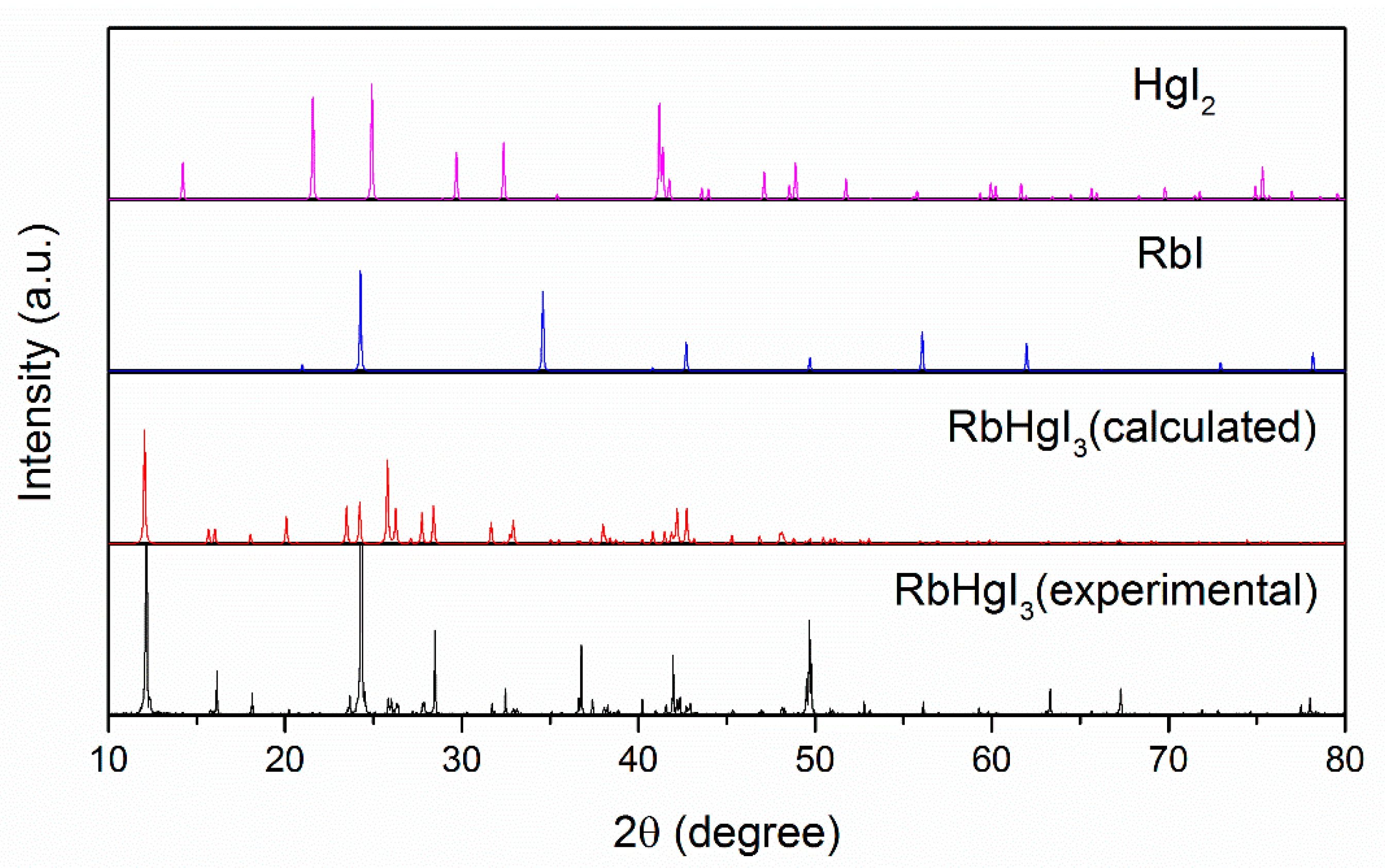
Figure 1. Comparison of experimental and calculated powder X-ray diffraction patterns: the experimental powder X-ray diffraction data of RbHgI3 powders obtained from the solution reaction; the calculated powder X-ray diffraction data of HgI2, RbI and RbHgI3 from single crystal structure data.
Crystals 07 00148 g001
Figure 2. (a) Ball-and-stick packing diagrams of RbHgI3 along bc plane; (b) [HgI4] tetrahedron in a unit cell. Each Hg atom is bonded to four iodine atoms with two types of the bond length, namely, two are short (2.730(3) and 2.724(3) Å) and the other two are longer (2.868(16) Å); (c) Ball-and-stick diagrams of [HgI4] tetrahedron (Rb atoms are omitted for clarity). All the longer Hg-I bonds are always located below the Hg atom in each [HgI4] tetrahedron, giving rise to a net dipole moment parallel to the c axis (indicated by the black arrow).
Crystals 07 00148 g002
Figure 3. Attenuated total reflection Fourier-transformed infrared (ATR-FTIR) spectrum for RbHgI3.
Crystals 07 00148 g003
Figure 4. UV-Vis diffuse reflectance spectroscopy data for RbHgI3.
Crystals 07 00148 g004
Figure 5. Second harmonic generation (SHG) intensity versus particle size for RbHgI3 and KH2PO4 (KDP).
Crystals 07 00148 g005
Figure 6. Thermogravimetric analysis (TGA) curve for RbHgI3.
Crystals 07 00148 g006
Figure 7. The partial density of states (PDOS) for RbHgI3.
Crystals 07 00148 g007
Table 1. Calculated Refractive Indices at Selected Wavelengths for RbHgI3.
WavelengthnxnynzΔn (nynx)
1000 nm2.012.092.070.08
1064 nm2.002.082.060.08
2000 nm1.972.052.030.08
~∞1.962.042.020.08
Table 2. Crystallographic data of RbHgI3.
Empirical FormulaRbHgI3
Formula weight666.76
Temperature296(2)
Wavelength0.71073
Crystal colorYellow
Crystal systemOrthorhombic
Space groupAma2 (No.40)
Crystal size (mm3)0.10 × 0.10 × 0.08
Unit cell dimensions (Å)a = 8.838(3), b = 9.824(3), c = 11.050(3)
Z4
V3)959.4(5)
Absorption coefficient (mm−1)30.640
Density (calculated)4.616
Goodness-of-fit on F21.149
Reflections collected1473
Independent reflection1337 [R (int) = 0.0293]
R1, wR1 [I > 2σ (I)]0.0682/0.1845
R2, wR2 (all data)0.0637/0.1818
Min/max Δρ/e·Å−3−3.472/3.022
ω = 1/[s2 (Fo2) + (0.0702P)2 + 51.3875P], where P = (Fo2 + 2Fc2)/3.