
Tri- and Mono-Nuclear Zinc(II) Complexes Based on Half- and Mono-Salamo Chelating Ligands


School of Chemical and Biological Engineering, Lanzhou Jiaotong University, Lanzhou 730070, China
*
Author to whom correspondence should be addressed.
Crystals 2017, 7(9), 267; https://doi.org/10.3390/cryst7090267
Submission received: 25 July 2017
/
Revised: 27 August 2017
/
Accepted: 30 August 2017
/
Published: 1 September 2017
(This article belongs to the Section Crystal Engineering)
Abstract
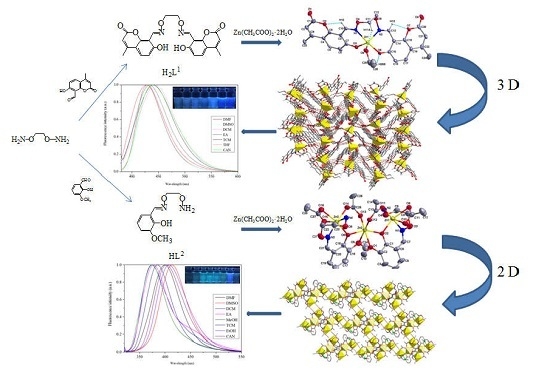
:Two newly designed complexes, [Zn(L1)(EtOH)] (1) and [{Zn(L2)(OAc)2}2Zn]·CHCl3 (2) derived from salamo and half-salamo chelating ligands (H2L1 and HL2) have been synthesized and characterized by elemental analyses, IR and UV-VIS spectra, fluorescence spectra, and X-ray crystallography. Complex 1 shows a slightly distorted tetragonal pyramid and forms an infinite 3D supramolecular structure. All of the Zn(II) ions in complex 2 are hexa-coordinated with slightly distorted octahedral geometries. Complex 2 possesses an infinite 2D space structure. The fluorescence titration experiments were used to characterize fluorescence properties of complexes 1 and 2. And the normalized fluorescent spectra exhibit that complexes 1 and 2 have favourable fluorescent emissions in different solvents.
1. Introduction
As we know, Salen-type ligands (R–CH=N–(CH2)2–N=CH–R) and their metal complexes have been extensively investigated in modern coordination chemistry for several decades [1,2,3,4,5], which have been extensively investigated in potential application in biological fields [6,7,8,9,10,11,12,13], electrochemical conducts [14,15], nonlinear optical materials [16,17,18,19,20], magnetic materials [21,22,23,24,25], luminescence properties [26,27,28,29,30,31,32], and supramolecular architecture [33,34,35,36,37], and so on. Chemical modifications of substituent or functional groups in the Salen N2O2 ligands are effective in exchanging the structures or the main functions of complexes, such as salamo ligand, a Salen analogue, (R–CH=N–O–(CH)n–O–N=CH–R) is one of the most versatile ligands and the large electronegativity of oxygen atoms is expected to strongly affect the electronic properties of the N2O2 coordination sphere, which can lead to different and novel structures and properties of the resulting complexes [38].
Due to the unique structure of salamo-type complexes, a study shown that it is at least 104 times more stable than salen-type complexes [39]. The Zn(II) ion does not produce spectroscopic or magnetic signals because of its 3d104s0 electronic configuration, when the Zn(II) ion forms complexes with ligands, the complexes generally have fluorescence properties [40,41]. Although these salamo-type Zn(II) complexes are currently being studied and developed, the solvent effects on the salamo-type Zn(II) complexes are still very rare. In order to further study the syntheses, crystal structures and fluorescence properties of the Zn(II) complexes with the salamo-type ligands, herein, two new complexes [Zn(L1)(EtOH)] (1) and [{Zn(L2)(OAc)2}2Zn]·CHCl3 (2) with salamo and half-salamo ligands H2L1 and HL2 have been reported, especially the study of the half-salamo ligand and its complex is reported firstly. Half-salen ligands and their metal complexes have been extensively investigated in modern coordination chemistry for a long time [42,43], but at present, no literature has shown that half-salamo ligands and their metal complexes have been synthesized.
2. Experimental
2.1. Materials and Methods
7-Hydroxyl-4-methyl-coumarin and 3-methoxysalicylaldehyde of 98% purity were purchased from Alfa Aesar and used without further purification (New York, NY, USA). The other reagents and solvents were analytical grade reagents from Tianjin Chemical Reagent Factory (Tianjin, China).
C, H and N analyses were obtained using a GmbH VarioEL V3.00 automatic elemental analysis instrument (Berlin, Germany). Elemental analyses for zinc were detected with an IRIS ER/S-WP–1 ICP atomic emission spectrometer (Berlin, Germany). Melting points were obtained by the use of a microscopic melting point apparatus made by Beijing Taike Instrument Company Limited and were uncorrected. IR spectra were recorded on a Vertex70 FT-IR spectrophotometer, with samples prepared as KBr (500–4000 cm−1) and CsI (100–500 cm−1) pellets (Bruker AVANCE, Billerica, MA, USA). UV-VIS absorption spectra were recorded on a Shimadzu UV-3900 spectrometer (Shimadzu, Japan). Luminescence spectra in solution were recorded on a Hitachi F-7000 spectrometer (Shimadzu, Japan). 1H NMR spectra were determined by a German Bruker AVANCE DRX-400 spectrometer (Bruker AVANCE, Billerica, MA, USA). X-ray single crystal structure determinations were carried out on a Bruker Smart Apex CCD diffractometer (Bruker AVANCE, Billerica, MA, USA).
2.2. Synthesis of H2L1
The major reaction steps involved in the synthesis of H2L1 and HL2 are given in Scheme 1. 8-Formyl-7-hydroxy-4-methylcoumarin was prepared according to reported procedure [44]. 1,2-Bis(aminooxy)ethane was synthesized following the literature [45,46,47].
H2L1: A solution of 8-formyl-7-hydroxyl-4-methyl-coumarin (768.76 mg, 2.9 mmol) in methanol (25 mL) was added to a solution of 1,2-bis(aminooxy)ethane (92.00 mg, 1.0 mmol) in methanol (25 mL). The suspension solution was stirred and refluxed at 65 °C for 4 h, and then a yellowish solid of the salamo-type ligand (H2L) was obtained, which was collected by suction filtration. Yield: 83.2%, m.p. 287-288 °C. Anal. Calcd for C24H20N2O8 (%): C, 62.07; H, 4.34; N, 6.03. Found: C, 61.84; H, 4.41; N, 6.06. 1H NMR (400 MHz, CDCl3), δ 10.72 (s, 2H), 8.95 (s, 2H), 7.50 (d, J = 8.9 Hz, 2H), 6.93 (d, J = 8.9 Hz, 2H), 6.14 (s, 2H), 4.54 (s, 4H), 2.40 (s, 6H).
2.3. Synthesis of HL2
HL2: An methanol solution (25 mL) of 3-methoxysalicylaldehyde (1 mmol, 152.6 mg) was added dropwisely to 1,2-bis(aminooxy)ethane (1.5 mmol, 138.0 mg) in methanol solution (25 mL). The resulting mixed solution was heated for 3 h between 55 and 60 °C temperature range. The solution was concentrated in vacuo and the residue was purified by column chromatography (SiO2, chloroform/ethyl acetate, 30:1) to afford a colourless flocculent crystalline solid, then the half-salamo-type ligand (HL2) was obtained, which was collected by suction filtration. Yield: 79.4%. m.p. 91-92 °C. Anal. Calc. for C10H14N2O4 (%): C 53.09; H 6.24; N 12.38. Found: C 53.21; H 6.19; N 12.29. 1H NMR (400 MHz, CDCl3), δ 9.87 (s, 1H), 8.22 (s, 1H), 6.91 (dd, J = 7.9, 1.5 Hz, 1H), 6.86 (s, 1H), 6.80 (dd, J = 7.7, 1.7 Hz, 1H), 5.52 (s, 2H), 4.40–4.33 (m, 2H), 4.00–3.94 (m, 2H), 3.91 (s, 3H).
2.4. Synthesis of Complex 1
To a ethanol solution (2 mL) of zinc(II) acetate dehydrate (0.01 mmol, 2.19 mg), and a solution of H2L1 (0.01 mmol, 4.64 mg) in 6 mL of dichloromethane was added dropwise, and immediately the mixed solution colour changed to yellow. The mixture solution was filtered and the filtrate was allowed to stand for two weeks. Through partial solvent evaporation, single crystals suitable for X-ray diffraction analysis were obtained after two weeks. Yield: 48.2%. Anal. Calcd for C26H24N2O9Zn ([Zn(L1)(EtOH)] (1)) (%): C, 54.42; H, 4.22; N, 4.88; Zn, 11.39. Found: C, 54.29; H, 4.29; N, 4.80; Zn, 11.25.
2.5. Synthesis of Complex 2
To a methanol solution (1 mL) of zinc(II) acetate dehydrate (0.03 mmol, 6.57 mg), and a solution of HL2 (0.02 mmol, 9.28 mg) in 2 mL of chloroform was added dropwise, The colour of the mixing solution turned to yellow immediately, then the mixture was filtered and the filtrate was obtained. The single crystals suitable for X-ray diffraction studies were obtained by vapour diffusion of diethyl ether into the filtrate for two days at room temperature. Yield: 52.6%. Anal. Calcd for C29H39Cl3N4O16Zn3 ([{ZnL2(OAc)2}2Zn]·CHCl3 (2)) (%): C, 34.76; H, 3.92; N, 5.59; Zn, 19.57. Found: C, 34.55; H, 3.98; N, 5.37; Zn, 19.26.
2.6. Crystal Structure Determinations of Complexes 1 and 2
The crystal diffractometer provides a monochromatic beam of Mo Kα radiation (0.71073 Å) produced using Graphite monochromator from a sealed Mo X-ray tube was used for obtaining crystal data for complexes 1 and 2 at 173.00(10) and 292.38(10), respectively. The LP factor semi-empirical absorption corrections were applied using the SADABS program. The structures were solved by the direct methods (SHELXS-2014) [48]. The H atoms were included at the calculated positions and constrained to ride on their parent atoms. All non-hydrogen atoms were refined anisotropically using a full-matrix least-squares procedure on F2 with SHELXL-2014 [48]. The crystal data and experimental parameters relevant to the structure determinations are listed in Table 1.
Crystallographic data have been deposited with the Cambridge Crystallographic Data Centre as supplementary publication, No. CCDC 1564063 and 1564062 for complexes 1 and 2. Copies of the data can be obtained free of charge on application to CCDC, 12 Union Road, Cambridge CB21EZ, UK (Telephone: (44) 01223 762910; Fax: +44-1223-336033; E-mail: deposit @ccdc.cam.ac.uk). These data can be also obtained free of charge at www.ccdc.cam. Ac.uk/conts/retrieving.html.
3. Results and Discussion
Complexes 1 and 2 constructed from salamo and half-salamo chelating ligands (H2L1 and HL2) have been synthesized, and characterized by IR spectra, UV-VIS spectra, and X-ray crystallography analyses. The fluorescence titration experiments were used to characterize fluorescence properties of complexes 1 and 2. The normalized fluorescent spectra exhibits that complexes 1 and 2 have favourable fluorescent emissions in different solvents.
3.1. IR Spectra
The FT-IR spectra of H2L1 and HL2 with their corresponding complexes 1 and 2 exhibit various bands in the 4000–400 cm−1 region (Figure 1). A typical C=N stretching band of the free ligands H2L1 and HL2 appears at 1619 and 1604 cm−1, and that of complexes 1 and 2 at 1581 and 1597 cm−1, respectively [49]. The C=N stretching frequencies are shifted to low frequencies, indicating that the Zn(II) atoms are coordinated by azomethine nitrogen atoms of (L1)2−and (L2)1− moieties. Therefore, the conclusion could be made that H2L1 and HL2 coordinated with Zn(II) atoms [50]. The typical C=O stretching band at 1728 cm−1 was exhibited by the free ligands H2L1, where at 1712 cm−1 show the C=O stretching band in complex 1. The free ligands H2L1 and HL2 exhibit Ar–O stretching frequencies at 1288 and 1249 cm−1, while the Ar–O stretching frequencies of the complexes 1 and 2 appear at 1226 and 1242 cm−1, respectively. The Ar–O stretching frequencies are shifted to low frequencies, which could be evidence of the Zn–O bond formation between Zn(II) atoms and oxygen atoms of phenolic groups [51].
The far-IR spectra (550–100 cm−1) of both complexes 1 and 2 were also obtained so as to identify the bonds of Zn–O and Zn–N frequencies. The bands at 447 and 463 cm−1 of complexes 1 and 2 can be attributed to ν(Zn–O), while the bands at 516 and 564 cm−1 are assigned to ν(Zn–N) [52].
3.2. Crystal Structure of Complex 1
As depicted in Figure 2 and Table 2, complex 1 crystallizes in the monoclinic space group P21/n, which consists of one Zn(II) ion, one completely deprotonated (L1)2− unit, and one coordinated ethanol molecule. The Zn(II) ion is penta-coordinated by two oxime nitrogen (N1 and N2) atoms and two phenoxo oxygen (O3 and O6) atoms, the four atoms are all from one deprotonated (L)2− unit, and one oxygen (O9) atom from the coordinated ethanol molecule (Figure 2a). The coordination environment around the Zn(II) ion is best described as a slightly distorted trigonal bipyramidal geometry, which obtains the geometry adopted by the Zn(II) ion, and the τ value was estimated to be τ = 0.845 (Figure 2b) [53,54]. The phenolic oxygen (O3) and the oxime nitrogen (N2) of the (L1)2− unit and one oxygen (O9) atom of the coordinated ethanol molecule constitute, together, the basal plane (Zn1-O3, 1.940(5) Å; Zn1-N2, 2.036(7) Å and Zn1-O9, 2.049(6) Å), and other phenolic oxygen (O6) and oxime nitrogen (N1) atoms of the (L1)2− unit occupy the axial positions (Zn1-O6, 1.994(5) and Zn1-N1, 2.187(7) Å). The three coordination atoms on the base plane and the Zn(II) ion is 0.062(3) Å displaced from the mean plane [55,56]. Additionally, four of the intramolecular C10–H10···O2, C11–H11A···O9, C13–H13···O7, and C25–H25B···O6 hydrogen bonds were formed (Table 3). The protons (-C10H10) and (-C13H13) of (L1)2− unit are hydrogen bonded to two of ester oxygen (O2 and O7) atoms of (L1)2− units, respectively, and the proton (-C11H11A) of the (L1)2− unit is hydrogen bonded to one oxygen (O9) atom of the coordinated ethanol molecule. Meanwhile, the proton (-C25H25B) of the coordinated ethanol molecule is hydrogen bonded to one phenoxo oxygen (O6) atom of the (L1)2− unit. The formation of intramolecular hydrogen bonds may result in a relatively stable chemical property of complex 1 [57,58].
As shown in Figure 3 and Table 3, six pairs of intermolecular hydrogen bonds, O9-H9···O8, C2-H2···O4, C11-H11B···O8, C12-H12A···O1, C12-H12B···O3, and C21-H21···O3 are formed. In addition, the Cg3 (O2–C1–C2–C3–C4–C9) of pyrone rings as acceptors forms one hydrogen bond with the protons (-C26H26B) of coordinated ethanol molecules. The space skeleton of complex 1 adopts a 3D supramolecular structure by the action of hydrogen bond and C-H···π stacking interactions [34,59,60].
3.3. Crystal Structure of Complex 2
X-ray crystallographic analysis of complex 2 reveals an asymmetric trinuclear structure. It crystallizes in the triclinic system, space group P-1, consists of three Zn(II) ions, two completely deprotonated (L2)1− units, four coordinated acetate ions. Selected bond lengths and angles are listed in Table 4.
As shown in Figure 4, the two terminal Zn(II) ions (Zn1 and Zn3) were both located in the cis-N2O coordination cavity of the deprotonated (L2)1− units, the carboxylate oxygen (O9 and O10) and (O15 and O16) atoms from coordinated acetate ions chelate to Zn1 and Zn3, and carboxylate oxygen (O11 and O14) atoms from the μ2-acetate bridge to Zn1 and Zn3 in axial positions (Figure 4a). The dihedral angle between the coordination planes of O10–Zn1–O2 and N2–Zn1–O9 is 4.18(2), the dihedral angle between the coordination planes of N4–Zn3–O16 and N6–Zn3–O15 is 2.12(2), indicating slight distortion octahedral geometry from the square planar structure. Then, the coordination sphere of the central Zn(II) (Zn2) atom is completed by double μ2-phenoxo oxygen (O2 and O6) atoms from two (L2)1− moieties, two μ2-acetato oxygen (O12 and O13) atoms and two oxygen (O5 and O1) atoms from methoxyl groups. As a result the central Zn2 atom finally has an O2O2O2 coordination environment. Then, all of the hexa-coordinated Zn(II) ions of complex 2 have slightly distorted octahedral symmetries (Figure 4b) [61,62].
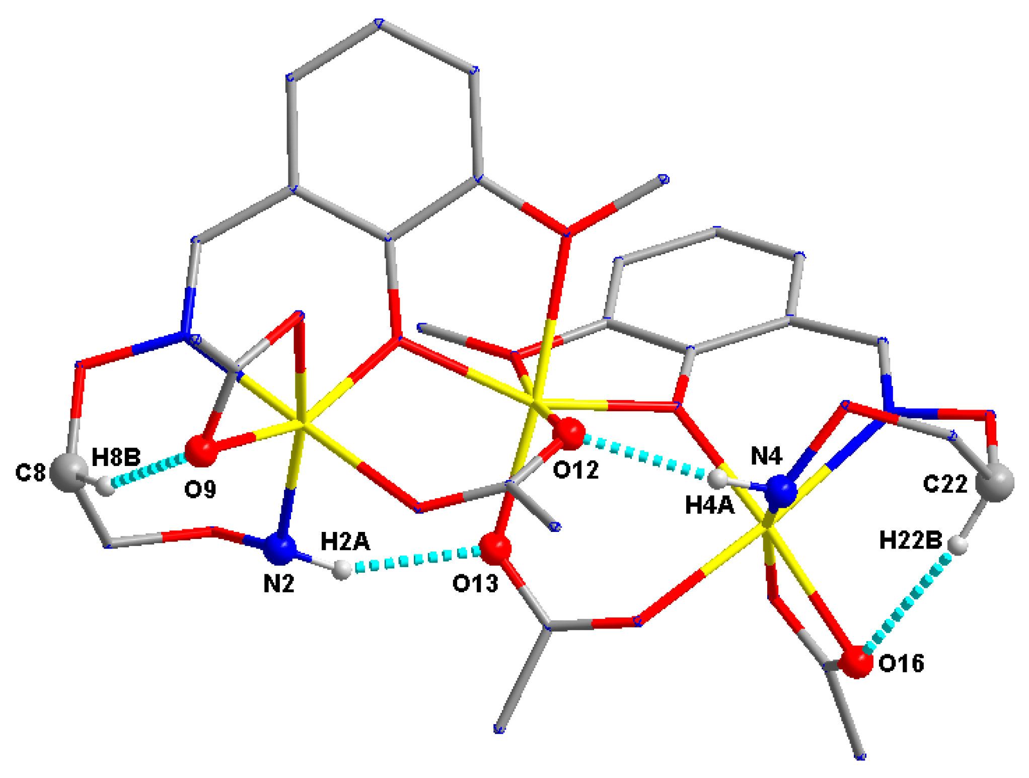
As depicted in Figure 5, in complex 2, four pairs of intramolecular hydrogen bonds N2-H2A···O13, N4-H4A···O12, C8-H8B···O9 and C22-H22B···O16 are formed. The protons (-N2H2A and -N4H4A) of (L2)1‒ units form hydrogen bonds with two oxygen (O13 and O12) atoms of μ2-acetate ions, respectively. The protons (-C8H8B and -C22H22B) from ethylenedioxime carbon atoms of (L2)1‒ units form hydrogen bonds with carboxylate oxygen (O9 and O16) atoms of coordinated acetate ions [63].
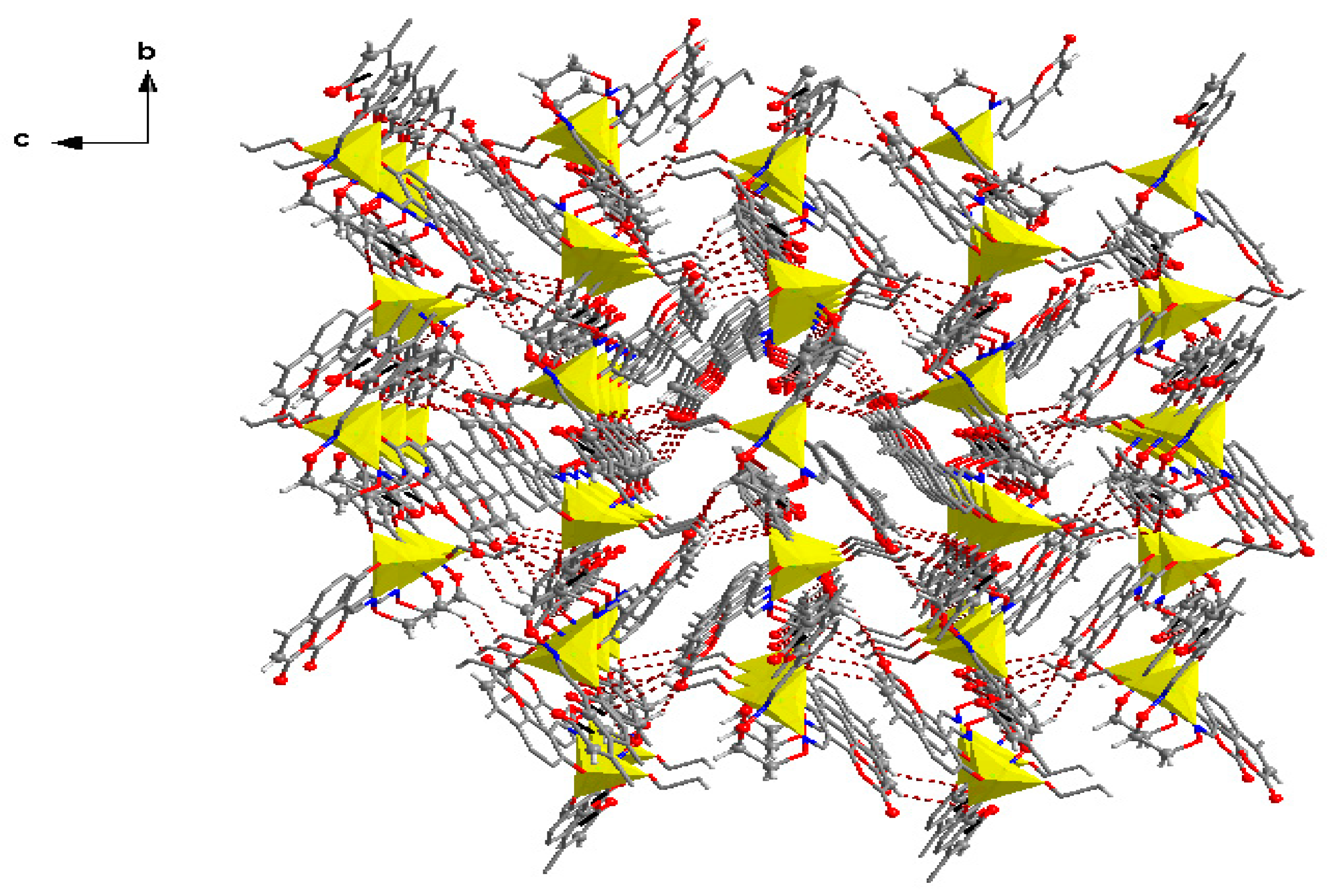
As illustrated in Figure 6 and Table 5, a large number of intermolecular hydrogen bonds and C-Cl···π, C-H···π stacking interactions in complex 2. The 2D supramolecular structure of complex 2 is composed of two parts. The first part was linked by intermolecular N2-H2B⋅⋅⋅O9, N4-H4B⋅⋅⋅O16 and C29-H29⋅⋅⋅O15 [41,64] hydrogen bonding interactions. The other part was made up of the C-Cl···π [27], C-H···π stacking interactions. The Cg7 (C15-C20) and Cg6 (C1-C6) of phenyl rings as acceptors form two hydrogen bonds with the protons (-C29Cl2 and -C10H10B) of adjacent molecules. The intermolecular hydrogen bonds and C-Cl···π, C-H···π stacking interactions of complex 2 not only make its spatial structure more diversified, but also may cause better chemical stability [27,59].
3.4. UV-VIS Spectra
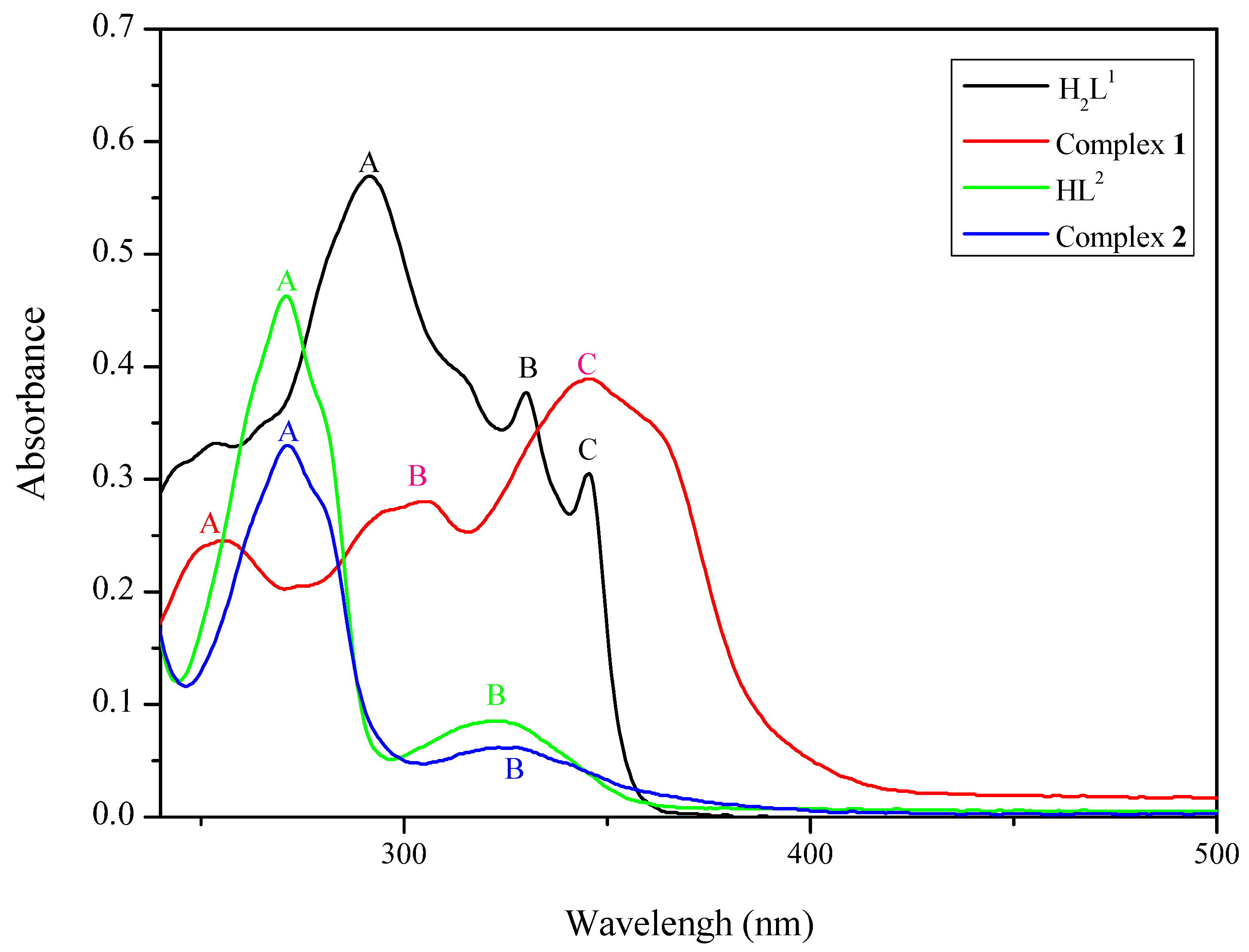
The UV-VIS absorption spectra of the free ligands H2L1 and HL2 with their corresponding complexes 1 and 2 in the dichloromethane solutions (1.0 × 10⁻⁵ mol/L) at 298 K are shown in Table 6 and Figure 7.
Obviously, the absorption peaks of the ligand H2L1 and HL2 differ from those of their corresponding complexes 1 and 2 . The absorption spectrum of the free salamo-type ligand H2L1 consists of three relatively intense bands centered at 291, 329 and 345 nm, which may be assigned to the π-π* transitions of the phenyl rings of coumarin and the oxime group [44,65]. Upon coordination of the ligand, the absorption intensities are weakened compared with the free ligand H2L1, which indicate that the oxime nitrogen atoms are involved in coordination to the Zn(II) atoms. Likewise, the absorption spectrum of the half-salamo ligand HL2 consists of two relatively intense bands centred at 271 and 323 nm, which may be assigned to the π-π* transitions of the phenyl rings and the oxime group [45,65]. On the other hand, because of complex 2 is synthesized by the half-salamo ligand HL2, when the Zn(II) atoms coordinated to HL2, the conjugate system of complex 2 not change greatly compared with complex 1, which leads to the absorption spectra were almost unchanged before and after the complexation. Upon coordination of the ligand HL2, the absorption intensities are weakened compared with the free ligand HL2, which indicate that the oxime nitrogen atoms are involved in coordination with the Zn(II) atoms [37,66].
3.5. Fluorescence Properties
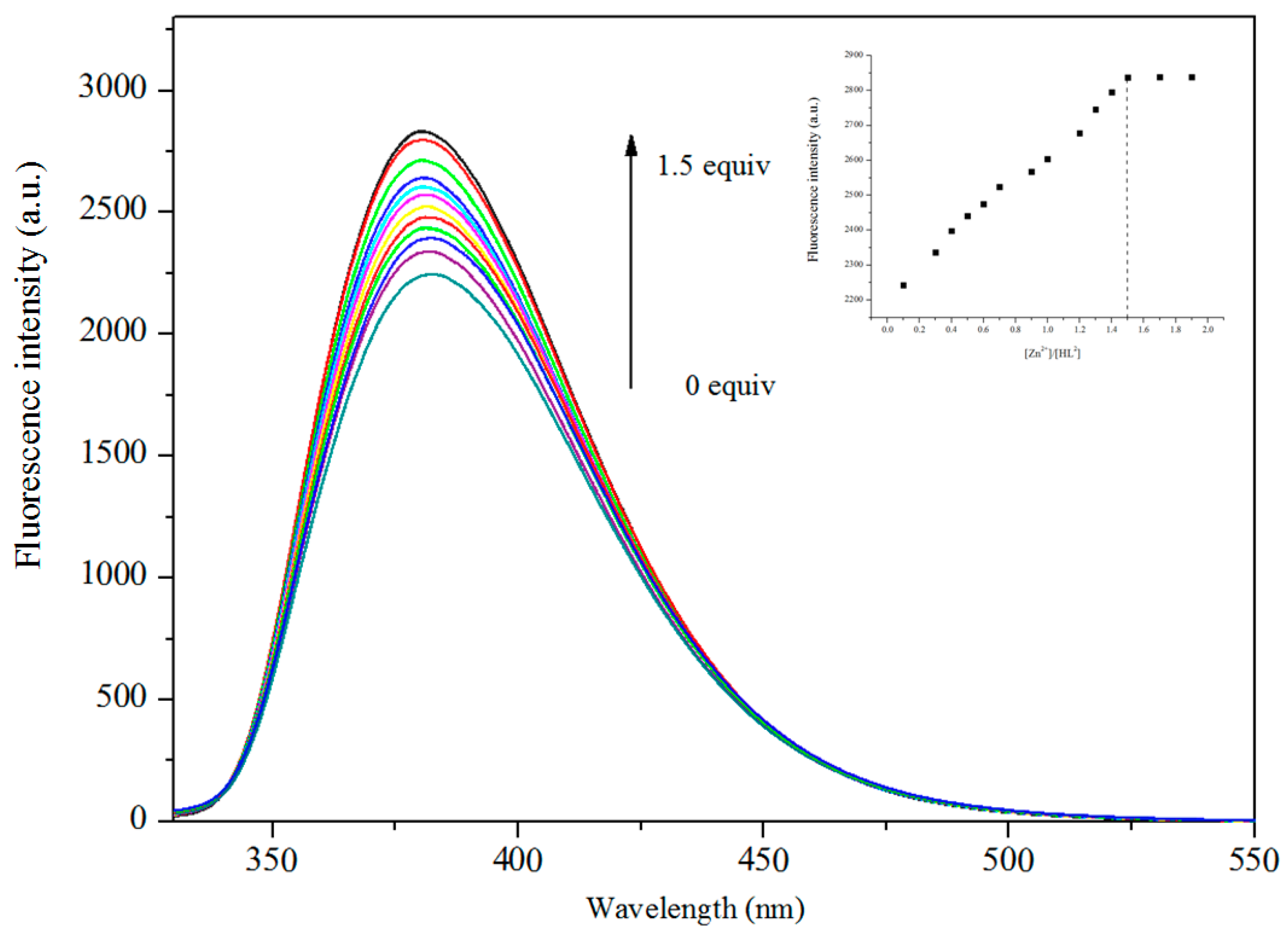
The fluorescence titration experiments of H2L1 and HL2 were determined in DMF solution (2.0 × 10⁻⁵ mol·L−1) with Zn(OAc)2·2H2O in methanol solution (1 × 10⁻³ mol·L−1) are shown in Figure 8 and Figure 9.
The free ligand H2L1 appears as an intense emission peak at 432 nm. With the fluorescence titration experiment, upon the addition of Zn2+, gradual changes in the fluorescence spectra. And the fluorescence intensity increased significantly. When the added amount of Zn2+ reached 1.0 equiv., the fluorescence emission intensity became stable, which indicates a 1:1 stoichiometry between Zn2+ and H2L1. The enhancement of fluorescence is due to the coordination of metal ions with ligands [67]. Likewise, Complex 2 displays enhanced emission intensities compared to the corresponding ligand (HL2) when excited at 380 nm. When the added amount of Zn2+ reached 1.5 equiv., the fluorescence emission intensity became steady. The result is corresponding to the crystal structure of complex 2 [68].
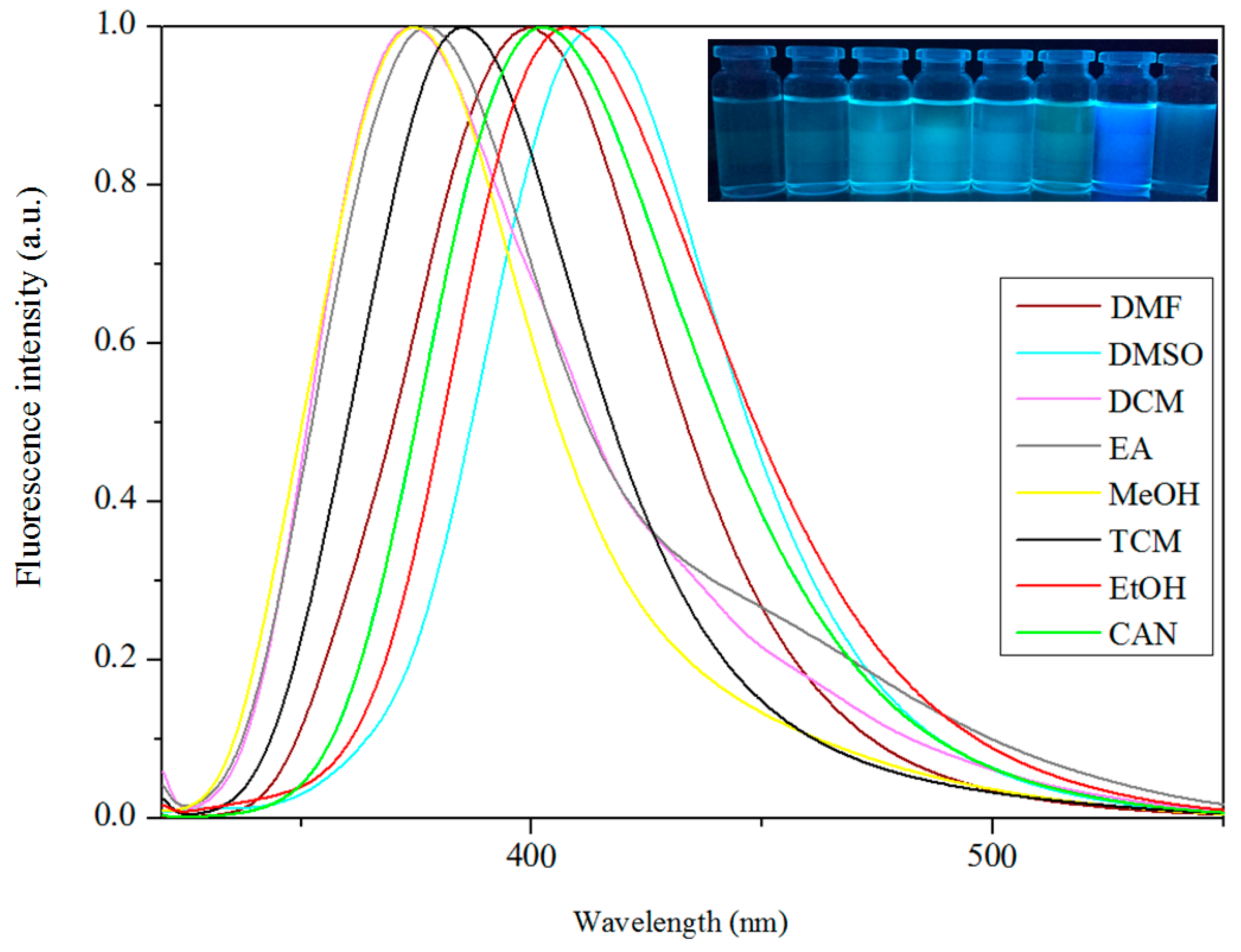
For research the solvent effect in fluorescence spectra of complexes 1 and 2, the fluorescence spectra of complex 1 and 2 in a series of solvents were examined and are shown in Table 7 and Figure 10 and Figure 11.
The normalized fluorescent spectra of complexes 1 and 2 are shown in Figure 12 and Figure 13. Additionally, the fluorescence image of complexes 1 and 2 upon irradiation with a 365 nm UV lamp also indicated that the metal complexes 1 and 2 have promising applications as fluorescent materials. The solvent effect brings pivotal effect to the photoluminescence of complexes 1 and 2.
As we know, many fluorescent complexes, especially those containing polar substituents on aromatic rings, are susceptible to solvents [27]. Due to the difference in polarity of solvents, complex 1 exhibits the relatively strong maximum fluorescence emission with relatively low solvent polarity in TCM and DCM at 439 and 440 nm, respectively. Additionally, in solvents THF, DMF, and DMSO with higher solvent polarity, the maximum fluorescence emission is relatively weak at 428, 429, and 431 nm, respectively. In solvents of medium polarity, EA and CAN, the maximum fluorescence emission was at 433 and 439 nm, respectively. Meanwhile, complex 2 exhibits the relatively strong maximum fluorescence emission with relatively high solvent polarity in DMF, CAN, EtOH, and DMSO at 399, 402, 407, and 414 nm, respectively. In solvents DCM, TCM, and EA with lower solvent polarity, the maximum fluorescence emission is relatively weak at 372, 384 and 377 nm, respectively. Furthermore, the maximum fluorescence emission unusually appears at 373 nm in MeOH solvent. The influence of the solvent effect changes the luminescent properties of complexes 1 and 2, making its application areas broad [27,69].
4. Conclusions
In summary, we have reported the successful syntheses and characterizations of two newly-designed complexes, [Zn(L1)(EtOH)] (1) and [{Zn(L2)(OAc)2}2Zn]·CHCl3 (2), derived from salamo and half-salamo chelating ligands (H2L1 and HL2). Complex 1 includes one Zn(II) ion, one completely deprotonated (L1)2− unit and one coordinated ethanol molecule, which shows a slightly distorted trigonal bipyramidal geometry and forms an infinite 3D supramolecular structure. Complex 2 includes three Zn(II) ions, two completely deprotonated (L2)1− moieties, four coordinated acetate ions, and possesses an infinite 2D space structure. The normalized fluorescent spectra exhibit that complexes 1 and 2 have favourable fluorescent emissions in different solvents.
Supplementary Materials
Supplementary File 1Acknowledgements
This work was supported by the National Natural Science Foundation of China (21361015, 21761018), the Outstanding Research Platform (Team), and the Graduate Student Guidance Team Building Fund of Lanzhou Jiaotong University (260001), which are gratefully acknowledged.
Author Contributions
Wen-Kui Dong and Lei Gao conceived and designed the experiments; Fei Wang performed the experiments; Yang Zhang analyzed the data; Wen-Kui Dong contributed reagents/materials/analysis tools; Xiu-Yan Dong wrote the paper.
Conflicts of Interest
The authors declare no competing financial interests.
References
- Dong, W.K.; Lan, P.F.; Zhou, W.M.; Zhang, Y. Salamo-type trinuclear and tetranuclear cobalt(II) complexes based on a new asymmetry salamo-type ligand: Syntheses, crystal structures and fluorescence properties. J. Coord. Chem. 2016, 65, 1272–1283. [Google Scholar] [CrossRef]
- Dong, X.Y.; Akogun, S.F.; Zhou, W.M.; Dong, W.K. Tetranuclear Zn(II) complex based on an asymmetrical Salamo-type chelating ligand: Synthesis, structural characterization, and fluorescence property. J. Chin. Chem. Soc. 2017, 64, 412–419. [Google Scholar] [CrossRef]
- Tao, C.H.; Ma, J.C.; Zhu, L.C.; Zhang, Y.; Dong, W.K. Heterobimetallic 3d–4f Zn(II)–Ln(III) (Ln = Sm, Eu, Tb and Dy) complexes with a N2O4 bisoxime chelate ligand and a simple auxiliary ligand Py: Syntheses, structures and luminescence properties. Polyhedron 2017, 128, 38–45. [Google Scholar] [CrossRef]
- Dong, Y.J.; Dong, X.Y.; Dong, W.K.; Zhang, Y.; Zhang, L.S. Three asymmetric Salamo-type copper(II) and cobalt(II) complexes: Syntheses, structures, fluorescent properties. Polyhedron 2017, 123, 305–315. [Google Scholar] [CrossRef]
- Dong, W.K.; Ma, J.C.; Dong, Y.J.; Zhao, L.; Zhu, L.C.; Sun, Y.X.; Zhang, Y. Two hetero-trinuclear Zn(II)-M(II) (M = Sr, Ba) complexes based on metallohost of mononuclear Zn(II) complex: Syntheses, structures and fluorescence properties. J. Coord. Chem. 2016, 69, 3231–3241. [Google Scholar] [CrossRef]
- Wu, H.L.; Wang, C.P.; Wang, F.; Peng, H.P.; Zhang, H.; Bai, Y.C. A new manganese(III) complex from bis(5-methylsalicylaldehyde)-3-oxapentane-1,5-diamine: Synthesis, characterization, antioxidant activity and luminescence. J. Chin. Chem. Soc. 2015, 62, 1028–1034. [Google Scholar] [CrossRef]
- Wu, H.L.; Bai, Y.C.; Zhang, Y.H.; Li, Z.; Wu, M.C.; Chen, C.Y.; Zhang, J.W. Synthesis, crystal structure, antioxidation and DNA-binding properties of a dinuclear copper(II) complex with bis(N-salicylidene)-3-oxapentane-1, 5-diamine. J. Coord. Chem. 2014, 67, 3054–3066. [Google Scholar] [CrossRef]
- Wu, H.L.; Bai, Y.; Yuan, J.K.; Wang, H.; Pan, G.L.; Fan, X.Y.; Kong, J. A zinc(II) complex with tris(2-(N-methyl)benzimidazlylmethyl)amine and salicylate: Synthesis, crystal structure, and DNA-binding. J. Coord. Chem. 2012, 65, 2839–2851. [Google Scholar] [CrossRef]
- Wu, H.L.; Pan, G.L.; Wang, H.; Wang, X.L.; Bai, Y.C.; Zhang, Y.H. Study on synthesis, crystal structure, antioxidant and DNA-binding of mono-, di- and poly-nuclear lanthanides complexes with bis(N-salicylidene)-3-oxapentane-1,5-diamine. J. Photochem. Photobiol. B Biol. 2014, 135, 33–43. [Google Scholar] [CrossRef] [PubMed]
- Wu, H.L.; Bai, Y.C.; Zhang, Y.H.; Pan, G.L.; Kong, J.; Shi, F.; Wang, X.L. Two lanthanide(III) complexes based on the schiff base N,N-Bis(salicylidene)-1,5-diamino-3-oxapentane: Synthesis, characterization, DNA-binding properties, and antioxidation. Z. Anorg. Allg. Chem. 2014, 640, 2062–2071. [Google Scholar] [CrossRef]
- Wu, H.L.; Pan, G.L.; Bai, Y.C.; Wang, H.; Kong, J.; Shi, F.; Zhang, Y.H.; Wang, X.L. Preparation, structure, DNA-binding properties, and antioxidant activities of a homodinuclear erbium(III) complex with a pentadentate Schiff base ligand. J. Chem. Res. 2014, 38, 211–217. [Google Scholar] [CrossRef]
- Wu, H.L.; Pan, G.L.; Bai, Y.C.; Wang, H.; Kong, J. Synthesis, structure, antioxidation, and DNA-bindingstudies of a binuclear ytterbium(III) complex with bis(N-salicylidene)-3-oxapentane-1,5-diamine. Res. Chem. Intermed. 2015, 41, 3375–3388. [Google Scholar] [CrossRef]
- Chen, C.Y.; Zhang, J.W.; Zhang, Y.H.; Yang, Z.H.; Wu, H.L. Gadolinium(III) and dysprosium(III) complexes with a Schiff base bis(N-salicylidene)-3-oxapentane-1,5-diamine: Synthesis, characterization, antioxidation, and DNA-binding studies. J. Coord. Chem. 2015, 68, 1054–1071. [Google Scholar] [CrossRef]
- Dong, W.K.; Ma, J.C.; Zhu, L.C.; Zhang, Y.; Li, X.L. Four new nickel(II) complexes based on an asymmetric Salamo-type ligand: Synthesis, structure, solvent effect and electrochemical property. Inorg. Chim. Acta 2016, 445, 140–148. [Google Scholar] [CrossRef]
- Chai, L.Q.; Zhang, K.Y.; Tang, L.J.; Zhang, J.Y.; Zhang, H.S. Two mono- and dinuclear ni(II) complexes constructed from quinazoline-type ligands: Synthesis, x-ray structures, spectroscopic, electrochemical, thermal, and antimicrobial studies. Polyhedron 2017, 130, 100–107. [Google Scholar] [CrossRef]
- Yu, T.Z.; Zhang, K.; Zhao, Y.L.; Yang, C.H.; Zhang, H.; Qian, L.; Fan, D.W.; Dong, W.K.; Chen, L.L.; Qiu, Y.Q. Synthesis, crystal structure and photoluminescent properties of an aromatic bridged Schiff base ligand and its zinc complex. Inorg. Chim. Acta 2008, 361, 233–240. [Google Scholar] [CrossRef]
- Dong, Y.J.; Ma, J.C.; Zhu, L.C.; Dong, W.K.; Zhang, Y. Four 3d–4f heteromultinuclear zinc(II)–lanthanide(III) complexes constructed from a distinct hexadentate N2O2-type ligand: Syntheses, structures and photophysical properties. J. Coord. Chem. 2017, 70, 103–115. [Google Scholar] [CrossRef]
- Wang, L.; Ma, J.C.; Dong, W.K.; Zhu, L.C.; Zhang, Y. A novel Self–assembled nickel(II)–cerium(III) heterotetranuclear dimer constructed from N2O2-type bisoxime and terephthalic acid: Synthesis, structure and photophysical properties. Z. Anorg. Allg. Chem. 2016, 642, 834–839. [Google Scholar] [CrossRef]
- Dong, W.K.; Chen, X.; Sun, Y.X.; Yang, Y.H.; Zhao, L.; Xu, L.; Yu, T.Z. Synthesis, structure and spectroscopic properties of two new trinuclear nickel(II) clusters possessing solvent effect. Spectrochim. Acta Part A 2009, 74, 719–725. [Google Scholar] [CrossRef] [PubMed]
- Dong, W.K.; Li, G.; Wang, Z.K.; Dong, X.Y. A novel trinuclear cobalt(II) complex derived from an asymmetric Salamo-type N2O3 bisoxime chelate ligand: Synthesis, structure and optical properties. Spectrochimica Acta Part A 2014, 133, 340–347. [Google Scholar] [CrossRef] [PubMed]
- Liu, Y.A.; Wang, C.Y.; Zhang, M.; Song, X.Q. Structures and magnetic properties of cyclic heterometallic tetranuclear clusters. Polyhedron 2017, 127, 278–286. [Google Scholar] [CrossRef]
- Dong, W.K.; Ma, J.C.; Dong, Y.J.; Zhu, L.C.; Zhang, Y. Di-and tetranuclear heterometallic 3d-4f cobalt(II)-lanthanide(III) complexes derived from a hexadentate bisoxime: Syntheses, structures and magnetic properties. Polyhedron 2016, 115, 228–235. [Google Scholar] [CrossRef]
- Song, X.Q.; Liu, P.P.; Xiao, Z.R.; Li, X.; Liu, Y.A. Four polynuclear complexes based on a versatile salicylamide salen-like ligand: Synthesis, structural variations and magnetic properties. Inorg. Chim. Acta 2015, 438, 232–244. [Google Scholar] [CrossRef]
- Liu, P.P.; Wang, C.Y.; Zhang, M.; Song, X.Q. Pentanuclear sandwich-type ZnII-LnIII clusters based on a new Salen-like salicylamide ligand: Structure, near-infrared emission and magnetic properties. Polyhedron 2017, 129, 133–140. [Google Scholar] [CrossRef]
- Dong, W.K.; Ma, J.C.; Zhu, L.C.; Zhang, Y. Nine self–assembled nickel(II)–lanthanide(III) heterometallic complexes constructed from a Salamo–type bisoxime and bearing N- or O-donor auxiliary ligand: Syntheses, structures and magnetic properties. New J. Chem. 2016, 40, 6998–7010. [Google Scholar] [CrossRef]
- Dong, W.K.; Ma, J.C.; Zhu, L.C.; Zhang, Y. Self-assembled zinc(II)-lanthanide(III) heteromultinuclear complexes constructed from 3-MeOsalamo ligand: Syntheses, structures and luminescent properties. Cryst. Growth Des. 2016, 16, 6903–6914. [Google Scholar] [CrossRef]
- Chen, L.; Dong, W.K.; Zhang, H.; Zhang, Y.; Sun, Y.X. Structural variation and luminescence properties of tri- and dinuclear CuII and ZnII complexes constructed from a naphthalenediol-based bis(Salamo)-type ligand. Cryst. Growth Des. 2017, 17, 3636–3648. [Google Scholar] [CrossRef]
- Song, X.Q.; Peng, Y.J.; Chen, G.Q.; Wang, X.R.; Liu, P.P.; Xu, W.Y. Substituted group-directed assembly of Zn(II) coordination complexes based on two new structural related pyrazolone based Salen ligands: Syntheses, structures and fluorescence properties. Inorg. Chim. Acta. 2015, 427, 13–21. [Google Scholar] [CrossRef]
- Dong, W.K.; Zhang, J.; Zhang, Y.; Li, N. Novel multinuclear transition metal(II) complexes based on an asymmetric Salamo-type ligand: Syntheses, structure characterizations and fluorescent properties. Inorg. Chim. Acta. 2016, 444, 95–102. [Google Scholar] [CrossRef]
- Dong, W.K.; Akogun, S.F.; Zhang, Y.; Dong, X.Y. A reversible “turn-on” fluorescent sensor for selective detection of Zn2+. Sensors Actuators B Chem. 2017, 238, 723–734. [Google Scholar] [CrossRef]
- Dong, Y.J.; Li, X.L.; Zhang, Y.; Dong, W.K. A highly selective visual and fluorescent sensor for Pb2+ and Zn2+ and crystal structure of Cu2+ complex based-on a novel single-armed Salamo-type bisoxime. Supramol. Chem. 2017, 29, 518–527. [Google Scholar] [CrossRef]
- Dong, W.K.; Li, X.L.; Wang, L.; Zhang, Y.; Ding, Y.J. A new application of Salamo-type bisoximes: as a relay-sensor for Zn2+/Cu2+ and its novel complexes for successive sensing of H+/OH‒. Sens. Actuators B Chem. 2016, 229, 370–378. [Google Scholar] [CrossRef]
- Zhao, L.; Dang, X.T.; Chen, Q.; Zhao, J.X.; Wang, L. Synthesis, crystal structure and spectral properties of a 2D supramolecular copper(II) complex with 1-(4-{[(E)-3-ethoxyl-2-hydroxybenzylidene]amino}phenyl)ethanone oxime. Synth. React. Inorg. Met. -Org. Nano-Met. Chem. 2013, 43, 1241–1246. [Google Scholar] [CrossRef]
- Sun, Y.X.; Xu, L.; Zhao, T.H.; Liu, S.H.; Liu, G.H.; Dong, X.T. Synthesis and crystal structure of a 3D supramolecular copper(II) complex with 1-(3-{[(E)-3-bromo-5-chloro-2-hydroxybenzylidene]amino}phenyl) ethanone oxime. Synth. React. Inorg. Met. -Org. Nano-Met. Chem. 2013, 43, 509–513. [Google Scholar] [CrossRef]
- Sun, Y.X.; Dong, W.K.; Wang, L.; Zhao, L.; Yang, Y.H. Synthesis and crystal structure of nickel(II) cluster with salen-type bisoxime ligand. Chinese J. Inorg. Chem. 2009, 25, 1478–1482. [Google Scholar]
- Sun, Y.X.; Zhang, S.T.; Ren, Z.L.; Dong, X.Y.; Wang, L. Synthesis, characterization, and crystal structure of a new supramolecular CdII complex with halogen-substituted salen-type bisoxime. Synth. React. Inorg. Met.-Org. Nano-Met Chem. 2013, 43, 995–1000. [Google Scholar] [CrossRef]
- Dong, W.K.; Zhang, X.Y.; Zhao, M.M.; Li, G.; Dong, X.Y. Syntheses and crystal structures of 5-Methoxy-6′-hydroxy-2,2′-[ethylenedioxybis(nitrilomethylidyne)]diphenol and its tetranuclear zinc(II) complex. Chin. J. Inorg. Chem. 2014, 30, 710–716. [Google Scholar]
- Zhou, J.A.; Tang, X.L.; Cheng, J.; Ju, Z.H.; Yang, L.Z.; Liu, W.S.; Chen, C.Y.; Bai, D.C. An 1 3,4-oxadiazole-based off-on fluorescent chemosensor for Zn2+ in aqueous solution and imaging application in living cells. Dalton Trans. 2012, 41, 10626–10632. [Google Scholar] [CrossRef] [PubMed]
- Akine, S.; Taniguchi, T.; Dong, W.K.; Masubuchi, S.; Nabeshima, T. Oxime-Based Salen-Type Tetradentate Ligands with High Stability against Imine Metathesis Reaction. J. Org. Chem. 2005, 70, 1704–1711. [Google Scholar] [CrossRef] [PubMed]
- Akine, S.; Dong, W.K.; Nabeshima, T. Octanuclear zinc(II) and cobalt(II) clusters produced by cooperative tetrameric assembling of oxime chelate ligands. Inorg. Chem. 2006, 454, 677–4684. [Google Scholar] [CrossRef] [PubMed]
- Song, X.Q.; Cheng, G.Q.; Liu, Y.A. Enhanced Tb(III) luminescence by d10 transition metal coordination. Inorg. Chim. Acta 2016, 450, 386–394. [Google Scholar] [CrossRef]
- Darensbourg, D.J.; Karroonnirun, O.; Wilson, S.J. Ring-Opening Polymerization of Cyclic Esters and Trimethylene Carbonate Catalyzed by Aluminum Half-Salen Complexes. Inorg. Chem. 2011, 50, 6775–6787. [Google Scholar] [CrossRef] [PubMed]
- Darensbourg, D.J.; Karroonnirun, O. Stereoselective Ring-Opening Polymerization of rac-Lactides Catalyzed by Chiral and Achiral Aluminum Half-Salen Complexes. Organometallics 2010, 29, 5627–5634. [Google Scholar] [CrossRef]
- Dong, Y.; Li, F.J.; Jiang, X.X.; Song, F.Y.; Cheng, Y.X.; Zhu, C.J. Na+ triggered fluorescence sensors for Mg2+ detection based on a coumarin salen moiety. Org. Lett. 2011, 9, 2252–2255. [Google Scholar] [CrossRef] [PubMed]
- Dong, W.K.; Wang, Z.K.; Li, G.; Zhao, M.M.; Dong, X.Y.; Liu, S.H. Syntheses, crystal structures, and properties of a Salamo-type tetradentate chelating ligand and its pentacoordinated copper(II) complex. Z. Anorg. Allg. Chem. 2013, 639, 2263–2268. [Google Scholar] [CrossRef]
- Dong, W.K.; Feng, J.H.; Yang, X.Q. Synthesis and crystal structure of a five-coordinated cu(II) dimer with 4,4′-Dibromo-2,2′-[ethylenedioxybis(nitrilomethylidyne)]diphenol. Synth. React. Inorg. Met. Org. Nano Met. Chem. 2007, 37, 189–192. [Google Scholar] [CrossRef]
- Ma, J.C.; Dong, X.Y.; Dong, W.K.; Zhang, Y.; Zhu, L.C.; Zhang, J.T. An unexpected dinuclear Cu(II) complex with a bis(Salamo) chelating ligand: synthesis, crystal structure, and photophysical properties. J. Coord. Chem. 2016, 69, 149–159. [Google Scholar] [CrossRef]
- Sheldrick, G.M. SHELXS-97. In Program for the Solution and the Refinement of Crystal Structures; University of Gottingen: Germany, 1997. [Google Scholar]
- Dong, W.K.; Zhang, F.; Li, N.; Xu, L.; Zhang, Y.; Zhang, J.; Zhu, L.C. Trinuclear cobalt(II) and zinc(II) salamo-type complexes: Syntheses, crystal structures, and fluorescent properties. Z. Anorg. Allg. Chem. 2016, 642, 532–538. [Google Scholar] [CrossRef]
- Dong, W.K.; Zhang, L.S.; Sun, Y.X.; Zhao, M.M.; Li, G.; Dong, X.Y. Synthesis, crystal structure and spectroscopic properties of a supramolecular zinc(II) complex with N2O2 coordination sphere. Spectrochim. Acta Part A 2014, 121, 324–329. [Google Scholar] [CrossRef] [PubMed]
- Hao, J.; Li, L.H.; Zhang, J.T.; Akogun, S.F.; Wang, L.; Dong, W.K. Four homo- and hetero-bismetallic 3d/3d-2s complexes constructed from a naphthalenediol-based acyclic bis(salamo)-type tetraoxime ligand. Polyhedron 2017, 134, 1–10. [Google Scholar] [CrossRef]
- Li, L.H.; Dong, W.K.; Zhang, Y.; Akogun, S.F.; Xu, L. Syntheses, structures and catecholase activities of homo-and hetero-trinuclear cobalt(II) complexes constructed from an acyclic naphthalenediol-based bis(salamo)-type ligand. Appl. Organomet. Chem. [CrossRef]
- Addison, A.W.; Rao, T.N.; Reedijk, J.; van Rijn, J.; Verschoor, G.C. Synthesis, structure, and spectroscopic properties of copper(II) compounds containing nitrogen–sulphur donor ligands; the crystal and molecular structure of aqua[1,7-bis(N-methylbenzimidazol-2′-yl)-2,6-dithiaheptane]copper(II) perchlorate. J. Chem. Soc. Dalton Trans. 1984, 7, 1349–1356. [Google Scholar] [CrossRef]
- Konno, T.; Tokuda, K.; Sakurai, J.; Okamoto, K.I. Five-Coordinate Geometry of Cadmium(II) with Octahedral Bidentate-S,S Complex-Ligand cis(S)-[Co(aet)2(en)]+ (aet = 2-aminoethanethiolate): Synthesis, Crystal Structures and Interconversion of S-Bridged CoIIICdII Polynuclear Complexes. Bull. Chem. Soc. Jpn. 2000, 73, 2767–2773. [Google Scholar] [CrossRef]
- Chai, L.Q.; Tang, L.J.; Chen, L.C.; Huang, J.J. Structural, spectral, electrochemical and DFT studies of two mononuclear manganese(II) and zinc(II) complexes. Polyhedron 2017, 122, 228–240. [Google Scholar] [CrossRef]
- Xu, L.; Zhu, L.C.; Ma, J.C.; Zhang, Y.; Zhang, J.; Dong, W.K. Syntheses, structures and spectral properties of mononuclear CuII and dimeric ZnII complexes based on an asymmetric Salamo-type N2O2 ligand. Z. Anorg. Allg. Chem. 2015, 641, 2520–2524. [Google Scholar] [CrossRef]
- Boggs, J.M. Lipid intermolecular hydrogen bonding: influence on structural organization and membrane function. Biochim. Biophys. Acta 1987, 906, 353–404. [Google Scholar] [CrossRef]
- Karas, L.J.; Batista, P.R.; Viesser, R.V.; Tormena, C.F.; Rittner, R.; de Oliveira, P.R. Trends of intramolecular hydrogen bonding in substituted alcohols: a deeper investigation. Phys. Chem. Chem.Phys. 2017, 19, 16904–16913. [Google Scholar] [CrossRef] [PubMed]
- Mathias, J.P.; Simanek, E.E.; Whitesides, G.M. Self-Assembly through Hydrogen Bonding: Peripheral Crowding-A new strategy for the preparation of stable supramolecular aggregates based on parallel, connected CA3.cntdot.M3 rosettes. J. Am. Chem. Soc. 1994, 116, 4326–4340. [Google Scholar]
- Yabuuchi, K.; Marfo-Owusu, E.; Kato, T. A new urea gelator: incorporation of intra- and intermolecular hydrogen bonding for stable 1D self-assembly. Org. Biomol. Chem. 2003, 1, 3464–3469. [Google Scholar] [CrossRef] [PubMed]
- Chai, L.Q.; Huang, J.J.; Zhang, J.Y.; Li, Y.X. Two 1-D and 2-D cobalt(II) complexes: Synthesis, crystal structures, spectroscopic and electrochemical properties. J. Coord. Chem. 2015, 68, 1224–1237. [Google Scholar] [CrossRef]
- Sun, Y.X.; Wang, L.; Dong, X.Y.; Ren, Z.L.; Meng, W.S. Synthesis, characterization, and crystal structure of a supramolecular CoII complex containing Salen-type bisoxime. Synth. React. Inorg. Met.-Org. Nano-Met. Chem. 2013, 43, 599–603. [Google Scholar] [CrossRef]
- Wang, P.; Zhao, L. Synthesis and crystal structure of supramolecular copper(II) complex based on N2O2 coordination Sphere. Asian J. Chem. 2015, 4, 1424–1426. [Google Scholar] [CrossRef]
- Liu, P.P.; Sheng, L.; Song, X.Q.; Xu, W.Y.; Liu, Y.A. Synthesis, structure and magnetic properties of a new one dimensional manganese coordination polymer constructed by a new asymmetrical ligand. Inorg. Chim. Acta 2015, 434, 252–257. [Google Scholar] [CrossRef]
- Wang, B.J.; Dong, W.K.; Zhang, Y.; Akogun, S.F. A novel relay-sensor for highly sensitive and selective detection of Zn2+/Pic− and fluorescence on/off switch response of H+/OH‒. Sens. Actuators B Chem. 2017, 247, 254–264. [Google Scholar] [CrossRef]
- Akine, S.; Morita, Y.; Utsuno, F.; Nabeshima, T. Multiple folding structures mediated by metal coordination of acyclic multidentate ligand. Inorg. Chem. 2009, 48, 10670–10678. [Google Scholar] [CrossRef] [PubMed]
- Song, X.Q.; Liu, P.P.; Liu, Y.A.; Zhou, J.J.; Wang, X.L. Two dodecanuclear heterometallic [Zn6Ln6] clusters constructed by a multidentate salicylamide salen-like ligand: Synthesis, structure, luminescence and magnetic properties. Dalton Trans. 2016, 45, 8154–8163. [Google Scholar] [CrossRef] [PubMed]
- Guo, C.Y.; Wang, Y.Y.; Xu, K.Z.; Zhu, H.L.; Liu, P.; Shi, Q.Z. Crystal structures, bioactivities and fluorescent properties of four diverse complexes with a new symmetric benzimidazolic ligand. Polyhedron 2008, 27, 3529–3536. [Google Scholar] [CrossRef]
- Che, G.B.; Liu, C.B.; Liu, B.; Wang, Q.W.; Xu, Z.L. Syntheses, structures and photoluminescence of a series of metal-organic complexes with 1,3,5-benzenetricarboxylate and pyrazino[2,3-f][1,10]-phenanthroline ligands. Cryst. Eng. Commun. 2008, 10, 184–191. [Google Scholar] [CrossRef]
Scheme 1.
Synthetic route to H2L1and HL2.
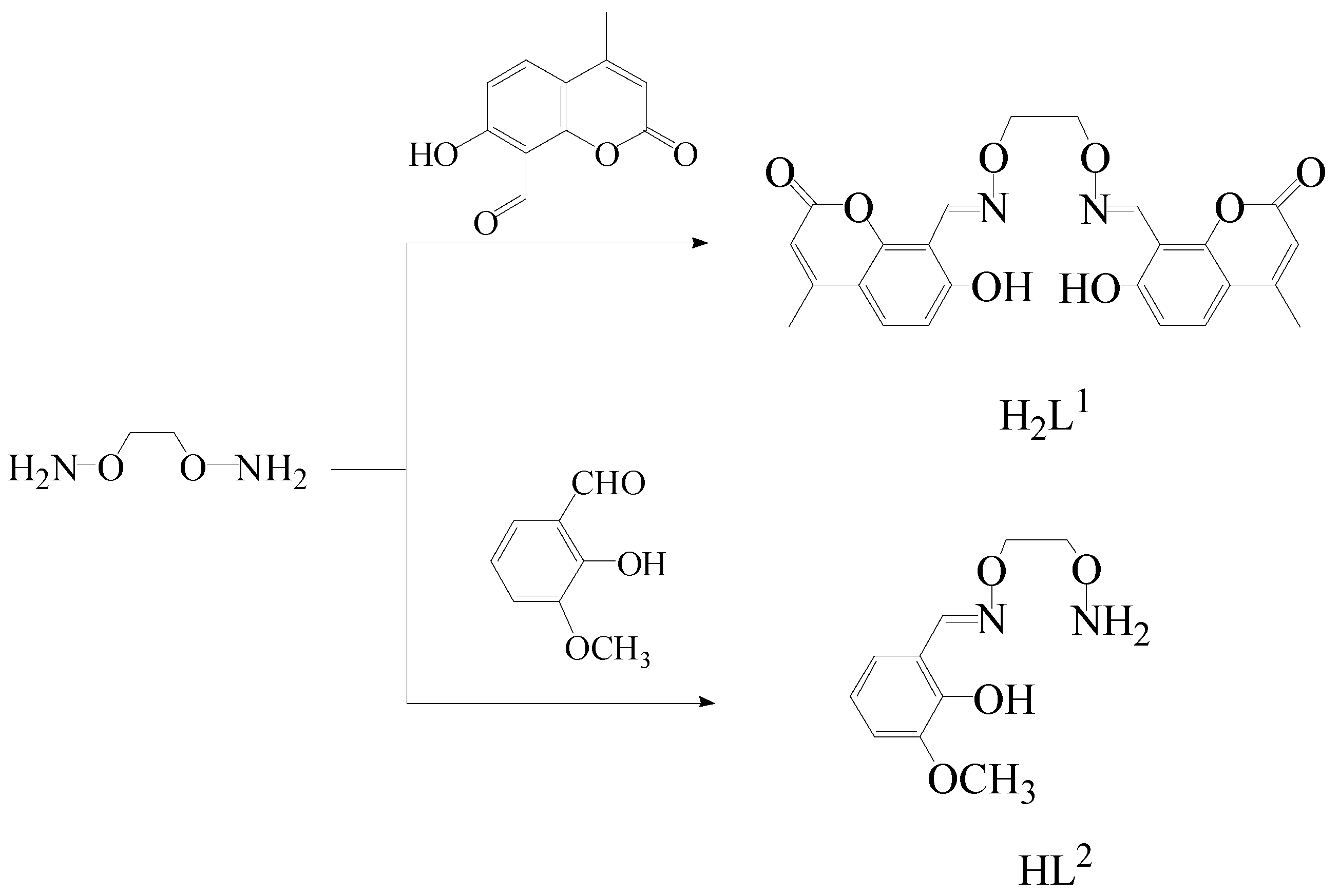
Figure 1.
The FT-IR spectra of the ligands and their complexes 1 and 2 (cm−1).
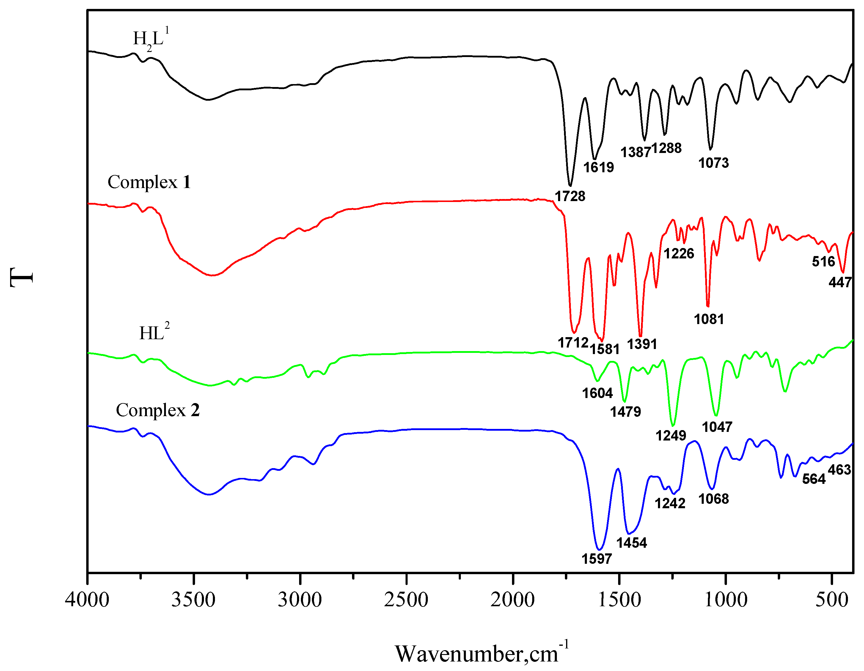
Figure 2.
(a) Molecular structure and atom numberings of complex 1 with 30% probability displacement ellipsoids (hydrogen atoms are omitted for clarity). (b) Coordination polyhedron for Zn(II) ion of complex 1.
Figure 2.
(a) Molecular structure and atom numberings of complex 1 with 30% probability displacement ellipsoids (hydrogen atoms are omitted for clarity). (b) Coordination polyhedron for Zn(II) ion of complex 1.
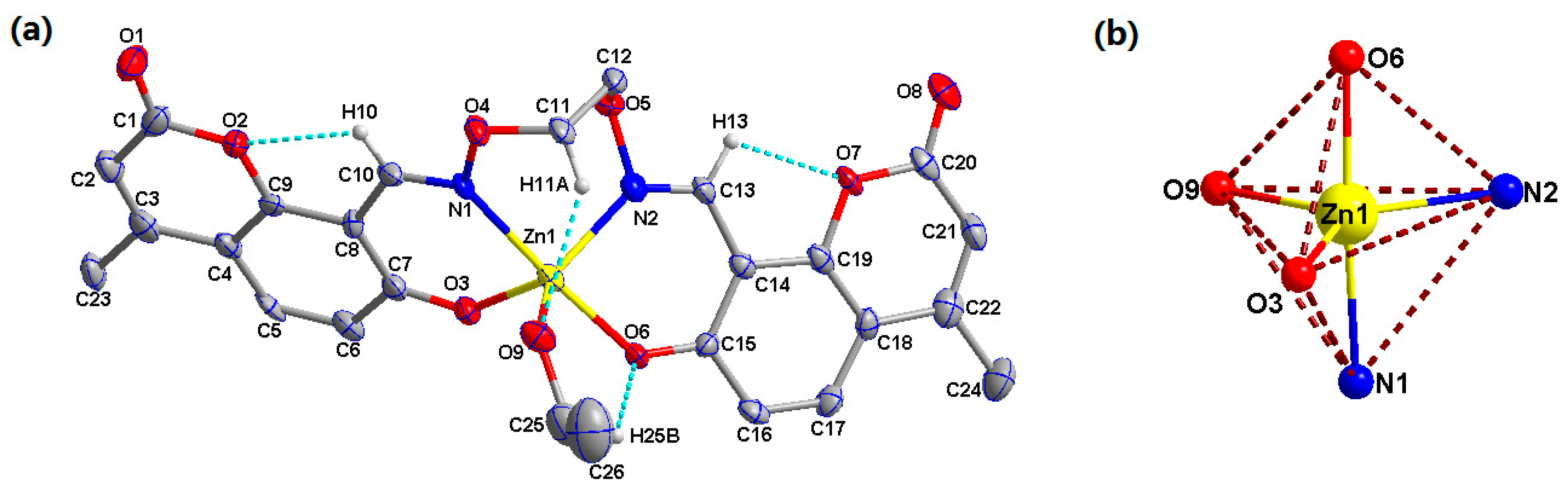
Figure 3.
View of the 3D supramolecular structure of complex 1 showing the O-H···O, C-H···O, hydrogen bonds, and C-H···π stacking interactions.
Figure 3.
View of the 3D supramolecular structure of complex 1 showing the O-H···O, C-H···O, hydrogen bonds, and C-H···π stacking interactions.

Figure 4.
(a) Molecular structure and atom numberings of complex 2 with 30% probability displacement ellipsoids (hydrogen atoms are omitted for clarity). (b) Coordination polyhedra for Zn1, Zn2, and Zn3 ions of complex 2.
Figure 4.
(a) Molecular structure and atom numberings of complex 2 with 30% probability displacement ellipsoids (hydrogen atoms are omitted for clarity). (b) Coordination polyhedra for Zn1, Zn2, and Zn3 ions of complex 2.
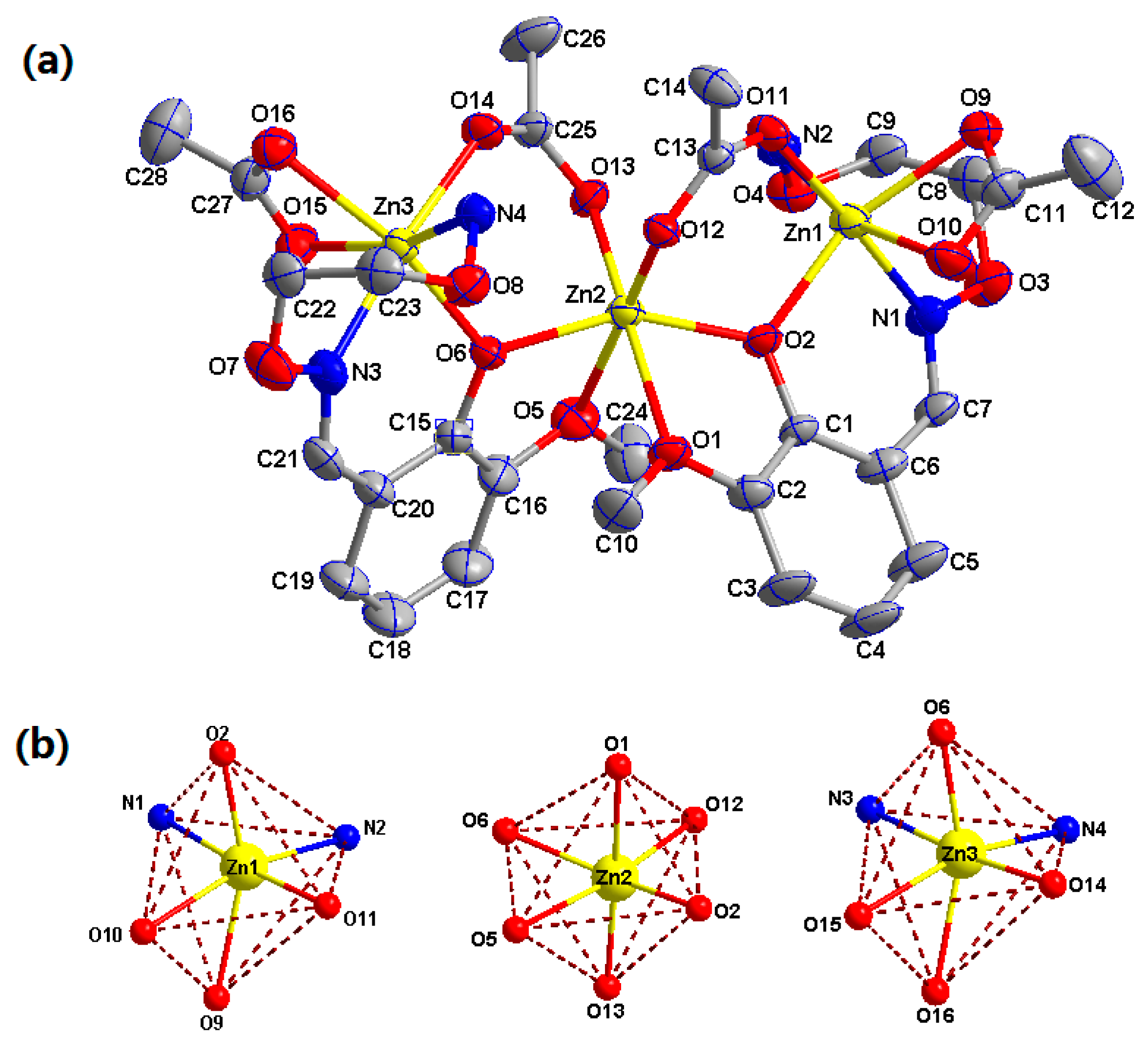
Figure 5.
Intramolecular C-H···O and N-H···O hydrogen bonds of complex 2 (hydrogen atoms, except those forming hydrogen bonds, are omitted for clarity).
Figure 5.
Intramolecular C-H···O and N-H···O hydrogen bonds of complex 2 (hydrogen atoms, except those forming hydrogen bonds, are omitted for clarity).

Figure 6.
View of the 2D supramolecular structure of complex 2 showing the N-H···O, C-H···O hydrogen bonding, and C-Cl···π, C-H···π stacking interactions.
Figure 6.
View of the 2D supramolecular structure of complex 2 showing the N-H···O, C-H···O hydrogen bonding, and C-Cl···π, C-H···π stacking interactions.
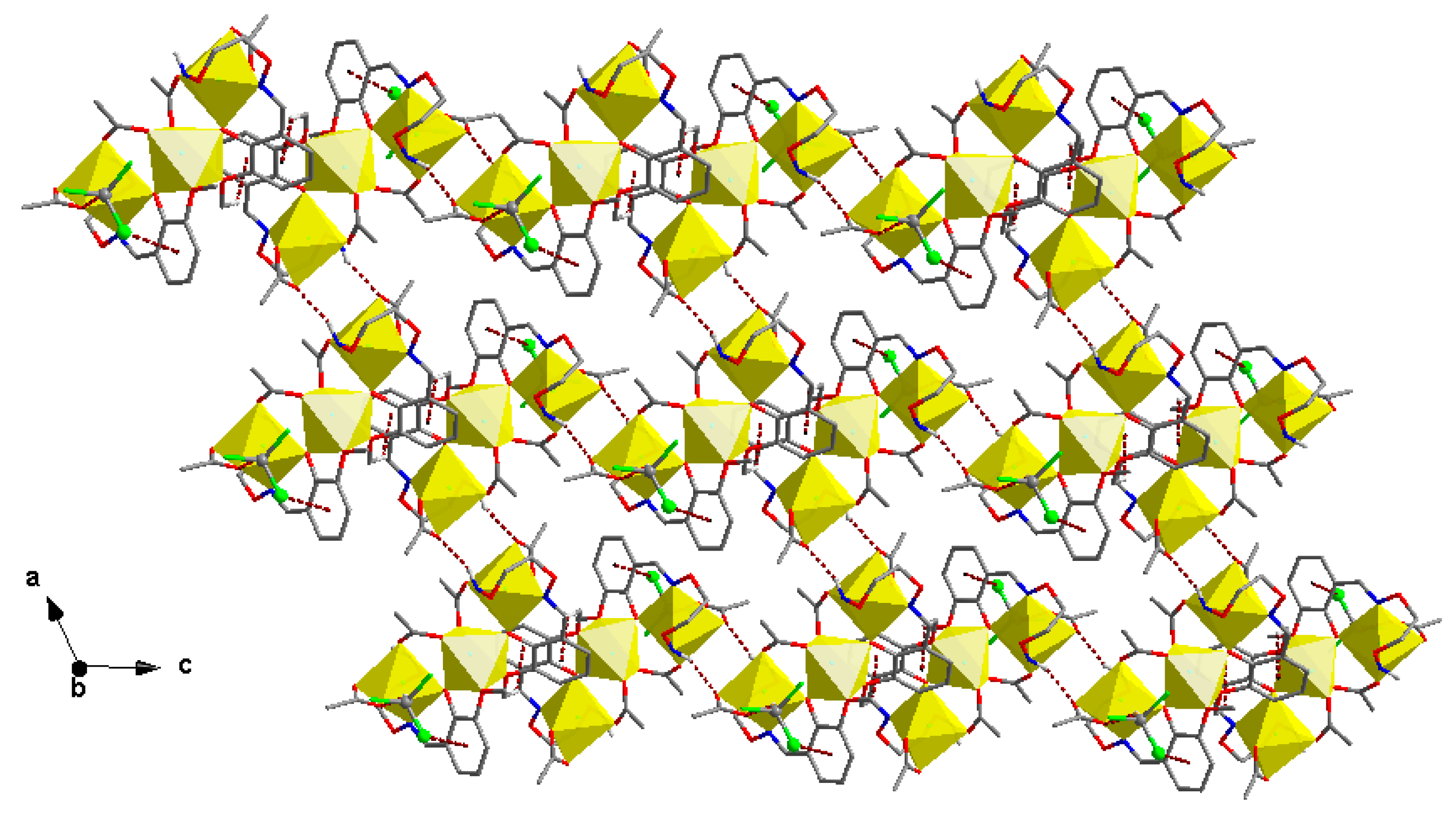
Figure 7.
The UV-VIS spectra of the free ligands H2L1 and HL2 with their corresponding complexes 1 and 2 (cm−1).
Figure 7.
The UV-VIS spectra of the free ligands H2L1 and HL2 with their corresponding complexes 1 and 2 (cm−1).

Figure 8.
Absorption spectra of H2L1 in DMF solution upon the addition of Zn2+. Inset: The absorbance at 432 nm varied as an interaction of [Zn2+]/[H2L1].
Figure 8.
Absorption spectra of H2L1 in DMF solution upon the addition of Zn2+. Inset: The absorbance at 432 nm varied as an interaction of [Zn2+]/[H2L1].
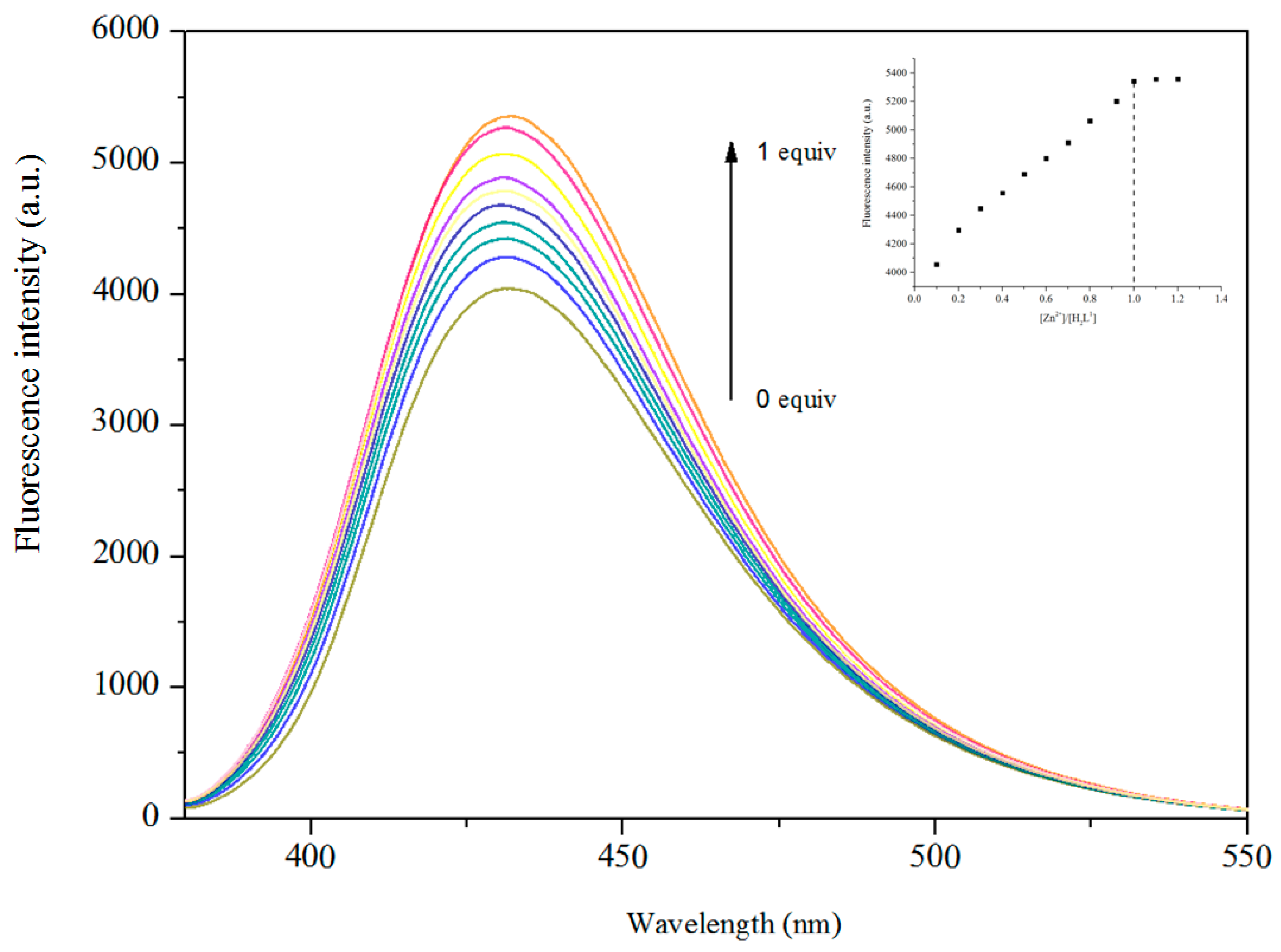
Figure 9.
Absorption spectra of HL2 in DMF solutions upon the addition of Zn2+. Inset: The absorbance at 380 nm varied as an interaction of [Zn2+]/[HL2].
Figure 9.
Absorption spectra of HL2 in DMF solutions upon the addition of Zn2+. Inset: The absorbance at 380 nm varied as an interaction of [Zn2+]/[HL2].

Figure 10.
The fluorescent (λex = 370 nm) spectra of complex 1 (2.5 × 10⁻⁵ M) in various solvents.
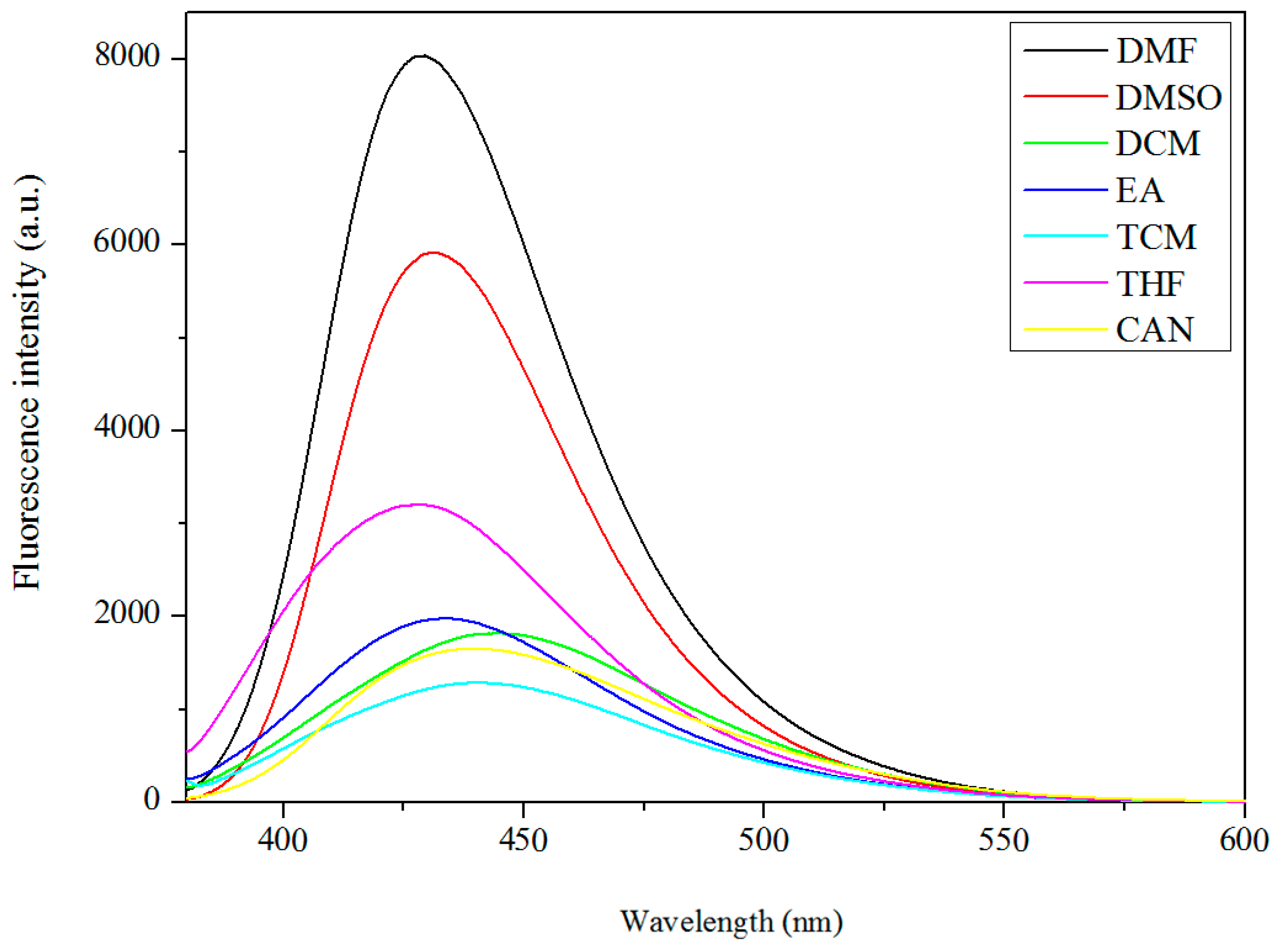
Figure 11.
The fluorescent (λex = 315 nm) spectra of complex 2 (2.5 × 10⁻⁵ M) in various solvents.
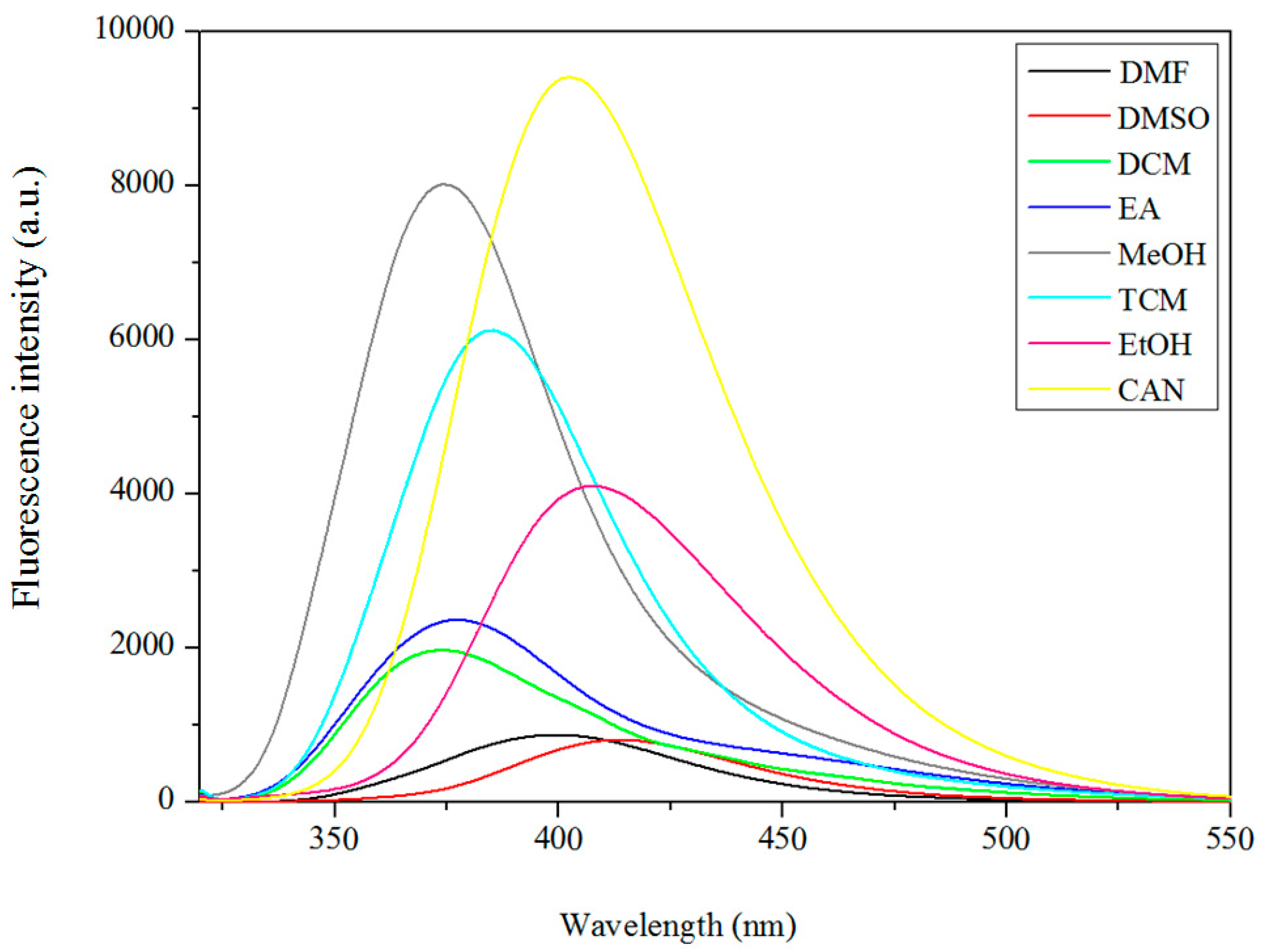
Figure 12.
The normalized fluorescent spectra of complex 1. Inset image: the fluorescence picture of complex 1 in various solvents upon irradiation with a 365 nm UV lamp.
Figure 12.
The normalized fluorescent spectra of complex 1. Inset image: the fluorescence picture of complex 1 in various solvents upon irradiation with a 365 nm UV lamp.
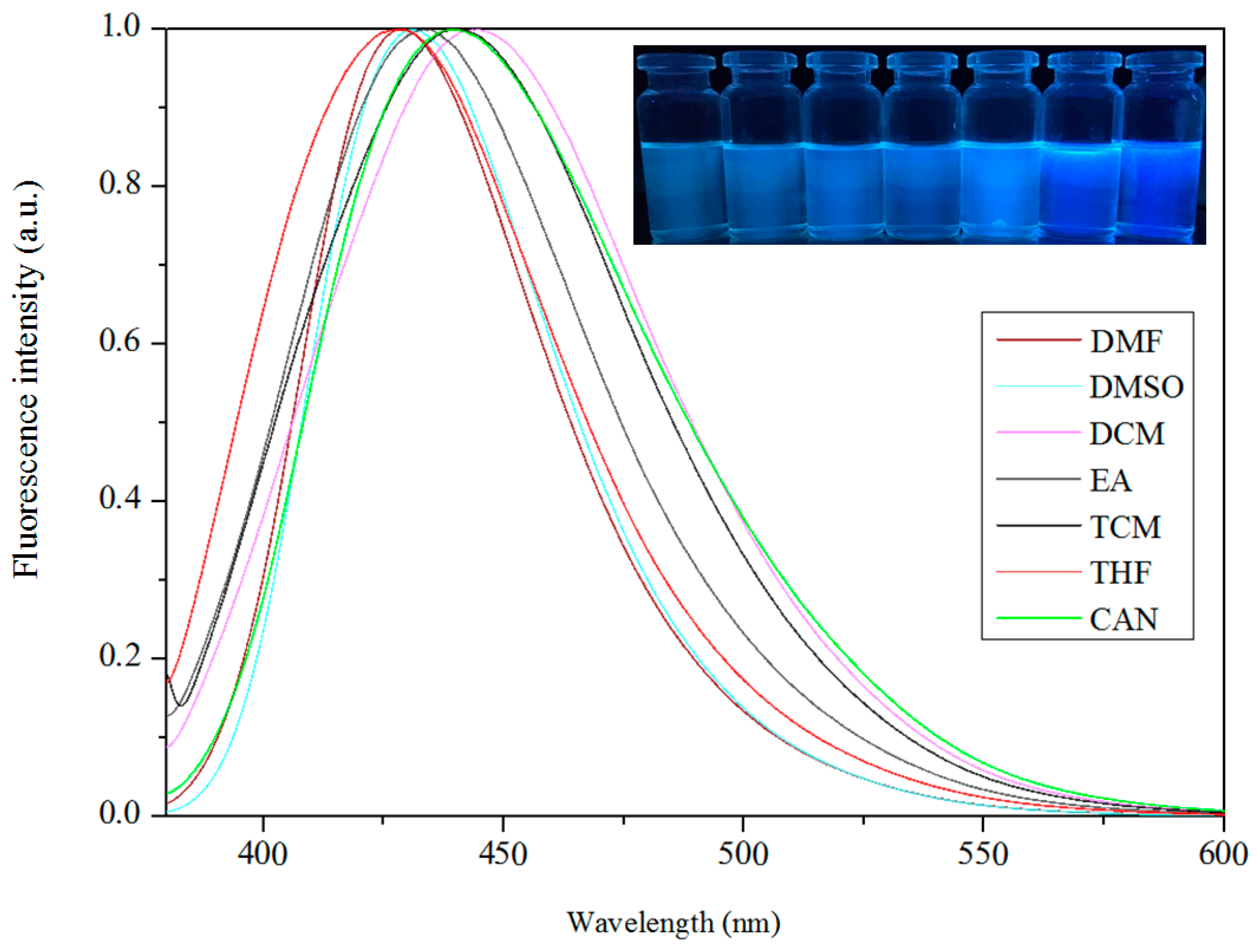
Figure 13.
The normalized fluorescent spectra of complex 2. Inset image: the fluorescence picture of complex 1 in various solvents upon irradiation with a 365 nm UV lamp.
Figure 13.
The normalized fluorescent spectra of complex 2. Inset image: the fluorescence picture of complex 1 in various solvents upon irradiation with a 365 nm UV lamp.


Table 1.
Crystal data and structure refinement parameters for complexes 1 and 2.
| Complex | 1 | 2 |
|---|---|---|
| Formula | C26H24N2O9Zn | C29H39Cl3N4O16Zn3 |
| Formula weight | 573.84 | 1002.16 |
| Temperature (K) | 173.00(10) | 292.38(10) |
| Wavelength (Å) | 0.71073 | 0.71073 |
| Crystal system | Monoclinic | Triclinic |
| Space group | P21/n | P–1 |
| Unit cell dimensions | ||
| a (Å) | 12.811(3) | 13.1694(13) |
| b (Å) | 13.4725(9) | 13.3568(13) |
| c (Å) | 15.393(3) | 16.1452(14) |
| α (°) | 90 | 94.664(8) |
| β (°) | 107.665(19) | 113.074(10) |
| γ (°) | 90 | 112.722(9) |
| V (Å3) | 2531.6(8) | 2317.6(4) |
| Z | 4 | 2 |
| Dc (g cm‒3) | 1.506 | 1.430 |
| μ (mm‒1) | 1.028 | 1.775 |
| F (000) | 1184 | 1012 |
| Crystal size (mm) | 0.24 × 0.19 × 0.17 | 0.22 × 0.18 × 0.14 |
| θ Range (°) | 3.338–25.008 | 3.34–26.02 |
| Index ranges | −13 ≤ h ≤ 15, | −10 ≤ h ≤ 16, |
| −16 ≤ k ≤ 14, | −16 ≤ k ≤ 16, | |
| −14 ≤ l ≤ 18 | −19 ≤ l ≤ 15 | |
| Reflections collected | 8334 | 16,912 |
| Independent reflections | 4446 | 9102 |
| Rint | 0.0996 | 0.0522 |
| Completeness | 99.6% | 99.79% |
| Data/restraints/parameters | 4446/16/349 | 9102/1/502 |
| GOF | 1.001 | 0.963 |
| Final R1, wR2 indices | 0.0581/0.0934 | 0.0633/0.1473 |
| R1, wR2 indices (all data) | 0.0721/0.1598 | 0.1236/0.1774 |
| Largest differences | 0.926/‒0.772 | 0.873/‒0.587 |
| peak and hole (e Å‒3) | ||
Table 2.
Selected bond lengths (Å) and angles (°) for complex 1.
| Bond | |||
|---|---|---|---|
| Zn1-O3 | 1.940(5) | Zn1-N1 | 2.187(7) |
| Zn1-O6 | 1.994(5) | Zn1-N2 | 2.036(7) |
| Zn1-O9 | 2.049(6) | ||
| Angles | |||
| O3-Zn1-O6 | 97.1(2) | O3-Zn1-O9 | 111.5(2) |
| O3-Zn1-N1 | 86.7(2) | O3-Zn1-N2 | 125.1(3) |
| O6-Zn1-O9 | 91.7(2) | O6-Zn1-N1 | 175.8(2) |
| O6-Zn1-N2 | 87.0(2) | N1-Zn1-O9 | 88.5(3) |
| N2-Zn1-O9 | 123.2(3) | N2-Zn1-N1 | 89.4(3) |
Table 3.
Hydrogen bonding and C-H···π stacking interactions (Å, °) for complex 1.
| D-H⋅⋅⋅A | D-H | H⋅⋅⋅A | D⋅⋅⋅A | D-H⋅⋅⋅A |
|---|---|---|---|---|
| C10-H10 ⋅⋅⋅O2 | 0.93 | 2.30 | 2.670(11) | 103 |
| C11-H11A⋅⋅⋅O9 | 0.97 | 2.59 | 3.421(10) | 143 |
| C13-H13⋅⋅⋅O7 | 0.93 | 2.25 | 2.636(10) | 104 |
| C25-H25B ⋅⋅⋅O6 | 0.97 | 2.46 | 3.055(14) | 119 |
| C2-H2 ⋅⋅⋅O4 | 0.93 | 2.58 | 3.454(12) | 158 |
| C11-H11B ⋅⋅⋅O8 | 0.97 | 2.49 | 3.281(9) | 139 |
| C12-H12A⋅⋅⋅O1 | 0.97 | 2.51 | 3.424(12) | 157 |
| C12-H12B⋅⋅⋅O3 | 0.97 | 2.56 | 3.494(11) | 161 |
| C21-H21⋅⋅⋅O3 | 0.93 | 2.54 | 3.277(11) | 137 |
| O9-H9⋅⋅⋅O8 | 0.86 | 1.99 | 2.725(10) | 144 |
| C26-H26B⋅⋅⋅Cg3 | 0.96 | 2.99 | 3.398(17) | 107 |
Note: Cg3 = O2–C1–C2–C3–C4–C9.
Table 4.
Selected bond lengths (Å) and angles (°) for complex 2.
| Bond | |||
|---|---|---|---|
| Zn1-O2 | 2.017(4) | Zn1-O9 | 2.203(4) |
| Zn1-O10 | 2.191(5) | Zn1-O11 | 2.112(4) |
| Zn1-N1 | 2.218(5) | Zn1-N2 | 2.077(5) |
| Zn2-O1 | 2.269(4) | Zn2-O2 | 2.006(4) |
| Zn2-O5 | 2.336(4) | Zn2-O6 | 2.001(4) |
| Zn2-O12 | 2.032(4) | Zn2-O13 | 2.022(4) |
| Zn3-O6 | 2.006(4) | Zn3-O14 | 2.147(4) |
| Zn3-O15 | 2.209(5) | Zn3-O16 | 2.160(5) |
| Zn3-N3 | 2.206(5) | Zn3-N4 | 2.085(5) |
| Angles | |||
| O2-Zn1-O9 | 155.36(16) | O2-Zn1-O10 | 96.75(16) |
| O2-Zn1-O11 | 95.76(15) | O2-Zn1-N1 | 81.53(17) |
| O2-Zn1-N2 | 99.03(17) | O9-Zn1-N1 | 94.48(18) |
| O10-Zn1-O9 | 59.04(16) | O10-Zn1-N1 | 93.23(19) |
| O11-Zn1-O9 | 89.09(16) | O11-Zn1-O10 | 89.72(18) |
| O11-Zn1-N1 | 176.21(18) | N2-Zn1-O9 | 105.28(18) |
| N2-Zn1-O10 | 164.21(18) | N2-Zn1-O11 | 88.08(17) |
| N2-Zn1-N1 | 89.72(18) | O1-Zn2-O5 | 84.12(15) |
| O2-Zn2-O1 | 74.99(15) | O2-Zn2-O5 | 85.59(16) |
| O2-Zn2-O12 | 97.76(16) | O2-Zn2-O13 | 100.51(15) |
| O6-Zn2-O1 | 84.90(16) | O6-Zn2-O2 | 152.58(15) |
| O6-Zn2-O5 | 73.79(16) | O6-Zn2-O12 | 101.12(15) |
| O6-Zn2-O13 | 96.80(16) | O12-Zn2-O1 | 91.08(15) |
| O12-Zn2-O5 | 173.26(15) | O13-Zn2-O1 | 171.40(15) |
| O13-Zn2-O5 | 88.25(16) | O13-Zn2-O12 | 96.84(15) |
| O6-Zn3-O14 | 92.82(15) | O6-Zn3-O15 | 95.64(16) |
| O6-Zn3-O16 | 155.32(16) | O6-Zn3-N3 | 82.60(17) |
| O6-Zn3-N4 | 99.71(18) | O14-Zn3-O15 | 88.68(17) |
| O14-Zn3-O16 | 88.30(16) | O14-Zn3-N3 | 174.96(19) |
| O16-Zn3-O15 | 59.72(16) | O16-Zn3-N3 | 96.74(19) |
| N3-Zn3-O15 | 93.87(19) | N4-Zn3-O14 | 88.59(17) |
| N4-Zn3-O15 | 164.52(19) | N4-Zn3-O16 | 104.96(18) |
| N4-Zn3-N3 | 90.08(19) |
Table 5.
Hydrogen bonding and C-H···π and C-Cl···π interactions (Å, °) for complex 2.
| D-H⋅⋅⋅A | D-H | H⋅⋅⋅A | D⋅⋅⋅A | D-H⋅⋅⋅A |
|---|---|---|---|---|
| N2-H2A ⋅⋅⋅O13 | 0.90 | 2.06 | 2.890(7) | 153 |
| N4-H4A⋅⋅⋅O12 | 0.90 | 2.10 | 2.945(8) | 155 |
| C8-H8B⋅⋅⋅O9 | 0.90 | 2.03 | 2.860(7) | 154 |
| C22-H22B ⋅⋅⋅O16 | 0.90 | 2.13 | 2.965(8) | 153 |
| N2-H2B ⋅⋅⋅O9 | 0.97 | 2.33 | 3.253(9) | 158 |
| N4-H4B ⋅⋅⋅O16 | 0.97 | 2.38 | 3.300(8) | 157 |
| C29-H29⋅⋅⋅O15 | 0.98 | 2.35 | 3.19(2) | 145 |
| N2-H2A ⋅⋅⋅O13 | 0.90 | 2.06 | 2.890(7) | 153 |
| C29-Cl2⋅⋅⋅Cg7 | 1.687 | 3.979 | 4.652(18) | 102.7 |
| C10-H10B ⋅⋅⋅Cg6 | 0.96 | 2.69 | 3.481(9) | 140 |
Note: Cg7 = C15–C20; Cg6=C1–C6.
Table 6.
Absorption maxima and molar extinction coefficients for complexes 1 and 2.
| Compound | c | A(ε) | B(ε) | C(ε) |
|---|---|---|---|---|
| H2L1 | 1.0 × 10‒5 | 291 (5.7 × 10‒4) | 329 (3.7 × 10‒4) | 345 (3.0 × 10‒4) |
| Complex 1 | 1.0 × 10‒5 | 252 (2.4 × 10‒4) | 304 (2.8 × 10‒4) | 344 (3.9 × 10‒4) |
| HL2 | 1.0 × 10‒5 | 270 (4.6 × 10‒4) | 322 (0.8 × 10‒4) | |
| Complex 2 | 1.0 × 10‒5 | 271 (3.3 × 10‒4) | 325 (0.6 × 10‒4) |
Table 7.
The maximum fluorescence emission in difference solvents for complexes 1 and 2.
| Compound | DMF | DMSO | DCM | EA | TCM | CAN | THF | MeOH | EtOH |
|---|---|---|---|---|---|---|---|---|---|
| Complex 1 | 429 | 431 | 440 | 433 | 439 | 439 | 428 | ||
| Complex 2 | 399 | 414 | 372 | 377 | 384 | 402 | 373 | 407 |
© 2017 by the authors. Licensee MDPI, Basel, Switzerland. This article is an open access article distributed under the terms and conditions of the Creative Commons Attribution (CC BY) license (http://creativecommons.org/licenses/by/4.0/).
Share and Cite
MDPI and ACS Style
Dong, X.-Y.; Gao, L.; Wang, F.; Zhang, Y.; Dong, W.-K. Tri- and Mono-Nuclear Zinc(II) Complexes Based on Half- and Mono-Salamo Chelating Ligands. Crystals 2017, 7, 267. https://doi.org/10.3390/cryst7090267
AMA Style
Dong X-Y, Gao L, Wang F, Zhang Y, Dong W-K. Tri- and Mono-Nuclear Zinc(II) Complexes Based on Half- and Mono-Salamo Chelating Ligands. Crystals. 2017; 7(9):267. https://doi.org/10.3390/cryst7090267
Chicago/Turabian StyleDong, Xiu-Yan, Lei Gao, Fei Wang, Yang Zhang, and Wen-Kui Dong. 2017. "Tri- and Mono-Nuclear Zinc(II) Complexes Based on Half- and Mono-Salamo Chelating Ligands" Crystals 7, no. 9: 267. https://doi.org/10.3390/cryst7090267
Note that from the first issue of 2016, this journal uses article numbers instead of page numbers. See further details here.