Article

In Vitro Antifungal Activity of Composites of AgNPs and Polyphenol Inclusion Compounds against Fusarium culmorum in Different Dispersion Media

by
Petruta Mihaela Matei
1,2,
Beatrice Michaela Iacomi
1,
Jesús Martín-Gil
2,
Eduardo Pérez-Lebeña
2,
M. Carmen Ramos-Sánchez
3,
M. Teresa Barrio-Arredondo
4 and
Pablo Martín-Ramos
5,*
1
Department of Bioengineering of Horticultural and Viticultural Systems, University of Agricultural Sciences and Veterinary Medicine of Bucharest, Bulevardul Mărăști 59, București 011464, Romania
2
Agriculture and Forestry Engineering Department, ETSIIAA, Universidad de Valladolid, Avenida de Madrid 44, 34004 Palencia, Spain
3
Microbiology and Parasitology Service, Hospital Universitario Rio Hortega, Sanidad de Castilla y León (SACYL), Calle Dulzaina, 2, 47012 Valladolid, Spain
4
Centro de Salud Barrio España, Sanidad de Castilla y León (SACYL), Calle Costa Brava, 4, 47010 Valladolid, Spain
5
Department of Agricultural and Environmental Sciences, EPS, Instituto Universitario de Investigación en Ciencias Ambientales de Aragón (IUCA), Universidad de Zaragoza, Carretera de Cuarte, s/n, 22071 Huesca, Spain
*
Author to whom correspondence should be addressed.
Agronomy 2018, 8(11), 239; https://doi.org/10.3390/agronomy8110239
Submission received: 27 September 2018 / Revised: 21 October 2018 / Accepted: 26 October 2018 / Published: 28 October 2018
(This article belongs to the Special Issue Nanotechnology Applications in Agriculture System)

Abstract

:
Fusarium culmorum is a soil-borne fungus able to cause Fusarium head blight, one of the most important cereal diseases worldwide, which can result in significant yield losses of up to 50% and which jeopardizes food and feed safety due to the mycotoxins produced. In the study presented herein, the enhancement of the antifungal activity against this pathogen, resulting from the addition of silver nanoparticles (AgNPs) to different polyphenol-stevioside inclusion compounds, dispersed either in a chitosan oligomers hydroalcoholic solution or in a choline chloride:urea:glycerol deep eutectic solvent, was investigated in vitro. The polyphenols assayed were curcumin, ferulic acid, gallic acid and silymarin. Four composite concentrations (62.5, 125, 250 and 500 µg·mL−1), with and without AgNPs, were assessed, finding noticeable differences in mycelial growth inhibition, with EC50 and EC90 values ranging from 118 to 579 µg·mL−1 and from 333 to 2604 µg·mL−1, respectively. The obtained results evidenced the improved efficacy of the composites with AgNPs, a superior performance of the composites based on curcumin and ferulic acid, and the advantages of the deep eutectic solvent-based dispersion medium over the chitosan oligomers-based one. The reported composites hold promise for crop protection applications.

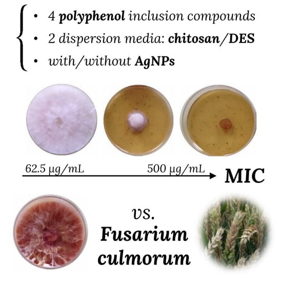
Graphical Abstract

1. Introduction

Fusarium culmorum (W.G. Smith) Sacc. is a soil-borne filamentous fungus that causes important diseases in cereals, grasses, and a wide variety of monocots and dicots. In wheat and barley, as well as in other small-grain cereals, it is able to produce Fusarium crown rot (FCR) and Fusarium head blight (FHB) [1]. FCR causes seedling blight, brown discolorations and rotting of root, crown and lower stem tissues [2]. FHB remains one of the most important diseases worldwide and continues to pose a major challenge in cereal production leading to severe yield reductions.
This fungus is also reported as a post-harvest pathogen, especially on freshly harvested grain that has not been properly dried or stored [1]. It produces type B trichothecenes (4-deoxynivalenol, 3-acetyl-4-deoxynivalenol, 15-acetyl-4-deoxynivalenol, nivalenol and 4-acetylnivalenol or fusarenone X), which may contaminate food and feed at high concentrations and cause serious poisoning in humans and animals [3]. Further, trichothecenes play an important role as virulence factors by inhibiting defense mechanisms activated by the plant [1].
As noted by Pani, et al. [4], the efficient control of F. culmorum associated disease and trichothecene contamination (to comply with EC No. 1881/2006 in the European Union) poses a major challenge and requires integrated management approaches, involving the choice of tolerant cultivars, the use of crop rotation, a reduction of nitrogen fertilization, crop residues plowing, and seed dressing with antifungal agents. With regard to this latter matter, although conventional azole and strobin fungicides can be effective at low disease pressure or on plant genotypes that present a moderate level of resistance, their repeated use may induce a selective pressure on fungal populations, hence favoring the onset of resistant mutants [5]. As a result, increasing efforts are being devoted to the identification of alternative approaches, in particular, regarding formulations based on biodegradable nanocomposite materials with biopolymers and on antioxidants, which can improve the natural resistance mechanisms of the host plant [6]. Nanoparticle formulations, specially silver nanoparticles, have also attracted considerable attention in plant protection applications [7].
Among the aforementioned biopolymers, chitosan, which features well-established antifungal properties [8], has been tested against F. culmorum and other Fusarium species by Bell, et al. [9], Park, et al. [10], Al-Hetar, et al. [11], Xing, et al. [12]. Specific and strong inhibitory activities against F. culmorum have also been reported for phenolic and polyphenolic natural compounds, such as ferulic, sinapic and p-coumaric acids [13]; gallic acid [14]; curcumin [15,16]; or caffeic acid [17]. A screening of 31 phenolic compounds (including gallic and ferulic acids) was also recently conducted by Pani, et al. [4]. Apropos of metal nanoparticles, silver nanoparticles [18,19,20,21,22] have shown promise as non-aggressive treatments against this fungus in sustainable agriculture. Other metal nanoparticles against other Fusarium species have been recently reviewed by Rai, et al. [23].
Although combinations of chitosan with encapsulated essential oils have been reported to have a fungistatic effect against Fusarium verticillioides [24] and F. graminearum [25], to the best of the authors’ knowledge, the efficacy of binary and ternary mixtures of the three bioactive compounds classes discussed above (biopolymers, phenolics and AgNPs) against Fusarium spp. has not been reported to date.
In an attempt to fill this research gap, the present work has aimed to evaluate the in vitro antifungal activity of composites consisting of silver nanoparticles and polyphenol inclusion compounds formed with stevioside (to improve their solubility and bioavailability [26,27]), with a view to evidencing the enhanced behavior resulting from the addition of AgNPs. Different concentrations, four polyphenols (curcumin, ferulic acid, gallic acid and silymarin) and two dispersion media (viz., chitosan oligomers in a hydroalcoholic solution, and a deep eutectic solvent (DES) based on a choline chloride and urea solution (1:2 v/v) in glycerol) have been investigated.

2. Materials and Methods

2.1. Reagents

Commercially-available silver nanoparticles (40 nm particle size (TEM), 20 µg·mL−1 in aqueous buffer, with sodium citrate as a stabilizer; PubChem Substance ID 329764597), curcumin (CAS 458-37-7), ferulic acid (CAS 1135-24-6), gallic acid (CAS 149-91-7), silymarin (MDL MFCD01776359), choline chloride (CAS 67-48-1), urea (CAS 5-13-6) and glycerol (CAS 56-81-5) were purchased from Sigma-Aldrich/Merck KGaA (Darmstadt, Germany). Stevioside (CAS 57817-89-7) was purchased from Wako (Osaka, Japan). All chemicals were used without further purification.
Chitosan oligomers were prepared from medium molecular weight chitosan (MMWC, supplied by Hangzhou Simit Chemical Technology Co. Ltd., Hangzhou, China) according to the procedure reported by Sun, et al. [28]: 10 g of MMWC was first solubilized in 500 mL of acetic acid (1%) under constant stirring at 60 °C and, once dissolved, chitosan oligomers (with MW < 2 kDa) were obtained by oxidative degradation with H2O2 (0.3 mol·L−1).
The choline chloride and urea (1:2 v/v) deep eutectic mixture was prepared according to Biswas, et al. [29], by heating at 80 °C under vigorous stirring in a hot-plate for 10 min.

2.2. Preparation of Polyphenol Inclusion Compounds

Polyphenol inclusion compounds were obtained by microwave-assisted aqueous biphasic system separation, according to the procedure described in references [30,31].

2.3. Preparation of Bioactive Composites with Chitosan Oligomers in Hydroalcoholic Solution

A series of inclusion compounds were synthesized using chitosan oligomers in hydroalcoholic solution as the dispersion medium. In a typical synthesis process, 10 mg of chitosan oligomers of 2 kDa, 60 mg of stevioside and 10 mg of one of the polyphenols (either curcumin, ferulic acid, gallic acid or silymarin) were added to 40 mL of hydroalcoholic solution (1:1 v/v distilled water and ethanol). The mixture was heated at 80 °C for 20 min and stirred in a microwave oven (Milestone Ethos-One, Sorisole, BG, Italy). The resulting products were isolated by centrifugation (2500 rpm) and stored at 4 °C. For the treatments with silver nanoparticles, 2 µg of AgNPs (0.1 mL from a 20 µg·mL−1 solution) were added dropwise to 0.9 mL of the microwave fractions, and the final solution, with pH 7.5, was sonicated with a probe-type UIP1000hdT ultrasonicator (Hielscher, Teltow, Germany; 1000 W, 20 kHz) for 5 min. The concentration in AgNPs of the doped composite was 2 µg·mL−1.

2.4. Preparation of Bioactive Composites in ChCl:urea Deep Eutectic Solvent

Thirty milligrams of stevioside and 10 mg of one of the polyphenols under study (either gallic acid, silymarin, ferulic acid or curcumin) were added to a dispersion medium consisting of 20 mL of choline chloride and urea (1:2 v/v) DES and 10 mL of glycerol. The mixture was subjected to microwave irradiation (at 80 °C; heating ramp: 5 °C·min−1) and stirring for 20 min. The bioactive products were again obtained by centrifugation at 2500 rpm. Part of the obtained solution—for each of the polyphenols—was used for the preparation of the treatments with nanosilver: in this case, 0.1 mL of AgNPs (20 µg·mL−1) were then added dropwise to 0.9 mL of the previously obtained solution, and the resulting mixtures, with pH 7.5, were subjected to sonication for 5 min at room temperature.

2.5. Characterization

The composites were characterized using Fourier-Transform Infrared spectroscopy (FTIR), scanning electron microscopy (SEM) and transmission electron microscopy (TEM). The vibrational spectrum in the 400–4000 cm−1 spectral range was characterized using a Nicolet iS50 FT-IR spectrometer (Thermo Scientific, Waltham, MA, USA), equipped with an in-built diamond attenuated total reflection (ATR) system, with a 1 cm−1 spectral resolution and 64 scans. SEM and TEM micrographs were collected with a Quanta 200FEG microscope (FEI, Hillsboro, OR, USA) and with a JEM-FS2200 HRP microscope (JEOL, Akishima, Tokyo, Japan), respectively, confirming good agreement with the characterization results recently reported in patent P201731489 [31].

2.6. Fungal Isolate and Growth Conditions

In vitro tests were conducted using Fusarium culmorum CS7071 isolated from infected wheat seeds. The isolate was identified based on the morphology of conidia and conidiophores as well as on cultural properties. A molecular identification was made using Fusarium−specific primers targeting translation elongation factor 1 (EF1): FUSAefF ATGGGTAAGGAGGACAAGAC and FUSAefR: GGAAGTACCAGTGATCATGTT [32,33]. A FASTA sequence was verified at NCBI Blast, confirming the correspondence with F. culmorum culture ICMP:15476 translation elongation factor 1 (EF1a) gene, partial cds, sequence ID: MG857546.1.
The isolate was purified by monospore isolation and maintained on malt agar slants, at 4 °C. Fresh subcultures were made by transferring hyphae plugs to Petri dishes containing potato dextrose agar (PDA, supplied by Scharlau, Barcelona, Spain) medium to obtain the inoculum for sensitivity tests.

2.7. In Vitro Tests of Mycelial Growth Inhibition

Tests were carried out to determine the biological activity of the nanocomposites using the agar dilution method. Aliquots of stock solutions were incorporated into the PDA medium to provide final concentrations of 62.5, 125, 250 and 500 µg·mL−1.
Mycelial disks of pathogen (8 mm in diameter), removed from the margins of a 7-day old culture, were transferred to PDA plates amended with the treatments under study at the aforementioned concentrations. Plates containing only the PDA medium served as the control. Three replicates were used per treatment and the screening was repeated twice.
Radial mycelial growth was determined after 5, 7 and 14 days by calculating the mean of two perpendicular colony diameters for each replicate. Mycelial growth inhibition (or the efficacy of the tested composite) for each treatment and concentration after 7 days of incubation, in the dark, was calculated according to the formula: ( ( d c d t ) / d c ) × 100 , where dc is the average diameter of fungal colony in the control and dt is the average diameter of the fungal colony treated with the tested composite. The data from two screening experiments were pooled and averaged.
Results were expressed as effective concentrations EC50 and EC90 (i.e., the concentrations which reduced mycelial growth by 50% and 90%, respectively) determined by regressing the inhibition of radial growth values (%) against the log10 values of the fungicide concentrations.

2.8. Statistical Analyses

Data were subjected to analysis of variance (ANOVA). For post hoc comparison of means, Tukey’s multiple range test at 0.05 probability level (p < 0.05) was used. All tests were made using IBM SPSS Statistics v.25 software.

3. Results

The bioactivity against F. culmorum of the different treatments, without and with AgNPs, was studied in vitro by monitoring the radial growth of the mycelium (Figure 1).
The radial growth of the mycelium results for the two dispersion media, i.e., chitosan oligomers in hydroalcoholic solution or the ChCl:urea:glycerol deep eutectic solvent are shown in Figure 2a,b, respectively. The increase in the concentration of the inclusion complexes from 62.5 µg·mL−1 to 500 µg·mL−1 resulted in a reduction in the radial growth of the mycelium in all cases.
It may be observed that in the chitosan oligomers dispersion medium, 100% mycelial growth inhibition without AgNPs only occurred for the composites with curcumin and ferulic acid at the highest concentration of 500 µg·mL−1. Upon addition of AgNPs, the efficacy of all composites was enhanced (for instance, the inhibition percentages at 250 µg·mL−1 increased from 40% to 49% for curcumin, from 10% to 40% for ferulic acid, from 18% to 33% for gallic acid, and from 9% to 36% for silymarin), but full inhibition was only attained by curcumin and ferulic at the highest dose, as it happened before. Significant differences between treatments with and without AgNPs were detected for all polyphenols except for curcumin (Table 1).
In the case of the composites with a DES dispersion medium, total inhibitory activities were attained for all the composites without AgNPs at a concentration of 500 µg·mL−1 (not only for curcumin and ferulic acid). When AgNPs were added, a synergistic effect was again observed, as total inhibition occurred for the curcumin-based treatment at 250 µg·mL−1, and inhibition percentages of 97%, 89% and 85% at the same dose were found for ferulic acid, silymarin and gallic acid composites, respectively. Significant differences between the treatments with and without AgNPs were detected in all cases (Table 1).
The product efficacies for the composites in DES were higher than those of the composites based on the hydroalcoholic solution of chitosan oligomers, as evidenced by the significant statistical differences in the radial growth of the mycelium (Table 2a), but all treatments showed significant differences vs. the control, at least at the highest two doses.
The results from the sensitivity tests may also be expressed in terms of EC50 and EC90 indicators (Table 3). The sensitivity of the isolate clearly varied as a function of the presence of AgNPs in the composites and of dispersion media, but also according to the phenolic compound used. Although in the case of the chitosan oligomers dispersion medium the addition of AgNPs barely changed the effective concentration values for curcumin and ferulic (the two polyphenols with the best activity), they noticeably improved those of silymarin and gallic acid composites. In the DES medium, the EC50 values were improved by 62%, 48% and 35% for curcumin, ferulic acid and silymarin, respectively, when AgNPs were incorporated. The enhancement for the EC90 values was less evident, except for curcumin, in which a 51% improvement was detected, pointing at a higher affinity between the two components. The difference between treatments with and without AgNPs was on the limit of significance (p = 0.055) for EC50 and was not significant (p = 0.198) for EC90.
In line with the discussion presented above, F. culmorum was found to be more sensitive to the treatments prepared in DES, with EC50 values ranging from 118 to 209 µg·mL−1 and EC90 values between 333 and 502 µg·mL−1. For comparison purposes, for the COS in hydroalcoholic solution medium treatments the EC50 values ranged from 227 to 579 µg·mL−1 and those of EC90 from 503 to 2604 µg·mL−1. Significant differences between the two dispersion media were found for both EC50 and EC90 (Table 2b).
With regard to the impact of the polyphenol choice, curcumin and ferulic acid generally performed better than gallic acid and silymarin (gallic acid would only be advised in the DES medium if AgNPs are not to be used), but no significant differences were observed.
The highest sensitivities of the F. culmorum corresponded to the inclusion compounds with curcumin and ferulic acid in DES (EC50 values of 129 and 118 µg·mL−1, respectively), followed by the inclusion compound with silymarin, which featured a similar EC90 (around 330 µg·mL−1).

4. Discussion

4.1. Efficacy of the Composites

In an attempt to enable comparisons with the literature in terms of inhibitory concentrations, the equivalent concentrations of each of the individual components of the composites are summarized in Table 4. Nonetheless, it should be stressed that Fusarium species do not have a normal minimum inhibitory concentration (MIC) and minimum effective concentration (MEC) distribution and thus prediction of antifungal susceptibility of a single strain is difficult ([34] and references therein). This same review paper concluded that, since most strains have high MICs (intrinsic resistance), different degrees of resistance at most can be determined in different strains. As the susceptibility profile is isolate dependent, antifungal susceptibility testing should be performed especially for any Fusarium involved in an invasive infection [34]. Comparisons of the effective concentrations below should therefore be taken with caution.
Concerning the efficacy of polyphenols against Fusarium spp., Sarrocco, et al. [16] studied the antifungal activity, main active components and mechanism of Curcuma longa extract on eleven fungi, including F. graminearum, F. chlamydosporum, F. tricinctum, F. culmorum and F. oxysporum, finding EC50 values (in µg·mL−1) of 108.8, 174.2, 254.7, 422.9 and 3883.3, respectively. Chowdhury, et al. [35] reported an IC50 of 135.9 μg·mL−1 for curcumin-I against F. solani, not included in the aforementioned study. Nguyen, et al. [14] tested the antifungal activity of gallic acid purified from Terminalia nigrovenulosa bark against this latter pathogen (F. solani), reporting 81% disease suppression for 1000 ppm (1000 μg·mL−1). Gauthier, et al. [17] found a higher sensitivity to caffeic acid, not assayed herein, for F. graminearum than for F. culmorum, reporting IC50 values >10 mM (1800 μg·mL−1) for the latter.
A thorough bibliographical survey yielded no studies on the effects of silymarin on Fusarium spp., but ferulic acid was tested by Pani, et al. [4]. Although they did not report EC50 values for the different polyphenols assayed against F. culmorum (they quantified the fungal growth in terms of dry fungal biomass), they found that while fungal growth was only slightly inhibited when 1.5 mM gallic acid was added to the liquid culture, ferulic acid at 0.5 mM showed a significant inhibitory activity, a result consistent with the behavior observed in this study.
From the values discussed in previous paragraphs, in which noticeably higher concentrations of polyphenols were required to attain inhibition, it may be inferred that the chosen approach, based on forming inclusion compounds, clearly improved their solubility and bioavailability, which are two of their inherent physicochemical characteristics that limit their applications [36]. This result would be consistent with other studies in which an enhancement of the solubility and bioavailability of polyphenols was attained by using terpene glycosides (such as rubusoside, stevioside, rebaudioside, or steviol monoside) or cyclodextrins to form inclusion compounds [26,27]. Moreover, the better behavior of DES composites as compared to those in the chitosan oligomers medium—in spite of having stevioside concentrations approximately half of those in the latter—points to the advantages associated with the fact that DES are an excellent extraction medium for phenolic compounds [37], either alone or in combination with inclusion compounds [38].
With regard to the efficacy of chitosan on Fusarium spp., disparate results have been reported in the literature: Xing, et al. [12] reported that F. culmorum was resistant to oleoyl-chitosan (O-chitosan) nanoparticles, finding no mycelial growth inhibition at concentrations of up to 2 mg·mL−1. In the same line, Bell, et al. [9], who studied the effects of chitin and chitosan on F. oxysporum in field tests, concluded that chitosan applied as a root dip alone did not reduce disease incidence (although it significantly reduced disease severity when used with a tolerant celery cultivar) and that chitosan treatment of transplants did not reduce soil populations of F. oxysporum. On the other hand, Al-Hetar, et al. [11] stated that chitosan at all concentrations tested (0.5, 1, 2, 4, and 8 mg·mL−1) reduced the hyphal growth of F. oxysporum f. sp. cubense, reporting an EC50 of 1.4 mg·mL−1 and a maximum inhibition of 76.36% at 8 mg·mL−1. Park, et al. [10] assessed the antifungal activity of water-soluble and low molecular weight chitosan against six plant pathogens, finding that F. oxysporum and F. graminearum were the most resistant. IC50 values ranging from 1.5 to 4.0 mg·mL−1 and from 1.8 to 3.2 mg·mL−1 were reported for F. graminearum and F. oxysporum, respectively, depending on the degree of acetylation of chitosan. Since all these concentration values were much higher than the ones used in this study (ranging from 7.5 to 62 µg·mL−1, Table 2), it appears that the contribution of chitosan to the antifungal activity of the composites would be pretty limited, mainly acting as a stabilizing agent and dispersion medium.
A similar situation to that described in the previous paragraph, with controversial and inconsistent data, can be found in terms of the effect of AgNPs. Kasprowicz, et al. [18] investigated the influence of AgNPs on F. culmorum spores vegetative growth rates, using AgNPs concentrations between 1 and 20 ppm. They found a significant reduction in mycelial growth for spores incubated with 5–10 ppm AgNPs solutions compared to the control, but no effective concentrations were reported. Venat, et al. [19] studied the fungitoxic properties of colloidal silver on mycelial growth of eight important plant pathogens, including F. culmorum and F. oxysporum. They found EC50 values of 11.99 ppm and 12.27 ppm, and EC90 values of 18.70 ppm and 18.19 ppm for F. culmorum and F. oxysporum, respectively, when colloidal silver prepared “in-house” was used. When commercial colloidal silver (supplied by Medicer Bios, Ploiești, Romania) was used instead, EC50 values of 26.83 ppm and 21.16 ppm, and EC90 values of 51.6 ppm and 34.66 ppm were determined for F. culmorum and F. oxysporum, respectively. Ziedan and Saad [39] reported full inhibition of F. oxysporum in the presence of AgNPs at a concentration of 15 ppm. Aleksandrowicz-Trzcińska, et al. [40] studied in vitro the effects of copper and silver nanoparticles on the growth of several species of pathogenic and wood-decay fungi, including two Fusarium spp., at concentrations of 5, 15, 25 or 35 ppm. In the case of AgNPs, at the highest concentration, they attained a 32.6% and a 41.2% inhibition for F. oxysporum and F. redolens, respectively. The efficacy of CuNPs was lower (19% and 21% inhibition, respectively). Conversely, Soltanloo, et al. [41] reported that concentrations of up to 100 ppm did not fully inhibit mycelial growth of F. graminearum, and Tutaj, et al. [22] reported that AgNPs at a concentration of 100 µg·mL−1 had no inhibitory effect on F. culmorum growth. Villamizar-Gallardo, et al. [42], working at similar concentrations (up to 100 ppm), concluded that, for F. solani, the AgNPs only caused texture and pigmentation changes, indicating resistance of this microorganism to the nanomaterial in question. In line with this last comment, it is worth noting that, as a result of AgNPs application, a change in mycelia pigmentation was also detected in this study. According to Kasprowicz, et al. [43], on the basis of an analysis of metabolites, this may be ascribed to a more intensive biosynthesis of aurofusarin and to the conversion of rubrofusarin to aurofusarin under the influence of nanosilver.
Considering that in tests conducted with AgNPs alone (not shown in Figure 2), at the same concentrations used in the composites (i.e., ranging from 0.25 to 2 ppm; Table 2), no inhibition was observed, and that those concentrations were approximately two orders of magnitude lower than the effective concentrations reported by the authors mentioned above, it becomes apparent that AgNPs could not be responsible for the bioactive behavior. However, as mentioned in the results section, even at these very low doses, AgNPs led to a noticeable improvement of the efficacy of the composites, pointing to a synergistic behavior with the polyphenols.
A relevant synergistic behavior between AgNPs and chitosan may be safely excluded, provided that much higher concentrations would be required: Dananjaya, et al. [44] reported that AgNPs improved the antifungal activities of chitosan nanoparticles against F. oxysporum, but a concentration of 1000 µg·mL−1 was needed to achieve a 81.52% growth inhibition.

4.2. Mechanism of Action

As noted above, the antifungal action of the composites should be mainly ascribed to the phenolic compounds. Their inhibition behavior would arise from their ability to disrupt the integrity of the plasma membrane and mitochondrial dysfunction, inducing metabolic stagnation [45]. For instance, curcumin can potentially disrupt the synthesis of critical proteins and enzymes that may ultimately inhibit the growth of fungi (in particular, of F. graminearum): it can downregulate D-Glyceraldehyde 3-phosphate:NAD+ oxidoreductase (GAPDH) (involved in glycolysis, gluconeogenesis, the Calvin cycle and other energetic metabolic pathways), it can inhibit the synthesis of ergosterol (so the membrane would be an important antifungal target), and it can suppress the activity of reduced B-nicotinamide adenine dinucleotide (NADH) oxidase and succinate dehydrogenase (SDH), interfering with the tricarboxylic acid (TCA) cycle and inhibiting the adenosine triphosphate (ATP) synthesis in the mitochondria [16]. Ferulic acid would also act on the cell membrane, provided that a significant change in intracellular ATP concentrations, a decrease in intracellular pH, cell membrane hyperpolarization, a reduction in bacterial membrane integrity, and morphological alterations were documented by Shi, et al. [46]. Gallic acid would exhibit both antioxidant as well as pro-oxidant characteristics, displaying a double-edged sword behavior, which turns it into an efficient apoptosis inducing agent [47]. Silymarin would also exert its antifungal activity by targeting the plasma membrane: Yun and Lee [48,49] reported that it induces an increase in permeability and physical perturbation of plasma membrane, allowing the penetration of molecules smaller than approximately 3.3 nm; it induces intracellular reactive oxygen species (ROS), which leads to the peroxidation of membrane lipids; and these membrane damages contributed to membrane malfunctions with membrane depolarization, K+ leakage, decrease in membrane fluidity, and consequently, to cell death.
Finally, with regard to the apparently synergistic behavior observed upon addition of AgNPs, it is worth noting that one of the possible mechanisms of action of silver requires that the silver ions enter the fungal cell for efficient killing. The enhancement of permeability driven by polyphenols would support that their interaction should be synergistic rather than simply additive. A tentative mechanism would be analogous to that described by Dananjaya, et al. [44] to explain the synergistic behavior between chitosan (which also enhances membrane permeability, in a similar fashion to polyphenols) and AgNPs: when the phenolic compounds interact with membrane proteins, causing disruption of the structures and functionality, they would induce an efflux of positively charged K+, resulting in a hyperpolarization of the plasma membrane, which increases the uptake of cations to balance the membrane potential [50]. Depending on the environmental conditions, AgNPs on the surfaces could be ionized and produce cationic silver (Ag+) traces [51]. This Ag+ traces would enter into the cell with the cationic influx generated due to the hyperpolarized cell membrane, subsequently binding with macromolecules such as protein and nucleic acids and further interfering with the metabolic pathways by inhibiting enzyme activities, further promoting the fungal growth inhibition.

5. Conclusions

Composites of four polyphenol inclusion compounds were assessed against Fusarium culmorum with and without AgNPs, at four different concentrations and in two dispersion media. The ChCl:urea:glycerol dispersion medium was found to perform better than the chitosan oligomers hydroalcoholic solution in all cases. The addition of nanosilver, even at very low doses (<2 µg·mL−1), significantly improved the bioactivity of the composites, in particular for the deep eutectic solvent medium, resulting in a decrease in the EC50 and EC90 values of up to 62% and 51% respectively, in the case of the curcumin-based treatment, suggesting synergistic behavior. Amongst the four polyphenols assayed, curcumin attained the best antifungal activity, closely followed by ferulic acid. For the best treatment (curcumin with AgNPs in DES), EC50 and EC90 values of 129 and 333 µg·mL−1, respectively, were obtained. The good efficacy of the proposed composites as compared to other alternatives reported in the literature advocates their potential for the protection of crops against FCR and FHB as an alternative to conventional azole and strobin fungicides.

6. Patents

The work reported in this manuscript is related to Spanish patent P201731489.

Author Contributions

Conceptualization, J.M.-G., E.P.-L., M.C.R.-S. and M.T.B.-A.; Formal analysis, P.M.-R.; Funding acquisition, E.P.-L.; Investigation, P.M.M., B.M.I. and J.M.-G.; Methodology, J.M.-G., E.P.-L., M.C.R.-S. and M.T.B.-A.; Resources, J.M.-G. and E.P.-L.; Supervision, J.M.-G.; Validation, P.M.M., B.M.I., M.C.R.-S. and M.T.B.-A.; Visualization, P.M.-R.; Writing—original draft, P.M.M., J.M.-G., M.C.R.-S., M.T.B.-A. and P.M.-R.; Writing—review & editing, P.M.-R.

Funding

This research was partly funded by Sistemas Biotecnología y Recursos Naturales. The APC was funded by Instituto de Investigación en Ciencias Ambientales de Aragón (IUCA), Universidad de Zaragoza.

Acknowledgments

P.M.-R. would like to thank Santander Universidades for its financial support through the “Becas Iberoamérica Jóvenes Profesores e Investigadores, España” scholarship program.

Conflicts of Interest

The authors declare no conflict of interest. The founding sponsors had no role in the design of the study; in the collection, analyses, or interpretation of data; in the writing of the manuscript, and in the decision to publish the results.

References

  1. Scherm, B.; Balmas, V.; Spanu, F.; Pani, G.; Delogu, G.; Pasquali, M.; Migheli, Q. Fusarium culmorum: Causal agent of foot and root rot and head blight on wheat. Mol. Plant Pathol. 2013, 14, 323–341. [Google Scholar] [CrossRef] [PubMed]
  2. Fernandez, M.R.; Jefferson, P.G. Fungal populations in roots and crowns of common and durum wheat in Saskatchewan. Can. J. Plant Pathol. 2004, 26, 325–334. [Google Scholar] [CrossRef]
  3. Pasquali, M.; Beyer, M.; Logrieco, A.; Audenaert, K.; Balmas, V.; Basler, R.; Boutigny, A.-L.; Chrpová, J.; Czembor, E.; Gagkaeva, T.; et al. A European database of Fusarium graminearum and F. culmorum trichothecene genotypes. Front. Microbiol. 2016, 7, 406. [Google Scholar] [CrossRef] [PubMed] [Green Version]
  4. Pani, G.; Scherm, B.; Azara, E.; Balmas, V.; Jahanshiri, Z.; Carta, P.; Fabbri, D.; Dettori, M.A.; Fadda, A.; Dessì, A.; et al. Natural and natural-like phenolic inhibitors of type B trichothecene in vitro production by the wheat (Triticum sp.) pathogen Fusarium culmorum. J. Agric. Food Chem. 2014, 62, 4969–4978. [Google Scholar] [CrossRef] [PubMed]
  5. Chala, A.; Weinert, J.; Wolf, G.A. An integrated approach to the evaluation of the efficacy of fungicides against Fusarium culmorum, the cause of head blight of wheat. J. Phytopathol. 2003, 151, 673–678. [Google Scholar] [CrossRef]
  6. Picot, A.; Atanasova-Pénichon, V.; Pons, S.; Marchegay, G.; Barreau, C.; Pinson-Gadais, L.; Roucolle, J.; Daveau, F.; Caron, D.; Richard-Forget, F. Maize kernel antioxidants and their potential involvement in Fusarium ear rot resistance. J. Agric. Food. Chem. 2013, 61, 3389–3395. [Google Scholar] [CrossRef] [PubMed]
  7. Kim, D.-Y.; Kadam, A.; Shinde, S.; Saratale, R.G.; Patra, J.; Ghodake, G. Recent developments in nanotechnology transforming the agricultural sector: A transition replete with opportunities. J. Sci. Food Agric. 2018, 98, 849–864. [Google Scholar] [CrossRef] [PubMed]
  8. Silva-Castro, I.; Martín-García, J.; Diez, J.J.; Flores-Pacheco, J.A.; Martín-Gil, J.; Martín-Ramos, P. Potential control of forest diseases by solutions of chitosan oligomers, propolis and nanosilver. Eur. J. Plant Pathol. 2017, 150, 401–411. [Google Scholar] [CrossRef]
  9. Bell, A.A.; Hubbard, J.C.; Liu, L.; Davis, R.M.; Subbarao, K.V. Effects of chitin and chitosan on the incidence and severity of Fusarium yellows of celery. Plant Dis. 1998, 82, 322–328. [Google Scholar] [CrossRef]
  10. Park, R.D.; Jo, K.J.; Jo, Y.; Jin, Y.L.; Kim, K.Y.; Shim, J.H. Variation of antifungal activities of chitosans on plant pathogens. J. Microbiol. Biotechnol. 2002, 12, 84–88. [Google Scholar]
  11. Al-Hetar, M.Y.; Zainal Abidin, M.A.; Sariah, M.; Wong, M.Y. Antifungal activity of chitosan against Fusarium oxysporum f. sp. cubense. J. Appl. Polym. Sci. 2011, 120, 2434–2439. [Google Scholar] [CrossRef]
  12. Xing, K.; Shen, X.; Zhu, X.; Ju, X.; Miao, X.; Tian, J.; Feng, Z.; Peng, X.; Jiang, J.; Qin, S. Synthesis and in vitro antifungal efficacy of oleoyl-chitosan nanoparticles against plant pathogenic fungi. Int. J. Biol. Macromol. 2016, 82, 830–836. [Google Scholar] [CrossRef] [PubMed]
  13. Boutigny, A.L.; Atanasova-Pénichon, V.; Benet, M.; Barreau, C.; Richard-Forget, F. Natural phenolic acids from wheat bran inhibit Fusarium culmorum trichothecene biosynthesis in vitro by repressing Tri gene expression. Eur. J. Plant Pathol. 2010, 127, 275–286. [Google Scholar] [CrossRef]
  14. Nguyen, D.M.C.; Seo, D.J.; Lee, H.B.; Kim, I.S.; Kim, K.Y.; Park, R.D.; Jung, W.J. Antifungal activity of gallic acid purified from Terminalia nigrovenulosa bark against Fusarium solani. Microb. Pathog. 2013, 56, 8–15. [Google Scholar] [CrossRef] [PubMed]
  15. Zorofchian Moghadamtousi, S.; Abdul Kadir, H.; Hassandarvish, P.; Tajik, H.; Abubakar, S.; Zandi, K. A review on antibacterial, antiviral, and antifungal activity of curcumin. BioMed Res. Int. 2014, 2014, 1–12. [Google Scholar] [CrossRef] [PubMed]
  16. Sarrocco, S.; Chen, C.; Long, L.; Zhang, F.; Chen, Q.; Chen, C.; Yu, X.; Liu, Q.; Bao, J.; Long, Z. Antifungal activity, main active components and mechanism of Curcuma longa extract against Fusarium graminearum. PLoS ONE 2018, 13, e0194284. [Google Scholar]
  17. Gauthier, L.; Bonnin-Verdal, M.N.; Marchegay, G.; Pinson-Gadais, L.; Ducos, C.; Richard-Forget, F.; Atanasova-Penichon, V. Fungal biotransformation of chlorogenic and caffeic acids by Fusarium graminearum: New insights in the contribution of phenolic acids to resistance to deoxynivalenol accumulation in cereals. Int. J. Food Microbiol. 2016, 221, 61–68. [Google Scholar] [CrossRef] [PubMed]
  18. Kasprowicz, M.J.; Kozioł, M.; Gorczyca, A. The effect of silver nanoparticles on phytopathogenic spores of Fusarium culmorum. Can. J. Microbiol. 2010, 56, 247–253. [Google Scholar] [CrossRef] [PubMed]
  19. Venat, O.; Iacomi, B.; PeticilĂ, A.G. In vitro studies of antifungal activity of colloidal silver against important plants pathogens. Not. Bot. Horti Agrobot. Cluj-Napoca 2018, 46. [Google Scholar] [CrossRef]
  20. Gorczyca, A.; Pociecha, E.; Kasprowicz, M.; Niemiec, M. Effect of nanosilver in wheat seedlings and Fusarium culmorum culture systems. Eur. J. Plant Pathol. 2015, 142, 251–261. [Google Scholar] [CrossRef]
  21. Rashed, A.O.M.; Mohamed, A.E.A.A.R.; Abobakr, M.M. Wheat protection from root rot caused by Fusarium culmorum using silver nanoparticles. J. Chem. Soc. Pak. 2016, 38, 898–903. [Google Scholar]
  22. Tutaj, K.; Szlazak, R.; Szalapata, K.; Starzyk, J.; Luchowski, R.; Grudzinski, W.; Osinska-Jaroszuk, M.; Jarosz-Wilkolazka, A.; Szuster-Ciesielska, A.; Gruszecki, W.I. Amphotericin B-silver hybrid nanoparticles: Synthesis, properties and antifungal activity. Nanomed. Nanotechnol. Biol. Med. 2016, 12, 1095–1103. [Google Scholar] [CrossRef] [PubMed]
  23. Rai, M.; Ingle, A.P.; Paralikar, P.; Anasane, N.; Gade, R.; Ingle, P. Effective management of soft rot of ginger caused by Pythium spp. and Fusarium spp.: Emerging role of nanotechnology. Appl. Microbiol. Biotechnol. 2018, 102, 6827–6839. [Google Scholar] [CrossRef] [PubMed]
  24. Villegas-Rascón, R.E.; López-Meneses, A.K.; Plascencia-Jatomea, M.; Cota-Arriola, O.; Moreno-Ibarra, G.M.; Castillón-Campaña, L.G.; Sánchez-Mariñez, R.I.; Cortez-Rocha, M.O. Control of mycotoxigenic fungi with microcapsules of essential oils encapsulated in chitosan. Food Sci. Technol. 2017, 38, 335–340. [Google Scholar] [CrossRef] [Green Version]
  25. Kalagatur, N.K.; Nirmal Ghosh, O.S.; Sundararaj, N.; Mudili, V. Antifungal activity of chitosan nanoparticles encapsulated with Cymbopogon martinii essential oil on plant pathogenic fungi Fusarium graminearum. Front. Pharmacol. 2018, 9, 610. [Google Scholar] [CrossRef] [PubMed]
  26. Nguyen, T.T.H.; Si, J.; Kang, C.; Chung, B.; Chung, D.; Kim, D. Facile preparation of water soluble curcuminoids extracted from turmeric (Curcuma longa L.) powder by using steviol glucosides. Food Chem. 2017, 214, 366–373. [Google Scholar] [CrossRef] [PubMed]
  27. Kadota, K.; Okamoto, D.; Sato, H.; Onoue, S.; Otsu, S.; Tozuka, Y. Hybridization of polyvinylpyrrolidone to a binary composite of curcumin/α-glucosyl stevia improves both oral absorption and photochemical stability of curcumin. Food Chem. 2016, 213, 668–674. [Google Scholar] [CrossRef] [PubMed]
  28. Sun, T.; Zhou, D.; Xie, J.; Mao, F. Preparation of chitosan oligomers and their antioxidant activity. Eur. Food Res. Technol. 2007, 225, 451–456. [Google Scholar] [CrossRef]
  29. Biswas, A.; Shogren, R.L.; Stevenson, D.G.; Willett, J.L.; Bhowmik, P.K. Ionic liquids as solvents for biopolymers: Acylation of starch and zein protein. Carbohydr. Polym. 2006, 66, 546–550. [Google Scholar] [CrossRef]
  30. Matei, P.; Martín-Gil, J.; Michaela Iacomi, B.; Pérez-Lebeña, E.; Barrio-Arredondo, M.; Martín-Ramos, P. Silver nanoparticles and polyphenol inclusion compounds composites for Phytophthora cinnamomi mycelial growth inhibition. Antibiotics 2018, 7, 76. [Google Scholar] [CrossRef] [PubMed]
  31. Martín-Gil, J.; Matei Petruta, M.; Pérez Lebeña, E. Complejo de Inclusión Para Mejorar la Biodisponibilidad de Compuestos Biológicamente Activos No Hidrosolubles. Patent P201731489, 28 December 2017. [Google Scholar]
  32. O’Donnell, K.; Kistler, H.C.; Cigelnik, E.; Ploetz, R.C. Multiple evolutionary origins of the fungus causing Panama disease of banana: Concordant evidence from nuclear and mitochondrial gene genealogies. Proc. Natl. Acad. Sci. USA 1998, 95, 2044–2049. [Google Scholar] [CrossRef] [PubMed] [Green Version]
  33. Karlsson, I.; Edel-Hermann, V.; Gautheron, N.; Durling, M.B.; Kolseth, A.-K.; Steinberg, C.; Persson, P.; Friberg, H.; Cullen, D. Genus-specific primers for study of Fusarium communities in field samples. Appl. Environ. Microbiol. 2016, 82, 491–501. [Google Scholar] [CrossRef] [PubMed]
  34. Al-Hatmi, A.; Curfs-Breuker, I.; de Hoog, G.; Meis, J.; Verweij, P. Antifungal susceptibility testing of Fusarium: A practical approach. J. Fungi 2017, 3, 19. [Google Scholar] [CrossRef] [PubMed]
  35. Chowdhury, H.; Banerjee, T.; Walia, S. In vitro screening of Curcuma longa L and its derivatives as antifungal agents against Helminthosporrum oryzae and Fusarium solani. Pestic. Res. J. 2008, 20, 6–9. [Google Scholar]
  36. Hussain, Z.; Thu, H.E.; Amjad, M.W.; Hussain, F.; Ahmed, T.A.; Khan, S. Exploring recent developments to improve antioxidant, anti-inflammatory and antimicrobial efficacy of curcumin: A review of new trends and future perspectives. Mater. Sci. Eng. C 2017, 77, 1316–1326. [Google Scholar] [CrossRef] [PubMed]
  37. Ruesgas-Ramón, M.; Figueroa-Espinoza, M.C.; Durand, E. Application of deep eutectic solvents (DES) for phenolic compounds extraction: Overview, challenges, and opportunities. J. Agric. Food. Chem. 2017, 65, 3591–3601. [Google Scholar] [CrossRef] [PubMed]
  38. Georgantzi, C.; Lioliou, A.E.; Paterakis, N.; Makris, D. Combination of lactic acid-based deep eutectic solvents (DES) with β-cyclodextrin: Performance screening using ultrasound-assisted extraction of polyphenols from selected native Greek medicinal plants. Agronomy 2017, 7, 54. [Google Scholar] [CrossRef]
  39. Ziedan, E.H.E.; Saad, M.M. Efficacy of nanoparticles on seed borne fungi and their pathological potential of cucumber. Int. J. PharmTech Res. 2016, 9, 16–24. [Google Scholar]
  40. Aleksandrowicz-Trzcińska, M.; Szaniawski, A.; Olchowik, J.; Drozdowski, S. Effects of copper and silver nanoparticles on growth of selected species of pathogenic and wood-decay fungi in vitro. For. Chron. 2018, 94, 109–116. [Google Scholar] [CrossRef]
  41. Soltanloo, H.; Alimohammadi, M.; Ramezanpour, S.; Bagher, M.; Najar, B. Nanosilver colloid: A novel antimicrobial candidate applicable in plant tissue culture. Aust. J. Basic Appl. Sci. 2010, 4, 5338–5345. [Google Scholar]
  42. Villamizar-Gallardo, R.; Cruz, J.F.O.; Ortíz-Rodriguez, O.O. Fungicidal effect of silver nanoparticles on toxigenic fungi in cocoa. Pesqui. Agropecu. Bras. 2016, 51, 1929–1936. [Google Scholar] [CrossRef]
  43. Kasprowicz, M.J.; Gorczyca, A.; Frandsen, R.J. The effect of nanosilver on pigments production by Fusarium culmorum (WG Sm.) Sacc. Pol. J. Microbiol. 2013, 62, 365–372. [Google Scholar] [PubMed]
  44. Dananjaya, S.H.S.; Erandani, W.K.C.U.; Kim, C.H.; Nikapitiya, C.; Lee, J.; De Zoysa, M. Comparative study on antifungal activities of chitosan nanoparticles and chitosan silver nano composites against Fusarium oxysporum species complex. Int. J. Biol. Macromol. 2017, 105, 478–488. [Google Scholar] [CrossRef] [PubMed]
  45. Hu, Y.; Zhang, J.; Kong, W.; Zhao, G.; Yang, M. Mechanisms of antifungal and anti-aflatoxigenic properties of essential oil derived from turmeric (Curcuma longa L.) on Aspergillus flavus. Food Chem. 2017, 220, 1–8. [Google Scholar] [CrossRef] [PubMed]
  46. Shi, C.; Zhang, X.; Sun, Y.; Yang, M.; Song, K.; Zheng, Z.; Chen, Y.; Liu, X.; Jia, Z.; Dong, R.; et al. Antimicrobial activity of ferulic acid against Cronobacter sakazakii and possible mechanism of action. Foodborne Pathog. Dis. 2016, 13, 196–204. [Google Scholar] [CrossRef] [PubMed]
  47. Badhani, B.; Sharma, N.; Kakkar, R. Gallic acid: A versatile antioxidant with promising therapeutic and industrial applications. RSC Adv. 2015, 5, 27540–27557. [Google Scholar] [CrossRef]
  48. Yun, D.G.; Lee, D.G. Silymarin exerts antifungal effects via membrane-targeted mode of action by increasing permeability and inducing oxidative stress. Biochim. Biophys. Acta 2017, 1859, 467–474. [Google Scholar] [CrossRef] [PubMed]
  49. Yun, D.G.; Lee, D.G. Assessment of silibinin as a potential antifungal agent and investigation of its mechanism of action. IUBMB Life 2017, 69, 631–637. [Google Scholar] [CrossRef] [PubMed]
  50. Peña, A.; Sánchez, N.S.; Calahorra, M. Effects of chitosan on Candida albicans: Conditions for its antifungal activity. BioMed Res. Int. 2013, 2013, 1–15. [Google Scholar]
  51. Ivask, A.; ElBadawy, A.; Kaweeteerawat, C.; Boren, D.; Fischer, H.; Ji, Z.; Chang, C.H.; Liu, R.; Tolaymat, T.; Telesca, D.; et al. Toxicity mechanisms in Escherichia coli vary for silver nanoparticles and differ from ionic silver. ACS Nano 2013, 8, 374–386. [Google Scholar] [CrossRef] [PubMed]
Figure 1. Example of sensitivity test. Radial growth of mycelium for treatments without AgNPs (top) and with AgNPs (bottom): (a,f) control (no gallic acid); (b,g) 62.5 µg·mL−1; (c,h) 125 µg·mL−1; (d,i) 250 µg·mL−1; and (e,j) 500 µg·mL−1 of gallic acid-based composite in DES dispersion medium.
Agronomy 08 00239 g001
Figure 2. Radial growth values of F. culmorum in the presence of the composites, which consisted of different polyphenol inclusion compounds without (w/o) and with (w/) AgNPs at different concentrations of the composites (in µg·mL−1), either in a chitosan hydroalcoholic solution (a) or in a deep eutectic solvent (b) dispersion medium. A 47-mm radial growth was obtained for the control and the AgNPs-only treatments (not shown) in all cases. Concentrations labelled with the same lowercase letters are not significantly different at p < 0.05 by Tukey’s test. All values are presented as the average of three repetitions. Error bars represent the standard deviation across three replicates.
Agronomy 08 00239 g002
Table 1. Analysis of the differences in radial growth values between treatments with and without AgNPs, for each dispersion medium and polyphenol, with a confidence interval of 95% by Tukey’s HSD test. COS and DES stand for chitosan hydroalcoholic solution and a deep eutectic solvent dispersion medium, respectively.
MediumPolyphenolDifferenceStandardized DifferenceCritical Valuep-Value Significant
COSCurcumin−1.042−1.6502.0930.115No
Ferulic acid−3.459−2.1822.0930.042Yes
Gallic acid−5.375−6.5212.093<0.0001Yes
Silymarin−3.333−2.1572.0930.044Yes
DESCurcumin−9.825−3.6972.0930.002Yes
Ferulic acid−6.979−5.1262.093<0.0001Yes
Gallic acid−4.771−4.4942.0930.000Yes
Silymarin−6.508−4.6032.0930.000Yes
Table 2. Analysis of the differences in radial growth values (a) and EC50 and EC90 values (b) between the two dispersion media with a confidence interval of 95% by Tukey’s HSD test.
(a)
(a)
ContrastDifferenceStandardized DifferenceCritical Valuep-ValueSignificant
DES vs. COS−12.599−13.8881.973<0.0001Yes
CategoryLS MeansStandard ErrorLower Bound (95%)Upper Bound (95%)Groups
DES20.6670.64119.40121.932A
COS33.2660.64132.00034.531 B
(b)
(b)
ContrastDifferenceStandardized DifferenceCritical Valuep-ValueSignificant
EC50DES vs. COS−164.188−4.6422.2280.001Yes
EC90DES vs. COS−617.013−2.7092.2280.022Yes
CategoryLS MeansStandard ErrorLower Bound (95%)Upper Bound (95%)Groups
EC50DES159.45025.012103.721215.179A
COS323.63825.012267.908379.367 B
EC90DES376.550161.05817.690735.410A
COS993.563161.058634.7021352.423 B
Table 3. Effective concentrations of the composites that inhibited mycelial growth by 50% and 90% (EC50 and EC90, respectively).
Treatments with Chitosan Oligomers
CurcuminFerulic AcidGallic AcidSilymarin
Without AgNPsWith AgNPsWithout AgNPsWith AgNPsWithout AgNPsWith AgNPsWithout AgNPsWith AgNPs
EC50 (µg·mL−1)239.4227.3291.7240.4579.1312.9396.2302.1
EC90 (µg·mL−1)540.5502.6712.9543.72604.5973.21240.5830.6
Treatments with DES
CurcuminFerulic AcidGallic AcidSilymarin
Without AgNPsWith AgNPsWithout AgNPsWith AgNPsWithout AgNPsWith AgNPsWithout AgNPsWith AgNPs
EC50 (µg·mL−1)208.8129.0174.4117.9167.5160.8182.4134.8
EC90 (µg·mL−1)501.6332.8376.5333.8365.3381.7393.6327.1
Table 4. Equivalent concentrations (in µg·mL−1) of each of the individual components in the bioactive composites under study.
Dispersion MediumCompositeChitosan OligomersDESPolyphenol *SteviosideAgNPs *
COS62.57.75-7.7546.750.25
12515.5-15.593.50.5
25031-311871
50062-623742
DES62.5-30.757.923.620.25
125-61.515.847.250.5
250-12331.594.51
500-246631892
* The synthesis procedure was designed for testing similar concentrations of polyphenols and AgNPs at the different concentrations for the two dispersion media.