Review

Immunochemical Analysis of the Antimalarial Drugs Artemisinin and Artesunate

by
Hiroyuki Tanaka
1,*,
Madan K. Paudel
1,
Ayako Takei
1,
Junichi Sakoda
1,
Thaweesak Juengwatanatrakul
3,
Kaori Sasaki-Tabata
1,
Waraporn Putalun
2,
Wanchai De-Eknamkul
4,
Oraphan Matangkasombut
5,
Yukihiro Shoyama
6 and
Satoshi Morimoto
1
1
Department of Pharmacognosy, Faculty of Pharmaceutical Sciences, Kyushu University, Fukuoka 812-8582, Japan
2
Faculty of Pharmaceutical Sciences, Khon Kaen University, Khon Kaen, 40002, Thailand
3
Faculty of Pharmaceutical Sciences, Ubon Ratchathani University, Ubon Ratchathani, 34190, Thailand
4
Faculty of Pharmaceutical Sciences, Chulalongkorn University, Bangkok, Thailand
5
Faculty of Pharmacy, Rangsit University, Pathumthani, Thailand
6
Faculty of Pharmaceutical Sciences, Nagasaki International University, Huis Ten Bosch 2825-7, Sasebo, Nagasaki 859-3298, Japan
*
Author to whom correspondence should be addressed.
Antibodies 2012, 1(3), 273-283; https://doi.org/10.3390/antib1030273
Submission received: 3 September 2012 / Revised: 19 October 2012 / Accepted: 29 October 2012 / Published: 2 November 2012

Abstract

:
We prepared a monoclonal antibody (mAb 1C1) showing specificity for artemisinin (AM) and artesunate (AS), and we developed an indirect competitive enzyme-linked immunosorbent assay (icELISA) using this novel mAb. Moreover, we prepared a recombinant antibody derived from mAb 1C1 in order to overcome insufficient mAb production by hybridoma culture. A recombinant antigen-binding fragment (Fab) was easily constructed using antibody manipulation technologies and was produced by microorganisms in high yield. We herein review immunochemical approaches for analysis of the antimalarial drugs AM and AS that were able to yield analysis results for multiple samples in a short period of time using simple and reliable protocols.

1. Introduction

Malaria is a devastating disease that represents a worldwide health threat, particularly in sub-Saharan Africa and South-East Asia. According to the 2010 World Malaria Report, the number of deaths due to malaria in 2009 was 781,000. Thus, international funding for malaria control has risen steeply in recent decades, reaching US $18 billion in 2010.
Artemisinin (AM), which is a sesquiterpene lactone, is a well-known antimalarial compound isolated from Artemisia annua [1,2,3]. Currently, AM and its related compounds (Figure 1), artesunate (AS) and artemether, are the most important drugs for the treatment of malaria and are used in artemisinin-based combination therapies (ACTs) recommended by the World Health Organization [4,5,6,7,8]. The number of patients receiving an ACT treatment course reached 158 million in 2009, and the cultivation of A. annua and production of artemisinin have expanded in line with the increase in global demand for ACTs.
Figure 1. Structures of artemisinin (AM) and artesunate (AS).
Antibodies 01 00273 g001
ACT first-line treatments are 4–22 times more expensive than non-AM-based drugs. Because analogs of AM, as a class, show the highest efficacy toward the prevention of malaria epidemics, several synthetic organic chemistry and chemical biology groups have undertaken programs to develop a scalable synthetic approach for the preparation of AM; however, these programs have not yet devised a practicable AM supply system [9,10,11]. Demand for AM may therefore be better met by isolating compounds from natural sources, such as A. annua.
In addition to its antimalarial properties, AM is of interest for its anticancer effects in vitro and in vivo [12]. Recent studies have shed light on the mechanisms underlying the anticancer activities of AM, and synthetic approaches to preparing AM derivatives that display better efficacy and tolerability profiles have been conducted by several groups [13,14,15,16,17,18,19,20]. The expanded medicinal applications of AM-related compounds may increase the demand for AM in the future. The efficient production of AM from good quality A. annua and the appropriate use of AM-related compounds is one practical strategy for alleviating the economic burden to patients and society.
Some research groups have pursued development of immunochemical approaches by designing a convenient indirect competitive enzyme-linked immunosorbent assay (icELISA) using polyclonal antibodies or monoclonal antibodies for the analysis of AM and its related compounds [21,22,23,24]. Immunoassays are convenient and enable the rapid analysis of multiple samples [25,26,27,28,29,30,31,32,33]. We have developed quality control methods for preparing AM from an Artemisia spp. selected for its ability to produce high yields of AM [34].
We recently devised and characterized two types of recombinant antibodies against AM and AS, and we evaluated their utility in an enzyme-linked immunosorbent assay (ELISA) for the detection of AM-related compounds produced in an Artemisia spp. culture [35]. We herein report the development of a sensitive and reliable immunochemical analysis using the mAband recombinant antibodiesfor the measurement of AMand its derivatives.

2. Establishment of icELISA Using Anti-AM mAb

2.1. Production and Characterization of Anti-AM mAb

AM is poorly immunogenic because it is a low molecular weight compound, a so-calledhapten. Generally, for the production of antibodies against haptens, a hapten-conjugate protein isused asan immunogen. Thus, we attempted to prepare an adequate immunogen for the production of an antibody against AM. AS, which is not only a structurally related compound of AM but is also one of the most important antimalarial drugs, was selected as an ideal hapten to prepare a conjugated protein, as AM has no functional groups. AS, which has a carboxylic group, could be easily conjugated with a carrier protein in thepresenceof EDC.
BALB/c mice hyperimmunized with AS-BSA yielded splenocytes producing an antibody specific to AM, and these were fused with SP2/0 myeloma cells using a routine procedure established in our laboratory. After testing the reactivity of each mAb with direct ELISA employing AS-HSA as a solid-phase antigen, hybridomas producing mAb reactive to AS-HSA were selected. The following icELISA was then performed in order to confirm the reactivity of each mAb against AM and eventually one hybridoma (1C1) secreting an mAb recognizing AM was obtained.
A purified mAb is required to confirm the reliability and reproducibility of the assay. After obtaining cloned hybridoma 1C1, a large-scale culture was implemented and an adequate amount of anti-AM mAb 1C1 was purified from the culture supernatant by Protein G affinity chromatography. The isotype of the anti-AM mAb 1C1 was then investigated using a purified mAb. Isotyping experiments classified the anti-AM mAb 1C1 having light chains into the IgG1 subclass antibody group.

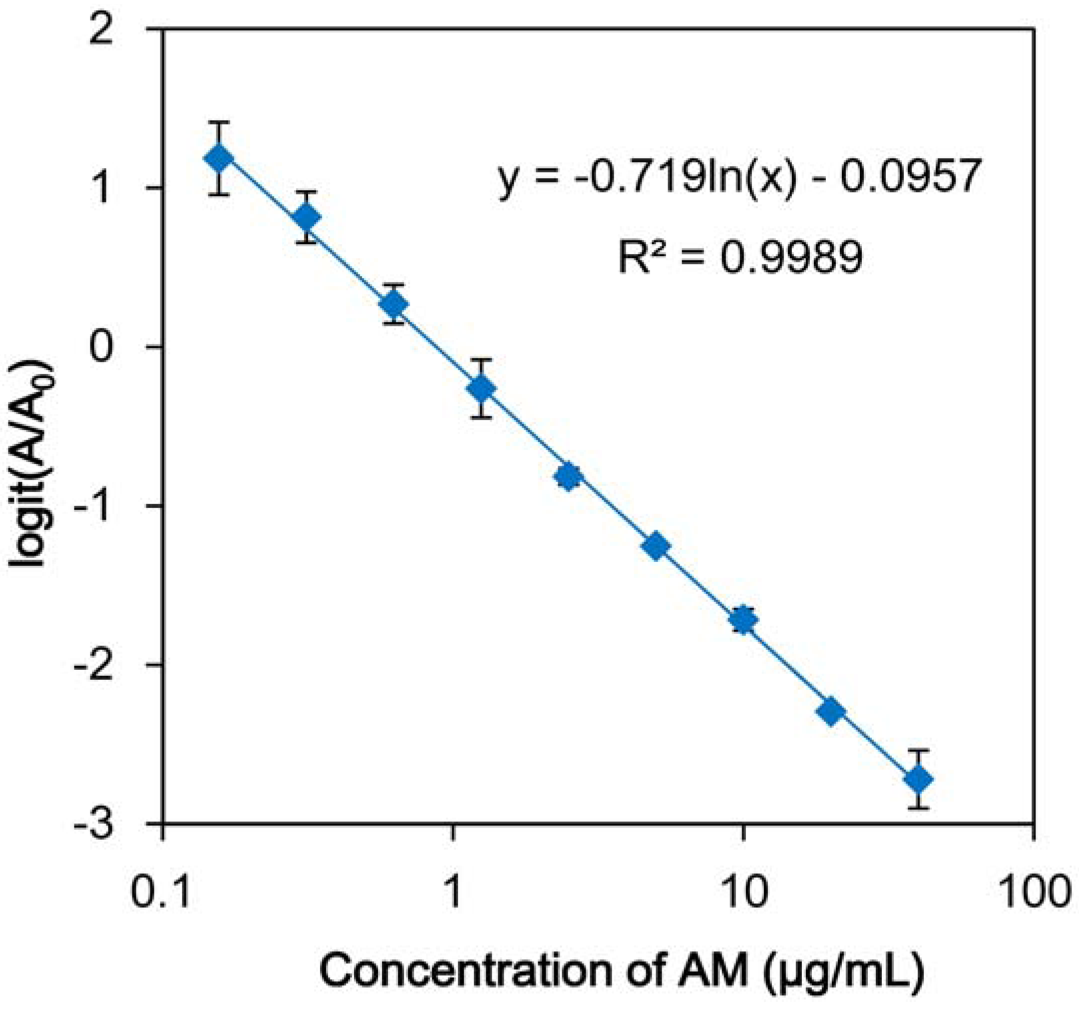
2.2. Evaluation and Application of icELISA Using Anti-AM mAb

An icELISA using anti-AM mAb 1C1 was developed based on the general format that we developed previously [34]. The free mAb 1C1 after competition with AM is bound to polystyrene microtiter plates precoated with AM-HSA (1 μg/mL) in the ELISA. During incubation with a secondary antibody, mAb 1C1 was labeled with peroxidase. After addition of substrate solution (ABTS), absorbance values were measured at 405 nm and a standard curve was prepared using the known concentration of AM. The standard curve exhibited a sigmoidal decrease according to the increase in AM concentration. Under these conditions, the full measurement range of the assay for AM extends from 2 to 20 μg/mL.
An ELISA for AM should be applicable to phytochemical investigations involving crude plant extracts containing numerous secondary metabolites. Cross-reactivities of the mAbs used in ELISA are thus an important factor to assess. The cross-reactivities of anti-AM mAb 1C1 were examined by competitive ELISA, and were calculated using the AMconcentrationyielding midrange valuesand the concentration of other compounds under investigation yielding midrange values by the method reported by Weiler and Zenk [36].
ELISA using mAb 1C1 was not specific for AM and showed cross-reactivities with AS and dihydroartemisinin, but did not react with other compounds. From these results, it appears that mAb 1C1 reacts with compounds whose basic skeleton is the same as that of AM and that it strongly recognizes the succinyl moiety of AS. The wide cross-reactivity of AM mAb 1C1 against AM-related compounds is a major advantage of the antibody reagent used in the ELISA, which is applied to both phytochemical investigations involving crude plant extracts and to therapeutic approaches with antimalarial drugs.
Table 1 shows the results of quantitative analysis of A. annua samples by ELISA. The results for AM content in Chinese wormwoods were similar to those reported previously [37,38]. However, other AM-related compounds which are recognized by the mAb 1C1 may affect these qualitative data. To confirm that these data reflect the content of AM in A. annua, we should compare them by the other analytical methods.
The benefits of this method are that it is easy to use and is capable of analyzing multiple samples at the same time without any sample pretreatment. The newly established ELISA using mAb-1C1may promote the breeding of high-AM-producing A. annua and prompt the appropriate therapeutic utility of antimalarial drugs.
Table 1. Content of artemisinin and its structurally related compoundsin Artemisia annua L. samples determined by icELISA using anti-AM mAb 1C1.
SampleContent (µg/mg dry wt.)
Chinese wormwood 1 3.21 ± 0.09
Chinese wormwood 21.02 ±0.23
Aerial part of regenerated A. annua1.07 ± 0.05
The samples of A. annua are prepared and provided by Dr. Waraporn Putalun at Khon Kaen University, Thailand.

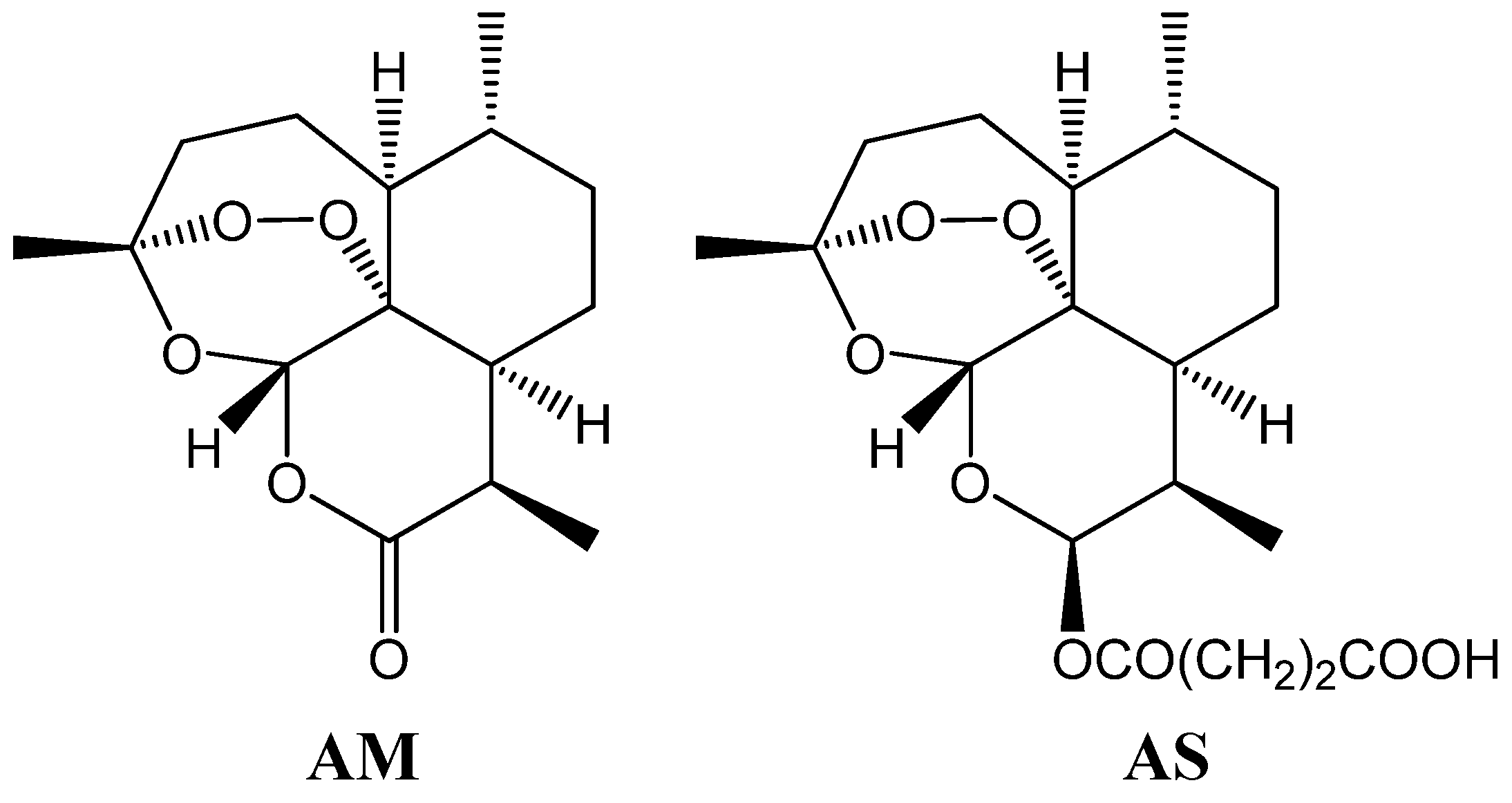
3. Production of Recombinant Fab against AM and AS

3.1. Cloning of cDNAs Encoding Fab and Bacterial Expression of Fab against AM and AS

We prepared a monoclonal antibody (mAb 1C1) and developed an icELISA using this novel mAb. However, the yield of mAb 1C1 was insufficient due to the low production characteristics of the hybridoma, and this hindered the facile application of the novel antibody to various immunochemical experiments. To overcome the insufficient production of the mAb using hybridoma culture, we prepared a recombinant Fab against the antimalarial drugs AM and AS.
The cDNAs encoding the VH-CH1 and VL-CL fragments of the anti-AM mAb were amplified using two sets of specific primers containing an appropriate restriction site. Both cDNAs were successfully cloned in this way. The amino acid sequences were aligned to determine the genes encoding the VH and VL fragments by reference to the N and C termini of each fragment and the complementarity-determining region (CDR) using the Kabat and Chothia numbering scheme (http://www.bioinf.org.uk/abs) [39,40]. Plausible genes encoding the VH and VL fragments of the anti-AM mAb 1C1 were identified. Both VH-CH1 and VL-CL genes consisted of 654 bp, and each fragment was fully consistent with the corresponding variable regions and constant regions of the IgG1 and a κ light chain of the anti-AM mAb, respectively, as determined by sequence analyses. The two cDNAs were inserted into a pET28a(+) vector for expression and characterization of the Fab against AM and AS.
Bacterial expression of VH1-CH1 and VL-CL using transformed Escherichia coli BL21 (DE3) was performed, and overexpressed proteins formed inclusion bodies. Each fragment with an N-terminal hexahistidine tag expressed in the insoluble fraction was resolved in an 8 M urea solution and was purified by metal chelator affinity chromatography. They were obtained in excellent yields, 19 mg per 1-L culture (VH1-CH1) and 18 mg per 1-L culture (VL-CL), respectively. A molecular weight of 27 kDa was determined by 12.5% SDS-PAGE (Figure 2B; Lanes 2 and 3), which was very similar to the theoretical values (27,305 Da for VH1-CH1 and 27,979 Da for VL-CL) predicted for each fragment with the tags.
Figure 2. SDS-PAGE analysis of recombinant antibodies against AM and AS.A) Analysis of the scFv. Lane 1, marker proteins; Lane 2, total protein before IPTG induction; Lane 3, total protein after IPTG induction; Lane 4, soluble fraction, Lane 5, insoluble fraction; Lane 6, purified scFv. B) Analysis of the Fab. Lane 1, marker proteins; Lane 2, purified VH-CH1; Lane 3, VL-CL; Lane 4, refolded Fab (non-reducing SDS-PAGE).
Antibodies 01 00273 g002

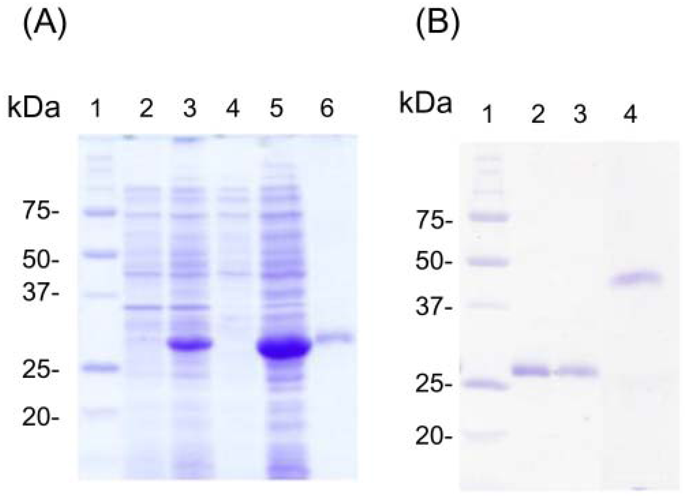
3.2. Refolding and Reactivity Tests for Fab against AM and AS

Because the purified recombinant antibodies were purified in their inactive forms, refolding was necessary in order to enable antigen recognition. We employed an efficient refolding method developed by Umetsu et al. [41] that is based on removal of the solubilizing agent by stepwise dialysis. Each recombinant antibody fragment that did not have a correct high-order structure was treated with β-ME in urea solution to cleave disulfide bonds, followed by dialysis against gradual tapering urea solutions. In the dialysis process, it is presumed that intramolecular hydrogen bonds are formed correctly and that hydrophobic interactions and van der Waals forces go on to facilitate their folding. At the 2–0.125 M urea stages, L-arginine was supplemented as a solubilizing agent, and reduced and oxidized glutathione were added to facilitate the formation of intra- and intermolecular disulfide bonds. Figure 2B (Lane 4) shows the results of nonreducing SDS-PAGE analysis of the refolded Fab, in which only one band was observed. The results reveal that each supplement played a role in line with expectations and disulfide bonds formed during the refolding of the Fab, and regeneration of the binding capabilities were expected.
The sample was dialyzed to refold the Fab, and its reactivities were evaluated by indirect ELISA using an AS-OVA conjugate as the antigen. The validation study by indirect ELISA indicated that recombinant Fab recognized the AS-OVA conjugate, and optical density (OD) increased in a concentration-dependent manner. Therefore, we focused on the development and evaluation of an icELISA using the Fab for applications related to analysis of AM-related compounds.

3.3. Development of icELISA Using Recombinant Fab against AM and AS

In order to develop analytical methods for use during production of AM-related compounds, refolded Fab was used in an icELISA using AS-OVA as a solid-phase antigen. Figure 3 shows that the standard curves generated for AM were linear from 0.16 μg/mL to 40 μg/mL. The cross-reactivities (CRs) of Fab against other natural products were similar to those observed for the anti-AM mAb, which recognizes only AM-related compounds (Table 2). In case of analysis of AS, the linearity between concentrations of AS and the signal was observed from 8.0 ng/mL to 60 ng/mL. These results confirmed that the sequences of the variable fragments of the Fab yielded properties consistent with those of mAb 1C1.
Figure 3. Standard curves for the inhibition by AM in an indirect competitive ELISA using the recombinant Fab. Various concentrations of AM was incubated with the recombinant Fab (0.5 µg/mL) against AM in a 96-well immunoplate precoated with AS-OVA (1 µg/mL).
Antibodies 01 00273 g003
Table 2. Cross-reactivities of anti-AM Fab against various natural products.
CompoundIC50 (µg/mL)
Artemisinin1.58
Artesunate0.022
Saikosaponin a>100
Ginsenoside Rb1>100
Baicalin>100
Quinine>100
Berberine>100
Arbutin>100
Amygdalin>100
Swertiamarin>100
Digitonin>100
We validated the icELISA using the Fab, as shown in Figure 3, for the quantitative identification of AM. The intra- and interassay variabilities were assessed for the icELISA by testing three samples of nine different AM concentrations performed together on the same day and on three consecutive days, respectively. The results showed that the maximum intraassay RSD was 4.7%, whereas the interassay RSD was 5.9%. The assay results were confirmed using the mAb, revealing that the developed icELISA was sufficiently accurate and reliable for quantitative analysis of AM.
After validating the icELISA as a quantitative method, the method was used for analysis of various Artemisia spp., which is the main source of AM. The leaves of five types of Artemisia spp. (A. annua A. absinthium A. abrotanum A. iudovicianna A. capillaris, A. pontica) were purchased from the market in Japan. They were dried and ground into powders, then extracted at room temperature with MeOH (0.5 mL) using an ultrasonic bath for 10 min. After centrifugation of the samples, the supernatant was transferred to a tube. This extraction step was repeated three times. After the extracted solutions had been concentrated in vacuo, the extract was dissolved in a small amount of MeOH and diluted appropriately to give 20% MeOH solutions for icELISA using the recombinant Fab against AM. As a result, AM content of A. annua was 5.40 μg/mg dwt., which was the highest value among the samples. The analytical data confirmed that A. absinthium and A. abrotanum contained AM. The experimental results were highly reproducible and agreed with previous reports, supporting the practical utility of the icELISA using the recombinant Fab against AM as a method for quantitatively analyzing AM [42].

4. Conclusions

Because AM and its structurally related compounds have poor UV chromophore or display fluorescence, previous quality control methods have involved derivatizing compounds in acidic or basic solutions for analysis using conventional TLC or HPLC techniques [43,44,45,46,47]. Recently, a liquid chromatographic tandem mass spectroscopy method for the quantification of AM was developed and was found to provide more sensitive quantitative analysis [48,49]. On the other hand, the benefits of this method also include its ease of use and its ability to analyze multiple samples at the same time without any sample pretreatment.
In addition, we developed expression systems that abundantly produce recombinant Fab against AM and AS, and we applied it to the icELISA in the first such approach to quantitative analysis of AM-related compounds. Given the poor production of anti-AM mAb by the hybridoma 1C1, the improved Fab production was significant for the development of a cost-effective immunoassay.
Our results suggest that the results encourage further evaluation of the technology as a potential method to assist with the development of AM plant breeding programs, quality checks in AM production operations, and techniques for therapeutic drug monitoring. The present immunoassay using antibodies possessing novel characteristics can potentially advance the clinical development of the anti-malarial drugs AM and AS.

Acknowledgments

This work was funded in part by Grants-in-Aid from the JSPS Asian CORE Program, the Ministry of Education, Culture, Sports, Science and Technology of Japan, and the National Center for Genetic and Biotechnology (BIOTEC), Thailand. We are also grateful for technical support from the Research Support Center, Graduate School of Medical Sciences of Kyushu University.

References

  1. Klayman, D.L. Qinghaosu (artemisinin): An antimalarial drug from China. Science 1985, 228, 1049–1055. [Google Scholar]
  2. Eckstein-Ludwig, U.; Webb, R.J.; van Goethem, I.D.A.; East, J.M.; Lee, A.G.; Kimura, M.; O’Neill, P.M.; Bray, P.G.; Ward, S.A.; Krishna, S. Artemisinins target the SERCA of Plasmodium falciparum. Nature 2003, 424, 957–961. [Google Scholar] [CrossRef]
  3. Miller, L.H.; Su, X. Artemisin: Discovery from the Chinese Herbal Garden. Cell 2011, 146, 855–858. [Google Scholar] [CrossRef]
  4. Smithuis, F.; Kyaw, M.K.; Phe, O.; Aye, K.Z.; Htet, L.; Barends, M.; Lindegardh, N.; Singtoroj, T.; Ashley, E.; Lwin, S.; et al. Efficacy and effectiveness of dihydroartemisinin-piperaquine versus artesunate-mefloquine in falciparum malaria: An open-label randomised comparison. Lancet 2006, 367, 2075–2085. [Google Scholar]
  5. Zongo, I.; Dorsey, G.; Rouamba, N.; Tinto, H.; Dokomajilar, C.; Guiguemde, R.T.; Rosenthal, P.J.; Ouedraogo, J.B. Artemether-lumefantrine versus amodiaquine plus sulfadoxine-pyrimethamine for uncomplicated falciparum malaria in Burkina Faso: A randomised non-inferiority trial. Lancet 2007, 369, 491–498. [Google Scholar] [CrossRef]
  6. Tshefu, A.K.; Gaye, O.; Kayentao, K.; Thompson, R.; Bhatt, K.M.; Sesay, S.S.; Bustos, D.G.; Tjitra, E.; Bedu-Addo, G.; Borghini-Fuhrer, I.; et al. Efficacy and safety of a fixed-dose oral combination of pyronaridine-artesunate compared with artemether-lumefantrine in children and adults with uncomplicated Plasmodium falciparum malaria: A randomised non-inferiority trial. Lancet 2010, 375, 1468–1481. [Google Scholar]
  7. Crawley, J.; Chu, C.; Mtove, G.; Nosten, F. Malaria in children. Lancet 2010, 375, 1457–1467. [Google Scholar] [CrossRef]
  8. Poespoprodjo, J.R.; Fobia, W.; Kenangalem, E.; Hasanuddin, A.; Sugiarto, P.; Tjitra, E.; Anstey, N.M.; Price, R.N. Highly effective therapy for maternal malaria associated with a lower risk of vertical transmission. J. Infect. Dis. 2011, 204, 1613–1619. [Google Scholar] [CrossRef]
  9. Ravindranathan, T.; Kumar, M.A.; Menon, R.B.; Hiremath, S.V. Stereoselective synthesis of artemisinin. Tetrahedron Lett. 1990, 31, 755–758. [Google Scholar] [CrossRef]
  10. Avery, M.A.; Chong, W.K.M.; Jenningswhite, C. Stereoselective total synthesis of (+)-artemisinin, the antimalarial constituent of Artemisia annua L. J. Am. Chem. Soc. 1992, 114, 974–979. [Google Scholar] [CrossRef]
  11. Ro, D.K.; Paradise, E.M.; Ouellet, M.; Fisher, K.J.; Newman, K.L.; Ndungu, J.M.; Ho, K.A.; Eachus, R.A.; Ham, T.S.; Kirby, J.; et al. Production of the antimalarial drug precursor artemisinic acid in engineered yeast. Nature 2006, 440, 940–943. [Google Scholar] [CrossRef]
  12. Woerdenbag, H.J.; Moskal, T.A.; Pras, N.; Malingre, T.M.; Elferaly, F.S.; Kampinga, H.H.; Konings, A.W.T. Cytotoxicity of artemisinin-related endoperoxides to Ehrlich ascites tumor-cells. J. Nat. Prod. 1993, 56, 849–856. [Google Scholar] [CrossRef]
  13. Mercer, A.E.; Copple, I.M.; Maggs, J.L.; O’Neill, P.M.; Park, B.K. The role of heme and the mitochondrion in the chemical and molecular mechanisms of mammalian cell death induced by the artemisinin antimalarials. J. Biol. Chem. 2011, 286, 987–996. [Google Scholar]
  14. Liu, W.M.; Gravett, A.M.; Dalgleish, A.G. The antimalarial agent artesunate possesses anticancer properties that can be enhanced by combination strategies. Int. J. Cancer 2011, 128, 1471–1480. [Google Scholar] [CrossRef]
  15. Disbrow, G.L.; Baege, A.C.; Kierpiec, K.A.; Yuan, H.; Centeno, J.A.; Thibodeaux, C.A.; Hartmann, D.; Schlegel, R. Dihydroartemisinin is cytotoxic to papillomavirus- expressing epithelial cells in vitro and in vivo. Cancer Res. 2005, 65, 10854–10861. [Google Scholar] [CrossRef]
  16. Hou, J.; Wang, D.; Zhang, R.; Wang, H. Experimental therapy of hepatoma with artemisinin and its derivatives: In vitro and in vivo activity, chemosensitization, and mechanisms of action. Clin. Cancer Res. 2008, 14, 5519–5530. [Google Scholar] [CrossRef]
  17. Posner, G.H.; McRiner, A.J.; Paik, I.H.; Sur, S.; Borstnik, K.; Xie, S.J.; Shapiro, T.A.; Alagbala, A.; Foster, B. Anticancer and antimalarial efficacy and safety of artemisinin-derived trioxane dimers in rodents. J. Med. Chem. 2004, 47, 1299–1301. [Google Scholar] [CrossRef]
  18. Jeyadevan, J.P.; Bray, P.G.; Chadwick, J.; Mercer, A.E.; Byrne, A.; Ward, S.A.; Park, B.K.; Williams, D.P.; Cosstick, R.; Davies, J.; et al. Antimalarial and antitumor evaluation of novel C-10 non-acetal dimers of 10beta-(2-hydroxyethyl)deoxoartemisinin. J. Med. Chem. 2004, 47, 1290–1298. [Google Scholar] [CrossRef]
  19. Galal, A.M.; Ross, S.A.; ElSohly, M.A.; ElSohly, H.N.; El-Feraly, F.S.; Ahmed, M.S.; McPhail, A.T. Deoxyartemisinin derivatives from photooxygenation of anhydrodeoxydihydroartemisinin and their cytotoxic evaluation. J. Nat. Prod. 2002, 65, 184–188. [Google Scholar] [CrossRef]
  20. Li, Y.; Shan, F.; Wu, J.M.; Wu, G.S.; Ding, J.; Xiao, D.; Yang, W.Y.; Atassi, G.; Leonce, S.; Caignard, D.H.; et al. Novel antitumor artemisinin derivatives targeting. G1 phase of the cell cycle. Bioorg. Med. Chem. Lett. 2001, 11, 5–8. [Google Scholar] [CrossRef]
  21. Jaziri, M.; Diallo, B.; Vanhaelen, M.; Homes, J.; Yoshimatsu, K.; Shimomura, K. Immunodetection of artemisinin in Artemisia annua cultivated in hydroponic conditions. Phytochemistry 1993, 33, 821–826. [Google Scholar]
  22. Ferreira, J.F.; Janick, J. Immunoquantitative analysis of artemisinin from Artemisia annua using polyclonal antibodies. Phytochemistry 1996, 41, 97–104. [Google Scholar] [CrossRef]
  23. Eggelte, T.A.; van Agtmael, M.A.; Vuong, T.D.; van Boxtel, C.J. The development of an immunoassay for the detection of artemisinin compounds in urine. Am. J. Trop. Med. Hyg. 1999, 61, 449–456. [Google Scholar]
  24. He, S.P.; Tan, G.Y.; Li, G.; Tan, W.M.; Nan, T.G.; Wang, B.M.; Li, Z.H.; Li, Q.X. Development of a sensitive monoclonal antibody-based enzyme-linked immunosorbent assay for the antimalaria active ingredient artemisinin in the Chinese herb Artemisia annua L. Anal. Bioanal. Chem. 2009, 393, 1297–1303. [Google Scholar] [CrossRef]
  25. Yanagihara, H.; Sakata, R.; Minami, H.; Tanaka, H.; Shoyama, Y.; Murakami, H. Immunoaffinity column chromatography against forskolin using an anti-forskolin monoclonal antibody and its application. Anal. Chim. Acta 1996, 335, 63–70. [Google Scholar] [CrossRef]
  26. Shan, S.J.; Tanaka, H.; Shoyama, Y. Enzyme-linked immunosorbent assayfor glycyrrhizin using anti-glycyrrhizin monoclonalantibody and an eastern blottingtechnique for glucuronides of glycyrrhetic acid. Anal. Chem. 2001, 73, 5784–5790. [Google Scholar] [CrossRef]
  27. Kim, J.S.; Tanaka, H.; Shoyama, Y. Immunoquantitative analysis for berberine and its related compounds using monoclonal antibodies in herbal medicines. Analyst 2004, 129, 87–91. [Google Scholar] [CrossRef]
  28. Phrompittayarat, W.; Putalun, W.; Tanaka, H.; Wittaya-Areekul, S.; Jetiyanon, K.; Ingkaninan, K. An enzyme-linked immunosorbant assay using polyclonal antibodies against bacopaside I. Anal. Chim. Acta 2007, 584, 1–6. [Google Scholar] [CrossRef]
  29. Sakamoto, S.; Putalun, W.; Tsuchihashi, R.; Morimoto, S.; Kinjo, J.; Tanaka, H. Development of an enzyme-linked immunosorbent assay (ELISA) using highly-specific monoclonal antibodies against plumbagin. Anal. Chim. Acta 2008, 607, 100–105. [Google Scholar] [CrossRef]
  30. Kido, K.; Edakuni, K.; Morinaga, O.; Tanaka, H.; Shoyama, Y. An enzyme-linked immunosorbent assay for aconitine-type alkaloids using an anti-aconitine monoclonal antibody. Anal. Chim. Acta 2008, 616, 109–114. [Google Scholar] [CrossRef]
  31. Juengwatanatrakul, T.; Sritularak, B.; Amornnopparattanakul, P.; Tassanawat, P.; Putalun, W.; Tanaka, H.; Morimoto, S. Preparation of a specificmonoclonal antibody to asiaticoside for the development of an enzyme-linked immunosorbent assay. Analyst 2011, 136, 1013–1017. [Google Scholar] [CrossRef]
  32. Kido, K.; Morinaga, O.; Shoyama, Y.; Tanaka, H. Quick analysis of baicalin in Scutellariae Radix by enzyme-linked immunosorbent assay using a monoclonal antibody. Talanta 2008, 77, 346–350. [Google Scholar] [CrossRef]
  33. Paudel, M.K.; Putalun, W.; Sritularak, B.; Morinaga, O.; Shoyama, Y.; Tanaka, H.; Morimoto, S. Development pf a combined technique using a rapid one-step immunochromatographic assay and indirect competitive ELISA for the rapid detection of bacalin. Anal. Chim. Acta 2011, 701, 189–193. [Google Scholar] [CrossRef]
  34. Tanaka, H.; Putalun, W.; De-Eknamkul, W.; Matangkasombut, O.; Shoyama, Y. Preparation of a novel monoclonal antibody against the antimalarial drugs artemisinin and artesunate. Planta Med. 2007, 73, 1127–1132. [Google Scholar] [CrossRef]
  35. Paudel, K.M.; Takei, A.; Sakoda, J.; Juengwatanatrakul, T.; Sasaki-Tabata, K.; Putalun, W.; Shoyama, Y.; Tanaka, H.; Morimoto, S. Preparation of a single-chain variable fragment and a recombinant antigen-binding Fragment against the anti-malarial drugs, artemisinin and artesunate, and their application in an ELISA. Anal. Chem. 2012, 84, 2002–2008. [Google Scholar] [CrossRef]
  36. Weiler, E.W.; Zenk, M.H. Radioimmunoassay for determination of digoxin and related compounds in Digitalis lanata. Phytochemistry 1976, 15, 1537–1545. [Google Scholar] [CrossRef]
  37. Peng, C.A.; Ferreira, J.F.S.; Wood, A.J. Direct analysis of artemisinin from Artemisia annua L. using high-performance liquid chromatography with evaporative light scattering detector and gas chromatography with flame ionization detector. J. Chromatogr. A 2006, 1133, 254–258. [Google Scholar] [CrossRef]
  38. Liu, C.Z.; Zhou, H.Y.; Zhao, Y. An effective method for fast determination of artemisinin in Artemisia annua L. by high performance liquid chromatography with evaporative light scattering detection. Anal. Chin. Acta 2007, 581, 298–302. [Google Scholar] [CrossRef]
  39. Kabat, E.A.; Wu, T.T. Identical V-region amino-acid sequences and segments of sequences in antibodies of VH and VL genes, minigenes, and complementarity-determining regions to binding of antibody-combining sites. J. Immun. 1991, 147, 1709–1719. [Google Scholar]
  40. Chothia, C.; Lesk, A.M.; Tramontano, A.; Levitt, M.; Smithgill, S.L.; Air, G.; Sheriff, S.; Padlan, E.A.; Davies, D.; Tulip, W.R.; et al. Conformations of immunoglobulin hypervariable regions. Nature 1989, 342, 877–8833. [Google Scholar] [CrossRef]
  41. Umetsu, M.; Tsumoto, K.; Hara, M.; Ashish, K.; Goda, S.; Adschiri, T.; Kumagai, I. How additives influence the refolding of immunoglobulin-folded proteins in a stepwise dialysis system. Spectroscopic evidence for highly efficient refolding of a single-chain Fv fragment. J. Biol. Chem. 2003, 278, 8979–8987. [Google Scholar]
  42. Abdul, M.; Ibrar, A.; Waheed, A.; Muhammad, F.A.; Rizwana, A.Q.; Izhar, H.; Bushra, M. Survey of artemisinin production by diverse Artemisia species in northern Pakistan. Malaria J. 2010, 9, 310. [Google Scholar] [CrossRef]
  43. Marchand, E.; Atemnkeng, M.A.; Vanermen, S.; Plaizier-Vercammen. Development and validation of a simple thin layer chromatographic method for the analysis of artemisinin in Artemisia annua L. plant extracts. J. Biomed. Chromatogr. 2008, 22, 454–459. [Google Scholar] [CrossRef]
  44. Koobkokkruad, T.; Chochai, A.; Kerdmanee, C.; De-Eknamkul, W. TLC-Densitometric analysis of artemisinin for the rapid screening of high-producing plantlets of Artemisia annua L. Phytochem. Anal. 2007, 18, 229–234. [Google Scholar] [CrossRef]
  45. Shishan, Z.; Mei-Yi, Z. Application of precolumn reaction to high-performance liquid chromatography of qinghaosu in animal plasma. Anal. Chem. 1986, 58, 289–292. [Google Scholar] [CrossRef]
  46. Lapkin, A.A.; Walker, A.; Sullivan, N.; Khambay, B.; Mlambo, B.; Chemat, S. Development of HPLC analytical protocols for quantification of artemisinin in biomass and extracts. J. Pharm. Biomed. Anal. 2009, 49, 908–915. [Google Scholar] [CrossRef]
  47. Diawara, H.Z.; Gbaguidi, F.; Evrard, B.; Leclercq, J.Q.; Moudachirou, M.; Debrus, B.; Hubert, P.; Rozet, E. Validation, transfer and measurement uncertainty estimation of an HPLC-UV method for the quantification of artemisinin in hydro alcoholic extracts of Artemisia annua L. J. Pharm. Biomed. Anal. 2011, 56, 7–15. [Google Scholar] [CrossRef]
  48. Lai, C.S.; Nair, N.K.; Mansor, S.M.; Olliaro, P.L.; Navaratnam, V. An analytical method with a single extraction procedure and two separate high performance liquid chromatographic systems for the determination of artesunate, dihydroartemisinin and mefloquine in human plasma for application in clinical pharmacological studies of the drug combination. J. Chromatogr. B. Analyt. Technol. Biomed. Life Sci. 2007, 857, 308–314. [Google Scholar] [CrossRef]
  49. Hodel, E.M.; Zanolari, B.; Mercier, T.; Biollaz, J.; Keiser, J.; Olliaro, P.; Genton, B.; Decosterd, L.A. A single LC-tandem mass spectrometry method for the simultaneous determination of 14 antimalarial drugs and their metabolites in human plasma. J. Chromatogr. B. Analyt. Technol. Biomed. Life Sci. 2009, 877, 867–886. [Google Scholar] [CrossRef]

