Review

Diving through Membranes: Molecular Cunning to Enforce the Endosomal Escape of Antibody-Targeted Anti-Tumor Toxins

by
Hendrik Fuchs
1,*,
Christopher Bachran
2 and
David J. Flavell
3
1
Institute for Laboratory Medicine, Clinical Chemistry and Pathobiochemistry, Charité – Universitätsmedizin Berlin, Campus Benjamin Franklin, Berlin 12200, Germany
2
Laboratory of Parasitic Diseases, National Institute of Allergy and Infectious Diseases, National Institutes of Health, Bethesda, MD 20892, USA
3
The Simon Flavell Leukaemia Research Unit, Southampton General Hospital, Southampton SO16 6YD, UK
*
Author to whom correspondence should be addressed.
Antibodies 2013, 2(2), 209-235; https://doi.org/10.3390/antib2020209
Submission received: 4 March 2013 / Revised: 27 March 2013 / Accepted: 5 April 2013 / Published: 17 April 2013
(This article belongs to the Special Issue Recombinant Immunotoxins)

Abstract

:
Membranes are vital barriers by which cells control the flux of molecules and energy between their exterior and interior and also between their various intracellular compartments. While numerous transport systems exist for ions and small molecules, the cytosolic uptake of larger biological molecules and in particular antibody-targeted drugs, is a big challenge. Inducing leakage of the plasma membrane is unfavorable since the target cell specificity mediated by the antibody would likely be lost in this case. After binding and internalization, the antibody drug conjugates reach the endosomes. Thus, enforcing the endosomal escape of anti-tumor toxins without affecting the integrity of other cellular membranes is of paramount importance. Different strategies have been developed in the last decades to overcome endosomal accumulation and subsequent lysosomal degradation of targeted protein-based drugs. In this review we summarize the various efforts made to establish efficient techniques to disrupt the endosomal membrane barrier including the use of molecular ferries such as cell penetrating peptides or viral membrane fusion proteins, endosomal leakage inducing molecules such as saponins or monensin and physicochemical methods as represented by photochemical internalization.

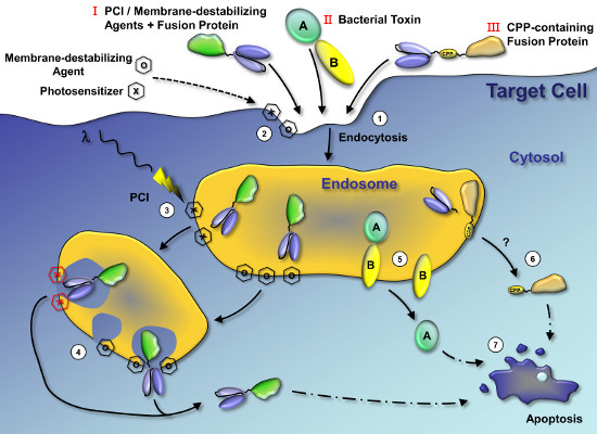
Graphical Abstract

1. Introduction

Antibody-based tumor targeted toxins are once again attracting increasing interest after their first description over two decades ago. The principle strength of targeted toxins is based on their bipartite structure, one component binds to a disease-specific cell-surface target molecule and the other confers cytotoxicity.
The targeting moiety allows the seeking out of small tumor cell clusters or single tumor cells present in the host. Targeted protein toxins therefore have greatest potential for the treatment of residual disease and metastases in addition to surgically inaccessible, disseminated and hematopoietic tumors. In the majority of cases, the targeting moiety is an antibody [1,2] or antibody fragment [1,3] but equally it can also be another protein species or a non-protein chemical structure that recognizes and binds to a suitable target cell-specific surface molecule. Advanced non-protein-based targeting moieties are, for instance, aptamers [4], which, however, have the disadvantage that they cannot be employed to produce a single recombinant drug. Besides antibodies, interleukins and growth factors are the most frequently used protein ligands for targeting toxins [5], but artificial ligands such as custom designed ankyrin repeat proteins have also been described [6]. One strategy to improve the target cell specificity of targeted toxins is through the use of a bispecific antibody. Such an example is the fusion protein DT2219ARL that consists of a catalytically active fragment of diphtheria toxin and two scFv ligands recognizing CD19 and CD22 [7]. Bispecific targeting can also be accomplished utilizing selected ligands as exemplified by a truncated Pseudomonas exotoxin A that was cloned onto the same single-chain molecule together with both human epidermal growth factor (EGF) and interleukin 4 [8].
The cell-killing moiety is commonly either a radioisotope, a small organic molecule that includes fungal poisons or a protein. Whereas radioisotopes are efficient independent of cellular uptake, a number of protein toxins used for tumor therapy typically exert their effect on a subcellular organelle within the cytosol. This requires binding of the targeting moiety to a cell membrane receptor that is then constitutively endocytosed after ligand binding followed by the subsequent transport of the toxin across a limiting cellular membrane [9]. Unfortunately, even if the toxins themselves are membrane permeable, their coupling to antibody or other targeting ligand renders them impermeable. The toxin is typically covalently linked to the antibody by chemical conjugation. Where the toxin is a protein, the drug can also be expressed as a fusion protein generated by recombinant DNA technology [5]. Insertion of an acid-sensitive bond or a cleavage site for endosomal proteases between the toxin and the antibody can result in the release of the toxin in acidic endosomes or lysosomes, thus allowing the passage of the toxin across the endosomal/lysosomal membrane where this is permissible [2,10]. Mutation of targeted toxins to avoid lysosomal degradation is another option to increase cytotoxicity, but seems to be limited to lymphocytes [11]. However, in many instances, protein-based drugs are employed that must be guided or assisted to pass across the limiting membrane to avoid endosomal and lysosomal accumulation and subsequent degradation. Direct visualization by confocal microscopy of the intracellular trafficking route of a targeted toxin showed that it accumulated preferentially in the lysosome in resistant cells but not in sensitive cells. These observations implicate the process of increased lysosomal degradation as the most likely basis for resistance [12] and underline the importance of a potent endosomal escape.
Protein toxins that evolved to pass cellular membranes are often very efficient with regards to their membrane translocation from the lumen of a vesicular compartment into the cytosol. The mechanism of the cytosolic transfer depends on the nature of the protein toxin and is only understood for a few toxins, which are derived from particular bacteria or plants. In recent years vertebrate and in particular human enzymes utilized as toxins have been gaining increasing attention as possible drug candidates [9]. The first toxin described for use in targeted tumor therapy was diphtheria toxin from Corynebacterium diphtheriae that was conjugated to antibodies directed against tumor cell surface antigens [13]. This toxin belongs to the class of ADP-ribosylating enzymes and transfers ADP-ribose to diphthamide, an unusually modified histidine of the mammalian elongation factor 2 [14]. This prevents the interaction with the binding cavern in the 60S ribosomal subunit resulting in the arrest of protein synthesis. Some of the proteins used in targeted toxins possess a natural cell binding domain that can bind to off-target cells and therefore mediate unwanted side effects [15]. These protein toxins are therefore utilized in a truncated form without the natural binding domain as first published for a monoclonal antibody conjugated to the isolated A-chain of diphtheria toxin [16]. Corresponding exotoxin A fragments without a binding domain from Pseudomonas aeruginosa are called PE40 and PE38 and have been used conjugated to various cell binding ligands in a number of clinical trials [17].
Another important group of proteins that are used for the construction of targeted toxins are the ribosome-inactivating proteins (RIPs) found in certain families of dicot plants [18,19]. Several have been investigated incorporated into targeted toxins in clinical trials [9]. RIPs can be subdivided into Type I consisting of a single catalytic A chain with a molecular mass of between 26–32 kDa and Type II species comprised of a catalytic A chain and a lectin-binding B chain of approximately 31–36 kDa that binds to specific cell-surface carbohydrate groups [20]. The catalytic A chain cleaves a specific N-glycosidic bond in mammalian 28S ribosomal RNA resulting in the release of an adenine that is required to bind the eukaryotic elongation factors 1 and 2 [21], resulting ultimately in the arrest of protein biosynthesis. Since Type-I-RIPs lack a cell binding domain, they are on the one hand ideal for the design of targeted toxins; however, they do not possess a domain for internalization or efficient transfer across the endosomal membrane; they are therefore on the other hand limited in their stand alone use, though they do efficiently mediate cell death once they have gained entry to the cytosol [19]. Thus, devising strategies to enhance the endosomal escape of Type-I-RIPs is of great practical interest.
All non-human proteins have the disadvantage of being potentially immunogenic, which raises the question whether human proteins with toxic potential are suitable to employ in targeted tumor therapies. Proapoptotic proteins appear to be the best candidates including proteins involved in microbial defense and developmental apoptosis. Indeed, the proapoptotic serine protease granzyme B that is released from cytoplasmic granules of cytotoxic T cells and natural killer cells [22], the RNase angiogenin [23] and the apoptosis inducing factor (AIF) [24] are human proteins that have been investigated as the toxic moiety in targeted strategies. Further ideas that aim to directly trigger apoptosis include the use of the tumor suppressor protein death-associated protein kinase 2 (DAPK2) [25,26] and of tumor necrosis factor-related apoptosis-inducing ligand (TRAIL) [27]. To reduce the immunogenicity of non-human proteins, several strategies have been successfully tested including the concomitant administration of immunosuppressive drugs, PEGylation of the targeted toxin, removal of immunogenic epitopes by site-directed mutagenesis, and the use of fully humanized antibodies [9]. Since the extent of an immune response is not only dependent on the antigen’s structure but also on the dose, it appears to be a major advantage to be able to reduce the systemic drug concentration. In the case of targeted toxins, the therapeutically effective concentration of drug in the body is largely dependent on the efficiency of cytosolic drug uptake [28], which includes target-specific cell binding, internalization, vesicular transport and endosomal escape. Thus, improving the efficiency of endosomal escape may also indirectly reduce immunogenicity by allowing for the administration of lower doses of drug, which nevertheless retain therapeutic effectiveness [29].
In addition to improving endosomal escape there are other possibilities that might improve the efficacy of targeted toxins. The elevated expression of certain matrix metalloproteases in the tumor environment can be utilized to activate the toxins at the tumor site [30] and their use in combination with other strategies present attractive possibilities. For instance, targeted protein toxins can be administered as an adjunct treatment for surgery or directly when combined with radio- or chemotherapy [2,3]. A number of preclinical and a few clinical studies are reported in the literature, most of which showing additive or synergistic effects [2,3]. A targeted toxin comprised of interleukin-13 and Pseudomonas exotoxin A showed synergistic cytotoxicity when combined with the cytostatic drug gemcitabine [31] and nude mice treated with a combination of radiation and a targeted toxin comprising a disulfide-linked Fv antibody and the Pseudomonas exotoxin A fragment PE38 had a statistically significant prolongation in time to tumor doubling or tripling compared with the targeted toxin or radiation alone [32].

2. Endosomal Escape

The task of getting a molecule that is unable to cross a limiting membrane into the cell interior is a widespread problem not only in applied drug delivery but also in basic science. For cell type independent uptake, various DNA [33] and protein transfection techniques [34] have been developed recently. Such a membrane transfer is typically achieved via the plasma membrane. In the majority of cases, cell specific drug delivery is mediated by the selective binding of the drug to a specific receptor on the target cell. Any system that unspecifically forms pores at the plasma membrane surface such as electroporation would undermine the specificity of selective delivery via a receptor by allowing for non-selective entry [35]. Therefore, alternative techniques must be developed to promote the membrane transfer of the receptor-bound molecule. Two strategies may be considered. In the first, the bound molecule itself harbors a domain that mediates the transfer after binding to the receptor, or in the second, other molecules located inside intracellular vesicles promote cytosolic entry after internalization of the bound drug. The former approach can be achieved by the insertion or addition of drug-releasing elements directly into the targeted toxin [36]. These additional elements can comprise peptides with new functions or chemical characteristics [10]. The latter approach can be specific for the internalized molecule or represent a more general leakage that tolerates the transfer of other molecules from the lumen of the vesicles. The chemical nature and toxicity profiles of the additives is in principal variable but they must fulfill the specific requirements to be approved as a drug. Endosomes are the most obvious vesicles to install a transfer mechanism into the cell, since these are the first reached after internalization: The shift to acidic pH within the endosomal lumen can be used as a trigger to the translocation process [37,38]. The ongoing challenge is therefore to develop strategies that are specifically restricted to endosomes.
Whilst binding and internalization are predominantly determined by the targeting moiety and the constitutive behavior of the targeted receptor, trafficking and cytosolic uptake are frequently guided by the properties of the toxin. As previously mentioned, bacterial toxins and Type-II-RIPs possess an intrinsic translocation domain in their B chain whereas Type-I-RIPs and vertebrate toxins do not [19,39]. Thus, in the majority of cases, diphtheria toxin and Pseudomonas exotoxin A are administered in a truncated form with the cell binding domain deleted and the membrane transfer domain retained [17,40]. In contrast, the vertebrate toxins and the isolated A chains of bacterial and plant toxins require an enhancement for their endosomal escape although these proteins exhibit some minor cytotoxicity due to presumptive non-specific membrane transfer via an unknown mechanism.
Whilst the latter toxins comprised of single chain proteins, another mechanism of efficient endosomal escape is used by anthrax toxin, a tripartite exotoxin from Bacillus anthracis. The receptor-binding component is termed protective antigen and binds to either of its two cellular receptors (capillary morphogenesis gene 2 and tumor endothelial marker 8), oligomerizes and is subsequently endocytosed. The two catalytically active proteins, lethal factor and edema factor, bind to the receptor-bound protective antigen and are co-internalized by endocytosis. Acidification in the endosome triggers insertion of the protective antigen into the membrane to form a pore that promotes the delivery of edema factor and lethal factor into the cytosol [41]. Edema factor is a calcium- and calmodulin-dependent adenylate cyclase that increases cAMP levels, thus interfering with cellular water homeostasis and intracellular signaling pathways, causing edema. Lethal factor is a zinc-dependent metalloprotease cleaving off the N-termini of several mitogen-activated protein kinase kinases. The resulting block in signal transduction leads to apoptosis in a variety of different cell types. The efficient endosomal escape of lethal factor and edema factor has been used for the delivery of fusion proteins containing the protective antigen-binding moiety of lethal factor to deliver other enzymes to the cytosol. These fusion proteins have been successfully utilized for various tumor-therapy approaches [30,42,43], the delivery of reporter proteins [44] and the delivery of anti-apoptotic Bcl-xL [45].
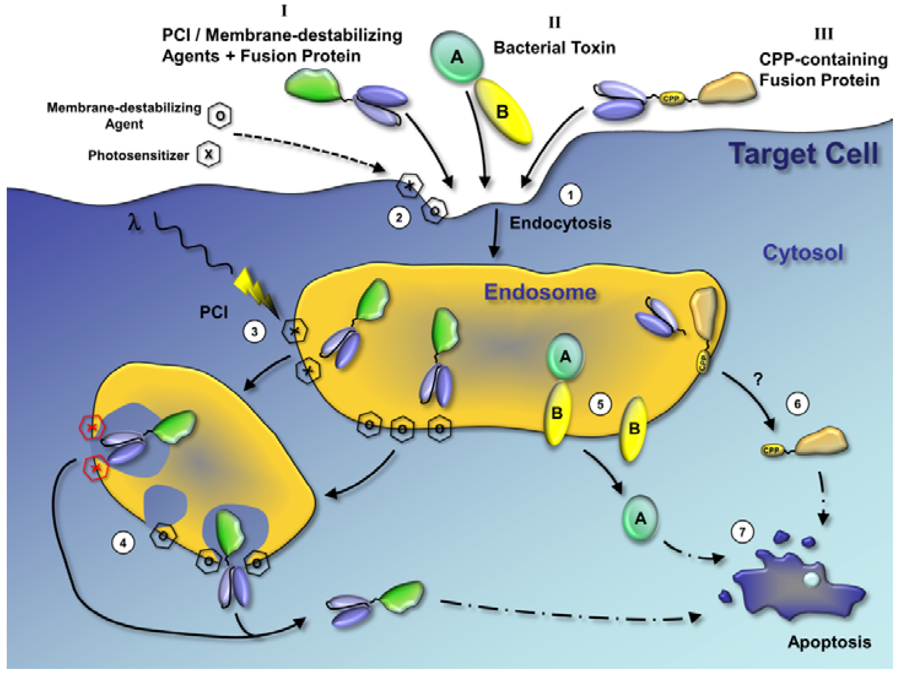
Three strategic categories to enhance the endosomal escape can be distinguished (Figure 1): Molecular ferries, leakage-inducing molecules and physicochemical techniques. Molecular ferries comprise molecules that are either part of the drug or bind to the drug and are able to dive through membranes taking with them cargo, in this case a drug. Typical examples are viral membrane fusion proteins and other cell penetrating peptides (CPP). Leakage-inducing molecules predominantly include substances that destabilize the endosomal membrane. This can be achieved by pore-forming substances, compounds with solubilizing effects and molecules that affect the endosomal pH. The third strategy comprises techniques that work through a direct physical effect, e.g., endosomal membrane disruption by light-induced effects.
Figure 1. Mechanisms of endosomal escape. The three most relevant mechanisms of endosomal escape are depicted in this figure: Endosomal escape of drugs by photochemical internalization (PCI) or membrane-destabilizing agents (I), endosomal escape of bacterial toxins (II), and cell penetrating peptides (CPP)-mediated release of drugs from endosomes (III). Endosomal escape by PCI, membrane-destabilizing agents and CPPs has been described for a variety of drugs and the fusion proteins in this figure serve only as examples. (1) The first step for all three mechanisms is the binding of fusion proteins or bacterial toxins to specific cell surface receptors and the internalization into the target cell by endocytosis. (2) Endosomal escape of fusion proteins by PCI or membrane-destabilizing agents (I): Photosensitizers (hexagon marked with an X, required for PCI) and membrane-destabilizing agents (hexagon marked with an O) are co-administered with fusion proteins and internalize in the plasma membrane and co-internalize with the fusion proteins in endosomes. (3) The photosensitizers are activated by light (red hexagons marked with X) and induce reactive oxygen species production. (4) Both activated photosensitizers and high local concentrations of membrane-destabilizing agents result in endosomal leakage and rupture. The fusion protein is released to the cytosol. (5) Endosomal release of bacterial toxins (II): Bacterial toxins usually contain one protein or a protein domain (marked as B in the figure) that binds the cell surface receptor and initiates translocation of the catalytic protein or other protein domain (marked as A in the figure) through the endosomal membrane into the cytosol. (6) CPP-mediated endosomal release: CPPs mediate the translocation of conjugated drugs or proteins through the endosomal membrane. The detailed mechanisms are unknown and most likely vary for different CPPs. The cell-targeting domain of a fusion protein may be released before the CPP is active in the endosome. (7) All three described mechanisms result in the release of the fusion proteins in the cytosol. The active domains act on their intracellular targets and induce various effects, such as apoptosis, in the target cell.
Antibodies 02 00209 g001
Regardless of the endosomal escape mechanism, drugs that exert their effect inside the cytosol or in organelles should accumulate suitably within the cell to achieve maximum toxicity. A promising strategy to achieve intracellular trapping is the inclusion of a cleavable peptide that, once cleaved inside the cytosol, converts the drug to a membrane-impermeable protein by removing any motif or domain used for membrane translocation [46,47]. An analogous procedure describes the insertion of a natural organelle-specific cleavage recognition site of the mitochondrial malate dehydrogenase signal sequence. Cleavage results in mitochondrial cargo accumulation making this approach suitable for use with targeted toxins that mediate their toxic effects inside mitochondria [48]. Another strategy is the utilization of a protease-cleavable disulfide-forming peptidic loop between two parts of the fusion protein [49]. When the targeting and toxic moieties were coupled chemically, a disulfide bridge might have fulfilled the role of a cytosolic cleavable peptide and an acid-cleavable hydrazone bond that of an endosomal cleavable peptide.

3. Molecular Ferries

3.1. Identifying Cell Penetrating Peptides from Various Organisms to Mediate Cytosolic Uptake

A number of molecules possess the characteristic of being able to take up cargo and to transport this like a ferry to other intracellular locations. Some of these molecular ferries also possess the characteristic that allows the cargo to be transported across limiting membranes. Although the molecular mechanism behind this process has in many cases still to be unraveled, these CPPs typically either traverse the membranes or fuse with the membrane.
CPPs are derived from parts of various proteins of diverse origins, including viral, bacterial, insect and mammalian sources. These peptides are generally 10–16 amino acids in length, with a maximum of about 45 amino acids. Most of them are structurally completely different from each other with no consensus sequence [50]. The most widely studied CPP is the protein transduction domain TAT from the human immunodeficiency virus 1 transcriptional activator protein Tat. Other examples include the Drosophila melanogaster homeotic transcription protein Antennapedia (penetratin), the herpes simplex virus structural protein VP22, a membrane translocation sequence (MTS) derived from the human Kaposi fibroblast growth factor, the synthetic peptides transportan and SynB1, a motif from the PreS2-domain of hepatitis-B virus surface antigen (TLM), the short amphipathic peptide Pep-1, and nuclear localization signals. CPPs are generally not specific for target cells by themselves but the specificity mediated by the targeting moiety is retained in targeted toxins equipped with such a peptide. Snyder et al. linked a chemokine receptor 4 ligand to two different anti-cancer peptides via TAT and demonstrated enhancement of tumor cell killing in vitro [51]. In another study three different CPPs were fused to the Type-I-RIP dianthin. Two of them proved to be able to increase the cytotoxicity in conjugation with transferrin in comparison to a transferrin-dianthin conjugate [52]. A fusion protein of the Type-I-RIP saporin, the above mentioned TLM motif from the PreS2-domain and EGF exhibited increased anti-tumor efficacy in a breast cancer model in mice [46]. The TLM was also successfully used in a targeted toxin composed of an anti-CD64 single chain Fv and angiogenin [53].
To further improve cytosolic drug delivery and to overcome the endosomal membrane barrier, the use of CPPs can be supplemented by other peptides that support drug activation, release and/or accumulation. Peptides containing a matrix metalloproteinase (MMP) recognition motif can be used for tumor site-specific activation. Olson et al. described an activatable CPP comprised of a polycationic peptide connected via a cleavable linker to a neutralizing polyanion. Adsorption and uptake into cells are inhibited until the linker is proteolyzed by MMP-2 or MMP-9. Accumulation was strongest at the tumor-stromal interface in primary tumors and associated metastases [54]. Alternatively, a CPP can be activated after target receptor-mediated endocytosis. Release of the targeting moiety inside the endosomes by furin-like proteases can result in the exposure and thus activation of the CPP [55]. An increase in efficacy can also be achieved when only the furin-site is exerted without a CPP, which is probably caused by the resulting drug release [56,57], however, this must be tested in individual cases since the addition of either one of two furin cleavage sites to a targeted gelonin was less efficient in a xenograft tumor model compared to a flexible non-cleavable linker [58]. Further strategies to increase the endosomal escape of targeted toxins include the use of the translocation motif of diphtheria toxin or Pseudomonas exotoxin A instead of a CPP demonstrated for ricin and granzyme B, and cellular protein retention signals such as REDLK and KDEL as shown for ricin and ribonucleases [10].

3.2. Exploiting Viruses to Augment Targeted Toxin Entry into Target Cells
Like any large macromolecule or multimeric complex, viruses also face the problem of breaching the plasma membrane of the host cell in order to achieve a productive infection. Consequently viruses have evolved a variety of highly efficient mechanisms that enable them to selectively disrupt the host cell membrane, either at the cell surface or within intracellular vesicular compartments that resultantly enables the viral genome to enter the cytosol and/or nucleus of the host cell. The molecular machinery that viruses have developed for this purpose can be exploited in the design of targeted toxins to improve the efficiency with which these molecules are translocated from an intracellular vesicular compartment to target ribosomes in the cytosol.
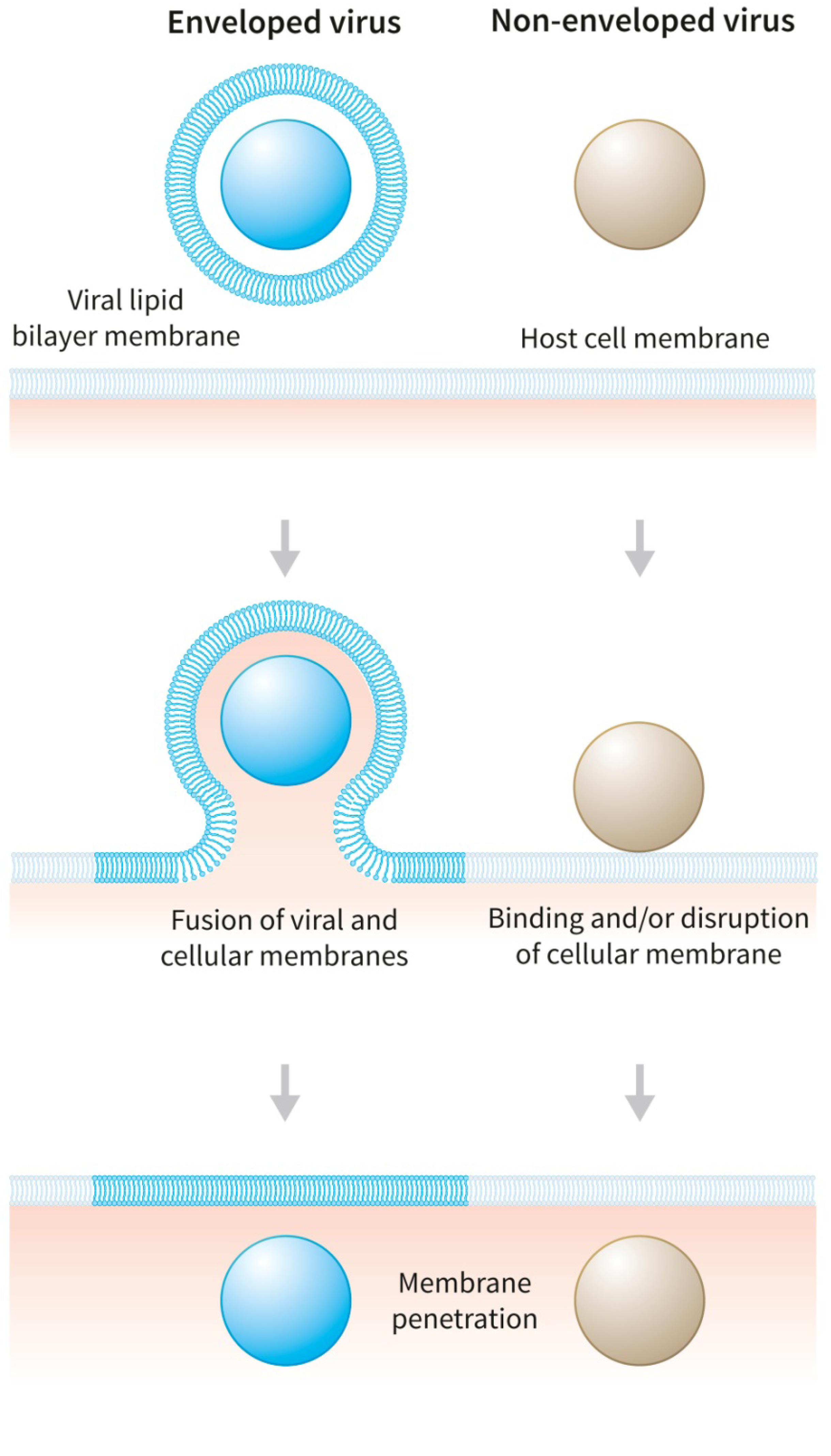
Viruses can be divided into two broad categories, enveloped and non-enveloped, each possessing a variety of different membrane disruptive machineries. A simple depiction of the differences in viral entry for the two categories of virus is shown in Figure 2.
Viral entry mechanisms were exploited initially for their ability to increase transfection efficiency rates for delivering packaged DNA to the target cell with adenovirus being the prototypical example [59]. In certain respects host cell and viral membrane fusion perpetrated by fusogenic proteins expressed by enveloped viruses recapitulates that of the normal cellular machinery involved in vesicle fusion driven by soluble N-ethylmaleimide-sensitive-factor attachment receptors (SNAREs) [60]. Our understanding of the molecular machinery that drives viral genome entry into the host cell has increased greatly in recent years and this knowledge provides us with the opportunity to exploit these as a tool to improve the efficiency of delivery of protein toxins to the cell interior where the target ribosomes reside. In this section we will review the work that has been conducted in this area.
Figure 2. A simplified cartoon representation of the differences in host cell membrane penetration by enveloped and non-enveloped viruses. Enveloped viruses such as influenza and vesicular stomatitis virus enter the cell by receptor mediated endocytosis and fuse their membrane with that of the host cell endosomal membrane using specialized fusogenic proteins which undergo a conformational change in the acidic endosomal environment. In contrast, upon binding to the host cell via specialized receptors, non-enveloped viruses undergo conformational changes in key virally encoded proteins such as protein VI from adenovirus that disrupt the host cell membrane. Host membrane disruption can occur either at the cell surface, as in poliovirus, from an endosome as in adenovirus, from the Golgi as in papillomavirus or from the endoplasmic reticulum as in polyomavirus. Adapted and redrawn from Tsai (2007) [61].
Antibodies 02 00209 g002
Enveloped viruses are surrounded by a bilipid membrane that is derived from the host cell. Enveloped viruses gain entry to the eukaryotic host cell exclusively through a process of membrane fusion utilizing specialized virally encoded glycoproteins for this purpose [62]. The bilipid membrane of enveloped viruses is acquired from the host cell membrane during virus assembly and budding during the infectivity cycle. In order to infect the host cell, the virus must release its genome into the cytosol of the host cell and in order to achieve this it must fuse its membrane with that of the host cell membrane. This results in the release of the nucleocapsid and accessory virally encoded proteins into the cytosol of the host cell to establish a new infectious cycle. Viral glycoproteins inserted into the virus envelope serve two purposes, firstly a receptor binding function that allows the virus to bind selectively to the target host cell that expresses the appropriate cognate ligand and secondly a fusion protein function that requires activation, generally, though not exclusively, after a pH-dependent conformational change in the glycoprotein exposes hydrophobic fusogenic residues capable of mediating membrane fusion [63]. These two separate functions can be mediated by a single glycoprotein or by two separate glycoproteins, depending on the virus family.
Also, depending on the type of virus, membrane fusion can occur either at the plasma membrane surface of the host cell upon virus binding that in turn initiates a conformational change in the glycoprotein to a fusogenic state or alternatively within an intracellular vesicular compartment following receptor mediated endocytosis of viral particles. Here, a lowering of pH in late endosomes [64] leads to protonation of key residues in the specialized viral fusion protein, notably histidines, which then induces the necessary conformational changes in the fusion protein to mediate membrane fusion between viral and host cell membranes. Figure 3 is a cartoon showing the general principle by which the fusion protein achieves this by bringing both membranes into close apposition by assuming a hairpin configuration resulting in the positioning of the two membrane anchors at the same end of a trimeric elongated rod-like structure. Ultimately the close positioning of the two membranes leads the two membranes to fuse through a mechanism that has yet to be fully described.
To date, three different classes of viral fusion proteins (Class I–III) [62] based on post-fusion structural motifs have been described and although there is a wide variation in their structural features overall, on triggering from a pre- to post-fusion state, they all have in common the pH-dependent exposure of a fusion peptide at the N-terminal end of the molecule and the subsequent formation of a stable hairpin-like configuration as shown in Figure 3 that is essential for bringing the two membranes into close apposition. Hemagglutinin (HA) expressed by influenza A virus is the best studied of the enveloped virus fusogenic glycoproteins [63,65]. Synthesized in the host cell firstly as a precursor HA0 it is cleaved late in assembly to HA1 and HA2. The N-terminus of HA2 contains the “fusion peptide” which upon acidification in the endosome triggers the fusion reaction. Wharton and coworkers [66] demonstrated that synthetic peptides derived from the N-terminal region of HA2 were capable of fusing cholesterol-free liposomes at neutral and acidic pH but could only fuse cholesterol containing liposomes at acid pH. Furthermore the fusogenic HA2-derived peptide caused leakage of the liposomal vesicles. It was later demonstrated that similar synthetic fusogenic peptides were capable of augmenting the transfection of human cell lines with a luciferase or β-galactosidase reporter gene delivered via the transferrin receptor (CD71) [67].
Figure 3. A model for membrane fusion by enveloped viruses by fusogenic viral proteins. Conformational changes in the fusogenic protein in the low pH endosomal compartment expose a fusogenic peptide which in Step 2 interacts with the host membrane followed by further structural rearrangements leading to the classical “hairpin” configuration as shown in Step 3. This brings the viral and host cell membranes into close proximity that results in their fusion shown in Step 4. Adapted and redrawn from Hughson (1997) [68].
Antibodies 02 00209 g003
We have previously explored the use of the same 26 amino acid HA2-derived fusogenic peptide described by Wharton on the cytotoxicity of anti-CD19 and anti-CD38 saporin-based targeted toxins for leukemia and lymphoma cell lines by covalently coupling the peptide via a cleavable disulfide bond to the antibody component of the targeted toxin. In both instances cytotoxicity for appropriate antigen expressing cell lines was increased by between 10 to 100-fold compared with the unmodified protein. However, immunospecificity was seriously reduced for the peptide modified targeted toxins, which were equally cytotoxic for target antigen negative cell lines (Flavell, unpublished data). Similarly, Chignola and coworkers [69] genetically fused a 19 amino acid peptide derived from the N-terminus of HA2 to ricin A chain (RTA) coupled to transferrin, but this failed to increase the cytotoxicity of this chimeric targeted toxin molecule for human transferrin receptor expressing cell lines. In contrast the same workers showed that KFT25, a 25 amino acid peptide derived from the N-terminal end of the fusogenic G protein from vesicular stomatitis virus when fused to RTA in turn conjugated to transferrin showed an almost four-fold increase in toxicity for the same cell lines over the unmodified targeted toxin transferrin-RTA.
Tolstikov and coworkers [70] investigated the effects of HA23 and HA24 fusogenic peptides derived from the N-terminus of HA2 on the potency of an anti-gp120-RTA immunotoxin targeting the gp120+ human immunodeficiency virus infected cell line H9/NL4-3. In this instance the HA23 peptide coupled to anti-gp120-RTA increased the potency of the targeted toxin against H9/NL4-3 cells four- to five-fold. Curiously the HA24 peptide had the opposite effect inhibiting anti-gp120-RTA activity.
Whilst the membrane disruptive machinery of enveloped viruses is understood in some considerable detail, the mechanism(s) employed by non-enveloped viruses is (are) less well understood [61]. However, the membrane penetration processes of enveloped and non-enveloped viruses do share some common features, particularly with respect to the possession of specialized proteins responsible for membrane disruption either at the cell surface as in the case of poliovirus or from within an intracellular vesicular compartment such as in the case of adenovirus (endosome), papillomavirus (Golgi) or polyomavirus (endoplasmic reticulum). Adenovirus is the best-studied non-enveloped virus whose infection pathway has been established in some considerable detail [71,72,73]. Most recent work points to the internal capsid protein VI as being responsible for endosomal membrane disruption that subsequently releases viral capsids to the cytosol [73]. In the intact virus, protein VI is associated with peripentonal hexons from which it is released following partial disassembly of the virus capsid following acidification in endosomes [74]. The N-terminus of mature protein VI contains a four-helix bundle that appears to be largely responsible for the pH-independent membrane disruption through a poorly defined mechanism. It is uncertain whether the protein VI interacts with lipid membranes in a similar way to influenza hemagglutinin with the helix lying parallel to the lipid bilayer or whether it might actually penetrate and span the lipid bilayer with the resultant formation of a pore structure [73].
The earliest work describing the effects of adenovirus on toxin delivery to target cells came from Fitzgerald et al. [75]. These workers demonstrated that inactivated adenovirus type 2 infection of KB cells promoted the release of EGF-coated gold particles and Pseudomonas exotoxin (PE) into the cytosol of KB cells. This was accompanied by a 100-fold increase in the cytotoxicity of PE for these cells. When PE was delivered to KB cells via the EGF receptor using an EGF-PE conjugate, adenovirus augmented the cytotoxicity of the conjugate by 10,000-fold. In additional work, Seth [76] showed that it was the major capsid penton base of adenovirus that seemed to be responsible for potentiation of an EGF-PE conjugate in KB cells. Fitzgerald et al. [77] also showed that inactivated human adenovirus type 2 infection of target cells augmented the cytotoxicity of an anti-transferrin-PE conjugate by 100 to 300-fold. Griffin et al. [78] were able to show that inactivated human adenovirus was able to specifically potentiate a targeted toxin comprised of carcinoembryonic antigen conjugated to ricin A chain for the human colorectal adenocarcinoma cell line LoVo shortening the length of time taken to reduce protein synthesis to fifty percent control cell levels from 10 h without virus to 0.5 h with virus, representing a twenty-fold increase in intoxification kinetics. Some years later Satyamoorthy et al. [79] were able to show that the potency of an fibroblast growth factor-saporin conjugate for a melanoma cell line expressing the cognate receptor was enhanced ten-fold by target melanoma cells infected with a replication deficient adenovirus. Goldmacher et al. [80] was able to show that adenovirus-2 potentiated the cytotoxicity of the ribosome inactivating protein gelonin for HeLa cells but not for the human lymphoblastoid cell line Namalwa. Adenovirus-2 did not affect the cytotoxicity of gelonin conjugated to J5, an anti-CD10 monoclonal antibody or to ricin B chain. When these workers examined the three major viral capsid proteins fiber, penton and hexon, only penton was capable of enhancing the cytotoxicity of gelonin for HeLa cells. Hexon and fiber had no discernible effects on gelonin cytotoxicity for either cell line. Like the whole virus, penton did not enhance the cytotoxicities of the gelonin-based anti-CD10 or anti-CD71 targeted toxins. The recent discovery that it is actually the minor capsid protein VI [73] that disrupts the endosomal membrane allowing virus entry to the cytosol suggests that the penton preparations described in earlier work were contaminated with small quantities of protein VI that were not detected by these workers. This would offer a plausible explanation with current data suggesting that protein VI is located in a region beneath the peripentonal hexons [74] with residues 48–74 and 235–239 necessary for binding to hexon [81,82]. Exploitation of adenovirus for delivery of various types of cargo including the ribotoxin saporin was taken a step further when the lytic domain of protein VI was genetically fused to either the poly(ADP-ribose) polymerase [83] or the major vault protein [84] of vault nanoparticles. Protein VI-modified vault nanoparticles were loaded with saporin or other tracker molecules and in all cases cytosolic delivery was increased demonstrating that endosomal membrane disruption and escape had occurred.
Using an entirely different genetic approach, Liu et al. [85] used a replication deficient adenoviral vector to deliver the targeted toxin gene for e23(scFv)-PE40 targeting the human epidermal growth factor receptor 2 (HER2) in human tumor cell lines. By combining the non-replicating targeted toxin gene carrying vector with a conditionally replicative adenovirus, these workers were able to successfully transfect the e23(scFv)-PE40 gene into target human cancer cell lines resulting in targeted toxin expression within the tumor cell population. Thus, recombinant targeted toxin secreted by tumor and also possibly by stroma cells within the tumor targeted adjacent tumor cells that expressed HER2. Using this approach, significant in vivo therapeutic effects were demonstrable in a nude mouse xenograft model of human gastric carcinoma with significant suppression of tumor growth following the intra-tumoral injection of vector plus conditionally replicative adenovirus in animals bearing established subcutaneous solid tumors. This work exemplifies an interesting alternative exploitation of adenovirus to breach the endosomal membrane to deliver a gene encoding a therapeutic protein molecule to a tumor cell population for localized delivery within that tumor. In this instance it is only necessary to achieve expression of the targeted toxin in a small fraction of the tumor cell population due to the bystander effect selectively delivered to HER2 expressing tumor cells by secreted targeted toxin.
There are many other fusogenic and membrane disruptive molecules from both enveloped and non-enveloped viruses that yet remain to be fully investigated, and as more information becomes available on these, novel applications for their exploitation in targeted toxin delivery to target cells are likely to follow.

4. Leakage-Inducing Molecules

A technology that differs from the idea of molecular ferries is the use of chemicals that alter the properties of the endosomal membrane so that molecules that are located in the lumen of the endosomes can either passively diffuse into the cytosol or be actively transported by mechanisms that are not accessible in intact membranes. Such chemicals destabilize the endosomal membrane by pore formation, partial solubilization or even disruption. This can be achieved by direct insertion into the endosomal membrane or indirectly either by affecting molecules that are important to maintain the integrity of the endosomal membrane or by inducing osmotic rupture (proton sponge).
Pore formation is based on the interplay between a membrane tension that enlarges the pore and a line tension that closes the pore [86]. Some substances lead to the reduction of the line tension resulting in a stable pore radius. These substances can be peptides, which can make it difficult to distinguish between CPPs and pore forming peptides, particularly when the mechanism of endosomal escape is largely unknown. Different models have described how cationic amphiphilic peptides can induce membrane pore formation. In the barrel-stave model peptides form a barrel from stave-like peptide clusters that insert themselves perpendicularly to the plane of the membrane to generate the pore [87]. In the toroidal pore model, aggregates of peptides enter into the membrane in a perpendicular orientation followed by membrane curving inward to form a hole [87]. An intensively investigated peptide is GALA, a 30 amino acid synthetic peptide with a glutamic acid-alanine-leucine-alanine repeat that also contains a histidine and tryptophan residue [88]. GALA converts from a random coil to an amphipathic alpha-helix after a pH decrease from 7.0 to 5.0. At neutral pH, GALA is water soluble while at acidic pH, GALA binds to bilayer membranes resulting in solubilization of neutral and negatively charged bilayers into peptide-lipid discs. GALA forms a transmembrane peptide pore comprised of an alpha-helical array of approximately 10 GALA monomers perpendicular to the plane of the membrane. Insertion of the pore into the membrane accelerates transmembrane phospholipid flip-flops [88].
Efficient endosomal escape can also be achieved by non-peptidic substances, such as tertiary amine groups that contain a hydrophobic chain, cationic polymers, polyamidoamine dendrimers [89] and other molecules of diverse chemical structures including chloroquine, monensin, wortmannin, retinoic acid and saponins [9,90,91]. The proton sponge effect is mediated by agents with a high buffering capacity and the flexibility to swell when protonated [92]. Protonation induces a substantial influx of ions and water into the endosomal lumen that subsequently results in the osmotic rupture of the membrane. Polyethylenimine (PEI) is one of the most common synthetic cationic polymers that possesses endosomal escape activity. PEI can effectively transfer DNA into a variety of cell lines and primary cells in vitro and in vivo [89]; however, the clinical application of PEI is limited by its substantial toxicity [93]. A receptor-specific cell transfection system (“transferrinfection”) uses a transferrin-polycation (i.e., polylysine) conjugate that is taken up into cells by receptor-mediated endocytosis where the polylysine mediates the endosomal escape of DNA through endosomal membrane disruption [94]. This method achieves a similar level of efficiency as chloroquine, a 4-aminoquinoline derivative that contains two tertiary and one secondary amine and a hydrophobic pentane chain. Chloroquine has already been employed in animal models to enhance the endosomal release of therapeutic molecules [95]. Retinoic acid also exerts a strong augmentative effect, increasing the cytotoxicity of the Type-II-RIP ricin more than 10,000-fold; however, this appears to be restricted to particular proteins since the Type-I-RIP saporin was not affected [91,96]. The phosphoinositide 3-kinase inhibitor wortmannin increased the cytotoxicity of targeted toxins containing Pseudomonas exotoxin A, saporin or gelonin both in vitro and in vivo [97]. Monensin is a polyether antibiotic that has similar properties in enhancing the efficacy of targeted toxins as chloroquine. Monensin displays optimal effects in form of long-circulating monensin nanoparticles [98,99].
Other powerful enhancers of endosomal escape are certain members of the group of saponins, plant glycosides containing a steroid or triterpene core structure to which one or two glycans are attached. These saponins increase the specific cytotoxicity of targeted saporin molecules, dependent on the cell line, by up to more than 100,000-fold in vitro, and up to 300-fold in vivo [29,100,101]. The effect is based on a poorly understood interaction between a few particular saponin species out of many thousands and the plant Type-I-RIPs saporin and dianthin [102]. Surface plasmon resonance measurements point to a transient binding of saponins to the toxin components of targeted toxins in a pH-dependent manner [103]. These saponins when used at sub-lytic concentrations specifically mediate the release of saporin from an intracellular vesicular compartment into the cytosol without affecting the integrity of the plasma membrane. The relevant intracellular compartments were identified as late endosomes and lysosomes [103]. Endosomal acidification is a prerequisite for the saponin-mediated release of saporin [103,104] and all saponins with endosomal escape enhancing properties exhibit a characteristic electrophoretic mobility [105]. Targeted toxins in combination with saponins enter cells via clathrin- and actin-dependent pathways [104]. A more than 90% reduction in the average tumor volume was observed in mouse models after combined treatment with a targeted saporin and either a particular saponin composite or a purified single saponin species [29,106].

5. Physicochemical Techniques

The majority of methods described for endosomal escape are based on modifications of proteins, nanoparticles or various molecules that affect endosomal membrane stability finally resulting in the increased delivery of drugs or DNA to the cytosol or the nucleus of targeted cells. While these methods are based on biochemical processes, a number of physicochemical processes have also been described that enhance endosomal escape. Most prominent amongst this group of methods is photodynamic therapy (PDT), which is based on the use of non-toxic photosensitive compounds that are activated when exposed to localized light. The activation results in generation of reactive oxygen species, which can efficiently eliminate cells [107]. PDT is useful in tumor therapy since the photosensitizers demonstrate a preferable retention in tumor tissue [108]. PDT is furthermore utilized for the endosomal escape triggered by photosensitizers that internalize into the endosomal membrane and are activated by light to disrupt this and release a drug into the cytosol [109]. This release method is usually referred to as photochemical internalization (PCI). Other methods for drug delivery by physicochemical methods described in this section include liposome delivery supported by ultrasound and the use of magnetic and redox properties of carrier substances for enhanced endosomal escape.
PCI has been shown to stimulate intracellular delivery of a large variety of molecules including proteins, liposomes, nucleic acids and antibiotics. The endosome release of proteins following PCI has been studied in great detail in recent years by Kristian Berg and Pål Selbo [110]. Fusion proteins of certain protein toxins (Type-I-RIPs such as saporin or gelonin) and tumor-targeting antibodies or cytokines in combination with PCI generated very potent anti-tumor treatments in animal therapy studies [111]. The combination of PCI with tumor-targeted drugs was highly synergistic and resulted in complete regressions in 33% of mice bearing melanoma tumors. Such approaches of PCI and tumor-targeted therapeutic proteins have been tested successfully for several fusion proteins including targeted toxins comprised of EGF and saporin or cetuximab and saporin [112]. Recently, the PCI effect was analyzed with regard to the optimal timing of the light pulse to release the fusion protein into the cytosol of targeted cells. This study on trastuzumab-saporin (an antibody against HER2 coupled to saporin) demonstrated that the fusion protein has to be administered to the cells prior to the light pulse to obtain a synergistic effects with PCI [113]. This study proves the mechanistic theory behind the combination of therapeutic fusion proteins and PCI.
PCI has been successfully studied in combination with a variety of molecules. Mathews et al. demonstrated the increased delivery of the antibiotic bleomycin to glioma cells by PCI and clearly demonstrated a decreased tumor cell survival [114]. The combination of PCI and bleomycin is currently investigated in a clinical phase I/II trial at the College Hospital in London with very promising initial results (clinical trial ID: NCT00953512, http://clinicaltrials.gov). In addition, four cytostatic agents (cisplatin, the cisplatin analog D prostanoid, bleomycin, and doxorubicin) have been tested in combination with PCI on breast cancer cell lines and PCI enhanced the cytotoxicity of bleomycin and D prostanoid synergistically, while it had no effect on the cytotoxicity of cisplatin or doxorubicin [115]. PCI has been used successfully for the delivery of liposomes into the cytosol of targeted cells. Fretz et al. loaded the protein toxin saporin into liposomes and incubated human ovarian carcinoma cells with these saporin loaded liposomes and the photosensitizer TPPS2a before applying varying light pulses [116]. The enhancement of cytotoxicity was dependent on the saporin concentration and illumination time with the highest efficacy at 120 s, the longest duration tested. Interestingly, the authors determined that after 120 s of illumination time the contribution to cell killing by reactive oxygen species (generated by the illumination of the photosensitizers) was in the range of 50% while after 75 or 45 s exposure the cell killing was mainly due to PCI releasing saporin from the endosome lumen to the cytosol, with the reactive oxygen species contributing less than 20% to overall cytotoxicity.
PCI has also been used to enhance the transfection efficacy of nucleic acids in several described studies. PCI enhanced the transfection rate of a plasmid encoding the tumor suppressor gene phosphatase and tensin homolog to glioma cells [117] and successfully inhibited tumor cell growth. In another study PCI failed to enhance the transfection rate of HepG2 cells transfected with polycationic amphiphilic cyclodextrin-DNA complexes but did enhance the transfection of the cells with complexed mRNA. The authors concluded that the low transfection rate for DNA was caused by problems with the complexed DNA entering the nucleus and may not be indicative that PCI per se failed to increase the delivery of complexed DNA to the cytosol [118]. An approach with low density lipoproteins as carriers for siRNA demonstrated only a moderate increase of transfection by PCI (38% gene knock-down without PCI versus 78% gene knock-down with PCI) [119]. Park et al. reported a 10-fold increase in transfection by PCI in mesenchymal stem cells [120]. In a study comparing the transfection rates of linear and branched chitosan as transfection vectors, PCI increased only the transfection rate of linear chitosan [121]. The two chitosan variants were delivered into cells via different pathways. Both were taken up by clathrin-independent endocytosis while the linear chitosan was additionally taken up by clathrin-dependent pathways. This result clearly indicates that PCI with the photosensitizers used in this study only affects endosomes involved in clathrin-dependent pathways. It is not surprising that different photosensitizers affect the efficacy of PCI by parameters such as illumination wavelength, internalization into specific endosomal compartments and isomer variation. Berg et al. presented disulfonated tetraphenyl chlorin as a photosensitizer suitable for clinical development due to the low batch-to-batch variation of different isomers [122]. The enhancing properties of this photosensitizer have been studied with therapeutic fusion proteins, cytostatics, and nucleic acids and have demonstrated the photosensitizer’s suitability for PCI. The same photosensitizer and two further photosensitizers were not transported out of cells by the ATP-binding cassette transporter ABCG2 [123]. This is important for the targeting of multidrug-resistant cells by PCI. Another photosensitizer, zinc(II) phthalocyanine, was described in complex with mesoporous silica nanoparticles and PEI, which enhances the endosomal escape by capturing protons in the endosome/lysosome. The complex with PEI demonstrated an improved phototoxicity [124]. Gold nanoparticles have been used in a study to induce endosome rupture by low-intensity laser and release of the gold nanoparticles into the cytosol [125].
Further physicochemical approaches for increased endosomal release use ultrasound. Gas-filled liposomes (bubble liposomes) have been used to disrupt endosomes upon ultrasound application to release the gas from the liposomes. While an effect is clearly demonstrable, the underlying mechanism is not well understood though it is likely to be simple mechanical disruption of the endosomal membrane. The endosomal release-effect has been shown for trans-activating transcriptor-conjugated liposomes [126] and for folate-conjugated liposomes [127]. A related method for increased endosomal escape was described by Lukianova-Hleb et al. by using plasmonic nanobubbles that increase the therapeutic efficacy 30-fold and allow for a 20-fold lower drug dose (doxorubicin was used in these studies) without the requirement of ultrasound to activate the endosomal release [128].
Magnetic nanoparticles (iron oxide nanoparticles) are a further tool to achieve efficient transfection of cells. Two groups recently designed magnetic nanoparticles containing pH-sensitive linkers that are cleaved in endosomes to allow endosomal escape of either a complexed siRNA [129] or a complexed cytostatic drug [130]. The magnetic particles allow for improved transfection via magnets (magnetofection) and the subsequent analysis of cells by electron microscopy. However, this method is currently limited to cell culture experiments and further translational development is unlikely.

6. Discussion

The various techniques that have been used to achieve endosomal escape that are described in this review are different from each other in numerous respects. Some individual methodological approaches may have advantages over others but some may prove impractical in clinical use. The challenge for scientists in exploiting the endosomal escape mechanisms described here is to find practical solutions that combine the positive properties of a particular method whilst eliminating any negative characteristic that could compromise their clinical utility. An optimal antibody-based tumor targeted drug system with a cytosolic mode of action should ideally possess the following characteristics (1) low or zero immunogenicity; (2) minimized non-specific off-target effects; (3) efficient penetration of the drug into the major target tissue but with a concomitantly sufficient distribution in the patient’s body to seek out occult metastases; (4) a highly specific, sufficient and homogeneous expression of the target receptor molecule on target tumor cells; (5) high affinity binding of the antibody to the target receptor; (6) efficient internalization of receptor bound drug and (7) highly efficient endosomal escape of the pharmacologically active substance into the cytosol of the target cell. When considering the various endosomal escape methods discussed in this review in order to improve drug delivery it is important not only to compare outcomes with respect to augmentation of endosomal escape but also to take into account the influence of the method employed on all the other seven variables mentioned above.
Immunogenicity is one of the major problems linked to protein-based drugs [131]. Thus, small molecule drugs used to rupture the endosomal limiting membrane as described in physicochemical methods and for leakage-inducing molecules are in one respect more favorable than molecular ferries based on viral proteins and non-human cell penetrating peptides that possess an inherent immunogenicity when presented to the human immune system. In their favor, viral systems are highly specific for their target cell whereas cell penetrating peptides and other small molecules lack target selectivity [132]. To overcome this problem, a number of strategies have been developed in recent years. For CPP-based drug delivery systems, specificity can be gained through various tumor-dependent stimuli-responsive mechanisms [132] or direct fusion of the CPP to the targeted toxin together with the inclusion of cleavable peptides to ensure endosomal activation and cytosolic trapping once internalized by the target cancer cell [46]. For leakage inducing systems, other solutions have been sought and found. For instance, a saponin was identified that specifically integrates into endosomes and thus retains the specificity of the targeted toxin, which only reaches the endosomes of tumor cells [103]. In case of physicochemical techniques, PCI itself has specificity for the tumor tissue. This is achieved by retention of the photosensitizer in tumor tissue and the local administration of light to the tumor area [108]. It is difficult to predict which of the different technologies will win the race and to what degree differing methods may even be combined to achieve the desired pharmacological clinical advantage for treatment. However, avoiding immunogenicity and retaining tumor specificity are the most important objectives and any endosomal disruption approach that seriously diminishes these two important characteristics is likely to be of little value. CPPs are per se non-specific and immunogenic and therefore the cards may be stacked against the use of this method in favor of other alternatives. However, new developments can turn the tables as exemplified by a recent publication from Xia et al. who describe activatable CPP-conjugated nanoparticles with an enhanced permeability for site-specific targeting delivery of an anticancer drug [133].

7. Conclusions

Techniques that enhance the endosomal escape of targeted drugs without affecting their specificity for the target cell are likely to be of great importance for the further successful clinical development of this class of therapeutic molecule. In the last decade, substantial progress has been made in developing new promising procedures that give rise to penetrability, leakage or disruption of endosomes finally resulting in increased cytosolic drug uptake and enhanced efficacy. Achieving more efficient drug release from endosomes would in principle allow for the use of reduced drug doses and subsequent systemic drug concentrations which would in turn lead to a wider therapeutic window and minimized side effects. A number of techniques are already being tested in clinical trials but the final outcome and benefits for patients are yet to be fully realized. Since the principles and methods behind the many approaches we have described in this brief review are completely different we express our hope that at least some will eventually find their way into routine treatment even if others fall by the way in clinical trials. Nevertheless, further research on endosomal escape mechanisms is urgently needed to understand the underlying molecular mechanisms involved and to exploit these in order to optimize uptake of cytosolically active drugs and thereby improve the therapeutic efficacy of targeted toxins and other therapeutics.

Acknowledgments

H.F. acknowledges the generous financial support of the Deutsche Forschungsgemeinschaft, the Deutsche Krebshilfe, the Alexander von Humboldt Stiftung and the Wilhelm Sander-Stiftung. Without these foundations the successful and continuous work on endosomal escape over a period of fifteen years in different projects would never have been possible.
D.F. is supported by the children’s leukaemia research charity, Leukaemia Busters.
C. B. acknowledges the support by the Intramural Research Program of the NIAID.

References

  1. Adair, J.R.; Howard, P.W.; Hartley, J.A.; Williams, D.G.; Chester, K.A. Antibody-drug conjugates—A perfect synergy. Expert Opin. Biol. Ther. 2012, 12, 1191–1206. [Google Scholar] [CrossRef]
  2. Govindan, S.V.; Goldenberg, D.M. Designing immunoconjugates for cancer therapy. Expert Opin. Biol. Ther. 2012, 12, 873–890. [Google Scholar] [CrossRef]
  3. Goldenberg, D.M.; Sharkey, R.M. Using antibodies to target cancer therapeutics. Expert Opin. Biol. Ther. 2012, 12, 1173–1190. [Google Scholar] [CrossRef]
  4. Chu, T.C.; Marks, J.W., 3rd; Lavery, L.A.; Faulkner, S.; Rosenblum, M.G.; Ellington, A.D.; Levy, M. Aptamer: Toxin conjugates that specifically target prostate tumor cells. Cancer Res. 2006, 66, 5989–5992. [Google Scholar] [CrossRef]
  5. Fuchs, H.; Bachran, C. Targeted tumor therapies at a glance. Curr. Drug Targets 2009, 10, 89–93. [Google Scholar] [CrossRef]
  6. Martin-Killias, P.; Stefan, N.; Rothschild, S.; Pluckthun, A.; Zangemeister-Wittke, U. A novel fusion toxin derived from an EpCAM-specific designed ankyrin repeat protein has potent antitumor activity. Clin. Cancer Res. 2011, 17, 100–110. [Google Scholar] [CrossRef]
  7. Vallera, D.A.; Chen, H.; Sicheneder, A.R.; Panoskaltsis-Mortari, A.; Taras, E.P. Genetic alteration of a bispecific ligand-directed toxin targeting human CD19 and CD22 receptors resulting in improved efficacy against systemic B cell malignancy. Leuk. Res. 2009, 33, 1233–1242. [Google Scholar] [CrossRef]
  8. Oh, S.; Stish, B.J.; Sachdev, D.; Chen, H.; Dudek, A.Z.; Vallera, D.A. A novel reduced immunogenicity bispecific targeted toxin simultaneously recognizing human epidermal growth factor and interleukin-4 receptors in a mouse model of metastatic breast carcinoma. Clin. Cancer Res. 2009, 15, 6137–6147. [Google Scholar] [CrossRef]
  9. Fuchs, H.; Bachran, C. Design of targeted protein toxins. In Drug Delivery in Oncology—From Basic Research to Cancer Therapy; Kratz, F., Senter, P., Steinhagen, H., Eds.; Wiley-VCH: Weinheim, Germany, 2011; Volume 3, pp. 1443–1487. [Google Scholar]
  10. Hetzel, C.; Bachran, C.; Tur, M.K.; Fuchs, H.; Stocker, M. Improved immunotoxins with novel functional elements. Curr. Pharm. Des. 2009, 15, 2700–2711. [Google Scholar] [CrossRef]
  11. Weldon, J.E.; Xiang, L.; Chertov, O.; Margulies, I.; Kreitman, R.J.; FitzGerald, D.J.; Pastan, I. A protease-resistant immunotoxin against CD22 with greatly increased activity against CLL and diminished animal toxicity. Blood 2009, 113, 3792–3800. [Google Scholar] [CrossRef]
  12. McGrath, M.S.; Rosenblum, M.G.; Philips, M.R.; Scheinberg, D.A. Immunotoxin resistance in multidrug resistant cells. Cancer Res. 2003, 63, 72–79. [Google Scholar]
  13. Moolten, F.L.; Cooperband, S.R. Selective destruction of target cells by diphtheria toxin conjugated to antibody directed against antigens on the cells. Science 1970, 169, 68–70. [Google Scholar]
  14. Liu, S.; Milne, G.T.; Kuremsky, J.G.; Fink, G.R.; Leppla, S.H. Identification of the proteins required for biosynthesis of diphthamide, the target of bacterial ADP-ribosylating toxins on translation elongation factor 2. Mol. Cell. Biol. 2004, 24, 9487–9497. [Google Scholar] [CrossRef]
  15. Kanellos, J.; MacKenzie, I.F.; Pietersz, G.A. In vivo studies of whole ricin monoclonal antibody immunoconjugates for the treatment of murine tumours. Immunol. Cell Biol. 1988, 66, 403–415. [Google Scholar] [CrossRef]
  16. Blythman, H.E.; Casellas, P.; Gros, O.; Gros, P.; Jansen, F.K.; Paolucci, F.; Pau, B.; Vidal, H. Immunotoxins: Hybrid molecules of monoclonal antibodies and a toxin subunit specifically kill tumour cells. Nature 1981, 290, 145–146. [Google Scholar]
  17. Weldon, J.E.; Pastan, I. A guide to taming a toxin—Recombinant immunotoxins constructed from Pseudomonas exotoxin A for the treatment of cancer. FEBS J. 2011, 278, 4683–4700. [Google Scholar] [CrossRef]
  18. Polito, L.; Bortolotti, M.; Pedrazzi, M.; Bolognesi, A. Immunotoxins and other conjugates containing saporin-s6 for cancer therapy. Toxins (Basel) 2011, 3, 697–720. [Google Scholar] [CrossRef]
  19. Puri, M.; Kaur, I.; Perugini, M.A.; Gupta, R.C. Ribosome-inactivating proteins: Current status and biomedical applications. Drug Discov. Today 2012, 17, 774–783. [Google Scholar]
  20. Stirpe, F. Ribosome-inactivating proteins. Toxicon 2004, 44, 371–383. [Google Scholar] [CrossRef]
  21. Endo, Y.; Mitsui, K.; Motizuki, M.; Tsurugi, K. The mechanism of action of ricin and related toxic lectins on eukaryotic ribosomes. The site and the characteristics of the modification in 28 S ribosomal RNA caused by the toxins. J. Biol. Chem. 1987, 262, 5908–5912. [Google Scholar]
  22. Rosenblum, M.G.; Barth, S. Development of novel, highly cytotoxic fusion constructs containing granzyme B: Unique mechanisms and functions. Curr. Pharm. Des. 2009, 15, 2676–2692. [Google Scholar] [CrossRef]
  23. Gao, X.; Xu, Z. Mechanisms of action of angiogenin. Acta Biochim. Biophys. Sin. (Shanghai) 2008, 40, 619–624. [Google Scholar] [CrossRef]
  24. Mahmud, H.; Dalken, B.; Wels, W.S. Induction of programmed cell death in ErbB2/HER2-expressing cancer cells by targeted delivery of apoptosis-inducing factor. Mol. Cancer Ther. 2009, 8, 1526–1535. [Google Scholar]
  25. Li, H.; Ray, G.; Yoo, B.H.; Erdogan, M.; Rosen, K.V. Down-regulation of death-associated protein kinase-2 is required for beta-catenin-induced anoikis resistance of malignant epithelial cells. J. Biol. Chem. 2009, 284, 2012–2022. [Google Scholar]
  26. Tur, M.K.; Neef, I.; Jost, E.; Galm, O.; Jager, G.; Stocker, M.; Ribbert, M.; Osieka, R.; Klinge, U.; Barth, S. Targeted restoration of down-regulated DAPK2 tumor suppressor activity induces apoptosis in Hodgkin lymphoma cells. J. Immunother. 2009, 32, 431–441. [Google Scholar] [CrossRef]
  27. Pennarun, B.; Meijer, A.; de Vries, E.G.; Kleibeuker, J.H.; Kruyt, F.; de Jong, S. Playing the DISC: turning on TRAIL death receptor-mediated apoptosis in cancer. Biochim. Biophys. Acta 2010, 1805, 123–140. [Google Scholar]
  28. Pirie, C.M.; Hackel, B.J.; Rosenblum, M.G.; Wittrup, K.D. Convergent potency of internalized gelonin immunotoxins across varied cell lines, antigens, and targeting moieties. J. Biol. Chem. 2011, 286, 4165–4172. [Google Scholar] [CrossRef]
  29. Bachran, C.; Durkop, H.; Sutherland, M.; Bachran, D.; Muller, C.; Weng, A.; Melzig, M.F.; Fuchs, H. Inhibition of tumor growth by targeted toxins in mice is dramatically improved by saponinum album in a synergistic way. J. Immunother. 2009, 32, 713–725. [Google Scholar] [CrossRef]
  30. Alfano, R.W.; Leppla, S.H.; Liu, S.; Bugge, T.H.; Ortiz, J.M.; Lairmore, T.C.; Duesbery, N.S.; Mitchell, I.C.; Nwariaku, F.; Frankel, A.E. Inhibition of tumor angiogenesis by the matrix metalloproteinase-activated anthrax lethal toxin in an orthotopic model of anaplastic thyroid carcinoma. Mol. Cancer Ther. 2010, 9, 190–201. [Google Scholar]
  31. Fujisawa, T.; Nakashima, H.; Nakajima, A.; Joshi, B.H.; Puri, R.K. Targeting IL-13Ralpha2 in human pancreatic ductal adenocarcinoma with combination therapy of IL-13-PE and gemcitabine. Int. J. Cancer 2010, 128, 1221–1231. [Google Scholar]
  32. Hassan, R.; Williams-Gould, J.; Steinberg, S.M.; Liewehr, D.J.; Yokokawa, J.; Tsang, K.Y.; Surawski, R.J.; Scott, T.; Camphausen, K. Tumor-directed radiation and the immunotoxin SS1P in the treatment of mesothelin-expressing tumor xenografts. Clin. Cancer Res. 2006, 12, 4983–4988. [Google Scholar]
  33. Gupta, M.; Tiwari, S.; Vyas, S. Structuring polymers for delivery of DNA-based therapeutics: Updated insights. Crit. Rev. Ther. Drug Carrier Syst. 2012, 29, 447–485. [Google Scholar] [CrossRef]
  34. Du, J.; Jin, J.; Yan, M.; Lu, Y. Synthetic nanocarriers for intracellular protein delivery. Curr. Drug Metab. 2012, 13, 82–92. [Google Scholar] [CrossRef]
  35. Pavlin, M.; Miklavcic, D. Theoretical and experimental analysis of conductivity, ion diffusion and molecular transport during cell electroporation—Relation between short-lived and long-lived pores. Bioelectrochemistry 2008, 74, 38–46. [Google Scholar] [CrossRef]
  36. Polson, A.G.; Calemine-Fenaux, J.; Chan, P.; Chang, W.; Christensen, E.; Clark, S.; de Sauvage, F.J.; Eaton, D.; Elkins, K.; Elliott, J.M.; et al. Antibody-drug conjugates for the treatment of non-Hodgkin's lymphoma: Target and linker-drug selection. Cancer Res. 2009, 69, 2358–2364. [Google Scholar] [CrossRef]
  37. Gan, Q.; Lu, X.; Dong, W.; Yuan, Y.; Qian, J.; Li, Y.; Shi, J.; Liu, C. Endosomal pH-activatable magnetic nanoparticle-capped mesoporous silica for intracellular controlled release. J. Materials Chem. 2012, 22, 15960–15968. [Google Scholar]
  38. Lee, E.S.; Gao, Z.; Bae, Y.H. Recent progress in tumor pH targeting nanotechnology. J. Control Release 2008, 132, 164–170. [Google Scholar] [CrossRef]
  39. Reyes, A.G.; Anne, J.; Mejia, A. Ribosome-inactivating proteins with an emphasis on bacterial RIPs and their potential medical applications. Future Microbiol. 2012, 7, 705–717. [Google Scholar] [CrossRef]
  40. Potala, S.; Sahoo, S.K.; Verma, R.S. Targeted therapy of cancer using diphtheria toxin-derived immunotoxins. Drug Discov. Today 2008, 13, 807–815. [Google Scholar] [CrossRef]
  41. Young, J.A.; Collier, R.J. Anthrax toxin: Receptor binding, internalization, pore formation, and translocation. Annu. Rev. Biochem. 2007, 76, 243–265. [Google Scholar] [CrossRef]
  42. Liu, S.; Redeye, V.; Kuremsky, J.G.; Kuhnen, M.; Molinolo, A.; Bugge, T.H.; Leppla, S.H. Intermolecular complementation achieves high-specificity tumor targeting by anthrax toxin. Nat. Biotechnol. 2005, 23, 725–730. [Google Scholar] [CrossRef]
  43. Schafer, J.M.; Peters, D.E.; Morley, T.; Liu, S.; Molinolo, A.A.; Leppla, S.H.; Bugge, T.H. Efficient targeting of head and neck squamous cell carcinoma by systemic administration of a dual uPA and MMP-activated engineered anthrax toxin. PLoS One 2011, 6, e20532. [Google Scholar]
  44. Hu, H.; Leppla, S.H. Anthrax toxin uptake by primary immune cells as determined with a lethal factor-beta-lactamase fusion protein. PLoS One 2009, 4, e7946. [Google Scholar] [CrossRef]
  45. Liu, X.H.; Collier, R.J.; Youle, R.J. Inhibition of axotomy-induced neuronal apoptosis by extracellular delivery of a Bcl-XL fusion protein. J. Biol. Chem. 2001, 276, 46326–46332. [Google Scholar] [CrossRef]
  46. Fuchs, H.; Bachran, C.; Li, T.; Heisler, I.; Durkop, H.; Sutherland, M. A cleavable molecular adapter reduces side effects and concomitantly enhances efficacy in tumor treatment by targeted toxins in mice. J. Control Release 2007, 117, 342–350. [Google Scholar] [CrossRef]
  47. Heisler, I.; Keller, J.; Tauber, R.; Sutherland, M.; Fuchs, H. A cleavable adapter to reduce nonspecific cytotoxicity of recombinant immunotoxins. Int. J. Cancer 2003, 103, 277–282. [Google Scholar] [CrossRef]
  48. Del Gaizo, V.; Payne, R.M. A novel TAT-mitochondrial signal sequence fusion protein is processed, stays in mitochondria, and crosses the placenta. Mol. Ther. 2003, 7, 720–730. [Google Scholar]
  49. Chen, X.; Bai, Y.; Zaro, J.L.; Shen, W.-C. Design of an in vivo cleavable disulfide linker in recombinant fusion proteins. BioTechniques 2010, 49, 513–518. [Google Scholar] [CrossRef]
  50. Sawant, R.; Torchilin, V. Intracellular transduction using cell-penetrating peptides. Mol. Biosyst. 2010, 6, 628–640. [Google Scholar] [CrossRef]
  51. Snyder, E.L.; Saenz, C.C.; Denicourt, C.; Meade, B.R.; Cui, X.S.; Kaplan, I.M.; Dowdy, S.F. Enhanced targeting and killing of tumor cells expressing the CXC chemokine receptor 4 by transducible anticancer peptides. Cancer Res. 2005, 65, 10646–10650. [Google Scholar]
  52. Lorenzetti, I.; Meneguzzi, A.; Fracasso, G.; Potrich, C.; Costantini, L.; Chiesa, E.; Legname, G.; Menestrina, G.; Tridente, G.; Colombatti, M. Genetic grafting of membrane-acting peptides to the cytotoxin dianthin augments its ability to de-stabilize lipid bilayers and enhances its cytotoxic potential as the component of transferrin-toxin conjugates. Int. J. Cancer 2000, 86, 582–589. [Google Scholar] [CrossRef]
  53. Hetzel, C.; Bachran, C.; Fischer, R.; Fuchs, H.; Barth, S.; Stocker, M. Small cleavable adapters enhance the specific cytotoxicity of a humanized immunotoxin directed against CD64-positive cells. J. Immunother. 2008, 31, 370–376. [Google Scholar] [CrossRef]
  54. Olson, E.S.; Aguilera, T.A.; Jiang, T.; Ellies, L.G.; Nguyen, Q.T.; Wong, E.H.; Gross, L.A.; Tsien, R.Y. In vivo characterization of activatable cell penetrating peptides for targeting protease activity in cancer. Integr. Biol. (Camb.) 2009, 1, 382–393. [Google Scholar] [CrossRef]
  55. Fuchs, H.; Bachran, C.; Heisler, I.; Sutherland, M. A closer look at protein transduction domains as a tool in drug delivery. Curr. Nanosci. 2005, 1, 117–124. [Google Scholar] [CrossRef]
  56. Cizeau, J.; Grenkow, D.M.; Brown, J.G.; Entwistle, J.; MacDonald, G.C. Engineering and biological characterization of VB6-845, an anti-EpCAM immunotoxin containing a T-cell epitope-depleted variant of the plant toxin bouganin. J. Immunother. 2009, 32, 574–584. [Google Scholar] [CrossRef]
  57. Zhang, L.; Zhao, J.; Wang, T.; Yu, C.J.; Jia, L.T.; Duan, Y.Y.; Yao, L.B.; Chen, S.Y.; Yang, A.G. HER2-targeting recombinant protein with truncated pseudomonas exotoxin A translocation domain efficiently kills breast cancer cells. Cancer Biol. Ther. 2008, 7, 1226–1231. [Google Scholar] [CrossRef]
  58. Cao, Y.; Marks, J.D.; Marks, J.W.; Cheung, L.H.; Kim, S.; Rosenblum, M.G. Construction and characterization of novel, recombinant immunotoxins targeting the Her2/neu oncogene product: In vitro and in vivo studies. Cancer Res. 2009, 69, 8987–8995. [Google Scholar] [CrossRef]
  59. Curiel, D.T.; Agarwal, S.; Wagner, E.; Cotten, M. Adenovirus enhancement of transferrin-polylysine-mediated gene delivery. Proc. Natl. Acad. Sci. USA 1991, 88, 8850–8854. [Google Scholar] [CrossRef]
  60. Jahn, R.; Scheller, R.H. SNAREs—Engines for membrane fusion. Nat. Rev. Mol. Cell Biol. 2006, 7, 631–643. [Google Scholar] [CrossRef]
  61. Tsai, B. Penetration of nonenveloped viruses into the cytoplasm. Annu. Rev. Cell Dev. Biol. 2007, 23, 23–43. [Google Scholar] [CrossRef]
  62. Weissenhorn, W.; Hinz, A.; Gaudin, Y. Virus membrane fusion. FEBS Lett. 2007, 581, 2150–2155. [Google Scholar] [CrossRef]
  63. Skehel, J.J.; Wiley, D.C. Receptor binding and membrane fusion in virus entry: The influenza hemagglutinin. Annu. Rev. Biochem. 2000, 69, 531–569. [Google Scholar] [CrossRef]
  64. Vazquez-Calvo, A.; Saiz, J.C.; McCullough, K.C.; Sobrino, F.; Martin-Acebes, M.A. Acid-dependent viral entry. Virus Res. 2012, 167, 125–137. [Google Scholar]
  65. Han, X.; Bushweller, J.H.; Cafiso, D.S.; Tamm, L.K. Membrane structure and fusion-triggering conformational change of the fusion domain from influenza hemagglutinin. Nat. Struct. Biol. 2001, 8, 715–720. [Google Scholar] [CrossRef]
  66. Wharton, S.A.; Martin, S.R.; Ruigrok, R.W.; Skehel, J.J.; Wiley, D.C. Membrane fusion by peptide analogues of influenza virus haemagglutinin. J. Gen. Virol. 1988, 69, 1847–1857. [Google Scholar] [CrossRef]
  67. Wagner, E.; Plank, C.; Zatloukal, K.; Cotten, M.; Birnstiel, M.L. Influenza virus hemagglutinin HA-2 N-terminal fusogenic peptides augment gene transfer by transferrin-polylysine-DNA complexes: Toward a synthetic virus-like gene-transfer vehicle. Proc. Natl. Acad. Sci. USA 1992, 89, 7934–7938. [Google Scholar] [CrossRef] [Green Version]
  68. Hughson, F.M. Enveloped viruses: A common mode of membrane fusion? Curr. Biol. 1997, 7, R565–R569. [Google Scholar] [CrossRef]
  69. Chignola, R.; Anselmi, C.; Dalla Serra, M.; Franceschi, A.; Fracasso, G.; Pasti, M.; Chiesa, E.; Lord, J.M.; Tridente, G.; Colombatti, M. Self-potentiation of ligand-toxin conjugates containing ricin A chain fused with viral structures. J. Biol. Chem. 1995, 270, 23345–23351. [Google Scholar] [CrossRef]
  70. Tolstikov, V.V.; Cole, R.; Fang, H.; Pincus, S.H. Influence of endosome-destabilizing peptides on efficacy of anti-HIV immunotoxins. Bioconjug. Chem. 1997, 8, 38–43. [Google Scholar] [CrossRef]
  71. Greber, U.F.; Webster, P.; Weber, J.; Helenius, A. The role of the adenovirus protease on virus entry into cells. EMBO J. 1996, 15, 1766–1777. [Google Scholar]
  72. Medina-Kauwe, L.K. Endocytosis of adenovirus and adenovirus capsid proteins. Adv. Drug Deliv. Rev. 2003, 55, 1485–1496. [Google Scholar] [CrossRef]
  73. Wiethoff, C.M.; Wodrich, H.; Gerace, L.; Nemerow, G.R. Adenovirus protein VI mediates membrane disruption following capsid disassembly. J. Virol. 2005, 79, 1992–2000. [Google Scholar] [CrossRef]
  74. Stewart, P.L.; Fuller, S.D.; Burnett, R.M. Difference imaging of adenovirus: Bridging the resolution gap between X-ray crystallography and electron microscopy. EMBO J. 1993, 12, 2589–2599. [Google Scholar]
  75. FitzGerald, D.J.; Padmanabhan, R.; Pastan, I.; Willingham, M.C. Adenovirus-induced release of epidermal growth factor and pseudomonas toxin into the cytosol of KB cells during receptor-mediated endocytosis. Cell 1983, 32, 607–617. [Google Scholar] [CrossRef]
  76. Seth, P.; Fitzgerald, D.; Ginsberg, H.; Willingham, M.; Pastan, I. Evidence that the penton base of adenovirus is involved in potentiation of toxicity of Pseudomonas exotoxin conjugated to epidermal growth factor. Mol. Cell. Biol. 1984, 4, 1528–1533. [Google Scholar]
  77. FitzGerald, D.J.; Trowbridge, I.S.; Pastan, I.; Willingham, M.C. Enhancement of toxicity of antitransferrin receptor antibody-Pseudomonas exotoxin conjugates by adenovirus. Proc. Natl. Acad. Sci. USA 1983, 80, 4134–4138. [Google Scholar] [CrossRef]
  78. Griffin, T.W.; Childs, L.R.; FitzGerald, D.J.; Levin, L.V. Enhancement of the cytotoxic effect of anti-carcinoembryonic antigen immunotoxins by adenovirus and carboxylic ionophores. J. Natl. Cancer Inst. 1987, 79, 679–685. [Google Scholar]
  79. Satyamoorthy, K.; Soballe, P.W.; Soans, F.; Herlyn, M. Adenovirus infection enhances killing of melanoma cells by a mitotoxin. Cancer Res. 1997, 57, 1873–1876. [Google Scholar]
  80. Goldmacher, V.S.; Blattler, W.A.; Lambert, J.M.; McIntyre, G.; Stewart, J. Cytotoxicity of gelonin conjugated to targeting molecules: Effects of weak amines, monensin, adenovirus, and adenoviral capsid proteins penton, hexon, and fiber. Mol. Pharmacol. 1989, 36, 818–822. [Google Scholar]
  81. Matthews, D.A.; Russell, W.C. Adenovirus protein-protein interactions: Hexon and protein VI. J. Gen. Virol. 1994, 75, 3365–3374. [Google Scholar] [CrossRef]
  82. Matthews, D.A.; Russell, W.C. Adenovirus protein-protein interactions: Molecular parameters governing the binding of protein VI to hexon and the activation of the adenovirus 23K protease. J. Gen. Virol. 1995, 76, 1959–1969. [Google Scholar] [CrossRef]
  83. Lai, C.Y.; Wiethoff, C.M.; Kickhoefer, V.A.; Rome, L.H.; Nemerow, G.R. Vault nanoparticles containing an adenovirus-derived membrane lytic protein facilitate toxin and gene transfer. ACS Nano 2009, 3, 691–699. [Google Scholar] [CrossRef]
  84. Kickhoefer, V.A.; Garcia, Y.; Mikyas, Y.; Johansson, E.; Zhou, J.C.; Raval-Fernandes, S.; Minoofar, P.; Zink, J.I.; Dunn, B.; Stewart, P.L.; et al. Engineering of vault nanocapsules with enzymatic and fluorescent properties. Proc. Natl. Acad. Sci. USA 2005, 102, 4348–4352. [Google Scholar] [CrossRef]
  85. Liu, X.; Wu, J.; Zhang, S.; Li, C.; Huang, Q. Novel strategies to augment genetically delivered immunotoxin molecular therapy for cancer therapy. Cancer Gene Ther. 2009, 16, 861–872. [Google Scholar] [CrossRef]
  86. Huang, H.W.; Chen, F.Y.; Lee, M.T. Molecular mechanism of Peptide-induced pores in membranes. Phys. Rev. Lett. 2004, 92, 198304. [Google Scholar]
  87. Jenssen, H.; Hamill, P.; Hancock, R.E. Peptide antimicrobial agents. Clin. Microbiol. Rev. 2006, 19, 491–511. [Google Scholar] [CrossRef]
  88. Li, W.; Nicol, F.; Szoka, F.C., Jr. GALA: A designed synthetic pH-responsive amphipathic peptide with applications in drug and gene delivery. Adv. Drug Deliv. Rev. 2004, 56, 967–985. [Google Scholar] [CrossRef]
  89. Liang, W.; Lam, J.K.W. Endosomal escape pathways for non-viral nucleic acid delivery systems. In Molecular Regulation of Endocytosis; Ceresa, B., Ed.; InTech: Rijeka, Croatia, 2012; pp. 429–456. [Google Scholar]
  90. Fuchs, H.; Bachran, D.; Panjideh, H.; Schellmann, N.; Weng, A.; Melzig, M.F.; Sutherland, M.; Bachran, C. Saponins as tool for improved targeted tumor therapies. Curr. Drug Targets 2009, 10, 140–151. [Google Scholar] [CrossRef]
  91. Wu, M. Enhancement of immunotoxin activity using chemical and biological reagents. Br. J. Cancer 1997, 75, 1347–1355. [Google Scholar] [CrossRef]
  92. Varkouhi, A.K.; Scholte, M.; Storm, G.; Haisma, H.J. Endosomal escape pathways for delivery of biologicals. J. Control Release 2011, 151, 220–228. [Google Scholar] [CrossRef]
  93. Chollet, P.; Favrot, M.C.; Hurbin, A.; Coll, J.L. Side-effects of a systemic injection of linear polyethylenimine-DNA complexes. J. Gene Med. 2002, 4, 84–91. [Google Scholar]
  94. Zenke, M.; Steinlein, P.; Wagner, E.; Cotten, M.; Beug, H.; Birnstiel, M.L. Receptor-mediated endocytosis of transferrin-polycation conjugates: An efficient way to introduce DNA into hematopoietic cells. Proc. Natl. Acad. Sci. USA 1990, 87, 3655–3659. [Google Scholar] [CrossRef] [Green Version]
  95. Zhang, X.; Sawyer, G.J.; Dong, X.; Qiu, Y.; Collins, L.; Fabre, J.W. The in vivo use of chloroquine to promote non-viral gene delivery to the liver via the portal vein and bile duct. J. Gene Med. 2003, 5, 209–218. [Google Scholar] [CrossRef]
  96. Vago, R.; Marsden, C.J.; Lord, J.M.; Ippoliti, R.; Flavell, D.J.; Flavell, S.U.; Ceriotti, A.; Fabbrini, M.S. Saporin and ricin A chain follow different intracellular routes to enter the cytosol of intoxicated cells. FEBS J. 2005, 272, 4983–4995. [Google Scholar] [CrossRef]
  97. Davol, P.A.; Bizuneh, A.; Frackelton, A.R., Jr. Wortmannin, a phosphoinositide 3-kinase inhibitor, selectively enhances cytotoxicity of receptor-directed-toxin chimeras in vitro and in vivo. Anticancer Res. 1999, 19, 1705–1713. [Google Scholar]
  98. Ippoliti, R.; Ginobbi, P.; Lendaro, E.; D'Agostino, I.; Ombres, D.; Benedetti, P.A.; Brunori, M.; Citro, G. The effect of monensin and chloroquine on the endocytosis and toxicity of chimeric toxins. Cell. Mol. Life Sci. 1998, 54, 866–875. [Google Scholar] [CrossRef]
  99. Shaik, M.S.; Ikediobi, O.; Turnage, V.D.; McSween, J.; Kanikkannan, N.; Singh, M. Long-circulating monensin nanoparticles for the potentiation of immunotoxin and anticancer drugs. J. Pharm. Pharmacol. 2001, 53, 617–627. [Google Scholar] [CrossRef]
  100. Bachran, C.; Weng, A.; Bachran, D.; Riese, S.B.; Schellmann, N.; Melzig, M.F.; Fuchs, H. The distribution of saponins in vivo affects their synergy with chimeric toxins against tumours expressing human epidermal growth factor receptors in mice. Br. J. Pharmacol. 2010, 159, 345–352. [Google Scholar] [CrossRef]
  101. Heisler, I.; Sutherland, M.; Bachran, C.; Hebestreit, P.; Schnitger, A.; Melzig, M.F.; Fuchs, H. Combined application of saponin and chimeric toxins drastically enhances the targeted cytotoxicity on tumor cells. J. Control Release 2005, 106, 123–137. [Google Scholar] [CrossRef]
  102. Weng, A.; Thakur, M.; Beceren-Braun, F.; Bachran, D.; Bachran, C.; Riese, S.B.; Jenett-Siems, K.; Gilabert-Oriol, R.; Melzig, M.F.; Fuchs, H. The toxin component of targeted anti-tumor toxins determines their efficacy increase by saponins. Mol. Oncol. 2012, 6, 323–332. [Google Scholar] [CrossRef]
  103. Weng, A.; Thakur, M.; von Mallinckrodt, B.; Beceren-Braun, F.; Gilabert-Oriol, R.; Wiesner, B.; Eichhorst, J.; Bottger, S.; Melzig, M.F.; Fuchs, H. Saponins modulate the intracellular trafficking of protein toxins. J. Control Release 2012, 164, 74–86. [Google Scholar] [CrossRef]
  104. Bachran, D.; Schneider, S.; Bachran, C.; Weng, A.; Melzig, M.F.; Fuchs, H. The endocytic uptake pathways of targeted toxins are influenced by synergistically acting Gypsophila saponins. Mol. Pharm. 2011, 8, 2262–2272. [Google Scholar] [CrossRef]
  105. Thakur, M.; Weng, A.; Bachran, D.; Riese, S.B.; Bottger, S.; Melzig, M.F.; Fuchs, H. Electrophoretic isolation of saponin fractions from Saponinum album and their evaluation in synergistically enhancing the receptor-specific cytotoxicity of targeted toxins. Electrophoresis 2011, 32, 3085–3089. [Google Scholar] [CrossRef]
  106. Thakur, M.; Mergel, K.; Weng, A.; von Mallinckrodt, B.; Gilabert-Oriol, R.; Durkop, H.; Melzig, M.F.; Fuchs, H. Targeted tumor therapy by epidermal growth factor appended toxin and purified saponin: An evaluation of toxicity and therapeutic potential in syngeneic tumor bearing mice. Mol. Oncol. 2012, in press. [Google Scholar]
  107. Master, A.; Livingston, M.; Sen Gupta, A. Photodynamic nanomedicine in the treatment of solid tumors: Perspectives and challenges. J. Control Release 2013, in press. [Google Scholar]
  108. Bossu, E.; A'Amar, O.; Parache, R.M.; Notter, D.; Labrude, P.; Vigneron, C.; Guillemin, F. Determination of the maximal tumor/normal skin ratio after HpD or m-THPC administration in hairless mouse (SKh-1) by fluorescence spectroscopy—A non-invasive method. Anticancer Drugs 1997, 8, 67–72. [Google Scholar] [CrossRef]
  109. Adigbli, D.K.; MacRobert, A.J. Photochemical internalisation: The journey from basic scientific concept to the threshold of clinical application. Curr. Opin. Pharmacol. 2012, 12, 434–438. [Google Scholar] [CrossRef]
  110. Weyergang, A.; Selbo, P.K.; Berstad, M.E.; Bostad, M.; Berg, K. Photochemical internalization of tumor-targeted protein toxins. Lasers Surg. Med. 2011, 43, 721–733. [Google Scholar]
  111. Selbo, P.K.; Rosenblum, M.G.; Cheung, L.H.; Zhang, W.; Berg, K. Multi-modality therapeutics with potent anti-tumor effects: Photochemical internalization enhances delivery of the fusion toxin scFvMEL/rGel. PLoS One 2009, 4, e6691. [Google Scholar]
  112. Yip, W.L.; Weyergang, A.; Berg, K.; Tonnesen, H.H.; Selbo, P.K. Targeted delivery and enhanced cytotoxicity of cetuximab-saporin by photochemical internalization in EGFR-positive cancer cells. Mol. Pharm. 2007, 4, 241–251. [Google Scholar]
  113. Berstad, M.B.; Weyergang, A.; Berg, K. Photochemical internalization (PCI) of HER2-targeted toxins: Synergy is dependent on the treatment sequence. Biochim. Biophys. Acta 2012, 1820, 1849–1858. [Google Scholar]
  114. Mathews, M.S.; Blickenstaff, J.W.; Shih, E.C.; Zamora, G.; Vo, V.; Sun, C.H.; Hirschberg, H.; Madsen, S.J. Photochemical internalization of bleomycin for glioma treatment. J. Biomed. Opt. 2012, 17, 058001. [Google Scholar] [CrossRef]
  115. Mathews, M.S.; Vo, V.; Shih, E.C.; Zamora, G.; Sun, C.H.; Madsen, S.J.; Hirschberg, H. Photochemical internalization-mediated delivery of chemotherapeutic agents in human breast tumor cell lines. J. Environ. Pathol. Toxicol. Oncol. 2012, 31, 49–59. [Google Scholar] [CrossRef]
  116. Fretz, M.M.; Hogset, A.; Koning, G.A.; Jiskoot, W.; Storm, G. Cytosolic delivery of liposomally targeted proteins induced by photochemical internalization. Pharm. Res. 2007, 24, 2040–2047. [Google Scholar]
  117. Mathews, M.S.; Shih, E.C.; Zamora, G.; Sun, C.H.; Cho, S.K.; Kwon, Y.J.; Hirschberg, H. Glioma cell growth inhibition following photochemical internalization enhanced non-viral PTEN gene transfection. Lasers Surg. Med. 2012, 44, 746–754. [Google Scholar] [CrossRef]
  118. Symens, N.; Mendez-Ardoy, A.; Diaz-Moscoso, A.; Sanchez-Fernandez, E.; Remaut, K.; Demeester, J.; Fernandez, J.M.; De Smedt, S.C.; Rejman, J. Efficient Transfection of Hepatocytes Mediated by mRNA Complexed to Galactosylated Cyclodextrins. Bioconjug. Chem. 2012, 23, 1276–1289. [Google Scholar] [CrossRef]
  119. Jin, H.; Lovell, J.F.; Chen, J.; Lin, Q.; Ding, L.; Ng, K.K.; Pandey, R.K.; Manoharan, M.; Zhang, Z.; Zheng, G. Mechanistic insights into LDL nanoparticle-mediated siRNA delivery. Bioconjug. Chem. 2012, 23, 33–41. [Google Scholar] [CrossRef]
  120. Park, S.J.; Na, K. The transfection efficiency of photosensitizer-induced gene delivery to human MSCs and internalization rates of EGFP and Runx2 genes. Biomaterials 2012, 33, 6485–6494. [Google Scholar] [CrossRef]
  121. Garaiova, Z.; Strand, S.P.; Reitan, N.K.; Lelu, S.; Storset, S.O.; Berg, K.; Malmo, J.; Folasire, O.; Bjorkoy, A.; Davies Cde, L. Cellular uptake of DNA-chitosan nanoparticles: The role of clathrin- and caveolae-mediated pathways. Int. J. Biol. Macromol. 2012, 51, 1043–1051. [Google Scholar] [CrossRef]
  122. Berg, K.; Nordstrand, S.; Selbo, P.K.; Tran, D.T.; Angell-Petersen, E.; Hogset, A. Disulfonated tetraphenyl chlorin (TPCS2a), a novel photosensitizer developed for clinical utilization of photochemical internalization. Photochem. Photobiol. Sci. 2011, 10, 1637–1651. [Google Scholar] [CrossRef]
  123. Selbo, P.K.; Weyergang, A.; Eng, M.S.; Bostad, M.; Maelandsmo, G.M.; Hogset, A.; Berg, K. Strongly amphiphilic photosensitizers are not substrates of the cancer stem cell marker ABCG2 and provides specific and efficient light-triggered drug delivery of an EGFR-targeted cytotoxic drug. J. Control Release 2012, 159, 197–203. [Google Scholar] [CrossRef]
  124. Tu, J.; Wang, T.; Shi, W.; Wu, G.; Tian, X.; Wang, Y.; Ge, D.; Ren, L. Multifunctional ZnPc-loaded mesoporous silica nanoparticles for enhancement of photodynamic therapy efficacy by endolysosomal escape. Biomaterials 2012, 33, 7903–7914. [Google Scholar] [CrossRef]
  125. Krpetic, Z.; Nativo, P.; See, V.; Prior, I.A.; Brust, M.; Volk, M. Inflicting controlled nonthermal damage to subcellular structures by laser-activated gold nanoparticles. Nano Lett. 2010, 10, 4549–4554. [Google Scholar] [CrossRef]
  126. Omata, D.; Negishi, Y.; Hagiwara, S.; Yamamura, S.; Endo-Takahashi, Y.; Suzuki, R.; Maruyama, K.; Nomizu, M.; Aramaki, Y. Bubble liposomes and ultrasound promoted endosomal escape of TAT-PEG liposomes as gene delivery carriers. Mol. Pharm. 2011, 8, 2416–2423. [Google Scholar] [CrossRef]
  127. Omata, D.; Negishi, Y.; Hagiwara, S.; Yamamura, S.; Endo-Takahashi, Y.; Suzuki, R.; Maruyama, K.; Aramaki, Y. Enhanced gene delivery using Bubble liposomes and ultrasound for folate-PEG liposomes. J. Drug Target. 2012, 20, 355–363. [Google Scholar] [CrossRef]
  128. Lukianova-Hleb, E.Y.; Belyanin, A.; Kashinath, S.; Wu, X.; Lapotko, D.O. Plasmonic nanobubble-enhanced endosomal escape processes for selective and guided intracellular delivery of chemotherapy to drug-resistant cancer cells. Biomaterials 2012, 33, 1821–1826. [Google Scholar] [CrossRef]
  129. Curcio, A.; Marotta, R.; Riedinger, A.; Palumberi, D.; Falqui, A.; Pellegrino, T. Magnetic pH-responsive nanogels as multifunctional delivery tools for small interfering RNA (siRNA) molecules and iron oxide nanoparticles (IONPs). Chem. Commun. (Camb.) 2012, 48, 2400–2402. [Google Scholar] [CrossRef]
  130. Luo, Z.; Cai, K.; Hu, Y.; Li, J.; Ding, X.; Zhang, B.; Xu, D.; Yang, W.; Liu, P. Redox-responsive Molecular nanoreservoirs for controlled intracellular anticancer drug delivery based on magnetic nanoparticles. Adv. Mater. 2011, 21, 431–435. [Google Scholar]
  131. De Groot, A.S.; Scott, D.W. Immunogenicity of protein therapeutics. Trends Immunol. 2007, 28, 482–490. [Google Scholar] [CrossRef]
  132. Huang, Y.; Jiang, Y.; Wang, H.; Wang, J.; Shin, M.C.; Byun, Y.; He, H.; Liang, Y.; Yang, V.C. Curb challenges of the "Trojan Horse" approach: Smart strategies in achieving effective yet safe cell-penetrating peptide-based drug delivery. Adv. Drug Deliv. Rev. 2013, in press. [Google Scholar]
  133. Xia, H.; Gu, G.; Hu, Q.; Liu, Z.; Jiang, M.; Kang, T.; Miao, D.; Song, Q.; Yao, L.; Tu, Y.; et al. Activatable cell penetrating peptide-conjugated nanoparticles with enhanced permeability for site-specific targeting delivery of anticancer drug. Bioconjug. Chem. 2013, 24, 419–430. [Google Scholar] [CrossRef]