Article

Bispecific CD3/HER2 Targeting FynomAb Induces Redirected T Cell-Mediated Cytolysis with High Potency and Enhanced Tumor Selectivity

Covagen AG, one of the Janssen Pharmaceutical Companies of Johnson & Johnson, Wagistrasse 25, 8952 Schlieren, Switzerland
*
Author to whom correspondence should be addressed.
Current address: Grabulovski Consulting Services GmbH, Riedhofstrasse 57, 8049 Zurich, Switzerland.
§
I.Z. passed away between completion of the study and manuscript submission.
These authors contributed equally to this work.
Antibodies 2015, 4(4), 426-440; https://doi.org/10.3390/antib4040426
Submission received: 13 October 2015 / Revised: 30 November 2015 / Accepted: 1 December 2015 / Published: 8 December 2015
(This article belongs to the Special Issue Advances in Bispecific Antibodies)

Abstract

:
CD3 bispecific therapies retargeting T cells to tumors have recently demonstrated striking activity in patients. Several CD3 bispecific antibodies directed against various tumor targets are currently being investigated in the clinic across different tumors. One limitation of these therapies is the risk of target-related toxicity due to low-level expression of tumor antigen in normal tissue. In this study we have engineered a bispecific CD3/HER2 FynomAb, COVA420, which redirects T cells with high potency and selectivity to tumor cells with high HER2 expression in vitro and in vivo. COVA420 activity depends on high HER2 density as no activity was observed on cells with lower HER2 levels as found in human normal tissue. These results suggest that COVA420 may spare normal tissue expressing low levels of HER2 while still having uncompromised efficacy on tumor cells with high HER2 expression. This concept may be applied to other cancer antigens that otherwise cannot be targeted by T cell redirecting approaches, and may therefore expand the applicability of CD3 bispecific FynomAbs to a larger number of solid tumors.

1. Introduction

In recent years, substantial progress has been made in the development of drugs that harness the therapeutic potential of T cells for the treatment of various cancers. Two different strategies to redirect T cells to lyse tumor cells are currently investigated in the clinic: (i) Chimeric antigen receptor (CAR) T cells make use of tumor targeting antibody fragments ex vivo grafted on donor T cell surfaces and (ii) recombinant CD3 bispecific protein therapeutics consisting of one binding moiety that targets CD3 on T cells while the second binding moiety is directed against a tumor-associated antigen [1,2]. Focusing on the latter, bispecific proteins allow efficient engagement of T cells with tumor cells and bring them in close proximity. This results in CD3 co-receptor crosslinking by tumor-bound bispecific molecules, which elicits an MHC-independent polyclonal T cell activation and a very potent tumor cell lysis via the release of Perforins and Granzymes [3,4]. Once a T cell is fully activated, it is capable of lysing several tumor cells in consecutive order, a process termed “serial killing” [5]. The most advanced CD3 bispecific protein is Blinatumomab, a CD3/CD19 Bispecific T cell Engager (BiTE®) that received FDA approval for the treatment of refractory B-precursor acute lymphoblastic leukemia (ALL) following impressive efficacy data observed in a phase II trial [6]. Due to its small size of around 50 kDa, Blinatumomab is rapidly cleared from circulation and therefore requires a continuous intravenous (i.v.) infusion over a period of four weeks per treatment cycle [7].
In the wake of the success of Blinatumomab, the field of bispecific CD3 targeting protein therapeutics is rapidly expanding, and a variety of antibody formats have been used to generate CD3 bispecific protein therapeutics against a growing number of tumor associated antigens. Some of the formats hold the promise of a longer serum half-life that may be compatible with weekly or even biweekly i.v. or s.c. administration, thereby circumventing the continuous infusion. For a comprehensive review about bispecific antibody formats see Spiess et al. [8].
Since T cell mediated responses are extremely potent, severe side effects can arise by directing T cells towards healthy tissues that express even low amounts of target antigen. For example, a HER2 CAR T cell therapy caused fatal toxicity in a cancer patient due to low levels of HER2 expressed on the lung epithelium [9]. Furthermore, a CD3/EGFR BiTE® caused acute toxicity in cynomolgus monkeys likely due to EGFR expression in normal tissue [10]. Not surprisingly, most CD3 bispecific proteins currently in clinical trials are targeting receptors whose expression is confined to the hematopoietic lineage (CD19, CD20, CD123) [6,11,12], or receptors with exceptionally high tumor specificity, such as carcinoembryonic antigen (CEA) [13], prostate-specific membrane antigen (PSMA) [14] or MHCI-gp100 complex [15]. Thus, the applicability of T cell redirection with currently available technologies is limited to antigens with extraordinarily high tumor specificity, which impedes the application to many solid tumor types, where no antigens with comparable high specificity are available.
Fynomers are small (6 kDa) engineered binding proteins that are derived from the SH3 domain of the Fyn kinase [16,17,18,19,20,21]. Fynomers can be genetically fused to different termini of antibodies resulting in bispecific proteins, termed FynomAbs, with different architecture, depending on the Fynomer fusion site. Notably, the architecture of FynomAbs targeting cell-surface receptors can greatly influence its bioactivity [17].
Here we describe a CD3/HER2 bispecific FynomAb that efficiently kills cancer cells with high expression levels of HER2 in vitro and in vivo, but spares cells with lower HER2 expression as found in healthy tissue. The design of CD3 bispecific FynomAbs with improved tumor selectivity represents a promising strategy to expand the applicability of CD3 bispecific therapies to targets that are deemed to be unsuitable for T cell retargeting approaches.

2. Results

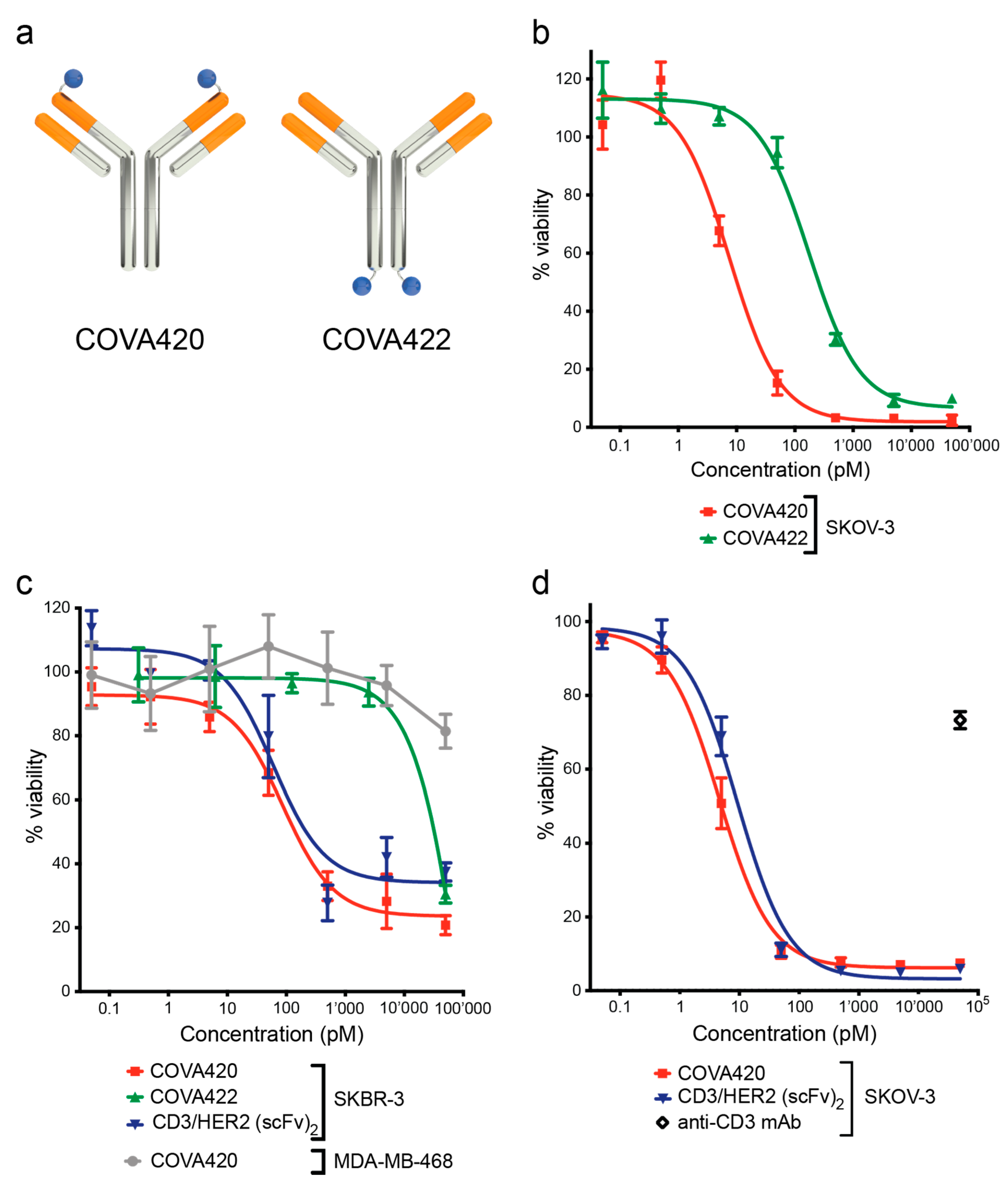
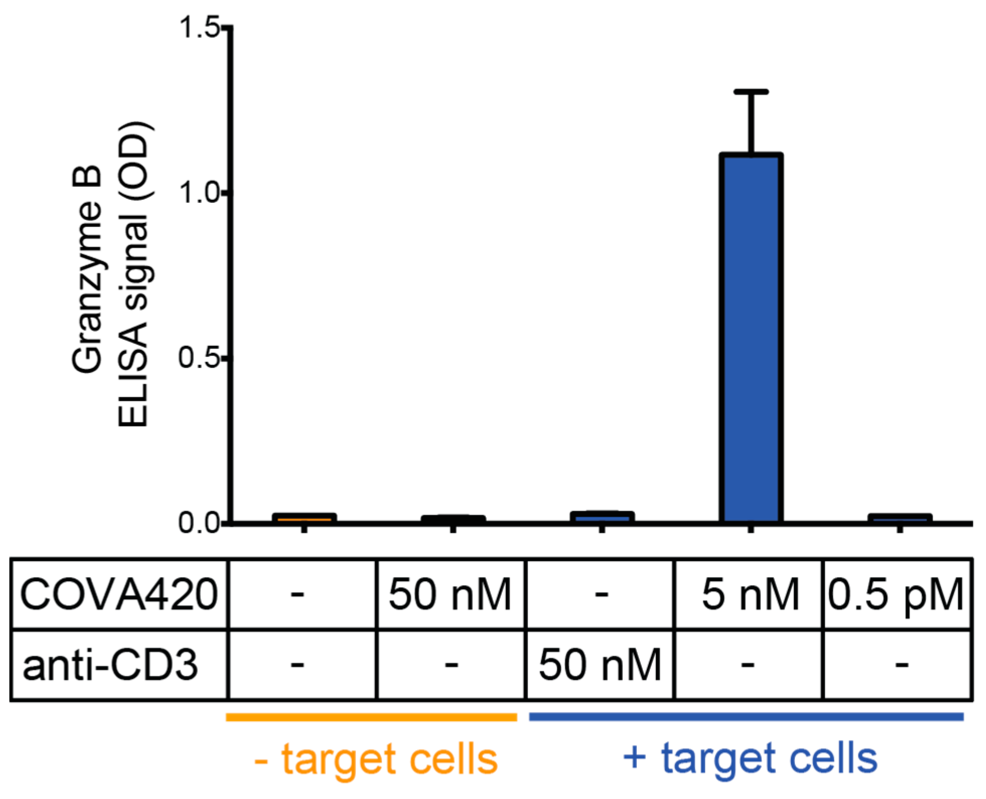
2.1. CD3/HER2 Bispecific FynomAb COVA420 Potently Induces T Cell Mediated Cytotoxicity

We have recently described the isolation and characterization of the anti-HER2 Fynomer C12 binding to the extracellular domain I of HER2 with a KD value of 80 nM [17]. By fusing this anti-HER2 Fynomer C12 to the N- or C-terminus of the anti-CD3 antibody hOKT3-Ala-Ala [22,23], we have generated two CD3/HER2 bispecific FynomAbs, COVA420 and COVA422, respectively (Figure 1a). Two FynomAb architectures were chosen in order to investigate whether the spatial arrangement of the two binding sites to each other had an impact on the bioactivity. Both FynomAbs were expressed in transiently transfected CHO-K1 cells and purified by protein A affinity chromatography. Cell binding was characterized in FACS titration experiments on HER2 and on CD3 expressing cells. COVA420 and COVA422 showed similar binding on both cell types (Supplementary Figure S1).
We assessed the activity of CD3/HER2 FynomAbs in an in vitro redirected T cell cytotoxicity assay on HER2 gene-amplified tumor cells with high HER2 expression. Human CD8+ purified T cells were co-cultured with SKOV-3 cells in the presence of COVA420 or COVA422. Both FynomAb architectures induced lysis of SKOV-3 cells, however, COVA420 showed more than 20-fold higher potency as compared to COVA422, with an EC50 of 7.5 pM versus 175 pM for COVA422 (Figure 1b). Similar observations were made on SKBR-3 cells, where the difference in potency between COVA420 and COVA422 was even greater (Figure 1c, EC50 87 pM versus EC50 > 10 nM).
COVA420 was selected for further characterization due to its potent activity. To benchmark COVA420 to (scFv)2-based formats (consisting out of two single chain Fv fragments connected by a flexible linker), we generated a CD3/HER2 bispecific (scFv)2 protein in a format highly similar to previously described BiTE® molecules [24]. The HER2 binding arm of the CD3/HER2 bispecific (scFv)2 was based on a trastuzumab-derived scFv sequence. The potency of COVA420 was equally high as for the CD3/HER2 (scFv)2 on SKBR-3 and SKOV-3 cells (Figure 1c,d). The bioactivity of COVA420 was HER2 target specific, since a HER2 negative control cell line, MDA-MB-468, was not lysed in the presence of COVA420 (Figure 1c). Furthermore, the parental anti-CD3 antibody hOKT3-Ala-Ala did not induce significant cytotoxicity on SKOV-3 cells, supporting T cell redirection as the mode of action of COVA420 (Figure 1d).
Figure 1. (a) Graphic representation of CD3/HER2 FynomAb architectures used in this study. COVA420: N-terminal heavy chain fusion; COVA422 C-terminal heavy chain fusion. (bd) Redirected T cell cytotoxicity assay on different HER2 positive or HER2 negative cell lines. Target cells were co-cultured with effector cells in the presence of FynomAbs or control molecules. Cytotoxicity was assessed in a colorimetric XTT assay. Data were normalized as described in the experimental section and are shown as mean % viability ± SD. Panel b shows COVA420 (red) and COVA422 (green) on HER2 positive SKOV-3 cells. Panel c shows COVA420, COVA422 and CD3/HER2 (scFv)2 (blue) on SKBR-3. EC50 values were 60 pM (CD3/HER2 (scFv)2); 87 pM (COVA420) and >10 nM (COVA422). No cytotoxicity was induced by COVA420 on HER2 negative MDA-MB-468 cells (grey). Panel d shows a comparison between COVA420 and CD3/HER2 (scFv)2 on SKOV-3 cells with EC50 values of 9.3 pM for COVA420 and 4.6 pM for the CD3/HER2 (scFv)2.
Antibodies 04 00426 g001
Next, we determined Granzyme B release, a central component of the cytotoxic T cell attack, in the supernatant of SKOV-3 cells incubated with COVA420 and CD8+ human T cells. Granzyme B release correlated with the cytotoxic effect of COVA420 and was strictly dependent on the presence of HER2 expressing target cells (Figure 2). No Granzyme B release was observed with the anti-CD3 antibody hOKT3-Ala-Ala. These findings indicate that the cytotoxic effect induced by COVA420 was mediated by T cells in a HER2-dependent manner.
Taken together, it was shown that the CD3/HER2 bispecific FynomAb COVA420 has a higher potency than COVA422, illustrating that the architecture of the FynomAb has an influence on its bioactivity. COVA420 induced T-cell-mediated cytotoxicity on SKBR-3 and SKOV-3 cells with similar potency to that of CD3/HER2 (scFv)2.
Figure 2. Granzyme B release into the cell culture supernatant was evaluated by ELISA. Controls were used at the highest assay concentration. For COVA420 concentrations where maximal cytotoxicity (5 nM) or no cytotoxicity (0.5 pM) was observed were analyzed. Results are shown as mean ELISA signal ± SD.
Antibodies 04 00426 g002

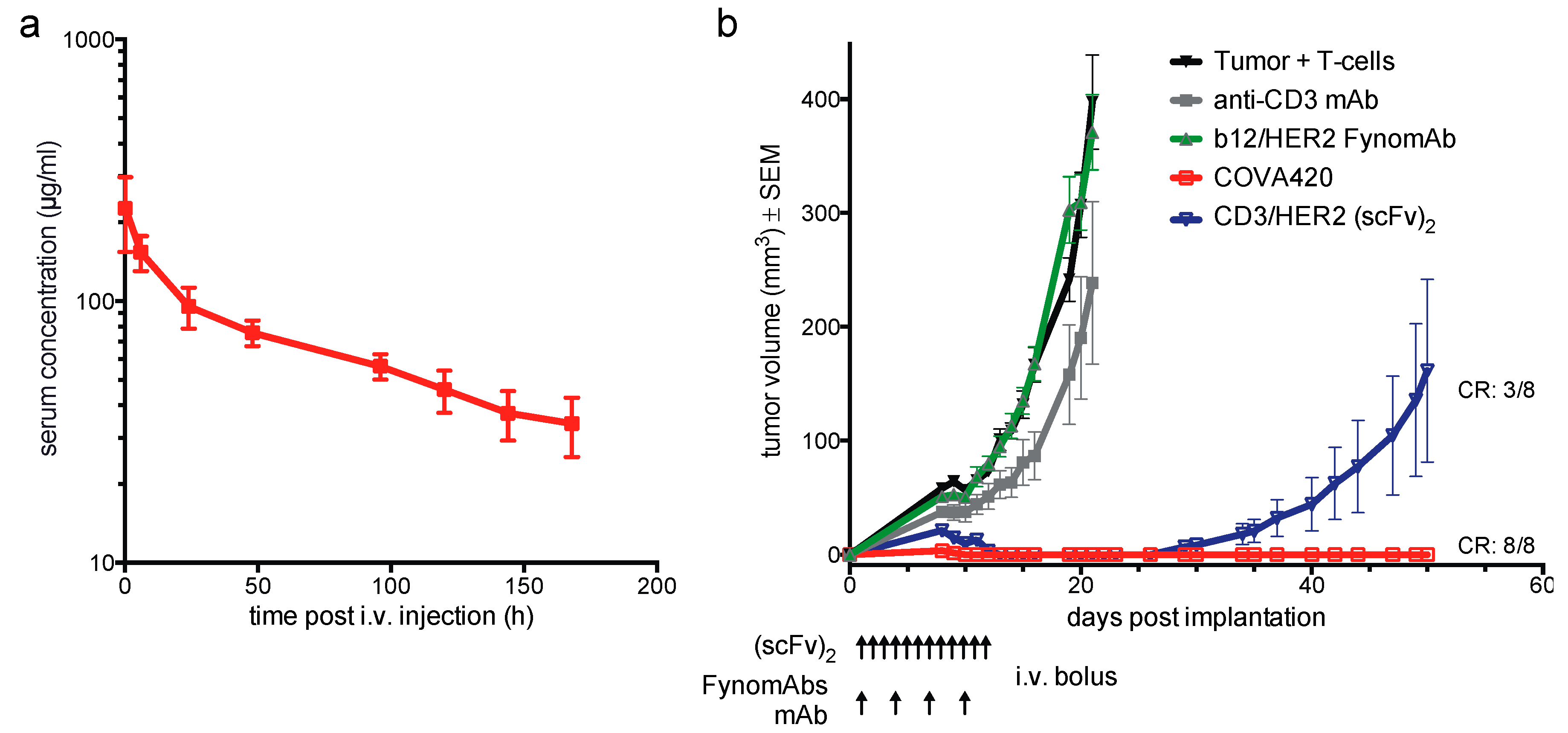
2.2. COVA420 Shows IgG-Like Pharmacokinetic Profile and Inhibits SKOV-3 Tumor Growth In Vivo

Pharmacokinetic (PK) properties of COVA420 were assessed in C57BL/6 mice using a HER2 ELISA to quantify COVA420 in the mouse serum. COVA420 demonstrated a biphasic IgG-like PK profile with an estimated terminal half-life of 140 h (Figure 3a), indicating good in vivo stability of COVA420 and of the HER2-specific Fynomers.
The in vivo efficacy of COVA420 was tested in a prophylactic setting in NOD.CB17 Prkdc mice. HER2 expressing SKOV-3 tumor cells were co-injected with ex vivo expanded human T cells. COVA420 was administered twice per week at a dose of 0.5 mg/kg. To account for the short serum half-life that was expected for CD3/HER2 (scFv)2 due to its small size of 52 kDa, a once-daily administration regimen of an equimolar dose of CD3/HER2 (scFv)2 was applied. COVA420 and CD3/HER2 (scFv)2 efficiently inhibited tumor outgrowth, while no significant anti-tumor activity was observed in the control groups treated with either the parental anti-CD3 antibody hOKT3-Ala-Ala or with a b12/HER2 control FynomAb in which the CD3 binding antibody has been replaced with an antibody specific to an irrelevant target (Figure 3b). At the end of the observation period, 8 out of 8 mice treated with COVA420 remained tumor-free, as compared to 3 out of 8 mice from the group treated with CD3/HER2 (scFv)2.
These findings show that COVA420 has promising anti-tumor activity and an IgG-like pharmacokinetic profile in mice.
Figure 3. (a) Pharmacokinetic analysis of COVA420 in C57BL/6 mice. Results are plotted as mean concentrations ± SD (n = 5). (b) Prophylactic SKOV-3 in vivo efficacy model. NOD.CB17 Prkdc mice were treated twice weekly with 0.5 mg/kg vehicle control (black), anti-CD3 mAb (grey), irrelevant FynomAb (green) or COVA420 (red) on days 1, 4, 7 and 10. CD3/HER2 (scFv)2 (blue) was injected daily at 0.16 mg/kg for 12 days, as indicated by the arrows. Tumor volume (mm3) is shown as mean ± SEM (n = 8); “CR” indicates the fraction of tumor-free mice in the COVA420 and in the CD3/HER2 (scFv)2 treatment groups on day 50.
Antibodies 04 00426 g003

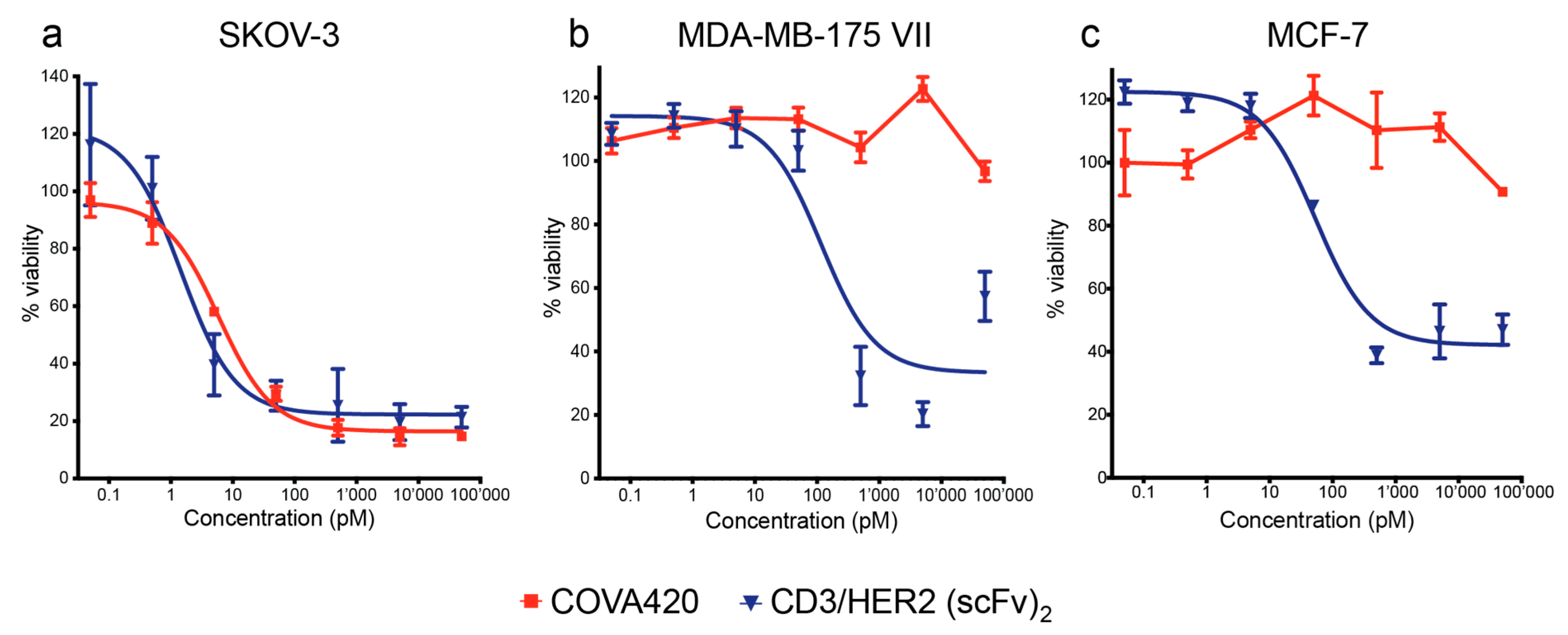
2.3. COVA420 Selectively Kills Cells with High HER2 Expression, but Spares Cells with Low HER2 Expression

After COVA420 demonstrated strong in vitro activity on SKOV-3 and SKBR-3 tumor cell lines expressing high levels of HER2, the activity of COVA420 on cell lines with lower HER2 expression levels was investigated. MDA-MB-175 VII, MCF-7 and MDA-MB-231 cell lines were chosen as models for low HER2 expression. The HER2 receptor densities on these cell lines quantified by FACS were 51,000 (MDA-MB-175 VII), 9500 (MCF-7) and 2600 (MDA-MB-231) receptors per cell, respectively (Supplementary Figure S2).
Interestingly, COVA420 did not exhibit any activity on MDA-MB-175 VII and MCF-7, in contrast to CD3/HER2 (scFv)2 which was still capable of inducing cytolysis of MDA-MB-175 VII and MCF-7 cells (Figure 4). The MDA-MB-231 cell line was neither affected by COVA420 nor by CD3/HER2 (scFv)2 (data not shown).
It was concluded from these experiments that despite its highly potent activity on SKOV-3 and SKBR-3 cells, COVA420 did not induce T cell mediated cytotoxicity on cells with low HER2 expression as found on MDA-MB-175 VII or MCF-7 cells.
Figure 4. Redirected T cell cytotoxicity assay in dependence of HER2 expression levels. Effector CD8+ T cells were incubated in the presence of COVA420 (red) or CD3/HER2 (scFv)2 (blue) with target cell lines with decreasing HER2 expression levels ((a) SKOV-3, HER2 high; (b) MDA-MB-175 VII, HER2 intermediate; (c) MCF-7, HER2 low). Redirected T cell cytotoxicity was assessed by XTT conversion. Data were normalized as described in the experimental section and plotted as mean ± SD.
Antibodies 04 00426 g004

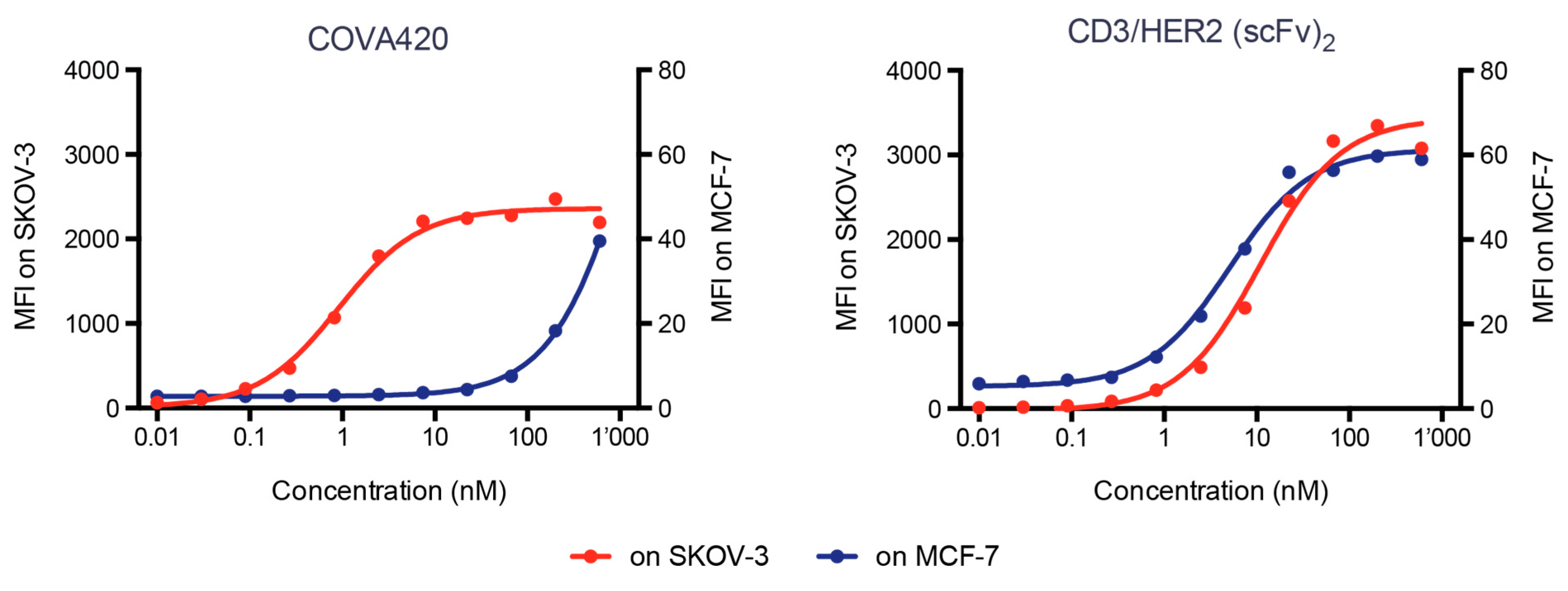
2.4. COVA420 Selectively Binds to Cells with High HER2 Expression

In order to understand the factors that contribute to the selectivity for high HER2 expression of COVA420, we compared binding of COVA420 and of CD3/HER2 (scFv)2 to SKOV-3 and MCF-7 cells. The CD3/HER2 (scFv)2 bound to both cell lines with comparable EC50 values, while COVA420 showed dramatically reduced binding to MCF-7 cells (Figure 5). This observation suggests that COVA420 binds in a HER2 density-dependent manner to tumor cells.
Figure 5. Flow cytometric binding analysis of COVA420 (left panel) and CD3/HER2 (scFv)2 (right panel) on SKOV-3 cells (high expression, red line, left y-Axis) or MCF-7 cells (low HER2 expression, blue line, right y-Axis). For each cell line the scale was adjusted to the maximum level of binding observed. EC50 of COVA420 were 0.9 nM on SKOV-3 cells and >200 nM on MCF-7 cells, EC50 of CD3/HER2 (scFv)2 was 11 nM on SKOV-3 and 5 nM on MCF-7 cells.
Antibodies 04 00426 g005

2.5. Discussion

In this study we describe a CD3/HER2 bispecific FynomAb, COVA420, that selectively induces potent T-cell-mediated cytotoxicity on tumor cells with high HER2 expression, but spares cells with low HER2 expression.
Notably, the architecture of the CD3/HER2 bispecific FynomAb has an impact on biological activity. COVA422, which was designed in a different architecture, also induces T-cell-mediated cytolysis, but is considerably less active than COVA420 despite comparable binding to HER2 and CD3 expressing cells. We hypothesize that the greater distance of the HER2 and the CD3 binding sites in COVA422 leads to a sub-optimal contact of the T cell with the target cell and subsequently to a less efficient target cell killing. These findings are in agreement with examples in the literature showing that the distance between T cells and target cell membrane determines the potency of T-cell-mediated lysis [25].
COVA420 is equally potent on SKOV-3 and SKBR-3 as a bispecific CD3/HER2 (scFv)2, a format that is well established for T cell redirection. The larger size of the FynomAb (162 kDa vs 52 kDa for the CD3/HER2(scFv)2) did not interfere with the formation of the immunological synapse between the T cell and target cell.
COVA420 shows strong anti-tumor activity in a prophylactic SKOV-3 tumor mouse model. At the end of the observation period, 8/8 mice treated with COVA420 were tumor-free as compared to 3/8 mice in the CD3/HER2 (scFv)2 treatment group. Because both molecules demonstrated similar potency on SKOV-3 cells in vitro, the superior in vivo efficacy of COVA420 is likely due to the longer serum half-life leading to higher exposure, despite less frequent dosing than the CD/HER2 (scFv)2. This result illustrates the promise of next generation T cell retargeting therapeutics with extended half-life to (i) circumvent continuous infusion that is currently required for BiTE® therapeutics, and (ii) to achieve better efficacy in solid tumors due to improved pharmacokinetic properties.
Unlike CD3/HER2 (scFv)2, COVA420 did not show any in vitro activity on MCF-7 and MDA-MB-175 VII cells that are models for tumors that lack HER2 amplification and show intermediate HER2 expression [26,27,28]. The HER2 levels found on these cells, 51,000 (MDA-MB-175 VII) or 9,500 (MCF-7) HER2 per cell - are similar to normal cells [29,30,31]. Cardiomyocytes, which are the primary site of toxicity caused by the anti-HER2 monoclonal antibody trastuzumab [32], are reported to express below 50,000 HER2 per cell [33]. Our results imply that COVA420 may show low or no activity on normal tissue. In vitro bioactivity assays on primary cells, including primary cardiomyocytes or human embryonic stem cell-derived cardiomyocytes, may provide further evidence that COVA420 is not active on normal tissues [34].
The finding that COVA420 strongly bound to SKOV-3 cells but weakly to MCF-7 cells suggests that the HER2 binding interaction of COVA420 with MCF-7 is not sufficient to elicit a cytotoxic T cell response towards the target cells. The selective binding of COVA420 for cells with high HER2 expression can be explained by the lower HER2 affinity of the Fynomer (KD = 80 nM) [17] paired with the bivalent binding interaction of COVA420. On cells with high HER2 density, bivalent binding of COVA420 is facilitated which promotes high avidity. In contrast, CD3/HER2 (scFv)2 is based on a monovalent high-affinity HER2 scFv derived from trastuzumab (Kd = 0.5 nM) [35], that binds to both cell types with similar affinity and hence is incapable of discriminating between high and low HER2 expressing cells.
A similar selective binding concept has been reported for an EGFR monoclonal antibody nimotuzumab [36]. Due to its reduced affinity for EGFR, nimotuzumab efficiently targets tumor cells with high EGFR densities, but shows minimal binding to cells with low EGFR densities. In clinical studies, nimotuzumab showed anti-tumor activity in the absence of skin toxicity, a side effect commonly observed with the higher affinity EGFR antibodies cetuximab and panitumumab that is presumably caused by low level EGFR expression in the human skin.
Because of the high potency of T-cell-mediated cytotoxicity, drug safety is a central aspect for the development of T cell engaging molecules. The vast majority of known solid tumor antigens such as HER2 or EGFR are overexpressed in the tumor, but are not entirely tumor-restricted. However, low target expression in healthy tissue can cause undesired on-target T cell activation leading to severe toxicities. One case has been reported in which a HER2 targeting CAR T cell therapy caused fatal toxicity in a tumor patient [9]. Also, a CD3/EGFR BiTE® therapy caused acute toxicity in cynomolgus monkeys likely due to EGFR expression in normal tissue [10].
Due to its selectivity advantage, COVA420 is expected to have a wider therapeutic window in patients that bear tumors with high HER2 levels. Two recent publications have adopted a similar strategy to develop EGFR and HER2 targeting CAR T cell therapies with a wider therapeutic window [29,37]. By tuning the affinity of the CAR antibody moiety, the authors generated CAR T cells that showed sensitivity to high target density and limited activity on normal tissue expressing the targets at a low density. Our study expands the concept to CD3 bispecific proteins.
The anti-CD3 antibody used for the construction of COVA420 bears two mutations in the lower hinge region, L234A and L235A (also referred as Ala-Ala), to reduce binding to Fc receptors (FcR) [38]. This hOKT3-Ala-Ala antibody was well tolerated in a phase III clinical trial enrolling more than 400 children and young adults with new-onset diabetes [23]. Therefore, we expect that tumor independent T cell activation and cytokine release-related syndromes induced by COVA420 through cross-linking of T cells with FcR-bearing accessory cells is unlikely to impede further development of COVA420. As the Fynomer technology offers a broad range of formatting flexibility [20], one could consider the possibility to generate scFv and/or Fab-based Fynomer fusions in order to further minimize the risk of cytokine release-related syndromes upon administration.
In conclusion, we have demonstrated that COVA420 is a very potent CD3/HER2 bispecific FynomAb designed for high selectivity towards tumor cells with high HER2 expression. While COVA420 potently induced T cell mediated cytolysis of cells with high HER2 levels, it did not impact cells with lower HER2 levels as found on healthy cells. The selectivity advantage holds the promise to lead to a wider therapeutic window and a safer application of COVA420 to HER2 overexpressing cancer patients. Due to the potential safety risks inherent to T cell re-directing therapies, clinical trials should be performed in metastatic cancer patients who failed on all prior lines of therapies, and close monitoring of cardiac function is warranted. We postulate that the FynomAb strategy presented in this study can be applied to other targets that are overexpressed in tumors in order to generate potent CD3 bispecific FynomAbs with an improved safety profile. This opens the potential to target solid tumor antigens that otherwise are not suited for T cell redirecting approaches and may expand the applicability of CD3 bispecific FynomAbs to a large number of solid tumor antigens and indications.

3. Experimental Section

3.1. Protein Purification

Proteins were expressed in CHO-K1 cells via transient transfection and secretion into the cell culture supernatant. For purification, medium was collected 7 days post transfection, sterile filtered using a 0.2 μm membrane and purified over a MabSelect SuRe column by the use of an Akta Purifier FPLC (both GE Healthcare) using PBS as running buffer and acid Glycine pH 2.7 elution.

3.2. Flow Cytometry, HER2 Quantification

Flow cytometric analysis was performed on a Guava EasyCyte flow cytometer (Millipore) using the EasyCyte software.
For cell staining experiments adherent cells were detached by Accutase treatment and resuspended in FACS buffer (PBS, 0.2% NaN3, 1% FCS). Staining was performed by incubating serially diluted FynomAbs or CD3/HER2 (scFv)2 in a concentration range from 500 to 0.01 nM. For FynomAbs an anti human IgG-Alexa488 conjugated secondary antibody (Invitrogen) was used, bound CD3/HER2 (scFv)2 was detected by the use of an anti-HIS6-Antibody (Fisher Scientific) followed by the addition of anti-mouse IgG-Alexa 488 conjugate. Stained cells were gated based on live cells and mean fluorescence was plotted against the effector molecule concentration. Data were further evaluated using Prism 5 (Graph Pad) by fitting a sigmoidal dose response. HER2 cell surface expression was quantified on various cell lines using the QIFIKIT® quantification kit (DAKO) according to manufacturer’s recommendation.

3.3. In Vitro Redirected T Cell Cytotoxicity Assays

Human PBMCs (SKBR-3 assay) or CD8+ T cells (all other in vitro assays) were used as effector cells in redirected T cell mediated cell cytotoxicity assays. PBMCs were isolated from fresh buffy coat preparations obtained from Blutspende Zürich or Bern by Ficoll Plaque plus (GE Healthcare) and density gradient centrifugation using standard procedures on the day before the experiment. Isolated PBMCs were incubated overnight in 10% FCS, RPMI and 37 °C, 5% CO2. The isolated PBMCs served as effector cells or were further purified to obtain enriched CD8+ T cells (Dynabeads Untouched Human CD8 T cells, Life Technologies, 1348D) according to the manufacturer’s recommendation. Alternatively, CD8+ T cells were isolated directly from fresh buffy coat preparations using the MACSxpress CD8 T cell isolation kit (Miltenyi Biotec 130-098-194) as recommended by the manufacturer.
Target tumor cell lines SKBR-3, SKOV-3, MDA-MB-175 VII, MCF-7, MDA-MB-231 and MDA-MB-468, all obtained from ATCC, were detached by Accutase treatment and seeded in 96 well plates at cell densities ranging from 5000–10,000 cells per well. The next day, serial dilutions of effector molecules in 10% FCS, RPMI were added together with the effector cells onto the adhered tumor cells. The final effector cell to target cell ratio was 25:1 for PBMC assays and 10:1 or 5:1 for CD8+ T cell assays.
After incubation between 24 h (PBMCs) and 66 h (CD8+ T cells) at 37 °C, 5% CO2, T cells were washed off and cell viability was evaluated using XTT-reagent (Sigma). A 100% lysis control was included by treating the target cells with 1% Triton-X100 (Sigma), and the value for spontaneous lysis was obtained by incubating the target cells with effector cells only (“spont lysis”). Absorbance at 450 nm was measured between 2.5 and 4 h after addition of XTT substrate. All measurements were done in triplicate. Percent cell viability was calculated using the following formula:
% viability = (value−100% lysis) / (spont lysis−100% lysis)*100
Percent cell viability was then plotted against the effector molecule concentration and data were evaluated using Prism 5 (GraphPad Software) by fitting a sigmoidal dose-response (three parameter equation).

3.4. Pharmacokinetic Analysis

To assess the pharmacokinetic properties of COVA420, 200 μg protein was injected into the tail vein of C57BL/6 (5 mice per group). Blood was withdrawn by saphenous bleeding after 10 min, 6, 24, 48, 96, 120, 144 and 168 h and processed into plasma by centrifugation. Plasma concentrations of COVA420 were measured by capture ELISA. In brief, plasma COVA420 was detected by sandwich ELISA, captured with the extracellular domain of HER2 (eBioscience) and detected with anti human IgG-HRP conjugate (Life Technologies). QuantaRed served as a substrate and fluorescence signals were read at 544 nm/590 nm. Serum concentrations are expressed in µg/mL, calculated based on a COVA420 standard curve. Serum concentration was analyzed using GraphPad Prism, version 5.

3.5. In Vivo Efficacy Studies

Human pan-CD3 T cells were isolated from fresh buffy coats obtained from healthy volunteers using the MACSxpress Pan T Cell Isolation Kit (Miltenyi, 130-098-193) and expanded (Miltenyi, T cell activation/expansion kit, 130-091-441) according to the manufacturer’s recommendations. After 20 days of expansion, activation beads were removed by means of magnetic separation. SKOV-3 tumor cells and T cells were mixed (E/T: 1:2, e.g., 1.5E6 T effector and 3E6 SKOV-3 cells) and injected into the right abdominal fat pad of 1.5 Gy irradiated NOD.CB17 Prkdc mice (Charles River, France; n = 8). Prior to cell injection, mice were NK-cell depleted by the i.p. injection of anti-asialo GM1 (WAKO: 986-10001). Treatments were initiated 24 h post E/T cell implantation; 0.5 mg/kg FynomAb was administered twice per week and equimolar doses of sc(Fv)2 molecules were administered daily by intravenous injection. Tumor volumes were calculated using the following formula: tumor volume (mm3) = 0.5 × length × (width)2. All mouse experiments conducted at Covagen AG were performed in accordance with Swiss guidelines and were approved by the Veterinarian Office of Zürich, Switzerland.

4. Conclusions

COVA420 is a highly potent CD3/HER2 bispecific FynomAb with enhanced selectivity towards tumor cells with high HER2 expression. While COVA420 potently induced T cell mediated cytolysis of cells with high HER2 levels, it did not impact cells with lower HER2 levels as found on healthy cells. The selectivity advantage holds the promise to lead to a wider therapeutic window and a safer application of COVA420 to HER2 overexpressing cancer patients.
The FynomAb strategy presented in this study may be applicable to other targets that are overexpressed in tumors in order to generate potent CD3 bispecific FynomAbs with an improved safety profile. This opens the potential to target solid tumor antigens that otherwise are not suited for T cell redirecting approaches due to target expression in normal tissues, and may expand the applicability of CD3 bispecific FynomAbs to a large number of solid tumor antigens and indications.

Supplementary Materials

Supplementary materials can be accessed at: https://www.mdpi.com/2073-4468/4/4/426/s1.

Author Contributions

Ulrich Wuellner, Kristina Klupsch, Fabian Buller, Isabella Attinger-Toller and Simon Brack designed and supervised experiments, performed experiments and wrote the manuscript. Dragan Grabulovski and Julian Bertschinger developed the concept, designed and supervised experiments and reviewed the manuscript. Roger Santimaria, Irene Zbinden and Patricia Henne performed the experiments.

Conflicts of Interest

All authors are or have been employees of Covagen AG.

References

  1. Nagorsen, D.; Baeuerle, P.A. Immunomodulatory therapy of cancer with T cell-engaging BiTE antibody blinatumomab. Exp. Cel. Res. 2011, 317, 1255–1260. [Google Scholar] [CrossRef] [PubMed]
  2. van der Stegen, S.J.; Hamieh, M.; Sadelain, M. The pharmacology of second-generation chimeric antigen receptors. Nat. Rev. Drug Discov. 2015, 14, 499–509. [Google Scholar] [CrossRef] [PubMed]
  3. Brischwein, K.; Schlereth, B.; Guller, B.; Steiger, C.; Wolf, A.; Lutterbuese, R.; Offner, S.; Locher, M.; Urbig, T.; Raum, T.; et al. MT110: A novel bispecific single-chain antibody construct with high efficacy in eradicating established tumors. Mol. Immunol. 2006, 43, 1129–1143. [Google Scholar] [CrossRef] [PubMed]
  4. Dreier, T.; Lorenczewski, G.; Brandl, C.; Hoffmann, P.; Syring, U.; Hanakam, F.; Kufer, P.; Riethmuller, G.; Bargou, R.; Baeuerle, P.A. Extremely potent, rapid and costimulation-independent cytotoxic T-cell response against lymphoma cells catalyzed by a single-chain bispecific antibody. Int. J. Cancer 2002, 100, 690–697. [Google Scholar] [CrossRef] [PubMed]
  5. Hoffmann, P.; Hofmeister, R.; Brischwein, K.; Brandl, C.; Crommer, S.; Bargou, R.; Itin, C.; Prang, N.; Baeuerle, P.A. Serial killing of tumor cells by cytotoxic T cells redirected with a CD19-/CD3-bispecific single-chain antibody construct. Int. J. Cancer 2005, 115, 98–104. [Google Scholar] [CrossRef] [PubMed]
  6. Przepiorka, D.; Ko, C.W.; Deisseroth, A.; Yancey, C.L.; Candau-Chacon, R.; Chiu, H.J.; Gehrke, B.J.; Gomez-Broughton, C.; Kane, R.C.; Kirshner, S.; et al. FDA Approval: Blinatumomab. Clin. Cancer Res. 2015, 21, 4035–4039. [Google Scholar] [CrossRef] [PubMed]
  7. Topp, M.S.; Gokbuget, N.; Zugmaier, G.; Klappers, P.; Stelljes, M.; Neumann, S.; Viardot, A.; Marks, R.; Diedrich, H.; Faul, C.; et al. Phase II trial of the anti-CD19 bispecific T cell-engager blinatumomab shows hematologic and molecular remissions in patients with relapsed or refractory B-precursor acute lymphoblastic leukemia. J. Clin. Oncol. 2014, 32, 4134–4140. [Google Scholar] [CrossRef] [PubMed]
  8. Spiess, C.; Zhai, Q.; Carter, P.J. Alternative molecular formats and therapeutic applications for bispecific antibodies. Mol. Immunol. 2015, 67, 95–106. [Google Scholar] [CrossRef] [PubMed]
  9. Morgan, R.A.; Yang, J.C.; Kitano, M.; Dudley, M.E.; Laurencot, C.M.; Rosenberg, S.A. Case report of a serious adverse event following the administration of T cells transduced with a chimeric antigen receptor recognizing ERBB2. Mol. Ther. 2010, 18, 843–851. [Google Scholar] [CrossRef] [PubMed]
  10. Lutterbuese, R.; Raum, T.; Kischel, R.; Hoffmann, P.; Mangold, S.; Rattel, B.; Friedrich, M.; Thomas, O.; Lorenczewski, G.; Rau, D.; et al. T cell-engaging BiTE antibodies specific for EGFR potently eliminate KRAS- and BRAF-mutated colorectal cancer cells. Proc. Natl. Acad. Sci. USA 2010, 107, 12605–12610. [Google Scholar] [CrossRef] [PubMed]
  11. Chichili, G.R.; Huang, L.; Li, H.; Burke, S.; He, L.; Tang, Q.; Jin, L.; Gorlatov, S.; Ciccarone, V.; Chen, F.; et al. A CD3xCD123 bispecific DART for redirecting host T cells to myelogenous leukemia: Preclinical activity and safety in nonhuman primates. Sci. Transl. Med. 2015, 7, 289ra282. [Google Scholar] [CrossRef] [PubMed]
  12. Gall, J.M.; Davol, P.A.; Grabert, R.C.; Deaver, M.; Lum, L.G. T cells armed with anti-CD3 x anti-CD20 bispecific antibody enhance killing of CD20+ malignant B cells and bypass complement-mediated rituximab resistance in vitro. Exp. Hematol. 2005, 33, 452–459. [Google Scholar] [CrossRef] [PubMed]
  13. Osada, T.; Hsu, D.; Hammond, S.; Hobeika, A.; Devi, G.; Clay, T.M.; Lyerly, H.K.; Morse, M.A. Metastatic colorectal cancer cells from patients previously treated with chemotherapy are sensitive to T-cell killing mediated by CEA/CD3-bispecific T-cell-engaging BiTE antibody. Br. J. Cancer 2010, 102, 124–133. [Google Scholar] [CrossRef] [PubMed]
  14. Friedrich, M.; Raum, T.; Lutterbuese, R.; Voelkel, M.; Deegen, P.; Rau, D.; Kischel, R.; Hoffmann, P.; Brandl, C.; Schuhmacher, J.; et al. Regression of human prostate cancer xenografts in mice by AMG 212/BAY2010112, a novel PSMA/CD3-Bispecific BiTE antibody cross-reactive with non-human primate antigens. Mol. Cancer Ther. 2012, 11, 2664–2673. [Google Scholar] [CrossRef] [PubMed]
  15. Liddy, N.; Bossi, G.; Adams, K.J.; Lissina, A.; Mahon, T.M.; Hassan, N.J.; Gavarret, J.; Bianchi, F.C.; Pumphrey, N.J.; Ladell, K.; et al. Monoclonal TCR-redirected tumor cell killing. Nat. Med. 2012, 18, 980–987. [Google Scholar] [CrossRef] [PubMed]
  16. Banner, D.W.; Gsell, B.; Benz, J.; Bertschinger, J.; Burger, D.; Brack, S.; Cuppuleri, S.; Debulpaep, M.; Gast, A.; Grabulovski, D.; et al. Mapping the conformational space accessible to BACE2 using surface mutants and cocrystals with Fab fragments, Fynomers and Xaperones. Acta Crystallogr. D Biol. Crystallogr. 2013, 69, 1124–1137. [Google Scholar] [CrossRef] [PubMed]
  17. Brack, S.; Attinger-Toller, I.; Schade, B.; Mourlane, F.; Klupsch, K.; Woods, R.; Hachemi, H.; von der Bey, U.; Koenig-Friedrich, S.; Bertschinger, J.; et al. A bispecific HER2-targeting FynomAb with superior antitumor activity and novel mode of action. Mol. Cancer Ther. 2014, 13, 2030–2039. [Google Scholar] [CrossRef] [PubMed]
  18. Grabulovski, D.; Kaspar, M.; Neri, D. A novel, non-immunogenic Fyn SH3-derived binding protein with tumor vascular targeting properties. J. Biol. Chem. 2007, 282, 3196–3204. [Google Scholar] [CrossRef] [PubMed]
  19. Schlatter, D.; Brack, S.; Banner, D.W.; Batey, S.; Benz, J.; Bertschinger, J.; Huber, W.; Joseph, C.; Rufer, A.; van der Klooster, A.; et al. Generation, characterization and structural data of chymase binding proteins based on the human Fyn kinase SH3 domain. MAbs 2012, 4, 497–508. [Google Scholar] [CrossRef] [PubMed]
  20. Silacci, M.; Baenziger-Tobler, N.; Lembke, W.; Zha, W.; Batey, S.; Bertschinger, J.; Grabulovski, D. Linker length matters, fynomer-Fc fusion with an optimized linker displaying picomolar IL-17A inhibition potency. J. Biol. Chem. 2014, 289, 14392–14398. [Google Scholar] [CrossRef] [PubMed]
  21. Silacci, M.; Lembke, W.; Woods, R.; Attinger-Toller, I.; Baenziger-Tobler, N.; Batey, S.; Santimaria, R.; von der Bey, U.; Koenig-Friedrich, S.; Zha, W.; et al. Discovery and characterization of COVA322, a clinical-stage bispecific TNF/IL-17A inhibitor for the treatment of inflammatory diseases. MAbs 2015, 1–9. [Google Scholar] [CrossRef] [PubMed]
  22. Herold, K.C.; Gitelman, S.E.; Masharani, U.; Hagopian, W.; Bisikirska, B.; Donaldson, D.; Rother, K.; Diamond, B.; Harlan, D.M.; Bluestone, J.A. A single course of anti-CD3 monoclonal antibody hOKT3gamma1(Ala-Ala) results in improvement in C-peptide responses and clinical parameters for at least 2 years after onset of type 1 diabetes. Diabetes 2005, 54, 1763–1769. [Google Scholar] [CrossRef] [PubMed]
  23. Sherry, N.; Hagopian, W.; Ludvigsson, J.; Jain, S.M.; Wahlen, J.; Ferry, R.J., Jr.; Bode, B.; Aronoff, S.; Holland, C.; Carlin, D.; et al. Teplizumab for treatment of type 1 diabetes (Protege study): 1-year results from a randomised, placebo-controlled trial. Lancet 2011, 378, 487–497. [Google Scholar] [CrossRef]
  24. Baeuerle, P.A.; Reinhardt, C. Bispecific T-cell engaging antibodies for cancer therapy. Cancer Res. 2009, 69, 4941–4944. [Google Scholar] [CrossRef] [PubMed]
  25. Bluemel, C.; Hausmann, S.; Fluhr, P.; Sriskandarajah, M.; Stallcup, W.B.; Baeuerle, P.A.; Kufer, P. Epitope distance to the target cell membrane and antigen size determine the potency of T cell-mediated lysis by BiTE antibodies specific for a large melanoma surface antigen. Cancer Immunol. Immunother. 2010, 59, 1197–1209. [Google Scholar] [CrossRef] [PubMed]
  26. Holliday, D.L.; Speirs, V. Choosing the right cell line for breast cancer research. Breast Cancer Res. 2011, 13, 215. [Google Scholar] [CrossRef] [PubMed]
  27. Wilson, T.R.; Lee, D.Y.; Berry, L.; Shames, D.S.; Settleman, J. Neuregulin-1-mediated autocrine signaling underlies sensitivity to HER2 kinase inhibitors in a subset of human cancers. Cancer Cell 2011, 20, 158–172. [Google Scholar] [CrossRef] [PubMed]
  28. Lee-Hoeflich, S.T.; Crocker, L.; Yao, E.; Pham, T.; Munroe, X.; Hoeflich, K.P.; Sliwkowski, M.X.; Stern, H.M. A central role for HER3 in HER2-amplified breast cancer: Implications for targeted therapy. Cancer Res. 2008, 68, 5878–5887. [Google Scholar] [CrossRef] [PubMed]
  29. Liu, X.; Jiang, S.; Fang, C.; Yang, S.; Olalere, D.; Pequignot, E.C.; Cogdill, A.P.; Li, N.; Ramones, M.; Granda, B.; et al. Affinity-Tuned ErbB2 or EGFR Chimeric Antigen Receptor T Cells Exhibit an Increased Therapeutic Index against Tumors in Mice. Cancer Res. 2015, 75, 3596–3607. [Google Scholar] [CrossRef] [PubMed]
  30. Lohrisch, C.; Piccart, M. An overview of HER2. Semin. Oncol. 2001, 28, 3–11. [Google Scholar] [CrossRef]
  31. Press, M.F.; Cordon-Cardo, C.; Slamon, D.J. Expression of the HER-2/neu proto-oncogene in normal human adult and fetal tissues. Oncogene 1990, 5, 953–962. [Google Scholar] [PubMed]
  32. Hynes, N.E.; Lane, H.A. ERBB receptors and cancer: the complexity of targeted inhibitors. Nat. Rev. Cancer 2005, 5, 341–354. [Google Scholar] [CrossRef] [PubMed]
  33. Onsum, M.D.; Geretti, E.; Paragas, V.; Kudla, A.J.; Moulis, S.P.; Luus, L.; Wickham, T.J.; McDonagh, C.F.; Macbeath, G.; Hendriks, B.S. Single-cell quantitative HER2 measurement identifies heterogeneity and distinct subgroups within traditionally defined HER2-positive patients. Am. J. Pathol. 2013, 183, 1446–1460. [Google Scholar] [CrossRef] [PubMed]
  34. Reynolds, J.G.; Geretti, E.; Hendriks, B.S.; Lee, H.; Leonard, S.C.; Klinz, S.G.; Noble, C.O.; Lucker, P.B.; Zandstra, P.W.; Drummond, D.C.; et al. HER2-targeted liposomal doxorubicin displays enhanced anti-tumorigenic effects without associated cardiotoxicity. Toxicol. Appl. Pharm. 2012, 262, 1–10. [Google Scholar] [CrossRef] [PubMed]
  35. Bostrom, J.; Haber, L.; Koenig, P.; Kelley, R.F.; Fuh, G. High affinity antigen recognition of the dual specific variants of herceptin is entropy-driven in spite of structural plasticity. PLoS ONE 2011, 6, e17887. [Google Scholar] [CrossRef] [PubMed]
  36. Garrido, G.; Tikhomirov, I.A.; Rabasa, A.; Yang, E.; Gracia, E.; Iznaga, N.; Fernandez, L.E.; Crombet, T.; Kerbel, R.S.; Perez, R. Bivalent binding by intermediate affinity of nimotuzumab: A contribution to explain antibody clinical profile. Cancer Biol. Ther. 2011, 11, 373–382. [Google Scholar] [CrossRef] [PubMed]
  37. Caruso, H.G.; Hurton, L.V.; Najjar, A.; Rushworth, D.; Ang, S.; Olivares, S.; Mi, T.; Switzer, K.; Singh, H.; Huls, H.; et al. Tuning Sensitivity of CAR to EGFR Density Limits Recognition of Normal Tissue While Maintaining Potent Antitumor Activity. Cancer Res. 2015, 75, 3505–3518. [Google Scholar] [CrossRef] [PubMed]
  38. Xu, D.; Alegre, M.L.; Varga, S.S.; Rothermel, A.L.; Collins, A.M.; Pulito, V.L.; Hanna, L.S.; Dolan, K.P.; Parren, P.W.; Bluestone, J.A.; et al. In vitro characterization of five humanized OKT3 effector function variant antibodies. Cell. Immunol. 2000, 200, 16–26. [Google Scholar] [CrossRef] [PubMed]

