Review

Generating Recombinant Antibodies to Membrane Proteins through Phage Display

1
Department of Biological Sciences, University of Illinois at Chicago, Chicago, IL 60607-7060, USA
2
AxioMx Inc., a subsidiary of Abcam Plc, Branford, CT 06405, USA
*
Author to whom correspondence should be addressed.
Current address: Meso Scale Diagnostics LLC, Rockville, MD 20850, USA
Antibodies 2016, 5(2), 11; https://doi.org/10.3390/antib5020011
Submission received: 25 March 2016 / Revised: 21 April 2016 / Accepted: 21 April 2016 / Published: 2 May 2016

Abstract

:
One of the most important classes of proteins in terms of drug targets is cell surface membrane proteins, and yet it is a challenging set of proteins for generating high-quality affinity reagents. In this review, we focus on the use of phage libraries, which display antibody fragments, for generating recombinant antibodies to membrane proteins. Such affinity reagents generally have high specificity and affinity for their targets. They have been used for cell staining, for promoting protein crystallization to solve three-dimensional structures, for diagnostics, and for treating diseases as therapeutics. We cover publications on this topic from the past 10 years, with a focus on the various formats of membrane proteins for affinity selection and the diverse affinity selection strategies used. Lastly, we discuss the challenges faced in this field and provide possible directions for future efforts.

1. Introduction

Recombinant affinity reagents offer many practical advantages compared to polyclonal or monoclonal antibodies [1]. First, as sequenced entities, they are renewable and easily shared as digital files (i.e., DNA sequences). Second, they are amenable to genetic engineering for epitope tagging and affinity maturation [2]. Third, they can be produced in sufficient quantities for thorough validation. Fourth, they can be generated through phage-display using high-throughput methods [3,4,5]. Across the world, there have been numerous efforts in improving technologies for proteome-scale production of recombinant affinity reagents [3,4,5,6,7,8,9,10], based on the expectation that, in the future, affinity reagents will be principally recombinant in nature.
The most common form of recombinant affinity reagents is the antibody fragment. This consists of single-chain Fragments of variable regions (scFvs), which are single polypeptides composed of two variable domains of the light and heavy chains of immunoglobulin G (IgG) molecule [11,12], or Fragments of antigen binding (Fabs), which consist of the entire light chain, and the N-terminal variable domain and first constant region of the heavy chain [13,14,15]. Another popular antibody fragment for phage-display is the single-domain antibody, also called the “nanobody”, which corresponds to the variable domain of the camelid heavy-chain antibody [16,17,18]. Other types of protein scaffolds, also currently in use as recombinant affinity reagents, include anticalins [19], the affibodies [20], designed ankyrin repeat proteins (DARPins) [21], and fibronectin type III (FN3) monobodies [22].
Over the years, phage-display technology has matured and techniques for constructing large libraries have improved. Consequently, it is straightforward to generate a recombinant affinity reagent to virtually any soluble and well-folded proteins. One class of targets that has been very challenging for phage-display experiments is the membrane protein. Generally, these proteins are difficult to overexpress in large amounts, and their stability normally requires the presence of artificial detergents or membranes. These challenges have led to a number of innovative methods for formatting membrane proteins as targets for phage-display experiments and affinity selection (Figure 1): they include recombinant proteins and synthetic peptides, detergent micelles, “nanodiscs", virus-like particles (VLPs), and intact cells. Work with each of these formats is described in more detail below.

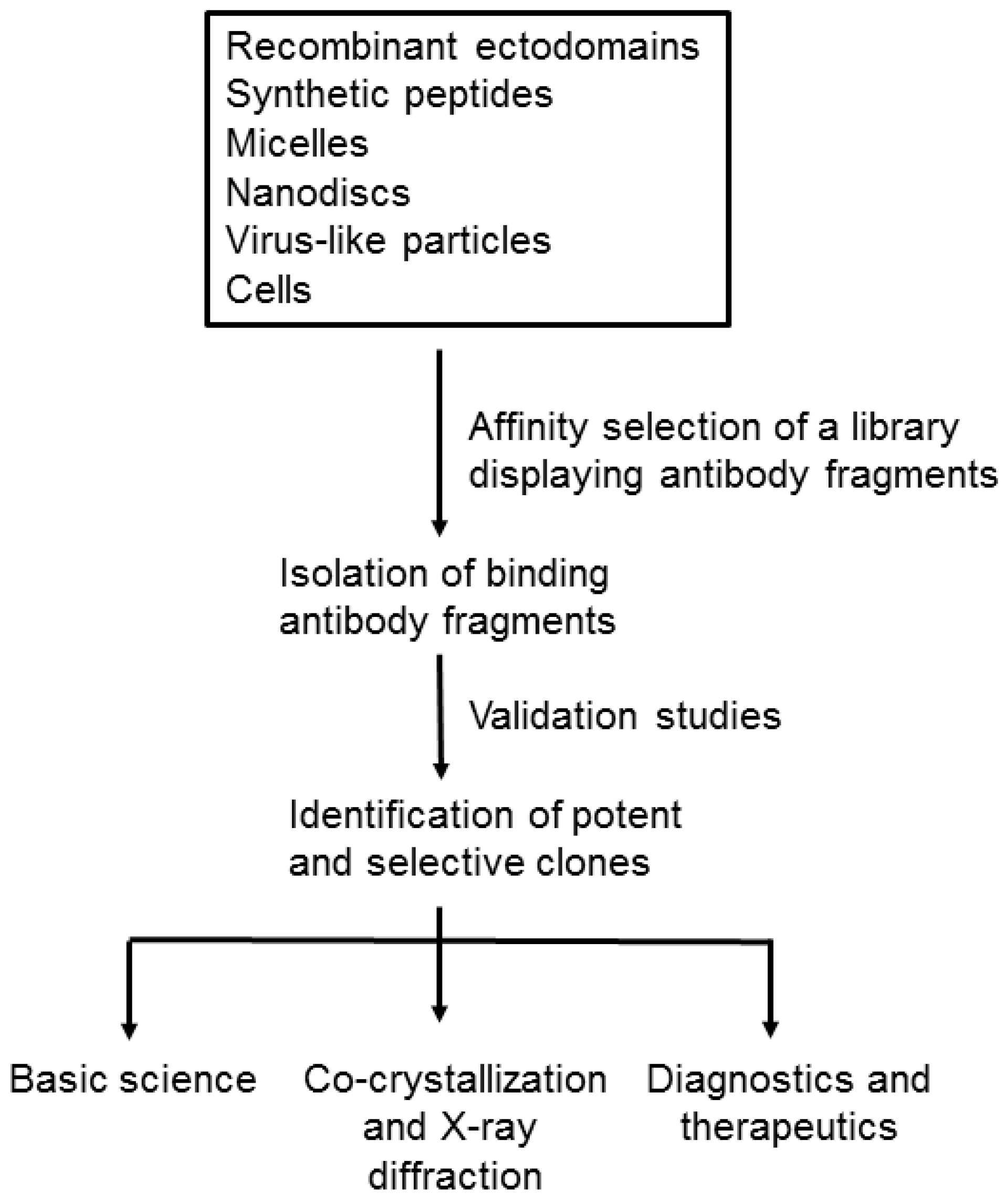
2. Formats of Membrane Proteins for Affinity Selection

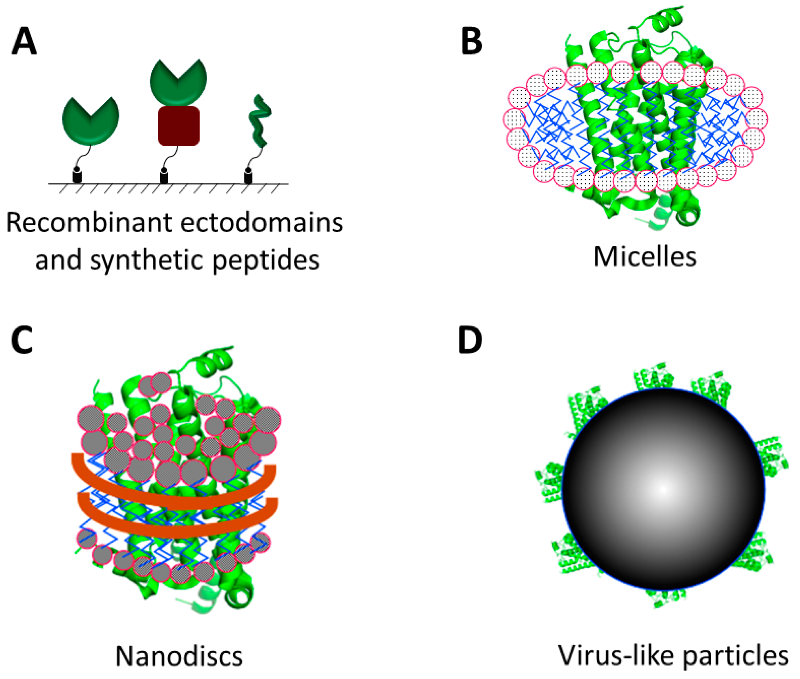
2.1. Recombinant Proteins and Synthetic Peptides

Membrane proteins often express poorly in heterologous cells [23]. One approach to circumvent this bottleneck is to express their extracellular domains (i.e., ectodomains) as soluble and secreted fusion proteins (Figure 2A) in mammalian [24,25,26,27], bacterial [28,29,30,31], yeast [32,33] and insect cells [27,34]. For example, as long as the fusion partner does not interfere with proper folding of the ectodomains, they can be expressed fused to the Fc region of IgG or an affinity tag, such as the His6-tag [35] and FLAG tag [36]. (Multi-pass membrane proteins, which lack a single ectodomain, cannot be overexpressed in this manner.) It should be noted that ectodomains overexpressed in bacterial systems will lack the post-translational modifications commonly found on mammalian membrane proteins.
Multi-pass membrane proteins, such as G-protein coupled receptors (GPCRs), are even more challenging to overexpress due to their high conformational complexity and low stability in heterologous cells. Multiple methods have been developed for improving the expression and stability of such membrane proteins [37,38,39]. In the first method, stable hydrophilic protein domains are fused to the intracellular loops of GPCRs to improve their expression and promote crystallization [37,40]. T4 Lysozyme (T4L) is the first and most widely used domain that has been used to engineer several GPCRs for crystallization [37,40,41,42,43]. Recently thermostabilized apocytochrome b562RIL (BRIL) has been used successfully to engineer well-expressed GPCR for crystallization [44,45]. In the second method, GPCR mutants were generated by alanine scanning [38,46] or leucine scanning [47], and were expressed in Escherichia coli (E. coli) cells. Mutants with higher stability were screened by binding assay with radiolabelled ligands after heat treatment. This is a simple and straightforward method and does not require special equipment for performing the assays. However, generating hundreds of individual alanine or leucine mutants can be time-consuming, thus this approach is not well-suited to high throughput. In the third method [39], GPCR mutants are created by error-prone PCR, and mutants with random mutations are expressed on the inner membrane of E. coli cells and screened by fluorescence-activated cell sorting (FACS) [48] with a fluorescent ligand. This method is predisposed to a much higher throughput, and multiple GPCRs with improved expression and stability [39,49,50] have been engineered. GPCR mutants with higher expression and stability engineered by the above three methods are useful for crystallization [51,52], biophysical characterization, and drug screening [53].
In lieu of overexpressed membrane proteins, short peptides (i.e., <15 amino acids) can serve as surrogate targets. They can be ordered from commercial vendors, and readily used in affinity selection experiments for generating recombinant antibodies to membrane proteins [54,55,56]. One drawback of using peptides as targets are that they represent linear, and not conformational, epitopes of the membrane protein and that such epitopes may not be accessible in the native state of the protein. Consequently, regions of membrane proteins that are thought to be extended, such as the N- and C-termini and loop sequences, are generally chosen for peptide design.
For selection of binders from a phage-displayed antibody library, the target protein or peptide typically contains an added biotin moiety, which permits simple capture by a streptavidin-coated magnetic bead. Alternatively, the target carries an epitope or fusion partner [57,58] that serves as a handle for recovery. Direct capture of the membrane protein to a solid support, such as a polystyrene well of microtiter plate, is problematic, as such direct absorption often leads to protein denaturation [59].

2.2. Detergent Micelles, Liposomes and Nanodiscs

Due to the hydrophobicity of their transmembrane regions, membrane proteins often misfold, denature, or aggregate when placed in an aqueous solution free of phospholipids. Therefore, for creating a membrane-like environment to preserve the stability and integrity of membrane proteins, membrane proteins are purified in the presence of detergents, excess of which are then dialyzed away for the subsequent studies [60]. Detergents form micelles (Figure 2B) when their concentrations reach or exceed their critical micelle concentration (CMC) [61]. Micelles-bearing membrane proteins have been used successfully to isolate phage-displayed antibodies to the sodium-citrate co-transporter [62,63] and to isolate monobodies to the fluoride ion channel [64]. However, applications of detergent micelles are limited to due to their instability and size heterogeneity, which may cause some solubilized membrane proteins to unfold and aggregate [65]. A second method for solubilizing and stabilizing the membrane protein is to insert them into liposomes [60,66,67], a bilayer structure that more closely mimics the native environment of the membrane proteins [67].
A third method to create a membrane-like environment for housing membrane proteins is by inserting them into nanodiscs [68]. Nanodiscs are macromolecular structures that spontaneously assemble when lipids are mixed with apolipoprotein A1 or B [69,70] (Figure 2C). Approximately 1000 phospholipids molecules associate as a lipid bilayer and two copies of apolipoprotein wrap around the bilayer, thereby protecting the hydrophobic chains of the phospholipid molecules. Membrane proteins can be inserted de novo into the nanodiscs through coupled in vitro transcription-translation [71]. Alternatively, detergent-soluble membrane proteins can be inserted into nanodiscs by dialyzing away the detergent. Due to the small size of the nanodiscs (i.e., 10 nm diameter), only one copy of a membrane protein is inserted per nanodisc [72,73,74]. A number of different membrane proteins have been reconstituted into nanodiscs and demonstrated to be functional [75,76,77,78].
While at this point in time, there are only three reports of using nanodiscs to affinity select recombinant antibodies to "native" membrane proteins [79,80,81], we anticipate that it will be a fruitful format in future efforts. To aid in the affinity selection process, a biotin tag can be enzymatically or chemically attached to the apolipoprotein A1 or B protein [82], before or after assembly of nanodiscs. This allows for the indirect capture of the nanodiscs to streptavidin-coated magnetic beads for affinity selection, while not altering epitopes in the target membrane protein. Affinity reagents that recognize the apolipoprotein or lipids in the nanodisc can be removed from the library by negative selection, where the phage library is incubated with nanodiscs that do not contain the target protein. This approach should yield affinity reagents that recognize the extracellular and intracellular domains of functional transmembrane proteins.

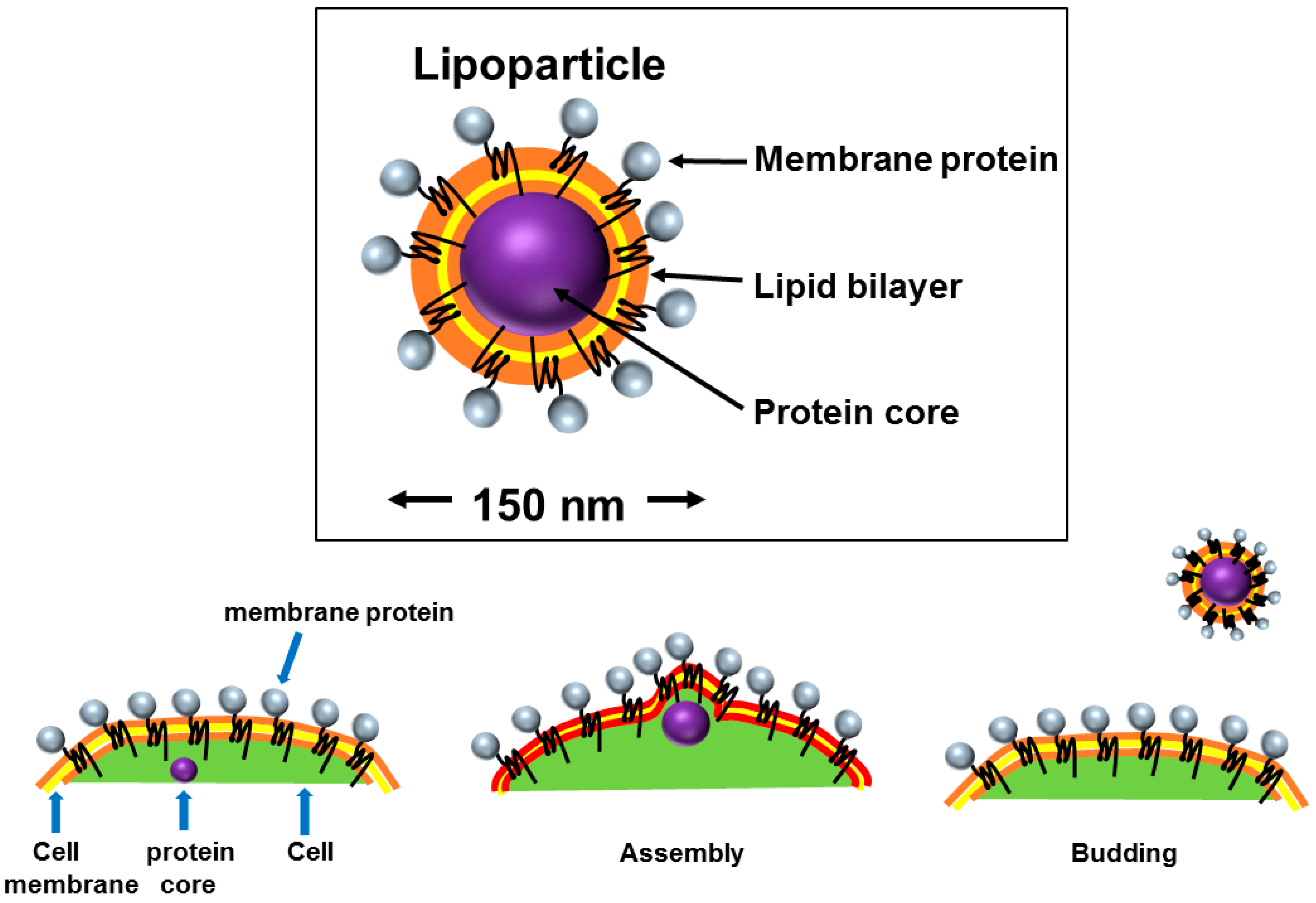
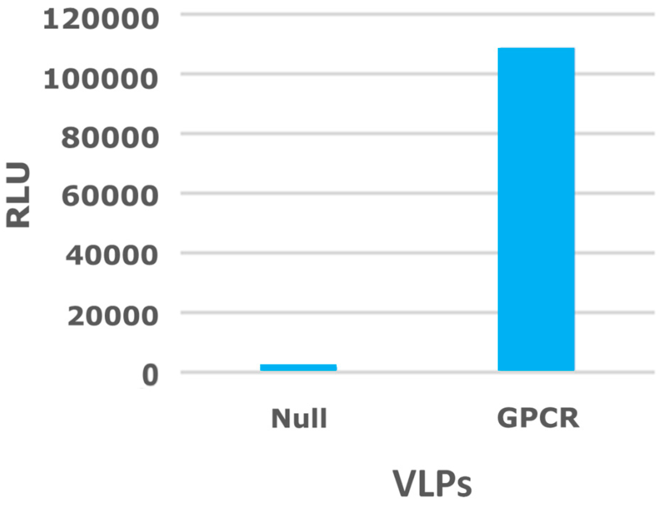
2.3. Virus-Like Particles

A virus-like particle (VLP) (Figure 2D) is similar to a budded enveloped virion that contains only the viral capsid protein that directs and executes viral budding from the cell membrane and is devoid of the viral genome and all other viral proteins (Figure 3). VLPs can be biotinylated either on the membrane lipid or on a membrane protein [83,84] and can be purchased commercially. VLPs are sturdier than intact mammalian cells, thus offering advantages during the affinity selection process by allowing for the addition of detergents, such as Triton X-100 [85,86,87] or Tween 20 [87,88], to the wash buffer to decrease non-specific binding. To test the expression of a GPCR protein on VLPs surface, an enzyme linked immunosorbent assay (ELISA) was performed with a monoclonal antibody, in which the antibody bound to GPCR-expressed VLPs but not to the null (i.e., not expressing the target receptor) VLPs (Figure 4).
Moreover, since VLPs can be biotinylated, competition with unmodified null VLPs can be performed during the affinity selection process to minimize recovery of virions that display antibodies binding to the VLPs themselves. There are multiple advantages associated with VLPs for phage display. VLPs are much more stable than micelles or liposomes [89,90] and can be stored for a long period of time without losing their functions. The multivalent display [91] of the membrane proteins on VLPs may help retain the binding clones through avidity during the affinity selection experiments, which may facilitate the discovery of even weak antibody binders. One major disadvantage of using VLPs is their cost. Commercially available VLPs are expensive, and, although they can be made in-house, it can be laborious to optimize each receptor target for high expression and to quality control for biotinylation levels.

2.4. Native Cells

Cultured cells, which endogenously express the target proteins, are often used in affinity selection experiments with phage-display libraries. The cells are kept intact, and virions displaying antibody fragments will only be retained, if they bind to an antigen on the surface of the cells. For example, monolayers of cells are incubated with a phage-display library, non-binders are washed away, and bound virions are recovered by acid elution (i.e., pH 2) or with a detergent that lyses the cells. After repeated rounds of affinity selection, the output is examined: binding of individual clones to cultured cells can be confirmed by an ELISA [92] or through phage titering [93,94]. Generally, one looks for a signal several fold higher for a particular cell line, compared to another cell line that does not express the membrane protein of interest. The main advantages of using native cells for generating antibodies against membrane proteins are that the target is in its native, physiologically-relevant state and is properly post-translationally modified. One disadvantage is due to the presence of many non-relevant targets on the cell surface, the affinity selection process is inefficient with high background of non-specific binders.

2.5. Engineered Cell Lines

In addition to working with native cells, one can transfect cells with plasmids that overexpress target membrane proteins on the cell surface. The major advantage of this approach is that matched cell lines can be generated—one expressing the cell surface protein of interest and the other one lacking the protein—which permits negative selection of phage-display antibodies that recognize non-relevant cell surface antigens.
In this approach, one can use either transiently transfected cells or stable cell lines (Figure 5). There are advantages and disadvantages with both types of cells. While target protein expression typically occurs within 24–48 h of transfection [95], the expression level is variable, with high levels of protein expression sometimes leading to cell death, thus decreasing the population of cells expressing the protein of interest over time [96]. The efficiency of transfection is also variable and is cell-type dependent. There are many approaches for transfection of mammalian cells, including cationic lipids, electroporation, etc. and each method will require optimization for the specific cell type [96,97]. Despite these disadvantages, transiently transfected cells can be used for affinity selection of phage-display antibodies that bind to cell surface receptors, especially when coupled with a multivalent phage display system, which increases the sensitivity of the phage for its target (Figure 6) through avidity [98].
For generating stable cells, it can take weeks to months to produce a cell line that expresses a satisfactory level of a membrane protein, but once identified, a single cell can be clonally expanded to express uniformly high levels of protein at sub-lethal levels with inducible promoter systems [99,100,101,102]. Stable cells can be stored and kept for indefinite amounts of time and may be a source of reliable antigen that allows for repeated experimentation [103]. Certain cell lines, such as Human embryonic kidney 293 (HEK-293) [104,105] and insect cells [34,106], have been used widely for making both transient and stable cells for overexpressing membrane proteins.

3. Challenges and Novel Methods for Affinity Selecting Recombinant Antibodies against Membrane Proteins

As the membrane protein of interest represents only a small fraction of available epitopes (i.e., extracellular matrix, phospholipids, glycolipids, etc.) on a cell surface, there are several challenges to generate recombinant affinity reagents to membrane proteins. First, when using cell lines in affinity selection experiments, it is difficult to wash cells adequately to eliminate non-specific binding virions; virions can stick to cell surfaces and be trapped by clumped cells. Second, experiments to compete or subtract common epitope between two cell lines are often incomplete. Third, generally the signal-to-noise ratio for binding to cells is often small (i.e., <2). Fourth, even when binders are isolated from whole cell screening, it is formidable to determine what the targets are for the binders.
To address the above challenges, several new affinity selection methods have been developed. We have previously described a novel approach to phage display that utilizes water-in-oil emulsions (Figure 7) for the identification and isolation of phage displayed scFvs [107]. Using this method, a large library of antibody-displaying virions is bound to beads in individual oil droplets rather than affinity selection on a large mixed population. This method allows one to query clonal populations of amplified phage against the antigen while eliminating competition from high affinity binders. In the emulsion droplets, a highly localized concentration of virions and antigen allows one to isolate a greater diversity of binders during the affinity selection process. This may be critical in the context of a complex antigen source, such as a whole cell or tissue sample, where antibodies against the highly expressed or immunodominant epitopes will outcompete the less abundant or less antigenic ones when bulk affinity selection is performed.
One new method in affinity selection of antibodies for efficiently partition binders over non-specific binding background is called “pathfinding”. This is a two-step method, in which cells are first incubated with a ligand or an antibody that interacts with protein of interest. Such a ligand or antibody is also conjugated to horseradish peroxidase (HRP), and after washing, the cells are incubated with the phage-display library. After eliminating non-specific binding virions with thorough washing, the cells are incubated with biotin-tyramide [108]. All free lysine residues near the HRP-labeled ligand or antibody, including the lysine residues on the virions that display the binding antibody fragments, are biotinylated in vitro. Such biotinylated virions can be partitioned away from non-specific binding virions with streptavidin-coated magnetic beads. This method has been successfully used to generate antibodies against C-C chemokine receptor type 5 (CCR5) [109]. One drawback of this method is that the ligand or the existing antibody may interfere with the binding of phage displayed antibodies to the target. As a result, instead of isolating antibodies to the target protein, antibodies binding to the cell surface antigens in close vicinity of the target protein are isolated [110].
To improve affinity selection process with cells grown in suspension, a method termed “Biopanning and Rapid Analysis of Selective Interactive Ligands” (BRASIL) has been developed [111]. This approach involves incubating a suspension of cultured cells with a phage library, followed by centrifuging the cells through an organic non-miscible lower phase, which washes cells free of all virions except those binding to a cell surface target. This method has been used to generate recombinant affinity reagents to an osteosarcoma cell surface antigen [112], Toll-like receptor 2 [113] and a thyroid tumor cell surface marker [114].
To remove the non-specific binders even more efficiently, microfluidic devices have been used for affinity selection [115,116]. Such microfluidic devices utilize continuous flow for washing, which prevents rebinding of non-specific binders. With one of such devices, a phage peptide library was affinity selected against neuropilin-1 on the surface of the human prostate carcinoma cells and several known and novel peptide sequences against neuropilin-1 were isolated [116]. In a side-by-side comparison, the microfluidic device removed non-specific binders and enriched binders much more efficiently than affinity selection with the regular cell suspension.
For multi-pass membrane proteins, here we propose an alternative approach for affinity selection. First, multiple peptide antigens, corresponding to the extracellular loops, N-termini and C-termini of the target proteins, can be designed with the aid of a variety of software tools for predicting protein structures [117,118]. One can use the peptides for affinity selection of a phage-displayed antibody library, and the enriched pool of clones can be mutated by chain shuffling and error-prone PCR [9]. The resulting mutant library can be used to affinity-select cells expressing the endogenous membrane protein of interest. There are several advantages to this approach. First, the initial rounds of affinity selection with peptides will help remove majority of the non-specific binding virions. Second, the copy number of a particular binder enriched by initial affinity selection is much higher than that in the non-selected naïve library, and the extra copies will increase the likelihood of this binder to bind its endogenous antigen on the cell surface and will help it retain during the extensive washing.

4. Various Applications of Recombinant Antibodies

Recombinant affinity reagents generated by phage display to membrane proteins have been widely used for basic science research, diagnostics, and therapeutics. One of their most common applications in basic science is for promoting crystallization of membrane proteins to solve three-dimensional structures. Membrane proteins are notoriously difficult to crystallize due to their heterogeneous nature. In addition, the detergent micelle or liposome used for solubilizing and stabilizing the membrane proteins can interfere with protein crystallization [119]. Recombinant affinity reagents not only stabilize the membrane proteins to increase conformational homogeneity but also create extra crystal contact surface, both of which promote crystallization [119]. With such an approach, Fabs have been generated by phage-display to assist the crystallization of K+ channel [120,121]. Nanobodies have been generated for crystalizing two membrane proteins, including the β2 adrenergic receptors at its active conformation [122] and the ATP-binding cassette (ABC) transporter P-glycoprotein [123]. This same approach has been applied to phage-displayed fibronectin type III (FN3) monobodies for solving structures of fluoride ion channel [64], and applied to designed ankyrin repeat proteins (DARPins) to crystallize multidrug exporter AcrB [124].
Efforts have also been undertaken to generate recombinant antibodies for diagnostic purposes. Recombinant antibodies have been generated for diagnosing infectious diseases, such as influenza [125,126,127] and encephalitis [128]. Antibodies [129,130,131] and nanobodies [132,133] have also been generated for cancer diagnosis. To aid cardiovascular imaging, a high affinity nanobody has been engineered to recognize vascular cell adhesion molecule-1 (VCAM1) [134].
One of the most important applications of recombinant affinity reagents is for treating diseases as therapeutics. As of 2016, there are seven phage-display derived drugs that have been approved by Food and Drug Administration (FDA) in the USA. The approved list includes the global best-selling drug, Adalimumab (also known as Humira), a recombinant antibody used to treating autoimmune diseases [135]. Two of the approved protein drugs targeting membrane proteins are Herceptin [136], which recognizes human epidermal growth factor receptor 2 (HER2) for treating breast cancer, and Romiplostim, which binds to thrombopoietin for treating Idiopathic thrombocytopenic purpura [137]. Besides the seven approved drugs, there are dozens of phage-display derived reagents in different phases of clinical trial [138]. In summary, phage display becomes one of the most popular display technologies in engineering proteins for therapeutic purpose.
In this review, we summarize the some of the latest progress of engineering antibody or its mimics against membrane proteins for applications in basic research, diagnostics and therapeutics. We give details of several formats for presenting the membrane proteins for affinity selection and several new methods to improve the efficiency of the affinity selection process. We expect that with all these progress, applications of recombinant antibodies generated by phage display will continue to expand and they will make significant impact on various fields of life science research.

Acknowledgments

Financial support was provided by a grant (U54 DK093444) from the Common Fund at the US National Institutes of Health.

Conflicts of Interest

The authors declare no conflict of interest. Renhua Huang is an employee of Meso Scale Diagnostics LLC, Rockville, MD, USA. Margaret Kiss, Melissa Batonick, and Michael P. Weiner are employees of AxioMx Inc., a subsidiary of Abcam Plc, Branford, CT, USA.

References

  1. Bradbury, A.; Pluckthun, A. Reproducibility: Standardize antibodies used in research. Nature 2015, 518, 27–29. [Google Scholar] [CrossRef] [PubMed]
  2. Gram, H.; Marconi, L.A.; Barbas, C.F., 3rd; Collet, T.A.; Lerner, R.A.; Kang, A.S. In vitro selection and affinity maturation of antibodies from a naive combinatorial immunoglobulin library. Proc. Natl. Acad. Sci. USA 1992, 89, 3576–3580. [Google Scholar] [PubMed]
  3. Schofield, D.J.; Pope, A.R.; Clementel, V.; Buckell, J.; Chapple, S.; Clarke, K.F.; Conquer, J.S.; Crofts, A.M.; Crowther, S.R.; Dyson, M.R.; et al. Application of phage display to high throughput antibody generation and characterization. Genome Biol. 2007, 8. [Google Scholar] [CrossRef] [PubMed]
  4. Colwill, K.; Graslund, S. A roadmap to generate renewable protein binders to the human proteome. Nat. Methods 2011, 8, 551–558. [Google Scholar] [CrossRef] [PubMed]
  5. Hust, M.; Meyer, T.; Voedisch, B.; Rulker, T.; Thie, H.; El-Ghezal, A.; Kirsch, M.I.; Schutte, M.; Helmsing, S.; Meier, D.; et al. A human scFv antibody generation pipeline for proteome research. J. Biotechnol. 2011, 152, 159–170. [Google Scholar] [CrossRef] [PubMed]
  6. Taussig, M.J.; Stoevesandt, O.; Borrebaeck, C.A.; Bradbury, A.R.; Cahill, D.; Cambillau, C.; de Daruvar, A.; Dubel, S.; Eichler, J.; Frank, R.; et al. Proteomebinders: Planning a European resource of affinity reagents for analysis of the human proteome. Nat. Methods 2007, 4, 13–17. [Google Scholar] [CrossRef] [PubMed]
  7. Stoevesandt, O.; Taussig, M.J. European and international collaboration in affinity proteomics. New Biotechnol. 2012, 29, 511–514. [Google Scholar] [CrossRef] [PubMed]
  8. Geyer, C.R.; McCafferty, J.; Dubel, S.; Bradbury, A.R.; Sidhu, S.S. Recombinant antibodies and in vitro selection technologies. Methods Mol. Biol. 2012, 901, 11–32. [Google Scholar] [PubMed]
  9. Huang, R.; Gorman, K.T.; Vinci, C.R.; Dobrovetsky, E.; Graslund, S.; Kay, B.K. Streamlining the pipeline for generation of recombinant affinity reagents by integrating the affinity maturation step. Int. J. Mol. Sci. 2015, 16, 23587–23603. [Google Scholar] [CrossRef] [PubMed]
  10. Zhong, N.; Loppnau, P.; Seitova, A.; Ravichandran, M.; Fenner, M.; Jain, H.; Bhattacharya, A.; Hutchinson, A.; Paduch, M.; Lu, V.; et al. Optimizing production of antigens and Fabs in the context of generating recombinant antibodies to human proteins. PLoS ONE 2015, 10. [Google Scholar] [CrossRef]
  11. Nelson, A.L. Antibody fragments: Hope and hype. MAbs 2010, 2, 77–83. [Google Scholar] [PubMed]
  12. Ahmad, Z.A.; Yeap, S.K.; Ali, A.M.; Ho, W.Y.; Alitheen, N.B.; Hamid, M. scFv antibody: Principles and clinical application. Clin. Dev. Immunol. 2012, 2012. [Google Scholar] [CrossRef]
  13. Fellouse, F.A.; Wiesmann, C.; Sidhu, S.S. Synthetic antibodies from a four-amino-acid code: A dominant role for tyrosine in antigen recognition. Proc. Natl. Acad. Sci. USA 2004, 101, 12467–12472. [Google Scholar] [CrossRef] [PubMed]
  14. Birtalan, S.; Zhang, Y.; Fellouse, F.A.; Shao, L.; Schaefer, G.; Sidhu, S.S. The intrinsic contributions of tyrosine, serine, glycine and arginine to the affinity and specificity of antibodies. J. Mol. Biol. 2008, 377, 1518–1528. [Google Scholar] [CrossRef] [PubMed]
  15. Miersch, S.; Sidhu, S.S. Synthetic antibodies: Concepts, potential and practical considerations. Methods 2012, 57, 486–498. [Google Scholar] [CrossRef] [PubMed]
  16. De Marco, A. Biotechnological applications of recombinant single-domain antibody fragments. Microb. Cell Fact. 2011, 10. [Google Scholar] [CrossRef] [PubMed]
  17. De Meyer, T.; Muyldermans, S.; Depicker, A. Nanobody-based products as research and diagnostic tools. Trends Biotechnol. 2014, 32, 263–270. [Google Scholar] [CrossRef] [PubMed]
  18. Helma, J.; Cardoso, M.C.; Muyldermans, S.; Leonhardt, H. Nanobodies and recombinant binders in cell biology. J. Cell Biol. 2015, 209, 633–644. [Google Scholar] [CrossRef] [PubMed]
  19. Skerra, A. Alternative binding proteins: Anticalins-harnessing the structural plasticity of the lipocalin ligand pocket to engineer novel binding activities. FEBS J. 2008, 275, 2677–2683. [Google Scholar] [CrossRef] [PubMed]
  20. Nilsson, F.Y.; Tolmachev, V. Affibody molecules: New protein domains for molecular imaging and targeted tumor therapy. Curr. Opin. Drug Discov. Dev. 2007, 10, 167–175. [Google Scholar]
  21. Boersma, Y.L.; Pluckthun, A. DARPins and other repeat protein scaffolds: Advances in engineering and applications. Curr. Opin. Biotechnol. 2011, 22, 849–857. [Google Scholar] [CrossRef] [PubMed]
  22. Koide, A.; Koide, S. Monobodies: Antibody mimics based on the scaffold of the fibronectin type iii domain. Methods Mol. Biol. 2007, 352, 95–109. [Google Scholar] [PubMed]
  23. Carpenter, E.P.; Beis, K.; Cameron, A.D.; Iwata, S. Overcoming the challenges of membrane protein crystallography. Curr. Opin. Struct. Biol. 2008, 18, 581–586. [Google Scholar] [CrossRef] [PubMed]
  24. Yasui, N.; Mihara, E.; Nampo, M.; Tamura-Kawakami, K.; Unno, H.; Matsumoto, K.; Takagi, J. Detection of endogenous LRP6 expressed on human cells by monoclonal antibodies specific for the native conformation. J. Immunol. Methods 2010, 352, 153–160. [Google Scholar] [CrossRef] [PubMed]
  25. Verstraete, K.; Remmerie, B.; Elegheert, J.; Lintermans, B.; Haegeman, G.; Vanhoenacker, P.; Van Craenenbroeck, K.; Savvides, S.N. Inducible production of recombinant human Flt3 ectodomain variants in mammalian cells and preliminary crystallographic analysis of Flt3 ligand-receptor complexes. Acta Crystallogr. Sect. F 2011, 67, 325–331. [Google Scholar] [CrossRef] [PubMed]
  26. Hsieh, S.C.; Tsai, W.Y.; Nerurkar, V.R.; Wang, W.K. Characterization of the ectodomain of the envelope protein of dengue virus type 4: Expression, membrane association, secretion and particle formation in the absence of precursor membrane protein. PLoS ONE 2014, 9. [Google Scholar] [CrossRef] [PubMed]
  27. Goehring, A.; Lee, C.H.; Wang, K.H.; Michel, J.C.; Claxton, D.P.; Baconguis, I.; Althoff, T.; Fischer, S.; Garcia, K.C.; Gouaux, E. Screening and large-scale expression of membrane proteins in mammalian cells for structural studies. Nat. Protoc. 2014, 9, 2574–2585. [Google Scholar] [CrossRef] [PubMed]
  28. Liu, J.; Zhang, X.X.; Zhang, S.Y.; Lu, M.; Kong, Y.Y.; Wang, Y.; Li, G.D. Expression of hepatitis C virus E2 ectodomain in E. Coli and its application in the detection of anti-E2 antibodies in human sera. Acta Biochim. Biophys. Sin. 2004, 36, 57–63. [Google Scholar] [CrossRef] [PubMed]
  29. Lay, C.S.; Wilson, K.A.; Kobe, B.; Kemp, B.E.; Drummer, H.E.; Poumbourios, P. Expression and biochemical analysis of the entire HIV-2 gp41 ectodomain: Determinants of stability map to N- and C-terminal sequences outside the 6-helix bundle core. FEBS Lett. 2004, 567, 183–188. [Google Scholar] [CrossRef] [PubMed]
  30. Laible, P.D.; Scott, H.N.; Henry, L.; Hanson, D.K. Towards higher-throughput membrane protein production for structural genomics initiatives. J. Struct. Funct. Genomics 2004, 5, 167–172. [Google Scholar] [CrossRef] [PubMed]
  31. Jacquez, P.; Lei, N.; Weigt, D.; Xiao, C.; Sun, J. Expression and purification of the functional ectodomain of human anthrax toxin receptor 2 in Escherichia coli Origami B cells with assistance of bacterial Trigger Factor. Protein Expr. Purif. 2014, 95, 149–155. [Google Scholar] [CrossRef] [PubMed]
  32. Hays, F.A.; Roe-Zurz, Z.; Stroud, R.M. Overexpression and purification of integral membrane proteins in yeast. Methods Enzymol. 2010, 470, 695–707. [Google Scholar] [PubMed]
  33. Mizutani, K.; Yoshioka, S.; Mizutani, Y.; Iwata, S.; Mikami, B. High-throughput construction of expression system using yeast pichia pastoris, and its application to membrane proteins. Protein Expr. Purif. 2011, 77, 1–8. [Google Scholar] [CrossRef] [PubMed]
  34. Trometer, C.; Falson, P. Mammalian membrane protein expression in baculovirus-infected insect cells. Methods Mol. Biol. 2010, 601, 105–117. [Google Scholar] [PubMed]
  35. Hengen, P. Purification of His-Tag fusion proteins from Escherichia coli. Trends Biochem. Sci. 1995, 20, 285–286. [Google Scholar] [CrossRef]
  36. Einhauer, A.; Jungbauer, A. The FLAG™ peptide, a versatile fusion tag for the purification of recombinant proteins. J. Biochem. Biophys. Methods 2001, 49, 455–465. [Google Scholar] [CrossRef]
  37. Engel, C.K.; Chen, L.; Prive, G.G. Insertion of carrier proteins into hydrophilic loops of the Escherichia coli lactose permease. Biochim. Biophys. Acta 2002, 1564, 38–46. [Google Scholar] [CrossRef]
  38. Serrano-Vega, M.J.; Magnani, F.; Shibata, Y.; Tate, C.G. Conformational thermostabilization of the β1-adrenergic receptor in a detergent-resistant form. Proc. Natl. Acad. Sci. USA 2008, 105, 877–882. [Google Scholar] [CrossRef] [PubMed]
  39. Sarkar, C.A.; Dodevski, I.; Kenig, M.; Dudli, S.; Mohr, A.; Hermans, E.; Pluckthun, A. Directed evolution of a G protein-coupled receptor for expression, stability, and binding selectivity. Proc. Natl. Acad. Sci. USA 2008, 105, 14808–14813. [Google Scholar] [CrossRef] [PubMed]
  40. Rosenbaum, D.M.; Cherezov, V.; Hanson, M.A.; Rasmussen, S.G.; Thian, F.S.; Kobilka, T.S.; Choi, H.J.; Yao, X.J.; Weis, W.I.; Stevens, R.C.; et al. GPCR engineering yields high-resolution structural insights into β2-adrenergic receptor function. Science 2007, 318, 1266–1273. [Google Scholar] [CrossRef] [PubMed]
  41. Cherezov, V.; Rosenbaum, D.M.; Hanson, M.A.; Rasmussen, S.G.; Thian, F.S.; Kobilka, T.S.; Choi, H.J.; Kuhn, P.; Weis, W.I.; Kobilka, B.K.; et al. High-resolution crystal structure of an engineered human β2-adrenergic G protein-coupled receptor. Science 2007, 318, 1258–1265. [Google Scholar] [CrossRef] [PubMed]
  42. Wu, B.; Chien, E.Y.; Mol, C.D.; Fenalti, G.; Liu, W.; Katritch, V.; Abagyan, R.; Brooun, A.; Wells, P.; Bi, F.C.; et al. Structures of the CXCR4 chemokine GPCR with small-molecule and cyclic peptide antagonists. Science 2010, 330, 1066–1071. [Google Scholar] [CrossRef] [PubMed]
  43. Manglik, A.; Kruse, A.C.; Kobilka, T.S.; Thian, F.S.; Mathiesen, J.M.; Sunahara, R.K.; Pardo, L.; Weis, W.I.; Kobilka, B.K.; Granier, S. Crystal structure of the micro-opioid receptor bound to a morphinan antagonist. Nature 2012, 485, 321–326. [Google Scholar] [CrossRef] [PubMed]
  44. Chun, E.; Thompson, A.A.; Liu, W.; Roth, C.B.; Griffith, M.T.; Katritch, V.; Kunken, J.; Xu, F.; Cherezov, V.; Hanson, M.A.; et al. Fusion partner toolchest for the stabilization and crystallization of G protein-coupled receptors. Structure 2012, 20, 967–976. [Google Scholar] [CrossRef] [PubMed]
  45. Thompson, A.A.; Liu, W.; Chun, E.; Katritch, V.; Wu, H.; Vardy, E.; Huang, X.P.; Trapella, C.; Guerrini, R.; Calo, G.; et al. Structure of the nociceptin/orphanin FQ receptor in complex with a peptide mimetic. Nature 2012, 485, 395–399. [Google Scholar] [CrossRef] [PubMed]
  46. Magnani, F.; Shibata, Y.; Serrano-Vega, M.J.; Tate, C.G. Co-evolving stability and conformational homogeneity of the human adenosine A2a receptor. Proc. Natl. Acad. Sci. USA 2008, 105, 10744–10749. [Google Scholar] [CrossRef] [PubMed]
  47. Miller, J.L.; Tate, C.G. Engineering an ultra-thermostable β1-adrenoceptor. J. Mol. Biol. 2011, 413, 628–638. [Google Scholar] [CrossRef] [PubMed]
  48. Herzenberg, L.A.; Parks, D.; Sahaf, B.; Perez, O.; Roederer, M. The history and future of the fluorescence activated cell sorter and flow cytometry: A view from stanford. Clin. Chem. 2002, 48, 1819–1827. [Google Scholar] [PubMed]
  49. Dodevski, I.; Pluckthun, A. Evolution of three human GPCRs for higher expression and stability. J. Mol. Biol. 2011, 408, 599–615. [Google Scholar] [CrossRef] [PubMed]
  50. Schlinkmann, K.M.; Hillenbrand, M.; Rittner, A.; Kunz, M.; Strohner, R.; Pluckthun, A. Maximizing detergent stability and functional expression of a GPCR by exhaustive recombination and evolution. J. Mol. Biol. 2012, 422, 414–428. [Google Scholar] [CrossRef] [PubMed]
  51. Egloff, P.; Hillenbrand, M.; Klenk, C.; Batyuk, A.; Heine, P.; Balada, S.; Schlinkmann, K.M.; Scott, D.J.; Schutz, M.; Pluckthun, A. Structure of signaling-competent neurotensin receptor 1 obtained by directed evolution in Escherichia coli. Proc. Natl. Acad. Sci. USA 2014, 111, E655–E662. [Google Scholar] [CrossRef] [PubMed]
  52. Ghosh, E.; Kumari, P.; Jaiman, D.; Shukla, A.K. Methodological advances: The unsung heroes of the GPCR structural revolution. Nat. Rev. Mol. Cell Biol. 2015, 16, 69–81. [Google Scholar] [CrossRef] [PubMed]
  53. Scott, D.J.; Kummer, L.; Tremmel, D.; Pluckthun, A. Stabilizing membrane proteins through protein engineering. Curr. Opin. Chem. Biol. 2013, 17, 427–435. [Google Scholar] [CrossRef] [PubMed]
  54. Memic, A.; Volgina, V.V.; Gussin, H.A.; Pepperberg, D.R.; Kay, B.K. Generation of recombinant guinea pig antibody fragments to the human GABAC receptor. J. Immunol. Methods 2011, 368, 36–44. [Google Scholar] [CrossRef] [PubMed]
  55. Koduvayur, S.P.; Gussin, H.A.; Parthasarathy, R.; Hao, Z.; Kay, B.K.; Pepperberg, D.R. Generation of recombinant antibodies to rat GABAA receptor subunits by affinity selection on synthetic peptides. PLoS ONE 2014, 9. [Google Scholar] [CrossRef] [PubMed]
  56. Kierny, M.R.; Cunningham, T.D.; Bouhenni, R.A.; Edward, D.P.; Kay, B.K. Generating recombinant antibodies against putative biomarkers of retinal injury. PLoS ONE 2015, 10. [Google Scholar] [CrossRef] [PubMed]
  57. Korepanova, A.; Moore, J.D.; Nguyen, H.B.; Hua, Y.; Cross, T.A.; Gao, F. Expression of membrane proteins from Mycobacterium tuberculosis in Escherichia coli as fusions with maltose binding protein. Protein Expr. Purif. 2007, 53, 24–30. [Google Scholar] [CrossRef] [PubMed]
  58. Wu, W.; Jin, J.; Liu, X.; Zhang, Y.; Li, M.; Shao, M.; Qian, Y.; Zhang, D.; Zhu, H.; Ruan, Y.; et al. Functional expression of the Fc-fused extracellular domains of group II membrane proteins. Glycoconj. J. 2015, 32, 69–76. [Google Scholar] [CrossRef] [PubMed]
  59. Kierstan, M.P.; Coughlan, M.P. Immobilization of proteins by noncovalent procedures: Principles and applications. Bioprocess Technol. 1991, 14, 13–71. [Google Scholar] [PubMed]
  60. Seddon, A.M.; Curnow, P.; Booth, P.J. Membrane proteins, lipids and detergents: Not just a soap opera. Biochim. Biophys. Acta 2004, 1666, 105–117. [Google Scholar] [CrossRef] [PubMed]
  61. Lin, C.E. Determination of critical micelle concentration of surfactants by capillary electrophoresis. J. Chromatogr. A 2004, 1037, 467–478. [Google Scholar] [CrossRef] [PubMed]
  62. Rothlisberger, D.; Pos, K.M.; Pluckthun, A. An antibody library for stabilizing and crystallizing membrane proteins—Selecting binders to the citrate carrier CitS. FEBS Lett. 2004, 564, 340–348. [Google Scholar] [CrossRef]
  63. Huber, T.; Steiner, D.; Rothlisberger, D.; Pluckthun, A. In vitro selection and characterization of DARPins and Fab fragments for the co-crystallization of membrane proteins: The Na+-citrate symporter CitS as an example. J. Struct. Biol. 2007, 159, 206–221. [Google Scholar] [CrossRef] [PubMed]
  64. Stockbridge, R.B.; Kolmakova-Partensky, L.; Shane, T.; Koide, A.; Koide, S.; Miller, C.; Newstead, S. Crystal structures of a double-barrelled fluoride ion channel. Nature 2015, 525, 548–551. [Google Scholar] [CrossRef] [PubMed]
  65. Raschle, T.; Hiller, S.; Etzkorn, M.; Wagner, G. Nonmicellar systems for solution NMR spectroscopy of membrane proteins. Curr. Opin. Struct. Biol. 2010, 20, 471–479. [Google Scholar] [CrossRef] [PubMed]
  66. Jesorka, A.; Orwar, O. Liposomes: Technologies and analytical applications. Annu. Rev. Anal. Chem. 2008, 1, 801–832. [Google Scholar] [CrossRef] [PubMed]
  67. Jespersen, L.K.; Kuusinen, A.; Orellana, A.; Keinanen, K.; Engberg, J. Use of proteoliposomes to generate phage antibodies against native AMPA receptor. Eur. J. Biochem. 2000, 267, 1382–1389. [Google Scholar] [CrossRef] [PubMed]
  68. Civjan, N.R.; Bayburt, T.H.; Schuler, M.A.; Sligar, S.G. Direct solubilization of heterologously expressed membrane proteins by incorporation into nanoscale lipid bilayers. Biotechniques 2003, 35, 556–563. [Google Scholar] [PubMed]
  69. Breslow, J.L.; Ross, D.; McPherson, J.; Williams, H.; Kurnit, D.; Nussbaum, A.L.; Karathanasis, S.K.; Zannis, V.I. Isolation and characterization of cDNA clones for human apolipoprotein A–I. Proc. Natl. Acad. Sci. USA 1982, 79, 6861–6865. [Google Scholar] [CrossRef] [PubMed]
  70. Knott, T.J.; Pease, R.J.; Powell, L.M.; Wallis, S.C.; Rall, S.C., Jr.; Innerarity, T.L.; Blackhart, B.; Taylor, W.H.; Marcel, Y.; Milne, R.; et al. Complete protein sequence and identification of structural domains of human apolipoprotein B. Nature 1986, 323, 734–738. [Google Scholar] [CrossRef] [PubMed]
  71. He, W.; Scharadin, T.M.; Saldana, M.; Gellner, C.; Hoang-Phou, S.; Takanishi, C.; Hura, G.L.; Tainer, J.A.; Carraway, K.L., 3rd; Henderson, P.T.; et al. Cell-free expression of functional receptor tyrosine kinases. Sci. Rep. 2015, 5. [Google Scholar] [CrossRef] [PubMed]
  72. Denisov, I.G.; Grinkova, Y.V.; Lazarides, A.A.; Sligar, S.G. Directed self-assembly of monodisperse phospholipid bilayer nanodiscs with controlled size. J. Am. Chem. Soc. 2004, 126, 3477–3487. [Google Scholar] [CrossRef] [PubMed]
  73. Bayburt, T.H.; Sligar, S.G. Membrane protein assembly into nanodiscs. FEBS Lett. 2010, 584, 1721–1727. [Google Scholar] [CrossRef] [PubMed]
  74. Schuler, M.A.; Denisov, I.G.; Sligar, S.G. Nanodiscs as a new tool to examine lipid-protein interactions. Methods Mol. Biol. 2013, 974, 415–433. [Google Scholar] [PubMed]
  75. Chromy, B.A.; Arroyo, E.; Blanchette, C.D.; Bench, G.; Benner, H.; Cappuccio, J.A.; Coleman, M.A.; Henderson, P.T.; Hinz, A.K.; Kuhn, E.A.; et al. Different apolipoproteins impact nanolipoprotein particle formation. J. Am. Chem. Soc. 2007, 129, 14348–14354. [Google Scholar] [CrossRef] [PubMed]
  76. Cappuccio, J.A.; Blanchette, C.D.; Sulchek, T.A.; Arroyo, E.S.; Kralj, J.M.; Hinz, A.K.; Kuhn, E.A.; Chromy, B.A.; Segelke, B.W.; Rothschild, K.J.; et al. Cell-free co-expression of functional membrane proteins and apolipoprotein, forming soluble nanolipoprotein particles. Mol. Cell Proteomics 2008, 7, 2246–2253. [Google Scholar] [CrossRef] [PubMed]
  77. Cappuccio, J.A.; Hinz, A.K.; Kuhn, E.A.; Fletcher, J.E.; Arroyo, E.S.; Henderson, P.T.; Blanchette, C.D.; Walsworth, V.L.; Corzett, M.H.; Law, R.J.; et al. Cell-free expression for nanolipoprotein particles: Building a high-throughput membrane protein solubility platform. Methods Mol. Biol. 2009, 498, 273–296. [Google Scholar] [PubMed]
  78. He, W.; Luo, J.; Bourguet, F.; Xing, L.; Yi, S.K.; Gao, T.; Blanchette, C.; Henderson, P.T.; Kuhn, E.; Malfatti, M.; et al. Controlling the diameter, monodispersity, and solubility of apoa1 nanolipoprotein particles using telodendrimer chemistry. Protein Sci. 2013, 22, 1078–1086. [Google Scholar] [CrossRef] [PubMed]
  79. Pavlidou, M.; Hanel, K.; Mockel, L.; Willbold, D. Nanodiscs allow phage display selection for ligands to non-linear epitopes on membrane proteins. PLoS ONE 2013, 8. [Google Scholar] [CrossRef] [PubMed]
  80. Dominik, P.K.; Kossiakoff, A.A. Phage display selections for affinity reagents to membrane proteins in nanodiscs. Methods Enzymol. 2015, 557, 219–245. [Google Scholar] [PubMed]
  81. Dominik, P.K.; Borowska, M.T.; Dalmas, O.; Kim, S.S.; Perozo, E.; Keenan, R.J.; Kossiakoff, A.A. Conformational chaperones for structural studies of membrane proteins using antibody phage display with nanodiscs. Structure 2016, 24, 300–309. [Google Scholar] [CrossRef] [PubMed]
  82. Kay, B.K.; Thai, S.; Volgina, V.V. High-throughput biotinylation of proteins. Methods Mol. Biol. 2009, 498, 185–196. [Google Scholar] [PubMed]
  83. Chackerian, B.; Rangel, M.; Hunter, Z.; Peabody, D.S. Virus and virus-like particle-based immunogens for Alzheimer's disease induce antibody responses against amyloid-β without concomitant T cell responses. Vaccine 2006, 24, 6321–6331. [Google Scholar] [CrossRef] [PubMed]
  84. Thrane, S.; Janitzek, C.M.; Agerbaek, M.O.; Ditlev, S.B.; Resende, M.; Nielsen, M.A.; Theander, T.G.; Salanti, A.; Sander, A.F. A novel virus-like particle based vaccine platform displaying the placental malaria antigen VAR2CSA. PLoS ONE 2015, 10. [Google Scholar] [CrossRef] [PubMed]
  85. Kee, G.S.; Pujar, N.S.; Titchener-Hooker, N.J. Study of detergent-mediated liberation of hepatitis B virus-like particles from S. cerevisiae homogenate: Identifying a framework for the design of future-generation lipoprotein vaccine processes. Biotechnol. Prog. 2008, 24, 623–631. [Google Scholar] [CrossRef] [PubMed]
  86. Burden, C.S.; Jin, J.; Podgornik, A.; Bracewell, D.G. A monolith purification process for virus-like particles from yeast homogenate. J. Chromatogr. B 2012, 880, 82–89. [Google Scholar] [CrossRef] [PubMed]
  87. Brune, K.D.; Leneghan, D.B.; Brian, I.J.; Ishizuka, A.S.; Bachmann, M.F.; Draper, S.J.; Biswas, S.; Howarth, M. Plug-and-display: Decoration of virus-like particles via isopeptide bonds for modular immunization. Sci. Rep. 2016, 6. [Google Scholar] [CrossRef] [PubMed]
  88. Gurramkonda, C.; Zahid, M.; Nemani, S.K.; Adnan, A.; Gudi, S.K.; Khanna, N.; Ebensen, T.; Lunsdorf, H.; Guzman, C.A.; Rinas, U. Purification of hepatitis B surface antigen virus-like particles from recombinant pichia pastoris and in vivo analysis of their immunogenic properties. J. Chromatogr. B 2013, 940, 104–111. [Google Scholar] [CrossRef] [PubMed]
  89. Caldeira, J.C.; Peabody, D.S. Stability and assembly in vitro of bacteriophage PP7 virus-like particles. J. Nanobiotechnol. 2007, 5. [Google Scholar] [CrossRef] [PubMed]
  90. Lynch, A.; Meyers, A.E.; Williamson, A.L.; Rybicki, E.P. Stability studies of HIV-1 Pr55gag virus-like particles made in insect cells after storage in various formulation media. Virol. J. 2012, 9. [Google Scholar] [CrossRef] [PubMed] [Green Version]
  91. Peabody, D.S. A viral platform for chemical modification and multivalent display. J. Nanobiotechnol. 2003, 1. [Google Scholar] [CrossRef] [PubMed] [Green Version]
  92. Even-Desrumeaux, K.; Chames, P. Phage display and selections on cells. Methods Mol. Biol. 2012, 907, 225–235. [Google Scholar] [PubMed]
  93. Dias-Neto, E.; Nunes, D.N.; Giordano, R.J.; Sun, J.; Botz, G.H.; Yang, K.; Setubal, J.C.; Pasqualini, R.; Arap, W. Next-generation phage display: Integrating and comparing available molecular tools to enable cost-effective high-throughput analysis. PLoS ONE 2009, 4. [Google Scholar] [CrossRef] [PubMed]
  94. Yoon, H.; Song, J.M.; Ryu, C.J.; Kim, Y.G.; Lee, E.K.; Kang, S.; Kim, S.J. An efficient strategy for cell-based antibody library selection using an integrated vector system. BMC Biotechnol. 2012, 12. [Google Scholar] [CrossRef] [PubMed]
  95. Pfeifer, A.; Verma, I.M. Gene therapy: Promises and problems. Annu. Rev. Genomics Hum. Genet. 2001, 2, 177–211. [Google Scholar] [CrossRef] [PubMed]
  96. Kim, T.K.; Eberwine, J.H. Mammalian cell transfection: The present and the future. Anal. Bioanal. Chem. 2010, 397, 3173–3178. [Google Scholar] [CrossRef] [PubMed]
  97. Recillas-Targa, F. Multiple strategies for gene transfer, expression, knockdown, and chromatin influence in mammalian cell lines and transgenic animals. Mol. Biotechnol. 2006, 34, 337–354. [Google Scholar] [CrossRef]
  98. Rondot, S.; Koch, J.; Breitling, F.; Dubel, S. A helper phage to improve single-chain antibody presentation in phage display. Nat. Biotechnol. 2001, 19, 75–78. [Google Scholar] [PubMed]
  99. Lama, J.; Carrasco, L. Inducible expression of a toxic poliovirus membrane protein in Escherichia coli: Comparative studies using different expression systems based on T7 promoters. Biochem. Biophys. Res. Commun. 1992, 188, 972–981. [Google Scholar] [CrossRef]
  100. Meyer-Ficca, M.L.; Meyer, R.G.; Kaiser, H.; Brack, A.R.; Kandolf, R.; Kupper, J.H. Comparative analysis of inducible expression systems in transient transfection studies. Anal. Biochem. 2004, 334, 9–19. [Google Scholar] [CrossRef] [PubMed]
  101. Bussow, K. Stable mammalian producer cell lines for structural biology. Curr. Opin. Struct. Biol. 2015, 32, 81–90. [Google Scholar] [CrossRef] [PubMed]
  102. Andrell, J.; Tate, C.G. Overexpression of membrane proteins in mammalian cells for structural studies. Mol. Membr. Biol. 2013, 30, 52–63. [Google Scholar] [CrossRef] [PubMed]
  103. Schiedner, G.; Hertel, S.; Bialek, C.; Kewes, H.; Waschutza, G.; Volpers, C. Efficient and reproducible generation of high-expressing, stable human cell lines without need for antibiotic selection. BMC Biotechnol. 2008, 8. [Google Scholar] [CrossRef] [PubMed]
  104. Chaudhary, S.; Pak, J.E.; Pedersen, B.P.; Bang, L.J.; Zhang, L.B.; Ngaw, S.M.; Green, R.G.; Sharma, V.; Stroud, R.M. Efficient expression screening of human membrane proteins in transiently transfected Human Embryonic Kidney 293S cells. Methods 2011, 55, 273–280. [Google Scholar] [CrossRef] [PubMed]
  105. Lin, C.Y.; Huang, Z.; Wen, W.; Wu, A.; Wang, C.; Niu, L. Enhancing protein expression in HEK-293 cells by lowering culture temperature. PLoS ONE 2015, 10. [Google Scholar] [CrossRef] [PubMed]
  106. Chen, H.; Shaffer, P.L.; Huang, X.; Rose, P.E. Rapid screening of membrane protein expression in transiently transfected insect cells. Protein Expr. Purif. 2013, 88, 134–142. [Google Scholar] [CrossRef] [PubMed]
  107. Buhr, D.L.; Acca, F.E.; Holland, E.G.; Johnson, K.; Maksymiuk, G.M.; Vaill, A.; Kay, B.K.; Weitz, D.A.; Weiner, M.P.; Kiss, M.M. Use of micro-emulsion technology for the directed evolution of antibodies. Methods 2012, 58, 28–33. [Google Scholar] [CrossRef] [PubMed]
  108. Mayer, G.; Bendayan, M. Biotinyl-tyramide: A novel approach for electron microscopic immunocytochemistry. J. Histochem. Cytochem. 1997, 45, 1449–1454. [Google Scholar] [CrossRef] [PubMed]
  109. Osbourn, J.K.; Earnshaw, J.C.; Johnson, K.S.; Parmentier, M.; Timmermans, V.; McCafferty, J. Directed selection of MIP-1α neutralizing CCR5 antibodies from a phage display human antibody library. Nat. Biotechnol. 1998, 16, 778–781. [Google Scholar] [CrossRef] [PubMed]
  110. Sui, J.; Bai, J.; St. Clair Tallarico, A.; Xu, C.; Marasco, W.A. Identification of CD4 and transferrin receptor antibodies by CXCR4 antibody-guided Pathfinder selection. Eur. J. Biochem. 2003, 270, 4497–4506. [Google Scholar] [CrossRef] [PubMed]
  111. Giordano, R.J.; Cardo-Vila, M.; Lahdenranta, J.; Pasqualini, R.; Arap, W. Biopanning and rapid analysis of selective interactive ligands. Nat. Med. 2001, 7, 1249–1253. [Google Scholar] [CrossRef] [PubMed]
  112. Pan, W.; Ye, Z. Auto T cells expressing chimeric antigen receptor derived from auto antibody might be a new treatment for osteosarcoma. Med. Hypotheses 2012, 78, 616–618. [Google Scholar]
  113. Lipes, B.D.; Chen, Y.H.; Ma, H.; Staats, H.F.; Kenan, D.J.; Gunn, M.D. An entirely cell-based system to generate single-chain antibodies against cell surface receptors. J. Mol. Biol. 2008, 379, 261–272. [Google Scholar] [CrossRef] [PubMed]
  114. Carneiro, A.P.; Reis, C.F.; Morari, E.C.; Maia, Y.C.; Nascimento, R.; Bonatto, J.M.; de Souza, M.A.; Goulart, L.R.; Ward, L.S. A putative OTU domain-containing protein 1 deubiquitinating enzyme is differentially expressed in thyroid cancer and identifies less-aggressive tumours. Br. J. Cancer 2014, 111, 551–558. [Google Scholar] [CrossRef] [PubMed]
  115. Liu, Y.; Adams, J.D.; Turner, K.; Cochran, F.V.; Gambhir, S.S.; Soh, H.T. Controlling the selection stringency of phage display using a microfluidic device. Lab. Chip 2009, 9, 1033–1036. [Google Scholar] [CrossRef] [PubMed]
  116. Wang, J.; Liu, Y.; Teesalu, T.; Sugahara, K.N.; Kotamrajua, V.R.; Adams, J.D.; Ferguson, B.S.; Gong, Q.; Oh, S.S.; Csordas, A.T.; et al. Selection of phage-displayed peptides on live adherent cells in microfluidic channels. Proc. Natl. Acad. Sci. USA 2011, 108, 6909–6914. [Google Scholar] [CrossRef] [PubMed]
  117. Hirokawa, T.; Boon-Chieng, S.; Mitaku, S. SOSUI: Classification and secondary structure prediction system for membrane proteins. Bioinformatics 1998, 14, 378–379. [Google Scholar] [CrossRef] [PubMed]
  118. Krogh, A.; Larsson, B.; von Heijne, G.; Sonnhammer, E.L. Predicting transmembrane protein topology with a hidden Markov model: Application to complete genomes. J. Mol. Biol. 2001, 305, 567–580. [Google Scholar] [CrossRef] [PubMed]
  119. Kim, J.; Stroud, R.M.; Craik, C.S. Rapid identification of recombinant Fabs that bind to membrane proteins. Methods 2011, 55, 303–309. [Google Scholar] [CrossRef] [PubMed]
  120. Zhou, Y.; Morais-Cabral, J.H.; Kaufman, A.; MacKinnon, R. Chemistry of ion coordination and hydration revealed by a K+ channel-Fab complex at 2.0 Å resolution. Nature 2001, 414, 43–48. [Google Scholar] [CrossRef] [PubMed]
  121. Uysal, S.; Vasquez, V.; Tereshko, V.; Esaki, K.; Fellouse, F.A.; Sidhu, S.S.; Koide, S.; Perozo, E.; Kossiakoff, A. Crystal structure of full-length KcsA in its closed conformation. Proc. Natl. Acad. Sci. USA 2009, 106, 6644–6649. [Google Scholar] [CrossRef] [PubMed]
  122. Rasmussen, S.G.; Choi, H.J.; Fung, J.J.; Pardon, E.; Casarosa, P.; Chae, P.S.; Devree, B.T.; Rosenbaum, D.M.; Thian, F.S.; Kobilka, T.S.; et al. Structure of a nanobody-stabilized active state of the β2 adrenoceptor. Nature 2011, 469, 175–180. [Google Scholar] [CrossRef] [PubMed]
  123. Ward, A.B.; Szewczyk, P.; Grimard, V.; Lee, C.W.; Martinez, L.; Doshi, R.; Caya, A.; Villaluz, M.; Pardon, E.; Cregger, C.; et al. Structures of P-glycoprotein reveal its conformational flexibility and an epitope on the nucleotide-binding domain. Proc. Natl. Acad. Sci. USA 2013, 110, 13386–13391. [Google Scholar] [CrossRef] [PubMed]
  124. Sennhauser, G.; Amstutz, P.; Briand, C.; Storchenegger, O.; Grutter, M.G. Drug export pathway of multidrug exporter AcrB revealed by DARPin inhibitors. PLoS Biol. 2007, 5. [Google Scholar] [CrossRef] [PubMed]
  125. Pitaksajjakul, P.; Lekcharoensuk, P.; Upragarin, N.; Barbas, C.F., 3rd; Ibrahim, M.S.; Ikuta, K.; Ramasoota, P. Fab MAbs specific to HA of influenza virus with H5N1 neutralizing activity selected from immunized chicken phage library. Biochem. Biophys. Res. Commun. 2010, 395, 496–501. [Google Scholar] [CrossRef] [PubMed]
  126. Dong, J.; Sakurai, A.; Nomura, N.; Park, E.Y.; Shibasaki, F.; Ueda, H. Isolation of recombinant phage antibodies targeting the hemagglutinin cleavage site of highly pathogenic avian influenza virus. PLoS ONE 2013, 8. [Google Scholar] [CrossRef] [PubMed]
  127. Wu, J.; Zeng, X.Q.; Zhang, H.B.; Ni, H.Z.; Pei, L.; Zou, L.R.; Liang, L.J.; Zhang, X.; Lin, J.Y.; Ke, C.W. Novel phage display-derived H5N1-specific scFvs with potential use in rapid avian flu diagnosis. J. Microbiol. Biotechnol. 2014, 24, 704–713. [Google Scholar] [CrossRef] [PubMed]
  128. Kirsch, M.I.; Hulseweh, B.; Nacke, C.; Rulker, T.; Schirrmann, T.; Marschall, H.J.; Hust, M.; Dubel, S. Development of human antibody fragments using antibody phage display for the detection and diagnosis of venezuelan equine encephalitis virus (VEEV). BMC Biotechnol. 2008, 8. [Google Scholar] [CrossRef] [PubMed]
  129. Rothe, A.; Klimka, A.; Tur, M.K.; Pfitzner, T.; Huhn, M.; Sasse, S.; Mallmann, P.; Engert, A.; Barth, S. Construction of phage display libraries from reactive lymph nodes of breast carcinoma patients and selection for specifically binding human single chain Fv on cell lines. Int. J. Mol. Med. 2004, 14, 729–735. [Google Scholar] [CrossRef] [PubMed]
  130. Jakobsen, C.G.; Rasmussen, N.; Laenkholm, A.V.; Ditzel, H.J. Phage display-derived human monoclonal antibodies isolated by binding to the surface of live primary breast cancer cells recognize GRP78. Cancer Res. 2007, 67, 9507–9517. [Google Scholar] [CrossRef] [PubMed]
  131. Araujo, T.G.; Paiva, C.E.; Rocha, R.M.; Maia, Y.C.; Sena, A.A.; Ueira-Vieira, C.; Carneiro, A.P.; Almeida, J.F.; de Faria, P.R.; Santos, D.W.; et al. A novel highly reactive Fab antibody for breast cancer tissue diagnostics and staging also discriminates a subset of good prognostic triple-negative breast cancers. Cancer Lett. 2014, 343, 275–285. [Google Scholar] [CrossRef] [PubMed]
  132. Huang, L.; Gainkam, L.O.; Caveliers, V.; Vanhove, C.; Keyaerts, M.; De Baetselier, P.; Bossuyt, A.; Revets, H.; Lahoutte, T. SPECT imaging with 99mTc-labeled EGFR-specific nanobody for in vivo monitoring of EGFR expression. Mol. Imaging Biol. 2008, 10, 167–175. [Google Scholar] [CrossRef] [PubMed]
  133. Vaneycken, I.; Devoogdt, N.; Van Gassen, N.; Vincke, C.; Xavier, C.; Wernery, U.; Muyldermans, S.; Lahoutte, T.; Caveliers, V. Preclinical screening of anti-HER2 nanobodies for molecular imaging of breast cancer. FASEB J. 2011, 25, 2433–2446. [Google Scholar] [CrossRef] [PubMed]
  134. Broisat, A.; Hernot, S.; Toczek, J.; De Vos, J.; Riou, L.M.; Martin, S.; Ahmadi, M.; Thielens, N.; Wernery, U.; Caveliers, V.; et al. Nanobodies targeting mouse/human VCAM1 for the nuclear imaging of atherosclerotic lesions. Circ. Res. 2012, 110, 927–937. [Google Scholar] [CrossRef] [PubMed]
  135. Monaco, C.; Nanchahal, J.; Taylor, P.; Feldmann, M. Anti-TNF therapy: Past, present and future. Int. Immunol. 2015, 27, 55–62. [Google Scholar] [CrossRef] [PubMed]
  136. Brenner, T.L.; Adams, V.R. First MAb approved for treatment of metastatic breast cancer. J. Am. Pharm. Assoc. 1999, 39, 236–238. [Google Scholar] [CrossRef]
  137. Molineux, G.; Newland, A. Development of romiplostim for the treatment of patients with chronic immune thrombocytopenia: From bench to bedside. Br. J. Haematol. 2010, 150, 9–20. [Google Scholar] [CrossRef] [PubMed]
  138. Nixon, A.E.; Sexton, D.J.; Ladner, R.C. Drugs derived from phage display: From candidate identification to clinical practice. MAbs 2014, 6, 73–85. [Google Scholar] [CrossRef] [PubMed]
Figure 1. Workflow for generating recombinant antibodies to membrane proteins. Purified membrane proteins, presented in different formats, are mixed with a library of virions displaying antibody fragments. The antibody binders are selected by affinity selection and the encoded antibody fragments are characterized biochemically. The most potent and selective binders are chosen for various applications, such as basic science, crystallography, diagnostics and therapeutics.
Antibodies 05 00011 g001
Figure 2. Different formats for presenting membrane proteins for affinity selection experiments. (A) The extracellular domains (green) of membrane proteins can be expressed alone or fused to another protein, such as Fc region of immunoglobulin G (IgG) (dark red). Portions of membrane proteins can also be chemically synthesized as peptides (green). These reagents can then be biotinylated and immobilized on streptavidin-coated surfaces (i.e., streptavidin-coated magnetic beads) for affinity selection. (B) Membrane proteins can be solubilized with detergents, which form micelles around the membrane proteins (e.g., G-protein coupled receptors shown here). (C) Apolipoproteins wrap around phospholipid molecules to form lipid-bilayer nanodiscs, in which membrane proteins insert. (D) The membrane proteins of interest can be displayed on the surface of virus-like particles (VLPs), which are sturdier than mammalian cells and permit harsh washing conditions during affinity selection.
Antibodies 05 00011 g002
Figure 3. The formation of a virus-like particle (VLP). Mammalian cells expressing the capsid of a retrovirus are transiently transfected with an expression vector for the membrane protein of interest. The retroviral capsid protein directs budding from the plasma membrane of the cell, taking membrane proteins with it, including the overexpressed membrane protein of interest.
Antibodies 05 00011 g003
Figure 4. Binding of an antibody to virus-like particles (VLPs) that express a G-protein coupled receptor (GPCR). A mouse monoclonal antibody was tested for binding to biotinylated null VLPs or biotinylated VLPs expressing a GPCR. VLPs were incubated with streptavidin-coated magnetic beads and blocked with milk. The monoclonal antibody was incubated with each VLPs attached to beads, washed with PBS + 0.01% tween (PBS-T), and then incubated with anti-mouse-Horseradish peroxidase (HRP). After final washes with PBS-T, the beads were transferred to an enzyme-linked immunosorbent assay (ELISA) plate and incubated with a chemiluminescent reagent, followed by reading of ELISA signal in a plate reader. RLU is the abbreviation for relative luminescence unit.
Antibodies 05 00011 g004
Figure 5. Cell surface expression in transiently-transfected or stably transfected cells. Human embryonic kidney 293 (HEK-293) cells were transiently transfected with an expression vector for a receptor tyrosine kinase (RTK) using a cationic lipid transfection reagent. Twenty-four hours post-transfection, the cells were stained with an anti-RTK monoclonal antibody and fluorescein isothiocyanate (FITC)-labeled anti-mouse antibody and analyzed by fluorescence-activated cell sorting (FACS). The FITC signal for the transfected cells was compared to non-transfected cells or cells stably transfected to express the target RTK. The percentage of FITC-positive cells was 94% for the stable cell line versus 27% for the transfected cells.
Antibodies 05 00011 g005
Figure 6. Cell enzyme-linked immunosorbent assay (ELISA) sensitivity using monovalent versus multivalent phage display system against transiently transfected cells. Human embryonic kidney 293 (HEK-293) cells were transiently transfected with an expression construct for a receptor tyrosine kinase (RTK). Phage displaying a scFv specific for the RTK was produced using either a standard helper phage (M13K07) or a modified helper phage (hyperphage) for monovalent or multivalent display of the scFv on the M13 virion, respectively. The phage produced using the monovalent display system showed no binding to the transfected cells in a cell ELISA. In contrast, the phage produced using the multivalent display system showed a dramatic increase in binding to the RTK-expressing cells.
Antibodies 05 00011 g006
Figure 7. Phage micro-emulsion technology. (A) Schematic of phage micro-emulsion technology. A heterodisperse emulsion is produced containing 5–10 phagemid-transduced E. coli and 3–6 antigen-coated beads (or target expressing cells) per microdroplet. Phage secreted from M13-transduced E. coli that encodes a recombinant antibody (rAb) against the target antigen will attach to the beads (or cells) within the droplet. After overnight incubation, the emulsion is broken, and the beads (or cells) are washed to remove unbound phage. A fluorescein isothiocyanate (FITC)-labelled anti-M13 secondary antibody is added to detect any bound phage, and the FITC-positive beads (or cells) are sorted by fluorescence-activated cell sorting (FACS). (B) An image of microdroplets under a light microscope.
Antibodies 05 00011 g007

