Magnetic and Structural Properties of Barium Hexaferrite Nanoparticles Doped with Titanium


1
Department of Physics, Yarmouk University, Irbid 21163, Jordan
2
Department of Electrical Power Engineering, Yarmouk University, Irbid 21163, Jordan
*
Author to whom correspondence should be addressed.
Symmetry 2019, 11(6), 732; https://doi.org/10.3390/sym11060732
Submission received: 6 May 2019
/
Revised: 21 May 2019
/
Accepted: 24 May 2019
/
Published: 28 May 2019
(This article belongs to the Special Issue Symmetry in Electromagnetism)
Abstract
:Samples of Barium Hexaferrite doped with Titanium BaFe12−xTixO19 with (x = 0.0, 0.2, 0.4, 0.6, 0.8, 1.0) were synthesized by the sol–gel auto-combustion technique. The powdered samples were divided into two parts, one sintered at 850 °C and another sintered 1000 °C for 1 h and samples were characterized by different experimental techniques. The XRD patterns confirmed the presence of M-type hexaferrite phase. The sizes of the crystallites were calculated by the Scherer equation, and the sizes were in the range of 27–42 nm. Using the hysteresis loops, the saturation magnetization Ms, remanence (Mr), the relative ratio (Mr/Ms), and the coercivity (Hc) were calculated. The study showed that the saturation magnetization (Ms) and remanence (Mr) decreased with increasing titanium concentration and were in the range from 44.65–17.17 emu/g and 23.1–7.7 emu/g, respectively. The coercivity (Hc) ranged between 0.583 and 4.51 (kOe). The magnetic properties of these Barium Hexaferrite doped with Titanium indicated that they could be used in the recording equipment and permanent magnets.
1. Introduction
There are vast numbers of applications based on magnetic materials and as a result of that; our lives have improved. They are used in the fabrication of many types of equipment and have a major role in the advancement of technological and industrial products. The types of magnetic materials, according to their magnetic behavior, are one of five types; Diamagnetic, Paramagnetic, Ferromagnetic, Antiferromagnetic, and Ferrimagnetic materials [1,2,3]. Magnetic ferrites, first discovered in the 1950s, are ceramics made from iron oxides with one or more additional metals chemically added [4,5]. These ferrites had been considered very highly valuable electronic materials for many decades. The ferrite compounds have a cubic structure, but there is also a group of ferrites with a hexagonal crystal structure, known as hexaferrites [6]. There was an increasing degree of interest in hexaferrites, and it is still growing today. They have been massively produced and became important materials; commercially and technologically used in many electrical systems, such as permanent magnets, magnetic recording and data storage devices [6,7].
The hexaferrites are complex oxide systems with a general chemical formula AO–Fe2O3–MO, where A is a large divalent cation, i.e., Ba, Sr, Ca, and M are a small divalent cation, i.e., Mn, Fe, Co, Ni, Cu, and Zn. They can be classified on the basis of their chemical composition and the A–M combination and the crystal structure into six fundamental, structural types: M, W, Y, X, U, and Z [8,9,10,11,12,13].
The ferromagnetic and ferromagnetic types show nonlinear relation between the magnetization M of the compound and the applied H. The hysteresis loop depicts the behavior of the magnetization M of the sample with the variation of the applied field H. As H increases, the magnetization increases up to its highest value; this defines an important characteristic parameter of the material called the saturation magnetization Ms at high enough applied fields. The value of the field needed to reach the saturation magnetization depends on the nature of the material, method of preparation, and other intrinsic and extrinsic parameters. The magnetization retains a value even at zero applied fields called the remnant magnetization or the remanence Mr. This parameter has particular importance in the case of permanent magnets production because it defines the magnetization of a magnet in the absence of an applied external field. The size of the opposite field needed to reduce M to zero is called the coercivity Hc. The value of the coercive field, the coercivity, defines the so-called magnetic hardness of the magnetic material [14].
Many research groups prepared these ferrites and investigated the effect of the dopants on the magnetic properties of such M-type magnetic hexaferrites [15,16,17,18,19,20,21,22,23,24,25,26,27,28,29,30,31,32,33]. The hexaferrites are of great interest for applications in the microwave technology and others. S.V. Trukhanov et al. studied the effect of gallium doping on the properties of barium hexaferrite, BaFe12-xGaxO19 (x ≤ 1.2), prepared by the ceramic technology method [34,35]. They showed that the unit cell monotonically decreases with increasing x and these Ga doped hexaferrites can effectively absorb high-frequency electromagnetic radiation. The maximum of the real part of permeability depends on the level of substitution by titanium cations and it is located in the region of 5–6 GHz. For gallium-substituted hexaferrites, the real part of permittivity decreases more slowly at low frequencies and almost monotonically with concentration. In last case, the real and imaginary parts of the permeability have a peak in the region of 49–51 GHz, which is determined by the level of diamagnetic substitution. Researchers have prepared (BaFe11.9Al0.1O19)1−x(BaTiO3)x with x = 0. 0.25, 0.5, 0.75, and 1 bicomponent compounds using the ceramic technique [36]. The researchers reported that these hexaferrites exhibited ferroelectricity at room temperature and the coercive field was lower due to the contribution of the microstructure-dependent shape anisotropy to the magnetic anisotropy energy and the behavior of these samples was discussed based on the grain size, density, and porosity. The magnetic and dipole moments in BaFe12-xInxO19 hexaferrites were studied by S. V. Trukhanov group [37]. These indium doped samples were prepared by solid reaction method as published by coworkers of the same group before [38]. The samples were studied by high resolution neutron powder diffraction and vibrating sample magnetometry in the temperature range of 4–730 K. They showed that spontaneous polarization was established due to the displacement of Fe3+(In3+) cations and the appearance of nonzero electric dipole moment, which causes the formation of the z-component of the spontaneous polarization. The crystal structure and magnetic properties of BaFe12-xMexO19 (Me = In3+ or Ga3+ and x = 0.1–1.2) solid solutions were studied using the time–of–flight neutron diffraction method [39]. The workers reported that the electric field–induced polarization was observed in these barium hexaferrite solid solutions at room temperature. Using the Gorter’s model, the researchers found that the magnetic moments of iron ions were oriented along the hexagonal axis which is the easy axis of magnetization. The previously mentioned works of S.V. Trukhanov and co-workers proved that the magnetoelectric characteristics of M-type hexaferrites fabricated by a modified ceramic technique can be more advanced than those for the well-known room temperature BiFeO3 orthoferrite multiferroic.
The main goal of this project was to study the effect of Titanium substitution on the magnetic and structural properties of the barium hexaferrites prepared according to the formula BaFe11.9TixO19 with x = 0.0, 0.2, 0.4, 0.6, 0.8, 1.0.
2. Experimental Techniques
A number of ferrite samples BaFe12-xTixO19 with (x = 0.0, 0.2, 0.4, 0.6, 0.8, 1.0) were prepared by using the sol-gel auto combustion technique. The chemicals used for the preparation of the samples were Fe(NO3)3, Ba(NO3)2, and TiCl3 and were dissolved in 100 ml of de-ionized water. Citric acid was added to the solution, and the molar ratio for the metallic mixture to acid was kept at 3:1 [33]. Using a hot plate and continuous stirring, the solution was heated up to a temperature of 80 °C. A solution of ammonia was added to the mixture to reach a pH value of 8.0. The solution was then heated slowly to 450 °C for several hours until a viscous brown gel mixture is formed. The gel was heated further to a temperature of 550 °C and the gel ignited and was burned out completely and formed a fluffy brown colored powder. This powder was divided into two samples, using a furnace one sample was sintered at 850 °C and the second sample was sintered at 1000 °C each for a period of one hour.
The structure of the samples was studied using X-ray powder diffraction (with Cu-Kα radiation λ = 1.5405 Å). Infrared spectroscopy was used to ensure the formation of M-type hexaferrite phase. The scanning electron microscope was used to study the microstructure of samples. Finally, the magnetic properties were studied at room temperature using a Vibrating Sample Magnetometer.
3. Results and Discussion
Figure 1 shows the XRD pattern for BaFe12O19 and the XRD patterns for doped BaFe12-xTixO19 samples sintered at 850 °C, while Figure 2 shows the XRD pattern for BaFe12O19 and those patterns for doped BaFe12-xTixO19 samples sintered at 1000 °C. It is clear that the XRD pattern for the BaFe12O19 (i. e., x = 0) as the prepared sample is consistent with the standard pattern (JPCDS #: 00-043-0002) for BaM compound but with a higher percentage of that phase at the higher sintering temperature. This leads to the conclusion that the samples must be sintered at temperatures even higher than 1000 °C. As seen from Figure 2, the patterns indicated the presence of α-Fe2O3 in samples sintered at 1000 °C as seen from the peak at 33.1 degree and other lower peaks at 24.2, 49.4, and 54 degree and the content of α-Fe2O3 changes with x and was the highest for x = 0.6. It is also clear from peaks at 53.2 and 61.8 degree that there are traces of FeTiO3 oxide and the peak at 28.8 degree indicated a presence of a small trace of BaTiO3.
The Scherrer formula was then used to calculate the average crystallite sizes for the samples:
Here, D is the average crystallite size, K is called the Scherrer's constant and is taken equal to 0.89 for the hexaferrite, λ is the x-ray wavelength and equals1.5405 Å, β is the width of the peak at half maximum and is measured in radians, and θ is the position of the peak. Using several peaks, the average crystallite size for all samples was calculated; the results and calculations are summarized in Table 1 and Table 2.
The average crystallite sizes were found to be in the range of (27–36) nm for samples sintered at 850 °C, and the average crystallite sizes for samples sintered at 1000 °C were found to be in the range of (35–41.5) nm. This may lead us to conclude that Ti substitution improved the BaM phase crystallinity especially at the sintering temperature of 1000 °C.
To prove the presence of M-type hexaferrite phase, Infra-Red (IR) analysis was used because the IR- spectra can point to its presence. The samples were mixed with 0.05% KBr in order to get an acceptable resolution of the compound bands. IR spectra of the as-prepared samples were obtained with a wave number varying between 300 cm−1 to 1000 cm−1 and the results are summarized in Figure 3 and Figure 4 for the two sintering temperatures, respectively.
As seen from Figure 3 and Figure 4, the IR spectra of the sintered samples have absorption bands at wave numbers in the range 430−590 cm−1, which are characteristic to the formation of ferrites. The peaks were clearer for those samples sintered at 1000 °C. The peak at about 430 cm−1 is due to bending of the absorption bands of the metal-oxygen and that at about 590 cm−1 is due stretching of these bands.
Figure 5 shows an SEM image for one of the samples, namely BaFe12-xTixO19 (with x = 0.2), which was sintered at T = 1000 °C. It shows that grains are ultrafine and almost have homogeneous distribution. The image also shows that the platelets have sizes in the range of 200 nm to 500 nm.
Based on X-ray results and IR data, we believe that samples are improved if heat-treated at 1000 °C.
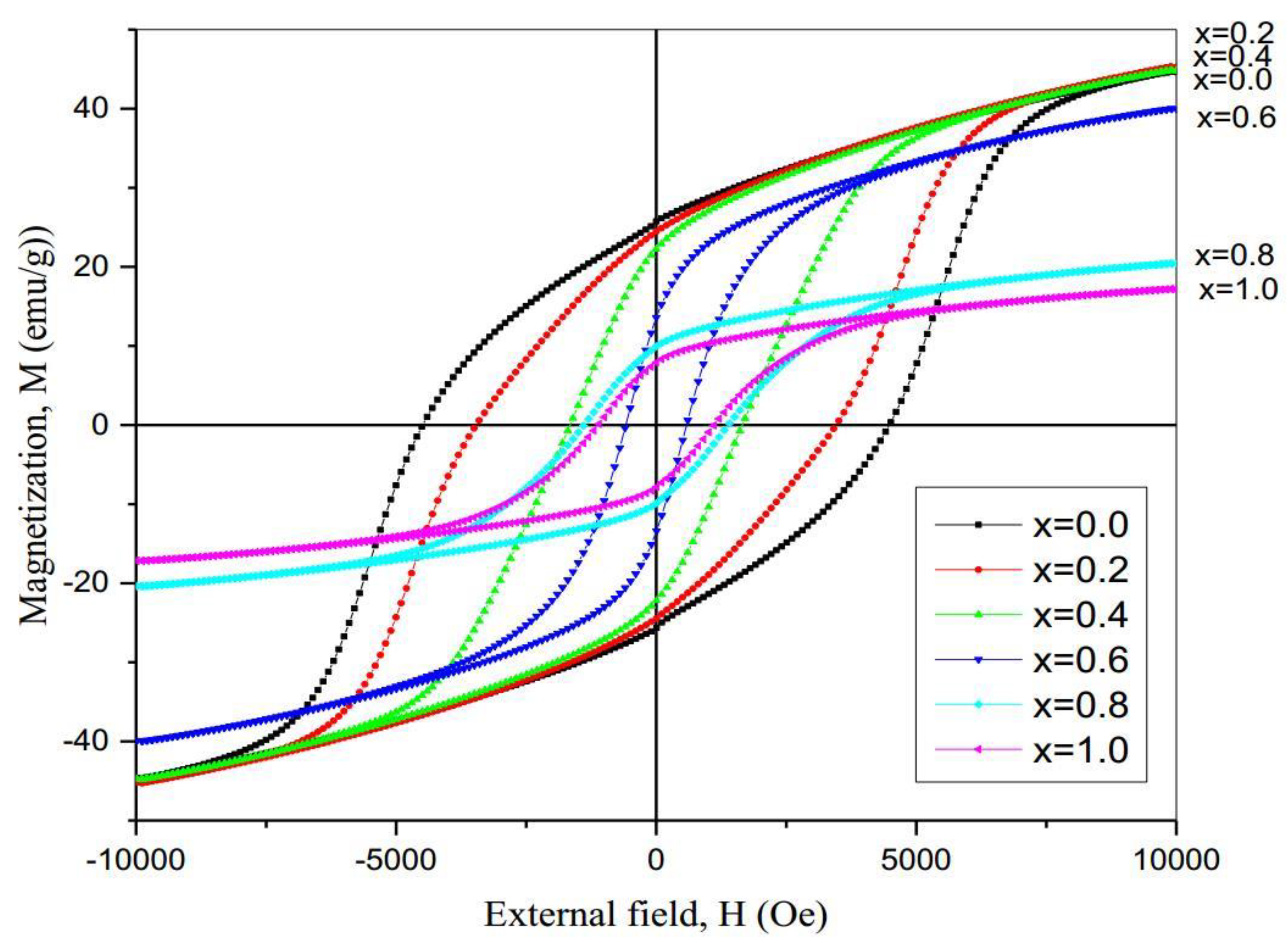
We studied only magnetic properties for samples sintered at 1000 °C. Magnetic measurements were carried out at room temperature by a VSM with applied field between 0 and 10 kOe. The group of hysteresis loops (HL) for all samples is shown in Figure 6. The loops do indicate that all the samples are indeed hard magnetic materials. The values of remanence magnetization and coercive field for each sample were read directly from graphs of loops. Since it is expected that samples have high magnetic anisotropy, we used the law of approach to saturation (LAS) to calculate the saturation magnetization. This is simply found by plotting M versus 1/H2 for values taken from high field region and all samples gave perfect straight linear graphs. The intercept of that line is equal to the saturation magnetization.
Table 3 shows summary of the calculated values for the saturation magnetization, the remanence magnetization, the reduced magnetization, and the coercivity.
The saturation magnetization shows inverse relation titanium concentration. The partial substitution of Fe by titanium ions in these hexaferrites has affected the values of MS, which depends on the amount of Ti ions diffusing into the BaM crystal in which Ti+3 ions partially replace Fe+3. However, the higher the Ti concentration results in lower Ms because other phases are then formed as was concluded from XRD data.
Hard ferrites typically have a high value of Mr, which is about 50% of the saturation magnetization (Ms), therefore the reduced remnant magnetization (Mrs = Mr/Ms) must be nearly 0.5, and this is true for samples consisting of single-domains and random orientations of grains [32]. Table 3 shows that the values of Mrs are very close to 50%. The coercive field, Hc, for the samples varied with Ti concentration and it ranged between 0.6 and 4.5 kOe as can be seen in Table 3. A plot of Ms and Hc versus the concentration of titanium is shown in Figure 7.
We calculated the average paramagnetic behavior for each sample by approximating the loop by a straight line as shown in Figure 8. Of course, this behavior is correct only for shallow applied fields, but we found that there is a general trend in these samples that can be depicted by the slopes of these lines. Slopes of these lines are listed in Table 4. The slopes are related to the magnetic susceptibility and increased slowly by increasing the concentration up to x = 0.6 and then decreases to half its value after that.
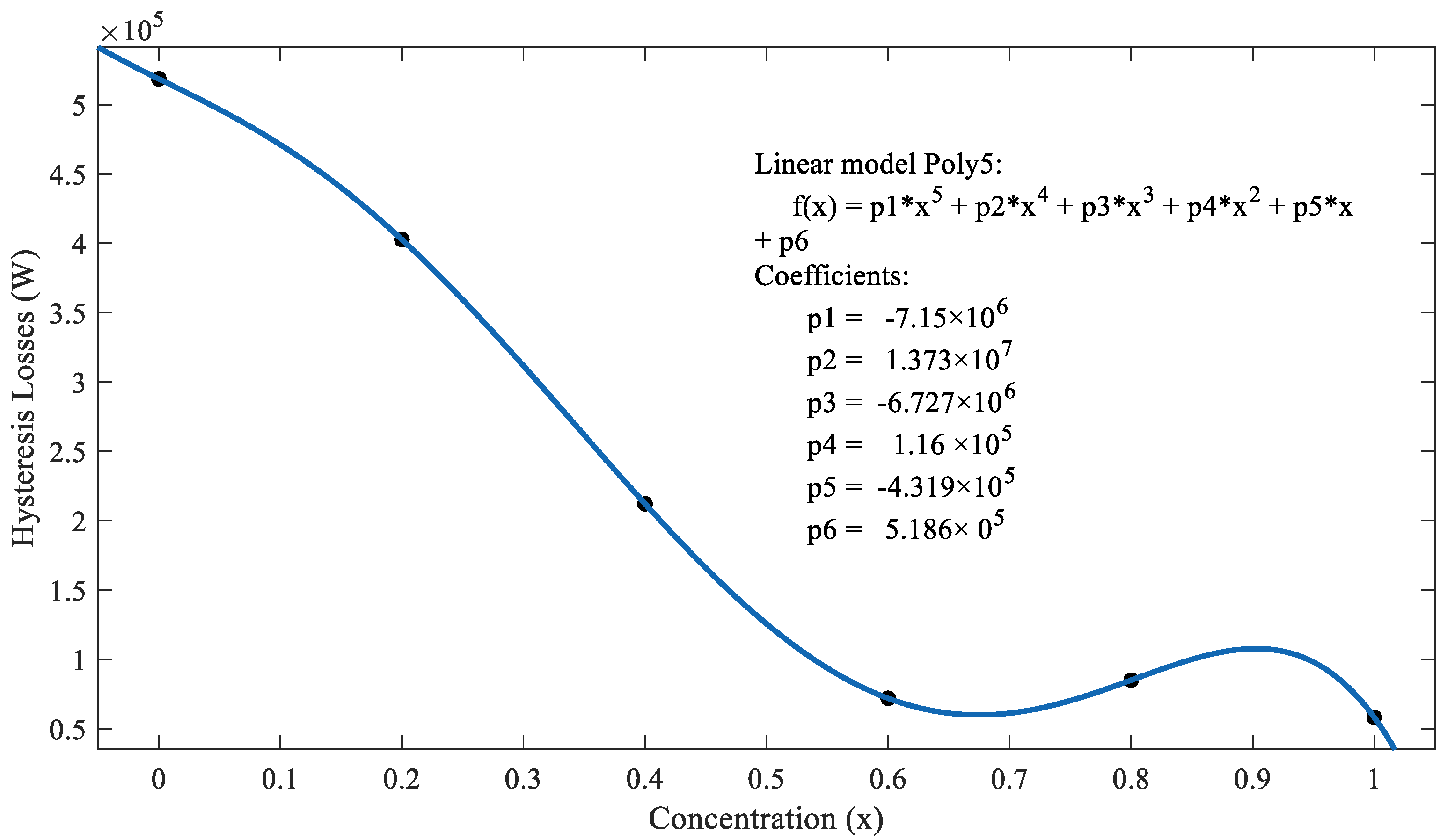
We also calculated hysteresis losses for each sample and the calculated values are listed in Table 4. It is clear from the values of losses that Ti concentration could be a scaling factor for magnetic energy stored when these materials are used in components of electromagnetic devices. This behavior is displaced in Figure 9. Losses are significant and almost linear for x ˂ 0.6.
4. Conclusions
A group of BaFe12-xTixO19 samples with (x = 0.0–1.0) were prepared by sol-gel-auto combustion method and were sintered at 850 °C and 1000 °C for 1 h in air. The XRD data and IR data analysis confirmed the presence of M type phase of the hexaferrite in these samples, and the higher sintering temperature showed an improvement of crystallinity. The average crystallite size for all samples was in the range of 27–42 nm. We believe that these are suitable to obtain the reasonable signal to noise ratio when used in a high-density recording medium. The values of saturation magnetization (Ms) and retentivity (Mr) calculated values decreased with increasing the Ti concentration, and this may result from particle size effects. The addition of titanium reduced the saturation magnetization from about 45 emu/g down to 17.2 emu/g. The rest of the magnetic properties that include coercivity, remanence magnetization, reduced remnant magnetization, and hysteresis losses suggest that these materials are beneficial in the components of electromagnetic devices that are used in recording technology and permanent magnets. We suggest that further work is needed to study the effect of sintering temperatures and very low controlled Ti concentrations. In addition, structural and magnetic studies at temperatures above and below room temperature using other techniques could be of great importance.
Author Contributions
Formal analysis, L.A.A.-H.; Methodology, H.A.K.; Supervision, A.R.A.D. All authors contribute equally to this manuscript.
Funding
This research received no external funding.
Acknowledgments
This research work was supported by the Deanship of Graduate Studies and Research, Yarmouk University, Irbid, Jordan; project 7/2017. We acknowledge the help of Prof. A. Hammoudeh in preparation of samples and IR data.
Conflicts of Interest
The authors declare no conflict of interest.
References
- Gutfleisch, O.; Willard, M.A.; Bruck, E.; Chen, C.H.; Sankar, S.G.; Liu, J.P. Magnetic materials and devices for the 21st century: Stronger, lighter, and more energy efficient. Adv. Mater. 2011, 23, 821–842. [Google Scholar] [CrossRef]
- Cullity, B.D.; Graham, C.D. Introduction to Magnetic Materials; John Wiley & Sons: New York, NY, USA, 2011. [Google Scholar]
- Bini, R.A.; Marques, R.F.C.; Santos, F.J.; Chaker, J.A.; Jafelicci, M., Jr. Synthesis and functionalization of magnetite nanoparticles with different amino-functional alkoxysilanes. J. Magn. Magn. Mater. 2012, 324, 534–539. [Google Scholar] [CrossRef] [Green Version]
- Cochardt, A. Recent ferrite magnet developments. J. Appl. Phys. 1966, 37, 1112–1115. [Google Scholar] [CrossRef]
- Sugimoto, M. The past, present, and future of ferrites. J. Am. Ceram. Soc. 1999, 82, 269–280. [Google Scholar] [CrossRef]
- Pullar, R.C. Hexagonal ferrites: A review of the synthesis, properties and applications of hexaferrite ceramics. Prog. Mater. Sci. 2012, 57, 1191–1334. [Google Scholar] [CrossRef]
- Koutzarova, T.; Kolev, S.; Ghelev, C.; Grigorov, K.; Nedkov, I. Structural and magnetic properties and preparation techniques of nanosized M-type hexaferrite powders. In Advances in Nanoscale Magnetism; Springer: Berlin, Germany, 2009; pp. 183–203. [Google Scholar]
- Maswadeh, Y.; Mahmood, S.H.; Awadallah, A.; Aloqaily, A.N. Synthesis and structural characterization of nonstoichiometric barium hexaferrite materials with Fe:Ba ratio of 11.5–16.16. In IOP Conference Series: Materials Science and Engineering; IOP Publishing Ltd.: Philadelphia, PA, USA, 2015. [Google Scholar]
- Radwan, M.; Rashad, M.M.; Hessien, M.M. Synthesis and characterization of barium hexaferrite nanoparticles. J. Mater. Process. Technol. 2007, 181, 106–109. [Google Scholar] [CrossRef]
- Martirosyan, K.S.; Galstyan, E.; Hossain, S.M.; Wang, Y.-J.; Litvinov, D. Barium hexaferrite nanoparticles: synthesis and magnetic properties. Mater. Sci. Eng. B 2011, 176, 8–13. [Google Scholar] [CrossRef]
- Meng, Y.Y.; He, M.H.; Zeng, Q.; Jiao, D.L.; Shukla, S.; Ramanujan, R.V.; Liu, Z.W. Synthesis of barium ferrite ultrafine powders by a sol–gel combustion method using glycine gels. J. Alloy. Compd. 2014, 583, 220–225. [Google Scholar] [CrossRef]
- Kouřil, K. Local structure of hexagonal ferrites studied by NMR. (2013). Available online: https://www.researchgate.net (accessed on 20 March 2019).
- Wu, M. M-Type barium hexagonal ferrite films. In Advanced Magnetic MaterialsAdv; InTech Open: London UK, 2012. [Google Scholar]
- Awadallah, A.M.; Sami, M. Effects of preparation conditions and metal ion substitutions for barium and iron on the properties of M-type barium hexaferrites. Ph.D. Research Proposal, The University of Jordan, Amman, Jordan, 2012. [Google Scholar] [CrossRef]
- Zhang, H.; Zeng, D.; Liu, Z. The law of approach to saturation in ferromagnets originating from the magnetocrystalline anisotropy. J. Magn. Magn. Mater. 2010, 322, 2375–2380. [Google Scholar] [CrossRef]
- Bsoul, I.; Mahmood, S.H. Magnetic and structural properties of BaFe12−xGaxO19 nanoparticles. J. Alloy. Compd. 2010, 489, 110–114. [Google Scholar] [CrossRef]
- Awawdeh, M.; Bsoul, I.; Mahmood, S.H. Magnetic properties and Mössbauer spectroscopy on Ga, Al, and Cr substituted hexaferrites. J. Alloy. Compd. 2014, 585, 465–473. [Google Scholar] [CrossRef]
- Packiaraj, G.; Panchal, N.R.; Jotania, R.B. Structural and Dielectric studies of Cu substituted Barium hexaferrite prepared by Sol-gel auto combustion technique. Solid State Phenom. 2014, 209, 102–106. [Google Scholar] [CrossRef]
- Mahmood, S.H.; Aloqaily, A.N.; Maswadeh, Y.; Awadallah, A.; Bsoul, I.; Juwhari, H. Structural and magnetic properties of Mo-Zn substituted (BaFe12-4xMoxZn3xO19) M-type hexaferrites. Mater. Sci. Res. India 2014, 11, 9–20. [Google Scholar] [CrossRef]
- Haneda, K.; Hiroshi, K. Intrinsic coercivity of substituted BaFe12O19. Japan. J. Appl. Phys. 1973, 12, 355. [Google Scholar] [CrossRef]
- Dushaq, G.H.; Mahmood, S.H.; Bsoul, I.; Juwhari, H.K.; Lahlouh, B.; AlDamen, M.A. Effects of molybdenum concentration and valence state on the structural and magnetic properties of BaFe11.6MoxZn0.4-xO19 hexaferrites. Acta Metall. Sin. (English Letters) 2013, 26, 509–516. [Google Scholar] [CrossRef]
- Ali, I.; Islam, M.U.; Awan, M.S.; Ahmad, M. Effects of Ga–Cr substitution on structural and magnetic properties of hexaferrite (BaFe12O19) synthesized by sol–gel auto-combustion route. J. Alloy. Compd. 2013, 547, 118–125. [Google Scholar] [CrossRef]
- Wang, S.; Ding, J.; Shi, Y.; Chen, Y.J. High coercivity in mechanically alloyed BaFe10Al2O19. J. Magn. Magn. Mater. 2000, 219, 206–212. [Google Scholar] [CrossRef]
- Ali, I.; Islam, M.U.; Awan, M.S.; Ahmad, M.; Ashiq, M.N.; Naseem, S. Effect of Tb3+ substitution on the structural and magnetic properties of M-type hexaferrites synthesized by sol–gel auto-combustion technique. J. Alloy. Compd. 2013, 550, 564–572. [Google Scholar] [CrossRef]
- Sharma, R.; Bisen, D.P.; Shukla, U.; Sharma, B.G. X-ray diffraction: a powerful method of characterizing nanomaterials. Recent Res. Sci. Technol. 2012, 4, 77–79. [Google Scholar]
- Uvarov, V.; Popov, I. Metrological characterization of X-ray diffraction methods at different acquisition geometries for determination of crystallite size in nano-scale materials. Mater. Charact. 2013, 85, 111–123. [Google Scholar] [CrossRef]
- Stuart, B.H. Experimental methods. In Infrared Spectroscopy: Fundamentals And Applications; John Wiley & Sons, Ltd.: Hoboken, NJ, USA, 2005; pp. 15–44. [Google Scholar]
- Coates, J. Interpretation of infrared spectra, a practical approach. In Encyclopedia of Analytical Chemistry; John Wiley & Sons Ltd.: Chichester, UK, 2000; pp. 10815–10837. [Google Scholar]
- Goldstein, J.; Yakowitz, H. Practical Scanning Electron Microscopy: Electron and Ion Microprobe Analysis; Springer: Boston, MA, USA, 1975. [Google Scholar]
- Aravind, A. Synthesis and characterization of 3d-transition metals doped ZnO thin films and nanostructures for possible spintronic applications. Ph.D. Thesis, Cochin University of Science and Technology, Kerala, India, 2012. [Google Scholar]
- Monshi, A.; Foroughi, M.R.; Monshi, M.R. Modified Scherrer equation to estimate more accurately nano-crystallite size using XRD. World J. Nano Sci. Eng. 2012, 2, 154–160. [Google Scholar] [CrossRef]
- Mahmood, S.H.; Dushaq, G.H.; Bsoul, I.; Awawdeh, M.A.; Juwhari, H.K.; Lahlouh, B.; AlDamen, M.A. Magnetic properties and hyperfine interactions in M-type BaFe12-2xMoxZnxO19 hexaferrites. J. Appl. Math. Phys. 2014, 2, 77–87. [Google Scholar] [CrossRef]
- Li, Y.; Wang, Q.; Yang, H. Synthesis, characterization, and magnetic properties of nanocrystalline BaFe12O19 Ferrite. Curr. Appl. Phys. 2009, 9, 1375–1380. [Google Scholar] [CrossRef]
- Trukhanov, S.V.; Trukhanov, A.V.; Kostishyn, V.G.; Panina, L.V.; Trukhanov, A.V.; Turchenko, V.A.; Tishkevich, D.I.; Trukhanova, E.L.; Yakovenko, O.S.; Matzui, L.Y.; et al. Effect of gallium doping on electromagnetic properties of barium hexaferrite. J. Phys. Chem. Solid. 2017, 111, 142–152. [Google Scholar] [CrossRef]
- Trukhanov, S.V.; Trukhanov, A.V.; Kostishyn, V.G.; Panina, L.V.; Trukhanov, An.V.; Turchenko, V.A.; Tishkevich, D.I.; Trukhanova, E.L.; Yakovenko, O.S.; Matzui, L.Yu. Investigation into the structural features and microwave absorption of doped barium hexaferrites. Dalton Trans. 2017, 46, 9010–9021. [Google Scholar] [CrossRef] [Green Version]
- Trukhanov, S.V.; Trukhanov, A.V.; Salem, M.M.; Trukhanova, E.L.; Panina, L.V.; Kostishyn, V.G.; Darwish, M.A.; Trukhanov, A.V.; Zubar, T.I.; Tishkevich, D.I.; et al. Preparation and investigation of structure, magnetic and dielectric properties of (BaFe11.9Al0.1O19)1-x-(BaTiO3)x bicomponent ceramics. Ceram. Int. 2018, 44, 21295–21302. [Google Scholar] [CrossRef]
- Trukhanov, S.V.; Trukhanov, A.V.; Turchenko, V.A.; Trukhanov, An.V.; Tishkevich, D.I.; Trukhanova, E.L.; Zubar, T.I.; Karpinsky, D.V.; Kostishyn, V.G.; Panina, L.V.; et al. Magnetic and dipole moments in indium doped barium hexaferrites. J. Magn. Magn. Mater. 2018, 457, 83–96. [Google Scholar] [CrossRef]
- Trukhanov, S.V.; Trukhanov, A.V.; Turchenko, V.A.; Kostishyn, V.G.; Panina, L.V.; Kazakevich, I.S.; Balagurov, A.M. Structure and magnetic properties of BaFe11.9In0.1O19 hexaferrite in a wide temperature range. J. Alloy. Compd. 2016, 689, 383–393. [Google Scholar] [CrossRef]
- Turchenko, V.A.; Trukhanov, S.V.; Balagurov, A.M.; Kostishyn, V.G.; Trukhanov, A.V.; Panina, L.V.; Trukhanova, E.L. Features of crystal structure and dual ferroic properties of BaFe12-xMexO19 (Me = In3+ and Ga3+; x = 0.1–1.2). J. Magn. Magn. Mater. 2018, 464, 139–147. [Google Scholar] [CrossRef]
Figure 1.
XRD patterns for BaFe12-xTixO19 samples with (x = 0.0–1.0) sintered at 850 °C for 1 h and shown the standard pattern of BaFe12O19 (file no.: 043-0002).
Figure 1.
XRD patterns for BaFe12-xTixO19 samples with (x = 0.0–1.0) sintered at 850 °C for 1 h and shown the standard pattern of BaFe12O19 (file no.: 043-0002).

Figure 2.
XRD patterns BaFe12-xTixO19 samples with (x = 0.0–1.0) sintered at 1000 °C for 1 h and shown the standard XRD pattern of BaFe12O19 (file no.: 043-0002).
Figure 2.
XRD patterns BaFe12-xTixO19 samples with (x = 0.0–1.0) sintered at 1000 °C for 1 h and shown the standard XRD pattern of BaFe12O19 (file no.: 043-0002).

Figure 3.
Infra-Red (IR) spectra of BaFe12-xTixO19 sintered at T = 850 °C.

Figure 4.
IR spectra of BaFe12-xTixO19 sintered at T = 1000 °C.
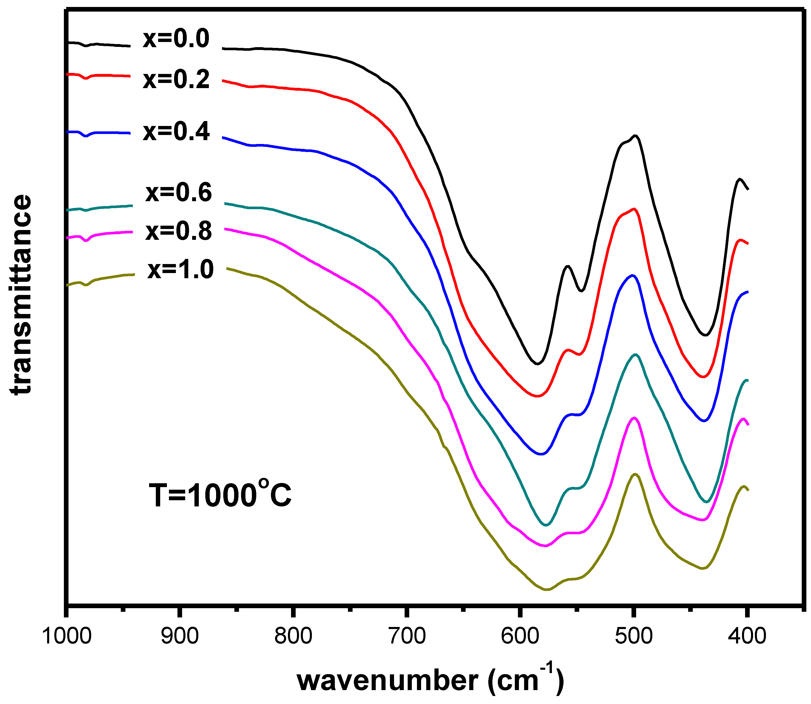
Figure 5.
SEM image for BaFe12-xTixO19 (x = 0.2) sample sintered at T = 1000 °C.
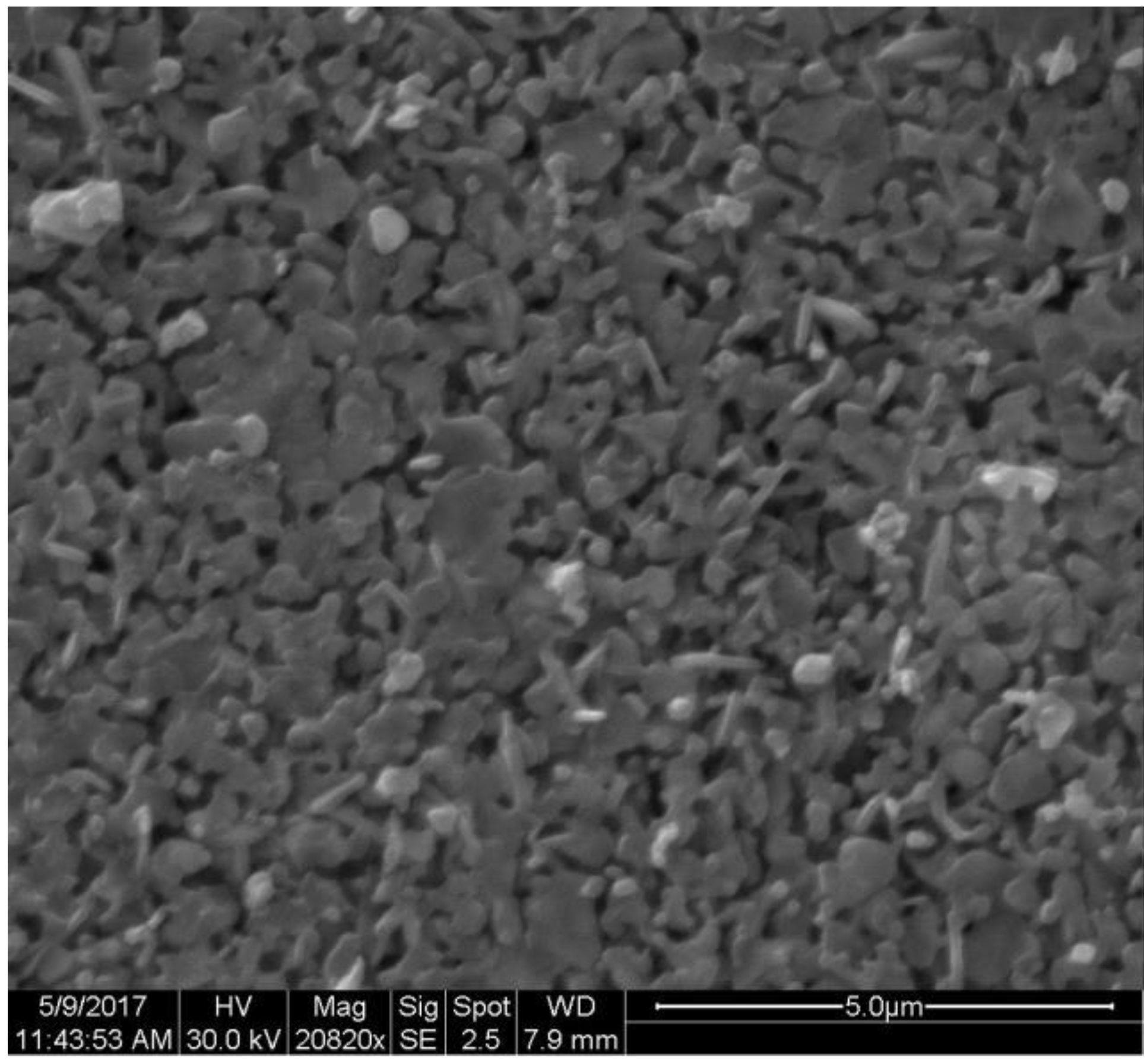
Figure 6.
Hysteresis loops for the samples BaFe12-xTixO19 (x = 0.0 − 1.0) sintered at 1000 °C.

Figure 7.
Saturation magnetization and coercive field versus Ti concentration.

Figure 8.
The hysteresis loop for the system BaFe12-xTixO19 (x = 0.2) approximated by a straight line.
Figure 8.
The hysteresis loop for the system BaFe12-xTixO19 (x = 0.2) approximated by a straight line.

Figure 9.
Hysteresis losses (in W) BaFe12-xTixO19 (x = 0.0–1.0) sintered at 1000 °C.

Table 1.
The calculated average crystallite sizes for the system BaFe12-xTixO19 with (x = 0.0–1.0) sintered at T = 850 °C.
Table 1.
The calculated average crystallite sizes for the system BaFe12-xTixO19 with (x = 0.0–1.0) sintered at T = 850 °C.
| Sample | (2θ)-Position (Degrees) | Size (nm) | Average Size (nm) |
|---|---|---|---|
(x = 0.0) | 32.19 | 24.8 | 27.2 |
| 34.14 | 29.3 | ||
| 37.1 | 29.6 | ||
| 40.33 | 27 | ||
| 55.1 | 25.3 | ||
(x = 0.2) | 33.1 | 27.3 | 30.3 |
| 35.67 | 33 | ||
| 49.51 | 25.4 | ||
| 54.1 | 35.3 | ||
(x = 0.4) | 33.3 | 34.1 | 34 |
| 35.8 | 35.9 | ||
| 49.65 | 33.3 | ||
| 54.26 | 32.7 | ||
(x = 0.6) | 33.31 | 39 | 36.5 |
| 35.79 | 41 | ||
| 49.69 | 34.6 | ||
| 54.24 | 31.5 | ||
(x= 0.8) | 33.39 | 34.2 | 34 |
| 35.86 | 35.9 | ||
| 49.7 | 33.3 | ||
| 54.3 | 32.7 | ||
(x = 1.0) | 33.31 | 39 | 36.3 |
| 35.69 | 41.3 | ||
| 49.54 | 34.6 | ||
| 54.15 | 30.4 |
Table 2.
The calculated average crystallite sizes for the system BaFe12-xTixO19 with (x = 0.0–1.0) sintered at T = 1000 °C.
Table 2.
The calculated average crystallite sizes for the system BaFe12-xTixO19 with (x = 0.0–1.0) sintered at T = 1000 °C.
| Sample | (2θ)-Position (Degrees) | Size (nm) | Average Size (nm) |
|---|---|---|---|
(x = 0.0) | 32.4 | 30.3 | 34.9 |
| 34.34 | 41 | ||
| 40.55 | 36.4 | ||
| 55.31 | 31.7 | ||
(x = 0.2) | 32.29 | 38.9 | 38.7 |
| 34.23 | 41.1 | ||
| 40.44 | 41.9 | ||
| 55.1 | 32.8 | ||
(x = 0.4) | 32.26 | 43.1 | 41.5 |
| 34.22 | 45.7 | ||
| 40.42 | 46.5 | ||
| 55.19 | 30.6 | ||
(x = 0.6) | 32.29 | 43 | 41 |
| 34.27 | 45.7 | ||
| 40.48 | 39.9 | ||
| 54.23 | 35.3 | ||
(x = 0.8) | 33.27 | 45.6 | 40.5 |
| 34.24 | 43.3 | ||
| 40.96 | 36.5 | ||
| 54.19 | 36.7 | ||
(x = 1.0) | 33.23 | 43.1 | 38 |
| 35.7 | 41.3 | ||
| 40.93 | 32.2 | ||
| 54.19 | 35.3 |
Table 3.
The saturation magnetization, remanence magnetization, reduced remnant magnetization and coercive field for the system BaFe12-xTixO19 (x = 0.0–1.0).
Table 3.
The saturation magnetization, remanence magnetization, reduced remnant magnetization and coercive field for the system BaFe12-xTixO19 (x = 0.0–1.0).
| x | Ms (emu/g) | Mr (emu/g) | Mrs (emu/g) | Hc (kOe) |
|---|---|---|---|---|
| 0.0 | 44.65 | 23.0606 | 0.5165 | 4.51 |
| 0.2 | 45.24 | 19.7474 | 0.4365 | 3.4557 |
| 0.4 | 44.83 | 19.5835 | 0.4368 | 1.6554 |
| 0.6 | 39.99 | 12.9342 | 0.3234 | 0.583 |
| 0.8 | 20.39 | 9.5829 | 0.47 | 1.385 |
| 1.0 | 17.17 | 7.7163 | 0.4494 | 1.11455 |
Table 4.
The slope and hysteresis losses (in W) for the system BaFe12-xTixO19 (x = 0.0–1.0).
| Concentration (x) | Slope | Hysteresis Losses (W) |
|---|---|---|
| 0.0 | 0.004908 | 518,580 |
| 0.2 | 0.005326 | 402,700 |
| 0.4 | 0.00571 | 212,180 |
| 0.6 | 0.005252 | 71,914 |
| 0.8 | 0.002623 | 84,982 |
| 1.0 | 0.002229 | 58,080 |
© 2019 by the authors. Licensee MDPI, Basel, Switzerland. This article is an open access article distributed under the terms and conditions of the Creative Commons Attribution (CC BY) license (http://creativecommons.org/licenses/by/4.0/).
Share and Cite
MDPI and ACS Style
Dairy, A.R.A.; Al-Hmoud, L.A.; Khatatbeh, H.A. Magnetic and Structural Properties of Barium Hexaferrite Nanoparticles Doped with Titanium. Symmetry 2019, 11, 732. https://doi.org/10.3390/sym11060732
AMA Style
Dairy ARA, Al-Hmoud LA, Khatatbeh HA. Magnetic and Structural Properties of Barium Hexaferrite Nanoparticles Doped with Titanium. Symmetry. 2019; 11(6):732. https://doi.org/10.3390/sym11060732
Chicago/Turabian StyleDairy, Abdul Raouf Al, Lina A. Al-Hmoud, and Heba A. Khatatbeh. 2019. "Magnetic and Structural Properties of Barium Hexaferrite Nanoparticles Doped with Titanium" Symmetry 11, no. 6: 732. https://doi.org/10.3390/sym11060732
Note that from the first issue of 2016, this journal uses article numbers instead of page numbers. See further details here.