Review

Symmetry Detection in Visual Impairment: Behavioral Evidence and Neural Correlates

1
Department of Psychology, University of Milano-Bicocca, 20126 Milano, Italy
2
Brain Connectivity Center, National Neurological Institute C. Mondino, 27100 Pavia, Italy
3
Brain Research Unit, OV Lounasmaa Laboratory, Aalto University School of Science, 02150 Espoo, Finland
4
Laboratory for Visual Neuroplasticity, Department of Ophthalmology, Massachusetts Eye and Ear Infirmary Harvard Medical School, Boston, MA 02115, USA
5
Department of Psychology, Faculty of Science and Technology, University of Westminster, London W1W 6UW, UK
6
Department of Psychology, Rochester Institute of Technology, Rochester, NY 14623, USA
7
Department of Brain and Behavioral Sciences, University of Pavia, 27100 Pavia, Italy
*
Author to whom correspondence should be addressed.
Symmetry 2014, 6(2), 427-443; https://doi.org/10.3390/sym6020427
Submission received: 19 February 2014 / Revised: 13 May 2014 / Accepted: 14 May 2014 / Published: 26 May 2014
(This article belongs to the Special Issue Visual Symmetry)

Abstract

:
Bilateral symmetry is an extremely salient feature for the human visual system. An interesting issue is whether the perceptual salience of symmetry is rooted in normal visual development. In this review, we discuss empirical work on visual and tactile symmetry detection in normally sighted and visually impaired individuals. On the one hand, available evidence suggests that efficient visual symmetry detection may need normal binocular vision development. On the other hand, converging evidence suggests that symmetry can develop as a principle of haptic perceptual organization in individuals lacking visual experience. Certain features of visual symmetry detection, however, such as the higher salience of the patterns containing a vertical axis of symmetry, do not systematically apply to the haptic modality. The neural correlates (revealed with neuroimaging) associated with visual and haptic symmetry detection are also discussed.

Graphical Abstract

1. Introduction

Bilateral symmetry is a prominent feature of the visual world. Not only are human faces and bodies symmetric, but most other living organisms (such as animals, trees, flowers and crystals) have at least one axis of symmetry, as do manufactured items, such as tools and buildings (for a recent review, see [1]). Psychophysical experiments mainly employing simple shapes or dense dot patterns as stimuli have shown that symmetry can be detected extremely quickly (within a few tens of milliseconds) and in an automatic (i.e., not mediated by a conscious cognitive effort; see [1] for review), pre-attentive manner [24]. Symmetry acts as a grouping principle of perceptual organization [5]: elements sharing symmetry relations tend to be aggregated, thus facilitating figure-ground segregation. It has also been suggested that symmetry detection may be a special case of orientation processing [6,7] in that once the axis of symmetry is determined, both this information and orientation information acquired from other contour classes converge at a common neural site where orientation information is processed irrespective of source. Further, Dakin and others have suggested that symmetry detection shares features with mechanisms responsible for processing orientation [810]. Symmetry is also thought to be important from an evolutionary perspective. Indeed, symmetry has been shown to play a role in mate selection and reproduction, likely acting as a marker of phenotypic and genetic quality (for a review, see [11]). Furthermore, patterns containing a vertical axis of symmetry seem to be most salient (i.e., easier to detect or perceptually “stronger”), followed by the horizontal and other oblique orientations [12]. The higher perceptual salience of vertical symmetry may depend on the fact that most objects in the world contain symmetry along the vertical axis (e.g., human faces and bodies, buildings, trees). The organization of the human visual system is likely optimized to process the structure of the visual world; thus, the predominance of vertical symmetry over other orientations in the external world may have made the vertical axis of symmetry perceptually stronger. Indeed, it has also been proposed that the higher salience of the vertical axis of symmetry may depend on the bilaterally symmetric organization of the visual system itself, as suggested by some psychophysical results in patients born without the corpus callosum [13].
In light of consistent evidence demonstrating the salience of symmetry in visual perception, a natural question that arises is whether symmetry salience depends on normal visual development for its emergence. Further, does symmetry act as a grouping principle of perceptual organization only in the visual domain? It may also be important in tactile shapes and object detection given that the sense of touch is also able to convey precise information regarding spatial features of external objects [14]. In this review, we address these issues by discussing available evidence regarding symmetry detection in sighted, visually impaired and blind individuals. The neural correlates of tactile symmetry detection in blind and sighted individuals is also reviewed in light of available knowledge on the neural underpinnings of visual symmetry detection. An in-depth discussion of models of visual symmetry detection is beyond the scope of this review, and it has already been covered in previous work (e.g., [1,15]). The experimental paradigms used across various studies will be described in general terms to ensure that results are understandable (the reader can find methodological details in the original empirical works).
Visual impairment and blindness can take many forms and can result from damage and complications localized to ocular, as well as cerebral structures. In this review, we first consider visual symmetry detection capacities in individuals affected by abnormal binocular visual development. This ranges from amblyopia (literally “blunt vision”), which is the reduction or partial loss of vision in one or both eyes in the absence of an ocular structural abnormality or pathology [16], to the extreme of monocular blindness. These conditions provide models to assess whether normal binocular vision is necessary for the salience of symmetry to emerge. It is important to stress that ocular forms of visual impairment directly affect brain structure and function via phenomena related to plasticity. Indeed, plastic changes occur in the brain as a natural consequence of learning and memory, development and ageing, but they can also be triggered by other factors, such as brain injury or, of interest here, congenital or acquired sensory deprivation [17,18]. We then consider the case of individuals affected by complete blindness due to peripheral (i.e., ocular) causes, comparing incidence at birth (or shortly thereafter) to blindness acquired later in life. In this latter group, it is presumed that the normal development of visual pathways has occurred prior to the loss of sight.

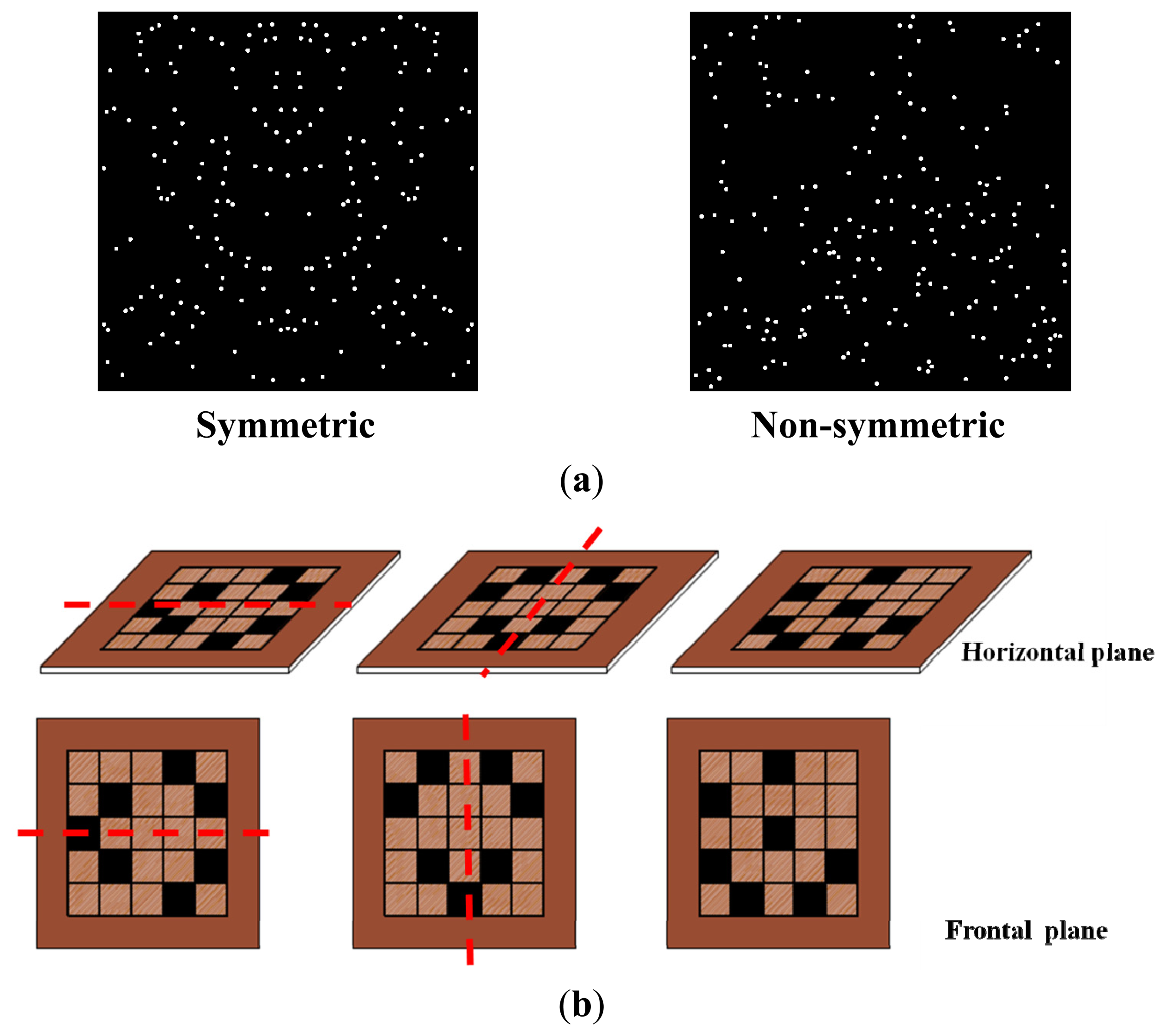
2. Visual Symmetry Detection in Individuals with Impaired Binocular Vision

An interesting question in studying visual symmetry detection is whether its salience depends on normal visual functioning and development, particularly with respect to binocular vision. Barlow and Reeves [2] reported equally efficient symmetry detection of dot patterns when viewed monocularly and binocularly by normally sighted participants. In a later study, Wenderoth [19] used visual stereoscopic patterns and demonstrated that monocular symmetry is not sufficient for (dichoptic) symmetry perception and that dichoptic symmetry can be perceived even if two monocular patterns were not symmetric per se. According to Wenderoth [19], these findings suggest that symmetry perception occurs at, or beyond, the site of the binocular combination of monocular signals, compatible with the idea that symmetry detection mainly occurs within higher-order (i.e., extrastriate) visual areas. However, testing normally sighted individuals under monocular viewing conditions does not allow one to test whether symmetry salience depends on normal binocular vision, since these individuals have normal binocularity. On the other hand, investigating conditions in which there is a deficit or lack of binocular vision (such as amblyopia or monocular blindness, respectively), would provide the opportunity to study whether symmetry detection depends on normal binocular development.
Amblyopia is the development of abnormal binocular vision due to strabismus, high anisometropia or form deprivation (such as a cataract) occurring during early life. In amblyopia, under normal binocular viewing conditions, one eye is favored while the information conveyed by the non-preferred eye is generally suppressed, at least within the central visual field [20]. Levi and Saarinen [21] have compared symmetry detection in amblyopes and normal observers under monocular viewing (i.e., with the non-tested eye covered by a patch). Both groups were presented with either symmetric or asymmetric visual stimuli consisting of Gabor patches. In the symmetric stimuli, the amount of symmetry could vary from 25% to 100% (specifically, four different symmetry conditions occurred: 25%, 50% or 75% or 100% of symmetry). The participants' task was to rate the correct amount of symmetry or identify the pattern as non-symmetric. When the amblyopic eye was tested, amblyopic observers showed a progressively higher symmetry threshold with increasing stimulus spatial frequency compared to normal sighted controls, whose ability in detecting symmetry was spatial frequency independent (consistent with [8], showing scale invariance in symmetry detection). Notably, symmetry thresholds did not vary with the manipulation of contrast features, suggesting that the impairment was not simply due to a general reduction in stimulus visibility. The amblyopic deficit in symmetry detection may indeed depend on the abnormal integration of local orientation information in amblyopic observers, which seems to be critical to form symmetry detection units and, therefore, process symmetric features [10]. Accordingly, amblyopic individuals also show abnormalities in other tasks requiring the integration of orientation features, such as contours [22,23]. These results suggest that a deficit in symmetry detection occurring in amblyopia is related to suboptimal processing of local orientation information and cannot be explained by a general impairment in lower-level visual functions. Furthermore, information about local orientation in symmetric patterns is most likely conveyed by higher-level visual areas [24] and supports the view that symmetry detection is mediated by extrastriate visual regions [2529]. In fact, these findings are also in line with Herbert et al.'s [30] data on visual symmetry detection in the elderly. They found an overall impairment in symmetry detection for individuals over 60 compared to participants younger than 60, although the vertical symmetry advantage was observed in all age groups. The impairment in symmetry detection did not covary with visual acuity loss, suggesting that the deficit observed in the older participants was not at a low level of processing in the visual system. Rather, Herbert et al. [30] interpreted their findings as suggesting that the general decline in performance was symptomatic of some unspecified changes in intermediate and higher level visual functions with aging. These changes are unlikely to be specific for symmetry detection, extending also to other complex visual processes. Habak and Faubert [31] found that the perception of simple motion stimuli (moving gratings) was comparable in young and elderly groups, whereas complex motion (moving dot textures) perception was selectively impaired in the older group. Similarly to the case of normal ageing, the deficit in symmetry detection observed in the amblyopic participants may be a manifestation of a broader deficit related to suboptimal high-level visual processing. In line with this, recent evidence has shown that amblyopic individuals require more samples in both eyes to detect biological motion compared to normal control observers [32]. These threshold differences did not depend on low-level losses, but likely reflected losses in feature integration due to undersampling in the amblyopic visual system. Indeed, it has been suggested that the deficits often reported in amblyopia for global form and motion integration are due to a selective difficulty in noise segregation [33].
The case of congenital monocular blindness represents a particular form of visual impairment characterized by profound blindness in one eye (or total absence of one eye, in cases of enucleation) and intact visual abilities in the fellow eye. Whereas some visual functions (such as motion discrimination capacities) are reduced in monocularly blind participants, other visuo-spatial functions appear to be enhanced in the functioning eye (for a review, see [34]). We have recently investigated symmetry detection abilities in monocularly blind individuals using dense dot patterns (see Figure 1A) [35]. Patterns were horizontally symmetric, vertically symmetric or non-symmetric. We found a similar level of detection sensitivity in monocularly blind and normally sighted controls (viewing monocularly, with their dominant eye covered). Moreover, the vertical axis of symmetry was more salient than the horizontal in all participants. However monocularly blind individuals were significantly slower in detecting symmetric patterns (irrespective of symmetry axis) compared to control participants. Although the monocularly blind also tended to be slower in a visual search task, the difference with controls was not significant. Overall, our findings suggest that normal binocular vision is necessary for optimal performance in detecting symmetry, whereas the salience of the vertical axis does not seem to depend on binocularity.
Findings from visually impaired individuals (at least for amblyopia and monocular blindness) suggest that the salience of bilateral mirror symmetry depends to a certain degree on normal binocular mechanisms and development. Although symmetry can still be perceived by individuals lacking binocular input (or with a deficit in binocular vision), processing appears to be less efficient than observed for individuals possessing a normally developed binocular vision system.

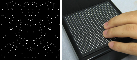
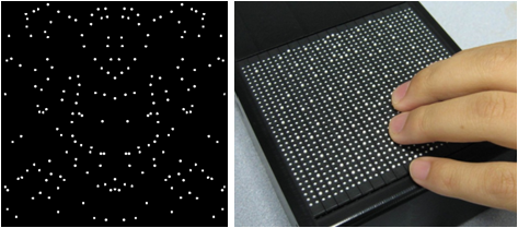
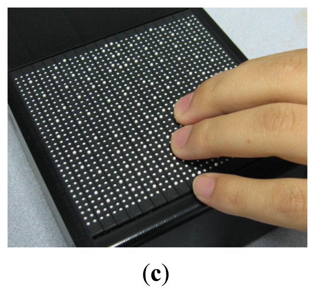
3. From Vision to Touch: Symmetry Detection in the Haptic Modality

3.1. Haptic Detection of Symmetry in Normally Sighted Participants

A number of studies have investigated symmetry processing in the haptic modality using two-dimensional (2D) line drawings or three-dimensional (3D) objects as stimuli [3944]. Overall, these studies show that although symmetry plays a role in the haptic recognition of 2D and 3D objects, symmetry is less salient for touch than it is for vision, and other stimulus properties, such as texture, may play a greater role for haptic shape discrimination compared to symmetry [43]. Indeed, the salience of symmetry in the haptic modality seems to be modulated by several different factors. This includes stimulus characteristics, such as material properties (e.g., texture and hardness), pattern complexity, size, dimensionality (2D/3D) and exploratory strategy [3943]. Regarding exploratory strategies, symmetry detection during haptic exploration is facilitated when two hands are used [3941]. This advantage is consistent with the reference-frame hypothesis [44], which suggests that bimanual exploration allows participants to relate the position of their hands to their own body midline providing for an effective frame of reference and also for coding the presence of symmetry in external objects (possibly via alignment of the axis of symmetry with the participants' body midline). However, the adoption of an efficient reference frame in processing symmetry also depends on the stimuli used (specifically, their size or whether stimuli consist in open shapes or closed shapes) [40,44]. Locher and Simmons [42] showed that when sighted (blindfolded) participants performed a haptic-symmetry discrimination task, the detection of symmetric shapes required more time than the detection of non-symmetrical ones. The strategy of contour following may be suboptimal in haptic symmetry detection. In a follow-up study, a simultaneous apprehension scanning strategy (i.e., smooth movements of both hands' thumb and index fingers over opposite sides of the shape) was found to provide superior symmetry detection [45]. Symmetry may also be more haptically salient for 3D objects than 2D patterns [39]. In considering these findings, it is important to note that most of the studies assessing symmetry detection in the haptic modality used raised line drawings or 3D objects. An exception to this was the study by Millar [43] that asked participants to decide whether two Braille-like configurations were identical or not. Both the symmetry of the configuration and dot numerosity were manipulated. Millar [43] found that dot numerosity affected judgments (the task was easier with fewer dots), whereas the presence of symmetry was irrelevant for the task. These findings suggest that dot density may be a more powerful cue than symmetry in haptic shape discrimination, at least for stimuli consisting of Braille-like small patterns, consistent with the idea that a perception of a global representation of shapes may be impaired when reference cues are missing. This finding is particularly of interest given accounts of visual symmetry detection arguing that grouping based on other basic visual properties (such as collinearity or proximity) may, in fact, precede and facilitate visual symmetry detection, at least in dense dot patterns [46,47]. In particular, Labonte et al. [47] suggested that symmetry detection (at least in dense dot patterns in which symmetric dots are embedded in noise) occurs in three stages: first, clusters are formed among local elements with sufficient mutual affinity (i.e., grouping by similarity or some other feature); second, pairs of symmetrical clusters are discovered and their axes of symmetry determined (symmetry detection); third, an attempt is made to detect more global symmetries by comparing the various axes of symmetry previously found (the so-called “symmetry-subsumption” stage). Given that touch affords sequential exploration of a stimulus rather than the parallel processing possible in vision, symmetry detection may follow other grouping mechanisms in touch. Differences in timing, the relative area that can be apprehended at one time and separation of the cortical pathways between these two sensory modalities likely contribute to symmetry being less salient in touch than in vision.
When investigating symmetry detection in the haptic modality in sighted individuals, it is important to consider that the haptic/tactile salience of symmetry may also indirectly depend on visual salience. In other words, the generation of a corresponding visual mental image to the tactile percept may play an important mediatory role [48]. We speculate that tactile exploration conveys a mental image corresponding to the percept analogous to exploring the same pattern with artificially-induced tunnel vision [49,50], something that would be worth investigating in future research. Nonetheless, while relevant in the case of sighted individuals, this potential confound (i.e., visual imagery mediating haptic symmetry detection) cannot play a role in the case of symmetry detection in congenitally blind individuals.

3.2. Haptic Detection of Symmetry in Blind Participants

Investigating individuals blind from birth or very early in life offers an opportunity to assess the dependence of tactile/haptic symmetry detection on prior visual experience. Although haptic object and picture recognition have been extensively studied in the blind, only recently has symmetry detection been specifically investigated in early and late blind individuals [3638].
In a set of studies by the co-authors of this review, Cattaneo et al. [36,37] presented blind participants with a short-term memory task in which they were required to memorize and retrieve a number of target-positions on a 2D matrix that was explored haptically (see details below). Using a similar task in the visual modality, Rossi-Arnaud and colleagues [51,52] found that the presence of symmetry (although irrelevant for the purpose of their task), facilitated the memory retrieval of target patterns. Our aim was to determine whether this was also the case in blind individuals when perceiving similar configurations in the haptic domain. In particular, we presented blind participants with wooden 2D matrices consisting of 25 tactilely perceivable cells, seven of which were covered with sandpaper, so that they could be easily recognized by touch [36,37]. Target cells could either form a symmetric configuration (either along the vertical or the horizontal axis) or be placed in a random non-symmetrical order (see Figure 1B). Participants were asked to explore the matrices using both hands to memorize the position of the target cells and, subsequently, to retrieve them on a test blank matrix. Sixteen seconds were given for exploration, and the recall phase immediately followed the encoding phase. Nothing was said about the fact that some patterns were symmetric and others were not, and no feedback was given to participants throughout the experiment. Configurations were presented both in the horizontal plane (i.e., placed on a table) and in the frontal plane (i.e., on a vertical panel facing the participants). The plane of presentation was manipulated to assess whether the vertical/horizontal presentation interacted with the perceived salience of the vertical/horizontal axis of symmetry. Indeed, given that we usually perceive vertically symmetric objects presented in front of us, the detection of patterns with a vertical axis may reinforce the salience of vertical symmetry when viewed in this plane. The congruency between the axis of symmetry and the plane of presentation may be especially relevant for haptic exploration, in light of previous evidence showing that during haptic exploration in the frontal plane, geocentric-based cues (i.e., the direction of the pull of gravity) offer the most powerful frame of reference [53,54].
Overall, early and late blind individuals (as well as blindfolded sighted controls) showed better memory recall for symmetrical rather than non-symmetrical configurations (irrespective of the plane of presentation), suggesting that symmetry facilitates the storage of spatial information in individuals lacking prior visual experience. Given that symmetry affords grouping, the effect may arise because the presence of paired structures increases the perceptual salience of the haptically explored pattern [36,37]. Our findings indicate that symmetry facilitates the tactile processing of stimuli, even when it is an incidental encoding property and participants' attention is not focused on this property, because participants were not informed that some configurations would be symmetric. This finding is consistent with other studies showing that symmetry enhances visual memory in an automatic fashion [51,52]. However, a critical difference emerged in the performance of sighted and blind participants related to the orientation of the symmetry axis. Specifically, sighted blindfolded participants showed better memory recall for vertically compared to horizontally symmetric configurations, confirming that the vertical axis of symmetry was more salient compared to the horizontal in the haptic modality, as well as in vision. In contrast, early blind individuals did not show any difference in memory recall performance related to the axis of orientation of the target pattern [36]. On the other hand, late blind subjects were significantly more accurate in remembering vertically compared to horizontally symmetric configurations, but only when patterns were presented in the frontal plane. For the horizontal plane, their accuracy was not affected by axis orientation [37]. We interpreted these findings as suggesting that blind individuals relied on an intrinsic reference frame (i.e., the spatial disposition of the target cells relative to each other) in which symmetry facilitated grouping, but was not affected by orientation. The higher salience of vertically symmetric patterns in the late blind for the frontal presentation of patterns likely reflects a sort of visual “imprinting” in these individuals. Thus, in the late blind, previous experiences of visual vertical symmetry were reinforced by the congruent external frame during frontal presentation and manifested as a tactile vertical symmetry advantage [37].
Importantly, the differences in performance we observed between the blind and the sighted groups did not depend on differences in their exploration strategies [36,37], since these were similar regardless of the participant's visual status. In fact, both congenitally and late blind participants were quite consistent in the way they scanned the patterns. Specifically, 15 out of the 16 blind participants [36] and eight out of the 12 late blind tested [37] used both hands in exploring the matrices throughout the experiment, and the majority of them started scanning from the upper row of the matrix, keeping the two hands approximately parallel, with the left hand exploring the left side of the row and the right hand exploring the right side of the row, before descending to the next row and going row-by-row to the bottom. Approximately 80% of the sighted participants (21 out of 26) used two hands in all experimental trials [36], mainly following the top-to-bottom search in parallel strategy employed by the blind. The other participants kept one hand (the left) anchored on the left side of the matrix and explored the matrix with their right hand only in all or most of the trials.
In the studies of Cattaneo et al. [36,37], the stimuli used were large in size, and the individual units composing the symmetrical or the non-symmetrical patterns were also few in number and comparatively large (see the methodological details reported above). In studies assessing visual symmetry detection (e.g., Sasaki et al. [27]), stimuli typically consist of dense dot patterns, with dot numerosity being much higher and with much smaller elements than in the aforementioned tactile studies. Moreover, in Cattaneo et al. [36,37], we investigated whether symmetry facilitated memory, but we did not assess the performance of detecting symmetry explicitly. In a recent study [38], we addressed these limitations by testing early blind and blindfolded sighted participants in a tactile/haptic symmetry detection task using stimuli that resembled those typically used in investigating visual symmetry detection. Tactile stimuli consisted of a 32 by 36 tactile pin array (inter-pin spacing of 2.4 mm) that covered an area of 8.5 by 7.5 cm (Figure 1C). Each pin could be raised and lowered independently in either a fully up or retracted position. In the experiment, all tactile patterns comprised 50 dots (i.e., tactile pins). For symmetric patterns, pins were arranged such that exactly 30% of the pins were located within the central three positions along the mid-axis of the display. The pattern was then reflected about the vertical (i.e., vertically symmetric) or horizontal (i.e., horizontally symmetric) axis. For random patterns, a completely asymmetric distribution of pins was used. Blindfolded sighted and early blind participants were asked to explore the display (presented for five seconds) using their dominant hand. Following an initial training period, we found that all participants were extremely accurate in detecting the presence of a symmetric pattern, achieving a level of performance around 80% correct identification. Interestingly, no advantage for the detection of vertical compared to horizontal axis of orientation was reported in either blindfolded sighted or blind participants. This finding corroborates the idea that symmetry is salient in haptic perception, irrespective of prior visual experience. The lack of an effect of orientation reported in this study may depend on the fact that unimanual exploration (rather than bimanual) was used. The lack of a vertical salience effect following unimanual exploration is in line with previous evidence in sighted participants [40].
On a behavioral level, in both the tactile memory task [36] and detection task [38], early blind participants were also found to outperform blindfolded sighted subjects. This result is in line with consistent evidence suggesting enhanced tactile discrimination abilities in the blind [55]. The lack of visual input is compensated for by enhanced capacities in the other sensory modalities, reflecting plasticity phenomena occurring at the neural level (for a review, see [56]). In particular, both “intramodal” and “crossmodal” types of plasticity have been well documented in blind individuals. Intramodal plasticity refers to changes occurring in the same cortical regions normally devoted to processing information in a specific sensory modality. Crossmodal plasticity occurs when areas usually devoted to processing a specific type of information are recruited to process other sensory information. For instance, Braille reading (as well as other active tactile discrimination tasks) have been found to activate the occipital cortex, including the striate cortex (i.e., areas V1 and V2), in blind participants [57].

4. Neural Correlates of Symmetry Detection in Sighted and Blind Individuals

The neural basis of visual symmetry detection has also been investigated using functional magnetic resonance imaging (fMRI) [2729]. In Sasaki's study [27], participants were presented with dense dot patterns and instructed to indicate whether the stimuli were symmetric or random. It was found that symmetry detection was associated with activation within higher-order extrastriate visual areas (including V3, V4, V5/MT, V7) and the lateral occipital (LO) complex, whereas early visual areas (V1 and V2) were minimally activated ([27]; see also [28]). Van der Zwan et al. [29] suggested that information about the orientation of axis of symmetry may be available as early as area V1, but that processing continues in extrastriate cortex. Interestingly, a stronger fMRI signal was found by Sasaki et al. [27] for vertical compared to horizontal symmetry, perhaps reflecting the enhanced salience of vertical patterns. Control experiments confirmed that the response pattern in extrastriate visual areas was specific to symmetry detection, since these regions were also active when attention was controlled for [27]. Moreover, the level of activity in extrastriate regions was found to positively correlate with the likelihood of symmetry detection [27]. The recruitment of LOC in visual symmetry detection may at least partially depend on the role of this region in object recognition [58,59] being that symmetry is an important cue in shape/object detection [5]. However, control experiments carried out by Sasaki et al. [27] suggest that areas V3A, V4d/v, V7 and LO responded predominately to symmetry and not to general object-like features presented in the stimuli.
Electrophysiological evidence supports a critical role of extrastriate visual areas in visual symmetry detection and adding information on chronometric aspects of symmetry processing. In particular, ERP research has consistently found that the sustained posterior negativity (SPN) component differs between symmetric and asymmetric patterns [6064]. Despite variations in the latency of the SPN component across different studies (ranging from 300 ms after stimulus onset [64] to 600–1100 ms after stimulus onset [60], its presence, together with the lack of symmetry-responses in earlier components (such as P1), is consistent with fMRI evidence that visual symmetry detection is mainly mediated by extrastriate visual activity and not by neurons in the primary visual cortex [63,64].
Although of significant importance, it is important to realize that neuroimaging and electrophysiological techniques can only offer correlational evidence regarding the relation between activation in a specific brain region and ongoing cognitive processes. Techniques of noninvasive brain stimulation can overcome these limitations. By directly affecting behavioral responses, the reversible disruption of a cortical area allows an investigator to assess whether a targeted region exerts a causal role in a task [65]. In line with this, some of the co-authors of this review have recently used transcranial magnetic stimulation (TMS) to assess the roles of striate and extrastriate regions in symmetry detection [25,26]. In Bona et al.'s study [25], participants were presented with dot patterns similar to those used in previous neuroimaging studies investigating symmetry detection (see Figure 1A). Patterns could be either vertically symmetric or non-symmetric. Participants were required to indicate (using a left/right key press) whether the presented pattern was symmetric or not. During this time, fMRI guided-TMS was applied over the right or left LO complex, bilaterally or over two corresponding control sites (i.e., stimulation was applied 2 cm up from LO in each hemisphere). While TMS delivered to either the left and right LO interfered with symmetry detection, the interference effect was stronger for right LO stimulation, suggesting that symmetry detection may be more right-lateralized. Indeed, evidence for a right-hemisphere lateralization in visual symmetry processing has also been reported in behavioral studies. For instance, using a divided visual field paradigm, Verma and colleagues [66] recently showed that symmetry detection is more efficient when stimuli are presented in the left hemifield, consistent with the hypothesis of a right-hemisphere dominance. Such a right-lateralized effect of symmetry detection might depend on a preferential global/local visual processing occurring within the right hemisphere [67,68]. Given the lexical ambiguity across studies for the terms global/local [69], it is worth specifying here that symmetry is considered to be the result of a “global” process in the sense that it can be immediately perceived as a form (Gestalt), without requiring a serial comparison between its local constituent elements. In other words, symmetry detection does not always require a point-by-point comparison of all elements of the pattern. Accordingly, two informative regions of information for symmetry perception seem to be the one around the symmetry axis and the one consisting of the stimulus outline [2,70,71]. The detection advantage for patterns presented in the left hemifield [66] may also depend on right hemispheric specialization in processing information conveyed by low spatial frequency channels [72], which have been shown to contribute more critically to symmetry detection [70]. Cattaneo et al. [26] also reported evidence for a bilateral involvement of the LO region in symmetry detection using more sparse symmetric (either vertically or horizontally) and non-symmetric patterns. No effect on symmetry discrimination was observed for TMS applied over V1/V2 regions [26].
Our group has recently investigated the neural underpinnings of tactile symmetry detection [38]. In this study, early blind subjects and blindfolded sighted participants were presented with either symmetric (vertically or horizontally) or asymmetric tactile configurations (see above for methodological details and behavioral results). We found brain regions activated during tactile symmetry detection to partially overlap in blind subjects and normal observers. In particular, both groups showed strong bilateral activation in parieto-frontal cortices. The LO region was also active in both groups, although more so in the blind. The identification of these areas in both sighted and blind subjects is suggestive of tactile symmetry detection networks that are common to both groups. Critically, however, blind individuals showed a robust bilateral activation within a wide region of the occipital pole (specifically including the pericalcarine cortex, LO, middle temporal cortex, inferior temporal and fusiform cortex) that was absent in the sighted (see Figure 2). Bauer et al.'s findings show that similar brain regions are active during tactile (in the blind) and visual detection (in the sighted) of symmetry. In particular, activity in the LO region appears important in mediating tactile symmetry detection. This finding is in line with previous evidence of common activation in the LO complex for visual and haptic object recognition [73]. Our findings also show that early blind participants appear to recruit the same brain regions as sighted for haptic symmetry detection, despite the absence of previous visual experience, in addition to other regions. In particular, the peri-calcarine visual areas were activated in blind observers, but not in sighted controls. This finding might be a result of the fact that tactile detection of symmetry requires spatial integration of both global and local features of the stimulus and such an integration might indeed take place in peri-calcarine areas. On the other hand, in sighted observers, symmetry detection has been shown to rely on the integration of stimulus features over a large area in the visual field [8,28] and on the comparison of information across relatively large distances rather than a point-by-point serial comparison [8]. The activation observed in early visual cortex that we observed in the blind during tactile discrimination is in line with previous evidence showing early visual cortex recruitment during Braille reading and other tactile discrimination tasks [57,74] and may be evidence of crossmodal cortical plasticity in the case of blindness [75].

5. Conclusions

The evidence reviewed here suggests that symmetry is a salient stimulus feature for sighted, as well as blind individuals. Haptic symmetry detection can occur very efficiently in both blind and sighted (blindfolded) individuals, although symmetry is certainly less salient for touch than for sight. However, prior visual experience seems to be crucial for the emergence of certain features (such as the vertical axis advantage) in driving haptic symmetry detection. Interestingly, visual and haptic symmetry detection appear to rely on common brain areas in sighted individuals, such as the lateral occipital (LO) complex. In the early blind, tactile symmetry detection activates extrastriate areas, but to a greater extent than what is observed in the sighted. Moreover, significant activations are also observed in the striate cortex of blind individuals during symmetry detection, reflecting underlying cortical neuroplastic changes. Visual symmetry detection efficiency also depends on normally developing binocular vision as evidenced by performance discrepancies in cases of visual impairment, such as amblyopia and monocular blindness. Overall, these data suggest that symmetry works as a key principle of perceptual organization in vision and touch, even in the absence of prior visual experience. However, its perceptual salience depends on normally developing binocular vision.

Acknowledgments

This work was supported by an NIH/NEI RO1 Grant EY019924 (L.B.M.) and by a MIUR FIRB Grant RBFR12F0BD (Z.C.).

Conflicts of Interest

The authors declare no conflict of interest.

References

  1. Treder, M.S. Behind the looking-glass: A review on human symmetry perception. Symmetry 2010, 2, 1510–1543. [Google Scholar]
  2. Barlow, H.B.; Reeves, B.C. The versatility and absolute efficiency of detecting mirror symmetry in random dot displays. Vis. Res. 1979, 19, 783–793. [Google Scholar]
  3. Driver, J.; Baylis, G.C.; Rafal, R.D. Preserved figure-ground segmentation and symmetry perception in visual neglect. Nature 1992, 360, 73–75. [Google Scholar]
  4. Wagemans, J. Parallel visual process in symmetry perception: Normality and pathology. Doc. Ophthalmol. 1998–1999, 95, 359–370. [Google Scholar]
  5. Machilsen, B.; Pauwels, M.; Wagemans, J. The role of vertical mirror symmetry in visual shape detection. J. Vis. 2009, 9, 1–11. [Google Scholar]
  6. Joung, W.; van der Zwan, R.; Latimer, C.R. Axes of symmetry produce tilt aftereffects. Spat. Vis. 1997, 13, 107–128. [Google Scholar]
  7. Joung, W.; Latimer, C.R. Tilt aftereffects generated by symmetrical dot patterns with two or four axes of symmetry. Spat. Vis. 2003, 16, 155–182. [Google Scholar]
  8. Dakin, S.C.; Herbert, A.M. The spatial region of integration for visual symmetry detection. Proc. Biol. Sci. 1998, 265, 659–664. [Google Scholar]
  9. Dakin, S.C.; Hess, R.F. The spatial mechanisms mediating symmetry perception. Vis. Res. 1997, 37, 2915–2930. [Google Scholar]
  10. Rainville, S.J.; Kingdom, F.A. The functional role of oriented spatial filters in the perception of mirror symmetry—Psychophysics and modeling. Vis. Res. 2000, 40, 2621–2644. [Google Scholar]
  11. Wade, T.J. The relationships between symmetry and attractiveness and mating relevant decisions and behavior: A review. Symmetry 2010, 2, 1081–1098. [Google Scholar]
  12. Wenderoth, P. The salience of vertical symmetry. Perception 1994, 23, 221–236. [Google Scholar]
  13. Herbert, A.M.; Humphrey, G.K. Bilateral symmetry detection: Testing a “callosal” hypothesis. Perception 1986, 25, 463–480. [Google Scholar]
  14. Gallace, A.; Spence, C. To what extent do Gestalt grouping principles influence tactile perception? Psychol. Bull. 2011, 137, 538–561. [Google Scholar]
  15. Wagemans, J. Detection of visual symmetries. Spat. Vis. 1995, 9, 9–32. [Google Scholar]
  16. Wong, A.M. New concepts concerning the neural mechanisms of amblyopia and their clinical implications. Can. J. Ophthalmol. 2012, 47, 399–409. [Google Scholar]
  17. Neville, H.; Bavelier, D. Human brain plasticity: Evidence from sensory deprivation and altered language experience. Prog. Brain Res. 2002, 138, 177–188. [Google Scholar]
  18. Martins Rosa, A.; Silva, M.F.; Ferreira, S.; Murta, J.; Castelo-Branco, M. Plasticity in the human visual cortex: An ophthalmology-based perspective. Biomed. Res. Int. 2013. [Google Scholar] [CrossRef]
  19. Wenderoth, P. Monocular symmetry is neither necessary nor sufficient for the dichoptic perception of bilateral symmetry. Vis. Res. 2000, 40, 2097–2100. [Google Scholar]
  20. McKee, S.P.; Levi, D.M.; Movshon, J.A. The pattern of visual deficits in amblyopia. J. Vis. 2003, 3, 380–405. [Google Scholar]
  21. Levi, D.M.; Saarinen, J. Perception of mirror symmetry in amblyopic vision. Vis. Res. 2004, 44, 2475–2482. [Google Scholar]
  22. Hess, R.F.; McIlhagga, W.; Field, D.J. Contour integration in strabismic amblyopia: The sufficiency of an explanation based on positional uncertainty. Vis. Res. 1997, 37, 3145–3161. [Google Scholar]
  23. Mussap, A.J.; Levi, D.M. Amblyopic deficits in detecting a dotted line in noise. Vis. Res. 2000, 40, 3297–3307. [Google Scholar]
  24. Saarinen, J.; Levi, M.D. Perception of mirror symmetry reveals long-range interactions between orientation-selective cortical filters. Neuroreport 2000, 11, 2133–2138. [Google Scholar]
  25. Bona, S.; Herbert, A.; Toneatto, C.; Silvanto, J.; Cattaneo, Z. The causal role of the lateral occipital complex in visual mirror symmetry detection and grouping: An fMRI-guided TMS study. Cortex 2014, 51, 46–55. [Google Scholar]
  26. Cattaneo, Z.; Mattavelli, G.; Papagno, C.; Herbert, A.; Silvanto, J. The role of the human extrastriate visual cortex in mirror symmetry discrimination: A TMS-adaptation study. Brain Cogn. 2011, 77, 120–127. [Google Scholar]
  27. Sasaki, Y.; Vanduffel, W.; Knutsen, T.; Tyler, C.; Tootell, R. Symmetry activates extrastriate visual cortex in human and nonhuman primates. Proc. Natl. Acad. Sci. USA 2005, 102, 3159–3163. [Google Scholar]
  28. Tyler, C.W.; Baseler, H.A.; Kontsevich, L.L.; Likova, L.T.; Wade, A.R.; Wandell, B.A. Predominantly extra-retinotopic cortical response to pattern symmetry. Neuroimage 2005, 24, 306–314. [Google Scholar]
  29. Van der Zwan, R.; Leo, E.; Joung, W.; Latimer, C.; Wenderoth, P. Evidence that both area V1 and extrastriate visual cortex contribute to symmetry perception. Curr. Biol. 1998, 8, 889–892. [Google Scholar]
  30. Herbert, A.M.; Overbury, O.; Singh, J.; Faubert, J. Aging and bilateral symmetry detection. J. Gerontol. B Psychol. Sci. Soc. Sci. 2002, 57, 241–245. [Google Scholar]
  31. Habak, C.; Faubert, J. Larger effect of aging on the perception of higher-order stimuli. Vis. Res. 2000, 40, 943–950. [Google Scholar]
  32. Luu, J.Y.; Levi, D.M. Sensitivity to synchronicity of biological motion in normal and amblyopic vision. Vis. Res. 2013, 83, 9–18. [Google Scholar]
  33. Mansouri, B.; Hess, R.F. The global processing deficit in amblyopia involves noise segregation. Vis. Res. 2006, 46, 4104–4117. [Google Scholar]
  34. Steeves, J.K.; González, E.G.; Steinbach, M.J. Vision with one eye: A review of visual function following unilateral enucleation. Spat. Vis. 2008, 21, 509–529. [Google Scholar]
  35. Cattaneo, Z.; Bona, S.; Monegato, M.; Pece, A.; Vecchi, T.; Herbert, A.M.; Merabet, L.B. Symmetry perception in monocular blindness. Vis. Cogn. 2014. submitted for publication. [Google Scholar]
  36. Cattaneo, Z.; Fantino, M.; Silvanto, J.; Tinti, C.; Pascual-Leone, A.; Vecchi, T. Symmetry perception in the blind. Acta Psychol. (Amst.) 2010, 134, 398–402. [Google Scholar]
  37. Cattaneo, Z.; Vecchi, T.; Fantino, M.; Herbert, A.M.; Merabet, L.B. The effect of vertical and horizontal symmetry on memory for tactile patterns in late blind individuals. Atten. Percept. Psychophys. 2013, 75, 375–382. [Google Scholar]
  38. Bauer, C.; Yazzolino, L.; Hirsch, G.; Cattaneo, Z.; Vecchi, T.; Merabet, L.B. Neural correlates associated with superior tactile symmetry perception in the early blind. Cortex 2014. submitted for publication. [Google Scholar]
  39. Ballesteros, S.; Manga, D.; Reales, J.M. Haptic discrimination of bilateral symmetry in 2-dimensional and 3-dimensional unfamiliar displays. Percept. Psychophys. 1997, 59, 37–50. [Google Scholar]
  40. Ballesteros, S.; Millar, S.; Reales, J.M. Symmetry in haptic and in visual shape perception. Percept. Psychophys 1998, 60, 389–404. [Google Scholar]
  41. Ballesteros, S.; Reales, J.M. Visual and haptic discrimination of symmetry in unfamiliar displays extended in the z-axis. Perception 2004, 33, 315–327. [Google Scholar]
  42. Locher, P.J.; Simmons, R.W. Influence of stimulus symmetry and complexity upon haptic scanning strategies during detection, learning, and recognition tasks. Percept. Psychophys. 1978, 23, 110–116. [Google Scholar]
  43. Millar, S. Short-term serial tactual recall: Effects of grouping on tactually probed recall of Braille letters and nonsense shapes by blind children. Br. J. Psychol. 1978, 69, 17–24. [Google Scholar]
  44. Millar, S. Understanding and Representing Space: Theory and Evidence from Studies with Blind and Sighted Children; Oxford University Press: Oxford, UK, 1994. [Google Scholar]
  45. Simmons, R.W.; Locher, P.J. Role of extended perceptual experience upon haptic perception of nonrepresentational shapes. Percept. Mot. Skills 1979, 48, 987–991. [Google Scholar]
  46. Jenkins, B. Spatial limits to the detection of transpositional symmetry in dynamic dot textures. J. Exp. Psychol. Hum. Percept. Perform. 1983, 9, 258–269. [Google Scholar]
  47. Labonté, F.; Shapira, Y.; Cohen, P.; Faubert, J. A model for global symmetry detection in dense images. Spat. Vis. 1995, 9, 33–55. [Google Scholar]
  48. Thinus-Blanc, C.; Gaunet, F. Representation of space in blind persons: Vision as a spatial sense? Psychol. Bull. 1997, 121, 20–42. [Google Scholar]
  49. Latimer, C.R. Eye-movement indices of form zerception: Some methods and preliminary results. In From Eye to Mind: Information Acquisition in Perception, Search and Reading; Groner, R., d'Ydewalle, G., Parham, R., Eds.; Elsevier Science: Amsterdam, The Netherlands, 1990; pp. 41–57. [Google Scholar]
  50. Latimer, C.R.; Joung, W.; van der Zwan, R.; Beh, H. Modelling experiential and task effects on attentional proceses in symmetry detection. In Current Oculomotor Research: Physiological and Psychological Aspects; Becker, W., Mergner, T., Eds.; Plenum: New York, NY, USA, 1999; pp. 309–312. [Google Scholar]
  51. Rossi-Arnaud, C.; Pieroni, L.; Baddeley, A. Symmetry and binding in visuo-spatial working memory. Neuroscience 2006, 139, 393–400. [Google Scholar]
  52. Rossi-Arnaud, C.; Pieroni, L.; Spataro, P.; Baddeley, A. Working memory and individual differences in the encoding of vertical, horizontal and diagonal symmetry. Acta Psychol. (Amst.) 2012, 141, 122–132. [Google Scholar]
  53. Gentaz, E.; Hatwell, Y. Role of gravitational cues in the haptic perception of orientation. Percept. Psychophys. 1996, 58, 1278–1292. [Google Scholar]
  54. Gentaz, E.; Hatwell, Y. The haptic oblique effect in the perception of rod orientation by blind adults. Percept. Psychophys. 1998, 60, 157–167. [Google Scholar]
  55. Goldreich, D.; Kanics, I.M. Performance of blind and sighted humans on a tactile grating detection task. Percept. Psychophys. 2006, 68, 1363–1371. [Google Scholar]
  56. Théoret, H.; Merabet, L.; Pascual-Leone, A. Behavioural and neuroplastic changes in the blind: Evidence from functionally relevant cross-modal interactions. J. Physiol. Paris 2004, 98, 221–233. [Google Scholar]
  57. Burton, H.; Snyder, A.Z.; Conturo, T.E.; Akbudak, E.; Ollinger, J.M.; Raichle, M.E. Adaptive changes in early and late blind: A fMRI study of Braille reading. J. Neurophysiol. 2002, 87, 589–607. [Google Scholar]
  58. Ales, J.M.; Appelbaum, L.G.; Cottereau, B.R.; Norcia, A.M. The time course of shape discrimination in the human brain. Neuroimage 2013, 67, 77–88. [Google Scholar]
  59. Grill-Spector, K.; Kourtzi, Z.; Kanwisher, N. The lateral occipital complex and its role in object recognition. Vis. Res. 2001, 41, 1409–1422. [Google Scholar]
  60. Höfel, L.; Jacobsen, T. Electrophysiological indices of processing aesthetics: Spontaneous or intentional processes? Int. J. Psychophysiol. 2007, 65, 20–31. [Google Scholar]
  61. Höfel, L.; Jacobsen, T. Electrophysiological indices of processing symmetry and aesthetics: A result of judgment categorization or judgment report? J. Psychophysiol. 2007, 21, 9–21. [Google Scholar]
  62. Jacobsen, T.; Höfel, L. Descriptive and evaluative judgment processes: Behavioral and electrophysiological indices of processing symmetry and aesthetics. Cogn. Affect. Behav. Neurosci. 2003, 3, 289–299. [Google Scholar]
  63. Makin, A.D.; Wilton, M.M.; Pecchinenda, A.; Bertamini, M. Symmetry perception and affective responses: A combined EEG/EMG study. Neuropsychologia 2012, 50, 3250–3261. [Google Scholar]
  64. Makin, A.D.; Rampone, G.; Pecchinenda, A.; Bertamini, M. Electrophysiological responses to visuospatial regularity. Psychophysiology 2013. [Google Scholar] [CrossRef]
  65. Robertson, E.M.; Théoret, H.; Pascual-Leone, A. Studies in cognition: The problems solved and created by transcranial magnetic stimulation. Cogn. Neurosci. 2003, 15, 948–960. [Google Scholar]
  66. Verma, A.; van der Haegen, L.; Brysbaert, M. Symmetry detection in typically and atypically speech lateralized individuals: A visual half-field study. Neuropsychologia 2013, 51, 2611–2619. [Google Scholar]
  67. Christie, J.; Ginsberg, J.P.; Steedman, J.; Fridriksson, J.; Bonilha, L.; Rorden, C. Global versus local processing: Seeing the left side of the forest and the right side of the trees. Front. Hum. Neurosci. 2012, 6. [Google Scholar] [CrossRef]
  68. Yovel, G.; Levy, J.; Yovel, I. Hemispheric asymmetries for global and local visual perception: Effects of stimulus and task factors. J. Exp. Psychol. Hum. Percept. Perform. 2001, 27, 1369–1385. [Google Scholar]
  69. Latimer, C.R.; Stevens, C.J. Some remarks on wholes, parts and their perception. Psycholoquy. 1997, 8. Available online: http://www.cogsci.ecs.soton.ac.uk/cgi/psyc/newpsy?8.13 (accessed on 26 May 2014).
  70. Julesz, B.; Chang, J.J. Symmetry perception and spatial-frequency channels. Perception 1979, 8, 711–718. [Google Scholar]
  71. Jenkins, B. Redundancy in the perception of bilateral symmetry in dot textures. Percept. Psychophys. 1982, 32, 171–177. [Google Scholar]
  72. Peyrin, C.; Chauvin, A.; Chokron, S.; Marendaz, C. Hemispheric specialization for spatial frequency processing in the analysis of natural scenes. Brain Cogn. 2003, 53, 278–282. [Google Scholar]
  73. Pietrini, P.; Furey, M.L.; Ricciardi, E.; Gobbini, M.I.; Wu, W.H.; Cohen, L.; Guazzelli, M.; Haxby, J.V. Beyond sensory images: Object-based representation in the human ventral pathway. Proc. Natl. Acad. Sci. USA 2004, 101, 5658–5663. [Google Scholar]
  74. Stilla, R.; Hanna, R.; Hu, X.; Mariola, E.; Deshpande, G.; Sathian, K. Neural processing underlying tactile microspatial discrimination in the blind: A functional magnetic resonance imaging study. J. Vis. 2008, 8, 1–19. [Google Scholar]
  75. Merabet, L.; Thut, G.; Murray, B.; Andrews, J.; Hsiao, S.; Pascual-Leone, A. Feeling by sight or seeing by touch? Neuron 2004, 42, 173–179. [Google Scholar]
Figure 1. (a) Examples of stimuli used in Bona et al. [25] and Cattaneo et al. [35], to assess visual symmetry detection: a vertically symmetric pattern (left) and a random non-symmetrical pattern (right) are shown here; (b) Example of haptic matrices used in Cattaneo et al. [36,37]. Matrices were presented either frontally (lower panel) or horizontally (upper panel). From left to right: examples of horizontally symmetric patterns, vertically symmetric patterns and non-symmetric patterns; (c) Custom made device used for the tactile symmetry detection task in Bauer et al. [38]. The picture shows a vertically symmetric configuration. The device is made of fMRI compliant material and, thus, can also be used in the scanner environment.
Symmetry 06 00427f1aSymmetry 06 00427f1b
Figure 2. Neural correlates (revealed by fMRI) associated with tactile symmetry detection in early compared to sighted (blindfolded) control subjects. The lateral view of the right hemisphere is shown superimposed over a staggered medial view of the left hemisphere. In both groups, task-related activation was associated with a distributed network implicating frontal and parietal cortical areas (e.g., medial frontal and superior parietal cortices). However, in the early blind, the detection of symmetrical tactile patterns was also associated with activation within retinotopic (e.g., primary visual cortex) and object-selective areas (e.g., lateral occipital and fusiform cortices).
Symmetry 06 00427f2

