Review

Concise Review: Asymmetric Cell Divisions in Stem Cell Biology

by
Florian Murke
1,†,
Symone Vitorianoda Conceição Castro
1,2,†,
Bernd Giebel
1 and
André Görgens
1,*
1
Institute for Transfusion Medicine, University Hospital Essen, University of Duisburg-Essen, Virchowstr. 179, Essen 45147, Germany
2
CAPES Foundation, Ministry of Education of Brazil, Brasília 70040-020, Brazil
*
Author to whom correspondence should be addressed.
These authors contributed equally to this work.
Symmetry 2015, 7(4), 2025-2037; https://doi.org/10.3390/sym7042025
Submission received: 29 July 2015 / Revised: 22 October 2015 / Accepted: 30 October 2015 / Published: 5 November 2015
(This article belongs to the Special Issue Symmetry and Asymmetry in Biology)

Abstract

:
Somatic stem cells are rare cells with unique properties residing in many organs and tissues. They are undifferentiated cells responsible for tissue regeneration and homeostasis, and contain both the capacity to self-renew in order to maintain their stem cell potential and to differentiate towards tissue-specific, specialized cells. However, the knowledge about the mechanisms controlling somatic stem cell fate decisions remains sparse. One mechanism which has been described to control daughter cell fates in selected somatic stem cell systems is the process of asymmetric cell division (ACD). ACD is a tightly regulated and evolutionary conserved process allowing a single stem or progenitor cell to produce two differently specified daughter cells. In this concise review, we will summarize and discuss current concepts about the process of ACD as well as different ACD modes. Finally, we will recapitulate the current knowledge and our recent findings about ACD in human hematopoiesis.

1. The Fate of Somatic Stem Cells is Tightly Controlled

Somatic stem cells are undifferentiated cells, which are required for tissue and organ regeneration and homeostasis. They are capable of differentiation towards multiple cell types of the corresponding tissue, thereby providing a means for replacing lost cells, regenerating damaged tissue and the natural tissue turnover. To perpetuate tissue regeneration and homeostasis during the entire life span of an organism, the pool of somatic stem cells has to be kept relatively constant in a highly controlled manner [1]. Uncontrolled expansion of stem cells could result in tumor formation [2,3,4], whereas decreased self-renewal or a bias towards differentiation could lead to tissue degeneration [5,6,7] as observed during aging. Thus, the stem cell fate decision between differentiation and self-renewal has to be highly regulated. It is an important issue in stem cell biology to unravel the underlying mechanisms. Elaborated knowledge in this context will certainly help to improve approaches in regenerative medicine and anti-tumor therapies. Even though the mechanisms behind cell fate decisions are incompletely understood, there is emerging evidence that the process of asymmetric cell division (ACD) provides an evolutionary conserved mechanism to regulate cell fate decisions in a number of different somatic stem cell systems in an intrinsic manner. Other processes involved in stem cell fate regulation comprise extrinsic cues provided by tissue-specific, specialized microenvironments being required for stem cell self-renewal, so-called stem cell niches [8,9,10]. Here, we summarize and discuss different types of ACD in model organisms and human hematopoiesis.

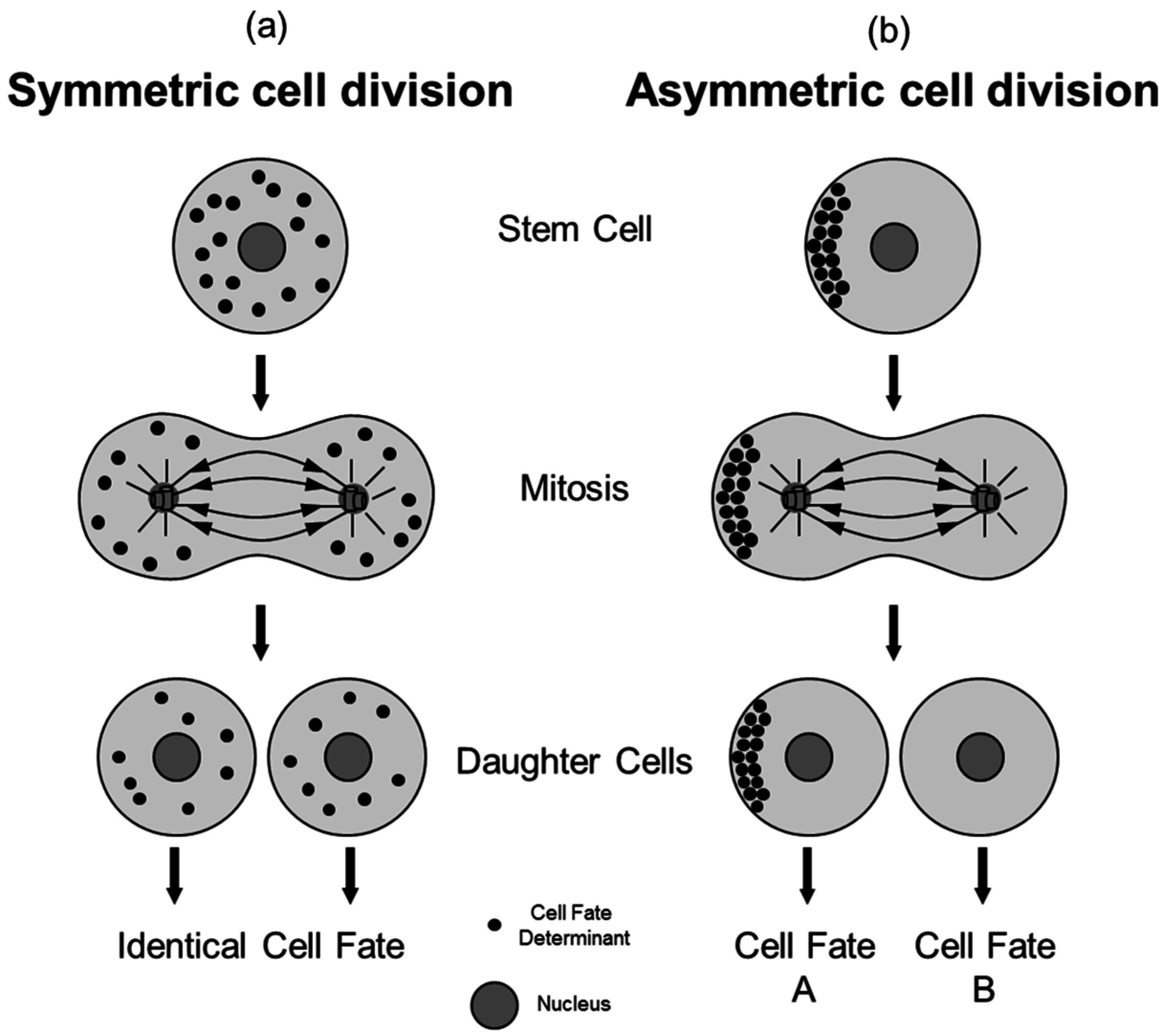
2. The Process of Asymmetric Cell Division

Stem cells can either divide symmetrically or asymmetrically. In a symmetric cell division (SCD), the mother cell creates two daughter cells with equal cell fates (Figure 1a). In contrast, an ACD is defined as cell division creating two qualitatively distinct daughter cells (Figure 1b). Differences can be mediated by asymmetric distribution of so-called cell fate determinants, e.g., membrane components, cell organelles, cytosolic components or proteins [1]. Generally, ACD is a fundamental process for controlling cell fates in somatic stem cell and developmental biology. In this process, a potential cell fate-determining factor needs to exceed a critical level of asymmetry to trigger a cell-fate decision. The asymmetric distribution to one daughter cell and not the other generally does not have to be perfect as long as the critical level of the respective factor (probably being different dependent on factor type and stem cell system) is accumulated in one and not in the other daughter cell.
While most studies on ACD focus on proteins acting as cell-fate determinants, asymmetric segregation of mRNA molecules was also found to regulate the decision between stem cell maintenance and differentiation, for example in mouse radial glial cells [11,12]. Recently, the asymmetric segregation of aged mitochondria was observed in human mammary stem-like cells. Here, the daughter cell receiving fewer aged mitochondria retains stem-like cell properties [13,14]. This shows that apart from proteins also other cell-intrinsic factors, like mitochondria or mRNA, can be distributed asymmetrically and act as cell-fate determinants.
Figure 1. Symmetric versus Asymmetric Cell Division. During a SCD (a), cell fate determining factors are distributed evenly to both arising daughter cells, resulting in identical cell fates. During ACD (b) cell fate determinants segregate unequally into both daughter cells, enabling them to realize different cell fates.
Symmetry 07 02025 g001
Following ACD of a stem cell, the presence or absence of the respective cell fate determining factor results in the activation or continuation of multifaceted (and poorly understood) programs. In turn, this can change the transcription and/or the epigenetic profile of the daughter cells. In this context, there are different concepts of whether the daughter cell, which inherited lesser amounts of a given cell fate determining factor, re-synthesizes this factor or changes its transcription. Additionally, other factors, e.g., extrinsic signals, might influence or regulate the outcome of the cell intrinsic cell-fate determination by ACD.
In recent years, there is accumulating evidence that ACD is an important mechanism for cell fate regulation in mammalian somatic stem cell systems. Up to now, the process of ACD has been barely analyzed and understood in mammals, especially in humans, but was studied extensively in different model organisms.

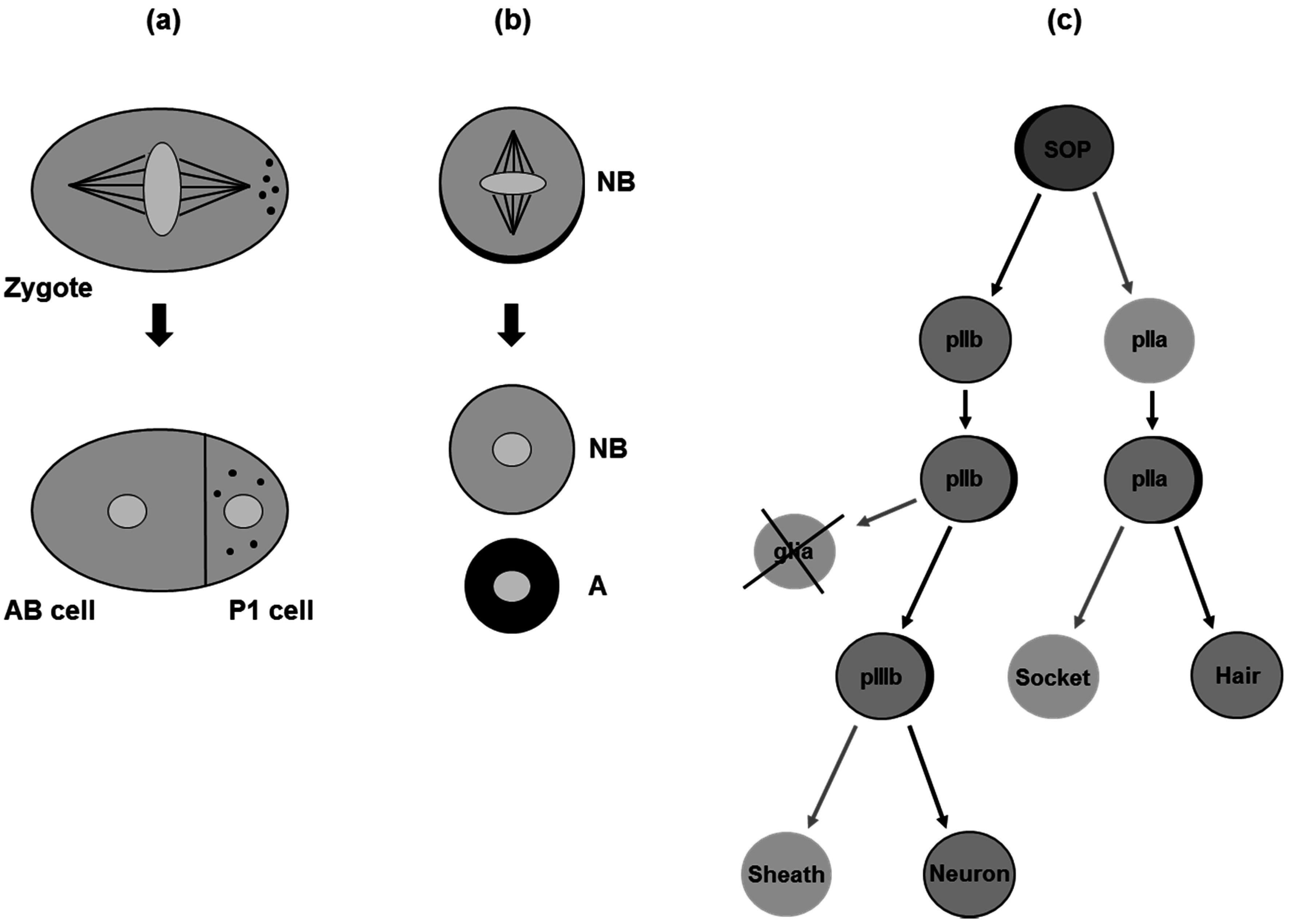
3. Asymmetric Cell Division in Model Organisms

Much of our current knowledge about ACD and its importance for stem cell fate specification has come from studies analyzing the development of model organisms, mainly the nematode Caenorhabditis elegans and the fruit fly Drosophila melanogaster. Here, we will briefly summarize three prominent examples which will illustrate the parallels and the differences of ACD regulation in different stem cell systems.
During embryogenesis of C. elegans, the zygote undergoes a series of ACDs. The zygote divides asymmetrically, resulting in the formation of a smaller posterior (P1 cell) and a larger anterior blastomere (AB cell). The localization of so-called P-granules to the posterior pole of the zygote determines the posterior cell as founder cell of the germline, while the AB cell does not inherit germline potential (Figure 2a, [15,16,17,18]). Genetic screens in this model have led to the identification of various factors regulating the asymmetric segregation of the P-granules, including proteins regulating cell polarity [19].
Figure 2. Examples for ACD in Model Organisms. (a) Initial ACD of a developing C. elegans zygote; (b) ACD of a Drosophila neuroblast (NB); (c) Drosophila sensory-organ precursor (SOP) development. Localization of cell fate determinants is indicated by black dots (a) or crescents (b/c).
Symmetry 07 02025 g002
In Drosophila, cell polarity organizing proteins were also found to regulate ACD of the neuroblast, a neural stem cell. Upon delamination of the neuroblast from the neuro-ectodermal epithelium, the maintenance of apico-basal cell polarity is required for the localization of the cell fate determinants Numb and Prospero at the basal membrane of mitotic neuroblasts and the proper orientation of the mitotic spindle [20,21]. The daughter cell inheriting Numb and Prospero becomes specified to differentiate, whereas the second daughter cell with low levels of these factors remains a neuroblast (Figure 2b, [22,23,24,25,26,27]).
The third example, the development of the peripheral nervous system of Drosophila, provides an excellent model that illustrates how extrinsic signals and ACD can orchestrate cell fate decisions. Each mechanoreceptor, the bristles, on the surface of an adult Drosophila fruit fly is created by a given sensory-organ precursor cell (SOP), which repetitively divides asymmetrically in a stereotypical manner (Figure 2c). At first, SOPs divide asymmetrically into the pIIa and pIIb daughter cells. The factor Numb is inherited by the pIIb cell, where it promotes the endocytosis and degradation of Notch, thereby removing Notch receptors from the pIIb membrane [28,29,30,31,32,33,34]. In consequence, the Notch signaling pathway is only activated in the pIIa cell. Using the same regulatory mechanism, the pIIa gives rise to a hair as well as a socket cell and the pIIb cell creates a glia cell, which undergoes apoptosis, and a pIIIb cell, which further divides asymmetrically to create the sheath cell and the neuron (reviewed comprehensively in [35,36]).

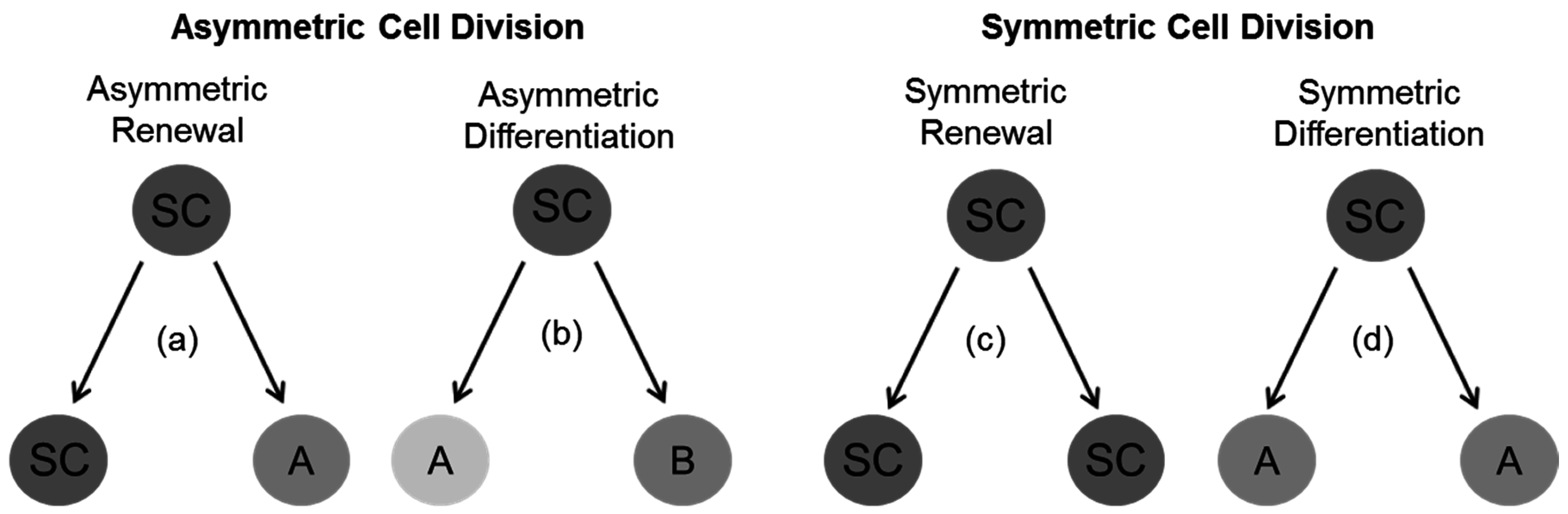
4. General Concepts for Divisional Modes in Stem and Progenitor Cells

According to the cell-fate determining mechanisms in C. elegans and Drosophila mentioned in the previous paragraph, two different types of ACD can be distinguished: asymmetric renewal and asymmetric differentiation. In the asymmetric renewal type (like in the Drosophila neuroblast ACD), the dividing stem cell gives rise to one committed daughter cell while the other retains the stem cell potential (Figure 3a). In contrast, during asymmetric differentiation like in the SOP, ACD creates two committed daughter cells with distinct potentials, resulting in the loss of the initial stem cell-like potential (Figure 3b).
Figure 3. Alternative Modes of Stem Cell Divisions. (a) Asymmetric renewal: One daughter cell becomes committed to differentiate (A), the other daughter cell retains the stem cell (SC) potential; (b) Asymmetric differentiation: Both daughter cells become committed to differentiate in order to realize different cell fates (A/B); (c) Symmetric renewal: Both daughter cells retain the stem cell potential of the mother cell; (d) Symmetric differentiation: Both daughter cells are committed to differentiate towards the same lineage (A).
Symmetry 07 02025 g003
In addition to ACD, SCD can control stem cell fates. Again, two different modes can be discriminated: symmetric renewal and symmetric differentiation. In case of symmetric renewal, the stem cell divides to create an equal pair of daughter cells, both retaining the mother cell’s potential (Figure 3c). Finally, a dividing stem cell giving rise to two equal daughter cells both being specified for differentiation towards the same cell fate would be symmetric differentiation (Figure 3d). In general, symmetric renewal can be assumed to increase the stem cell pool, whereas asymmetric renewal can simultaneously maintain the stem cell pool and facilitate differentiation [37,38].

5. Cell Polarity as a Prerequisite for ACD

As briefly mentioned above (see Chapter 3), the establishment of cell polarity and the definition of a polarity axis are important prerequisites for ACD. In general, cell polarity comprises morphological as well as intra-cellular properties [39]. In order to establish cellular polarity, different filaments, e.g., actin and tubulin filaments, form a highly dynamic and physical resistant network organizing and shaping the cell. In fact the localization and organization of all cell components (e.g., nucleus, cell membrane and organelles) is impressively structured. The following steps are essential in order to realize a proper ACD. At first, a cell polarity axis has to be defined. Subsequently, cell fate determinants have to be located according to the defined cell poles. Next, during mitosis, the spindle apparatus needs to be orientated according to the pre-established axis to ensure the segregation of the cell-fate determinants to mainly one of the two daughter cells, respectively. Finally, the two daughter cells have to be separated successfully by means of cytokinesis [1].
Genetic screens in C. elegans revealed defects impairing the initial ACD of the zygote and corresponding genes were called partitioning defect (par) genes [19]. Several of these PAR proteins, e.g., Par3 and Par6, were found to be part of the cell polarity machinery. In C. elegans, Par3 and Par6 form a complex with an atypical protein kinase C (aPKC) that concentrates in the anterior half of the embryo maintaining the anterior-posterior polarity axis [40,41]. The zygote of C. elegans divides along an anterior-posterior axis [15,16,17,18], whereas the Drosophila neuroblast segregates along an apico-basal axis [20,21]. The SOP and the pIIa cell divide along an anterior-posterior axis whereas the pIIb and pIIIb have an apico-basal separation axis [34]. The distinct orientations suggest a regulating impact of cell polarity which can be influenced by cell-cell contacts. Indeed, extensive studies of the Par/aPKC complex (reviewed in [42]) in C. elegans and Drosophila showed its essential functions in the three presented cellular models for ACD [40,43,44].

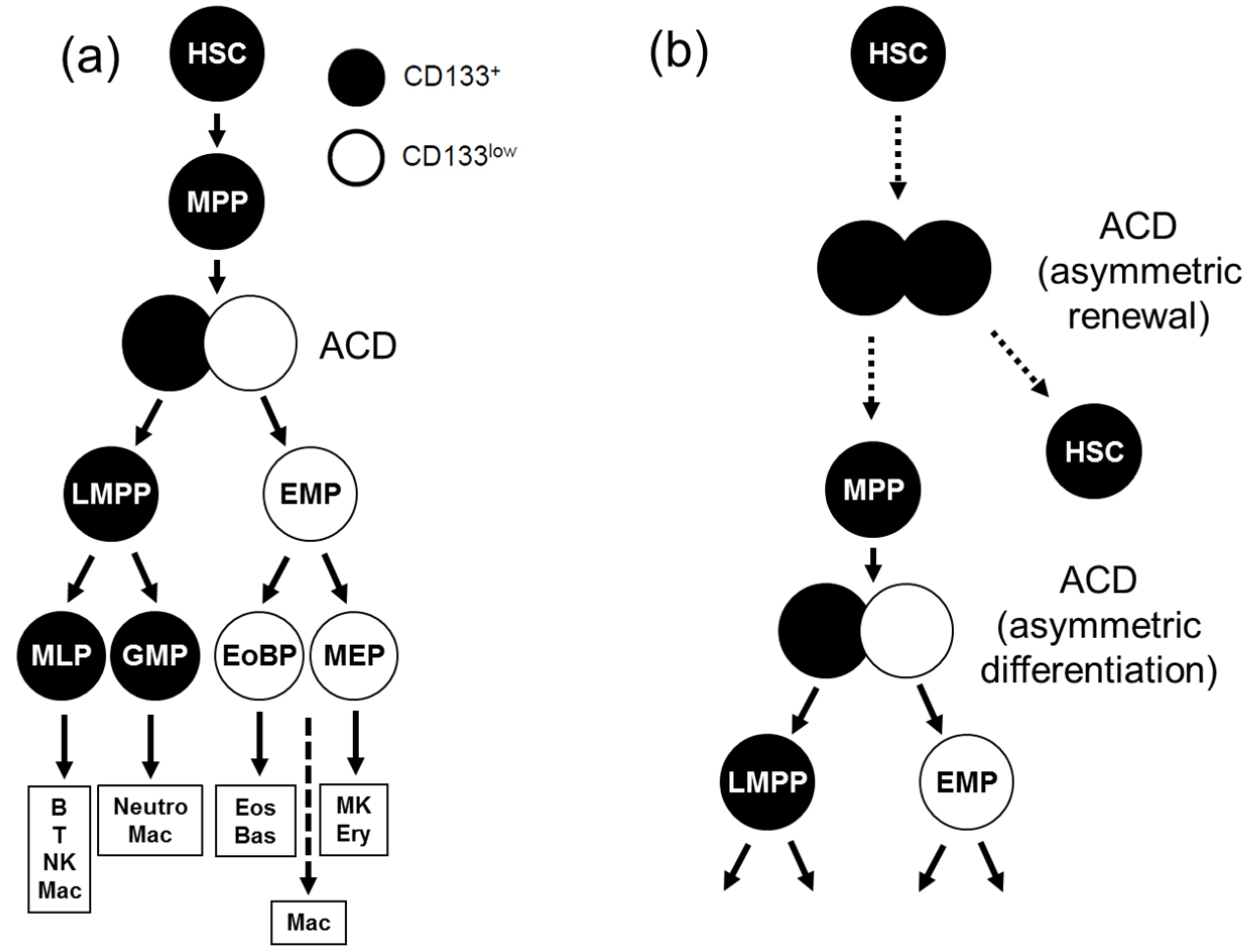
6. ACD in Human Hematopoiesis

Even though studies in model organisms have shed light on the regulation and have uncovered different modes of ACD, knowledge about its role in mammalian and especially human somatic stem cells remains sparse. So far, the hematopoietic stem cell (HSC) system is the best investigated human somatic stem cell system. HSCs are able to accomplish the dual task of self-renewal during the complete lifespan of an individual organism, and to develop all differentiated blood cell types. However, our understanding about the mechanisms regulating the cell fate of HSCs is fragmentary.
Regarding the regulation of such cell fate decisions, the previously mentioned stem cell niches were first described to consist mainly of osteoblasts in the endosteum of the bone marrow and sinusoidal endothelial cells in the spleen and bone marrow [45,46,47]. More recently, it was shown that mesenchymal stromal cells and endothelial cells contribute to the HSC niche as well [48,49,50]. These findings, including current controversies, are comprehensively discussed in numerous excellent reviews dealing about HSC niches [8,10,51,52].
In addition, it has been widely assumed since many years that HSCs divide asymmetrically to give rise to one daughter cell that retains the stem cell potential and to another cell being committed to differentiation [37,38,53]. This assumption was initially based on the outcome of so-called paired daughter cell experiments. Different groups separated daughter cells of single human hematopoietic stem and progenitor cells (HSPCs), and consistently reported that in 20%–40% of the cases both daughter cells showed different proliferation rates and differentiation capacities [54,55,56,57]. Even though this functional asymmetry of arising daughter cells was explained by ACD, theoretically it could have been established by post-mitotic communication processes between identical daughter cells [1]. Thus, it was a question whether HSPCs indeed can divide asymmetrically [55]. Focused on this question, our group started to search for asymmetrically segregating marker proteins.

7. Identification of ACD Markers in Human Hematopoiesis

Considering that asymmetrically dividing cells need to be polarized, we initially focused on cell polarity cues in human HSPCs and identified a number of proteins that were localized at higher concentrations at the one or the other pole of cytokine-stimulated HSPCs [39,58]. However, upon studying these cell polarity markers in mitotic HSPCs including the stem cell marker CD133/Prominin-1, we did not observe any indication for an asymmetric segregation of any of these markers [59].
In our studies, we and others had observed that most freshly isolated HSPCs express high levels of the protein CD133 (CD133+) on their cell surface [55,60,61]. Upon cultivation and the onset of cell division a proportion of HSPCs with strongly decreased levels of CD133 cell surface expression (CD133low) arises (approx. 40% after cultivation for 50–60 h) and increases over time [1,55,58,59]. Since the observed kinetics corresponded to a speculative model proposing that CD133 negative cells can only arise as a result of ACD—which in relation to the paired daughter cell assays was assumed to have a frequency of 30%—we searched for other cell surface markers being differently expressed on CD133+ and CD133low cells. In a subsequent flow-cytometric screen, we identified four markers (CD53, CD62L, CD63 and CD71) that were differently expressed on cells of both populations. Upon studying their subcellular distribution in dividing HSPCs, we observed (in good agreement with the hypothesis) asymmetric distribution of all four markers in 20%–30% of the mitotic cells and demonstrated for the first time that primitive human HSPCs indeed can divide asymmetrically [59].
Interestingly, three of these four identified proteins, CD53, CD63 and CD71, were linked to the endosomal compartment, which plays an important role in the molecular trafficking within cells. In addition, endosomes were found to segregate asymmetrically in developing C. elegans embryos [62]. Thus, the asymmetric segregation of endosomes might provide a more general and evolutionary conserved mechanism in ACD. This idea has been reviewed and discussed at greater detail in [63,64,65].
Since differences between CD133+ and CD133low populations led to the identification of the first ACD markers in human hematopoiesis, and since it has also been reported that an intracellular portion of CD133 does segregate asymmetrically in a proportion of human HSPCs [65], we continued to investigate the relevance of CD133 surface expression in human hematopoiesis. First, we comprehensively analyzed the differentiation potential of HSPC subsets differently expressing CD133 on their surface, which led to the discovery of new and unexpected lineage relationships in human hematopoiesis [66,67,68]. According to this revised model of human hematopoiesis (Figure 4a), CD133+ multipotent progenitors (MPP) can give rise to lymphomyeloid primed progenitors (LMPP) which retain CD133 surface expression, while cells which have lost CD133 expression correspond to erythromyeloid progenitors (EMP).
Figure 4. ACD in Human Hematopoiesis. (a) Revised model of human hematopoiesis. MPPs divide asymmetrically with regard to CD133 to create two daughter cells, a lymphomyeloid-primed progenitor (LMPP) and an erythromyeloid-primed progenitor (EMP) [67,69]; (b) Hypothetical model assuming the existence of further ACD during human hematopoiesis. In addition to the asymmetric differentiation mode described in (a), there are probably further branching points being regulated by ACD, e.g., the previously postulated asymmetric renewal type of ACD [37,38,53,70] illustrated here (MLP: Mixed lymphoid progenitor; GMP: Granulocyte-macrophage progenitor; EoBP: Eosinophil-basophil progenitor; MEP: Megakaryocyte-erythrocyte progenitor; Mac: Macrophage; Neutro: Neutrophil; Eos: Eosinophil; Bas: Basophil; MK: Megakaryocyte; Ery: Erythrocyte).
Symmetry 07 02025 g004
Since the kinetics of CD133 expression initially enabled us to discover the asymmetrically segregating proteins, we investigated whether LMPPs and EMPs arise by means of ACD of MPPs, next. In previous studies we noticed that the CD133 epitopes that are recognized by classically used anti-CD133 antibodies (AC133, AC141), which previously had shown symmetric CD133 distributions in mitotic HSPCs [59], are sensitive to fixation [58]. Additionally, both AC133 and AC141 have been reported to recognize glycosylation-dependent residues on the extracellular domains of CD133 [60,71]. When we tested a novel anti-CD133 antibody (HC7) reported in 2010 to bind glycosylation-independent protein residues of CD133 and being insensitive to fixation [72], we found clear asymmetric HC7 epitope distributions in approximately 30% of the mitotic human HSPCs [69]. Using fluorescence-labeled anti-CD133 (HC7) antibodies combined with time-lapse fluorescence microscopy, we developed single cell assays to study the mitotic distribution of CD133 on viable, dividing human MPPs, and the cell fate of both paired daughter cells, respectively. In this study, we demonstrated that with regard to the subcellular distribution of CD133 most human MPPs divide asymmetrically. The daughter cells receiving higher amounts of CD133 during ACD acquire LMPP-like potentials, while the daughter cells receiving lower amounts of CD133 are specified towards the erythromyeloid lineage (Figure 4a; [69]).
In summary, our data show that the initial lineage separation of lymphomyeloid and erythromyeloid lineages in human hematopoiesis is realized through ACD. Since we very rarely observed MPPs creating daughter cells with MPP potentials, we assume that under conventional culture conditions, ACD in human hematopoiesis rather follows the asymmetric differentiation type than the asymmetric renewal type. However, it still remains an open question whether ACDs are involved at other branching points of the human hematopoietic tree. It might be possible that HSPCs showing symmetric segregation of CD133 still divide asymmetrically, for example HSCs might create one HSC and one MPP (Figure 4b). To learn whether such self-renewing ACDs occur, additional asymmetrically segregating markers need to be identified. Future studies might indicate whether ACDs in human hematopoiesis also can be self-renewing [69].

8. Outlook

In general, our knowledge about the role of ACD in regulating somatic cell fate decisions is relatively sparse, and especially in humans limited to few stem cell systems. A main reason probably is that the analysis of single somatic stem cells is methodologically and technically challenging, mainly because of the relatively short time frame of mitosis. Technical advances of imaging systems and the application of new biological tools or probes to visualize ACD in living cells (like the fluorescence-labeled anti-CD133 antibodies described above) hopefully will make such studies more feasible in the future.

Acknowledgments

Own projects summarized and discussed in this review were supported by grants from the Deutsche Forschungsgemeinschaft (GI 336/4-1 to B.G.) and from the Ministry of Science, Innovation and Research of the German State of North Rhine-Westphalia/the Stem Cell Network North Rhine-Westphalia (323-400 012 13 to A.G.).

Author Contributions

F.M. and A.G. prepared all figures. All authors contributed to manuscript writing and critically revised the submitted version.

Conflicts of Interest

The authors declare no conflict of interest.

References

  1. Görgens, A.; Giebel, B. Self-renewal of primitive hematopoietic cells: A focus on asymmetric cell division. In Umbilical Cord Blood: A Future for Regenerative Medicine? Kadereit, S., Udolph, G., Eds.; World Scientific Publishing Company, Incorporated: Singapore, 2010; pp. 51–75. [Google Scholar]
  2. Al-Hajj, M. Cancer stem cells and oncology therapeutics. Curr. Opin. Oncol. 2007, 19, 61–64. [Google Scholar] [CrossRef] [PubMed]
  3. Savona, M.; Talpaz, M. Getting to the stem of chronic myeloid leukaemia. Nat. Rev. Cancer 2008, 8, 341–350. [Google Scholar] [CrossRef] [PubMed]
  4. Visvader, J.E.; Lindeman, G.J. Cancer stem cells in solid tumours: Accumulating evidence and unresolved questions. Nature Rev. Cancer 2008, 8, 755–768. [Google Scholar] [CrossRef] [PubMed]
  5. Rossi, D.J.; Jamieson, C.H.M.; Weissman, I.L. Stems cells and the pathways to aging and cancer. Cell 2008, 132, 681–696. [Google Scholar] [CrossRef] [PubMed]
  6. Ruzankina, Y.; Brown, E.J. Relationships between stem cell exhaustion, tumour suppression and ageing. Br. J. Cancer 2007, 97, 1189–1193. [Google Scholar] [CrossRef] [PubMed]
  7. Sharpless, N.E.; DePinho, R.A. How stem cells age and why this makes us grow old. Nature Rev. Mol. Cell Biol. 2007, 8, 703–713. [Google Scholar] [CrossRef] [PubMed]
  8. Boulais, P.E.; Frenette, P.S. Making sense of hematopoietic stem cell niches. Blood 2015, 125, 2621–2629. [Google Scholar] [CrossRef] [PubMed]
  9. Mesa, K.R.; Rompolas, P.; Greco, V. The dynamic duo: Niche/stem cell interdependency. Stem Cell Rep. 2015, 4, 961–966. [Google Scholar] [CrossRef] [PubMed]
  10. Mendez-Ferrer, S.; Scadden, D.T.; Sanchez-Aguilera, A. Bone marrow stem cells: Current and emerging concepts. Ann. NY Acad. Sci. 2015, 1335, 32–44. [Google Scholar] [CrossRef] [PubMed]
  11. Kusek, G.; Campbell, M.; Doyle, F.; Tenenbaum, S.A.; Kiebler, M.; Temple, S. Asymmetric segregation of the double-stranded RNA binding protein staufen2 during mammalian neural stem cell divisions promotes lineage progression. Cell Stem Cell 2012, 11, 505–516. [Google Scholar] [CrossRef] [PubMed]
  12. Vessey, J.P.; Amadei, G.; Burns, S.E.; Kiebler, M.A.; Kaplan, D.R.; Miller, F.D. An asymmetrically localized staufen2-dependent RNA complex regulates maintenance of mammalian neural stem cells. Cell Stem Cell 2012, 11, 517–528. [Google Scholar] [CrossRef] [PubMed]
  13. Katajisto, P.; Döhla, J.; Chaffer, C.L.; Pentinmikko, N.; Marjanovic, N.; Iqbal, S.; Zoncu, R.; Chen, W.; Weinberg, R.A.; Sabatini, D.M. Asymmetric apportioning of aged mitochondria between daughter cells is required for stemness. Science 2015, 348, 340–343. [Google Scholar] [CrossRef] [PubMed]
  14. Suomalainen, A. Stem cells: Asymmetric rejuvenation. Nature 2015, 521, 296–298. [Google Scholar] [CrossRef] [PubMed]
  15. Strome, S.; Wood, W.B. Immunofluorescence visualization of germ-line-specific cytoplasmic granules in embryos, larvae, and adults of caenorhabditis elegans. Proc. Natl. Acad. Sci. USA 1982, 79, 1558–1562. [Google Scholar] [CrossRef] [PubMed]
  16. Hird, S.N.; Paulsen, J.E.; Strome, S. Segregation of germ granules in living caenorhabditis elegans embryos: Cell-type-specific mechanisms for cytoplasmic localisation. Development 1996, 122, 1303–1312. [Google Scholar] [PubMed]
  17. Priess, J.R.; Thomson, J.N. Cellular interactions in early C. elegans embryos. Cell 1987, 48, 241–250. [Google Scholar] [CrossRef]
  18. Sulston, J.E.; Schierenberg, E.; White, J.G.; Thomson, J.N. The embryonic cell lineage of the nematode caenorhabditis elegans. Dev. Biol. 1983, 100, 64–119. [Google Scholar] [CrossRef]
  19. Kemphues, K.J.; Priess, J.R.; Morton, D.G.; Cheng, N.S. Identification of genes required for cytoplasmic localization in early C. elegans embryos. Cell 1988, 52, 311–320. [Google Scholar] [CrossRef]
  20. Hirata, J.; Nakagoshi, H.; Nabeshima, Y.; Matsuzaki, F. Asymmetric segregation of the homeodomain protein prospero during drosophila development. Nature 1995, 377, 627–630. [Google Scholar] [CrossRef] [PubMed]
  21. Spana, E.P.; Doe, C.Q. The prospero transcription factor is asymmetrically localized to the cell cortex during neuroblast mitosis in drosophila. Development 1995, 121, 3187–3195. [Google Scholar] [PubMed]
  22. Kaltschmidt, J.A.; Davidson, C.M.; Brown, N.H.; Brand, A.H. Rotation and asymmetry of the mitotic spindle direct asymmetric cell division in the developing central nervous system. Nature Cell Biol. 2000, 2, 7–12. [Google Scholar] [PubMed]
  23. Kraut, R.; Campos-Ortega, J.A. Inscuteable, a neural precursor gene of drosophila, encodes a candidate for a cytoskeleton adaptor protein. Dev. Biol. 1996, 174, 65–81. [Google Scholar] [CrossRef] [PubMed]
  24. Kraut, R.; Chia, W.; Jan, L.Y.; Jan, Y.N.; Knoblich, J.A. Role of inscuteable in orienting asymmetric cell divisions in drosophila. Nature 1996, 383, 50–55. [Google Scholar] [CrossRef] [PubMed]
  25. Schober, M.; Schaefer, M.; Knoblich, J.A. Bazooka recruits inscuteable to orient asymmetric cell divisions in drosophila neuroblasts. Nature 1999, 402, 548–551. [Google Scholar] [PubMed]
  26. Wodarz, A.; Ramrath, A.; Grimm, A.; Knust, E. Drosophila atypical protein kinase C associates with bazooka and controls polarity of epithelia and neuroblasts. J. Cell Biol. 2000, 150, 1361–1374. [Google Scholar] [CrossRef] [PubMed]
  27. Wodarz, A.; Ramrath, A.; Kuchinke, U.; Knust, E. Bazooka provides an apical cue for inscuteable localization in drosophila neuroblasts. Nature 1999, 402, 544–547. [Google Scholar] [CrossRef] [PubMed]
  28. Frise, E.; Knoblich, J.A.; Younger-Shepherd, S.; Jan, L.Y.; Jan, Y.N. The drosophila numb protein inhibits signaling of the notch receptor during cell-cell interaction in sensory organ lineage. Proc. Natl. Acad. Sci. USA 1996, 93, 11925–11932. [Google Scholar] [CrossRef] [PubMed]
  29. Gho, M.; Bellaïche, Y.; Schweisguth, F. Revisiting the drosophila microchaete lineage: A novel intrinsically asymmetric cell division generates a glial cell. Development 1999, 126, 3573–3584. [Google Scholar] [PubMed]
  30. Guo, M.; Jan, L.Y.; Jan, Y.N. Control of daughter cell fates during asymmetric division: Interaction of numb and notch. Neuron 1996, 17, 27–41. [Google Scholar] [CrossRef]
  31. Uemura, T.; Shepherd, S.; Ackerman, L.; Jan, L.Y.; Jan, Y.N. Numb, a gene required in determination of cell fate during sensory organ formation in drosophila embryos. Cell 1989, 58, 349–360. [Google Scholar] [CrossRef]
  32. Couturier, L.; Vodovar, N.; Schweisguth, F. Endocytosis by numb breaks notch symmetry at cytokinesis. Nat. Cell. Biol. 2012, 14, 131–139. [Google Scholar] [CrossRef] [PubMed]
  33. McGill, M.A.; Dho, S.E.; Weinmaster, G.; McGlade, C.J. Numb regulates post-endocytic trafficking and degradation of notch1. J. Biol. Chem. 2009, 284, 26427–26438. [Google Scholar] [CrossRef] [PubMed]
  34. Rhyu, M.S.; Jan, L.Y.; Jan, Y.N. Asymmetric distribution of numb protein during division of the sensory organ precursor cell confers distinct fates to daughter cells. Cell 1994, 76, 477–491. [Google Scholar] [CrossRef]
  35. Furman, D.P.; Bukharina, T.A. Drosophila mechanoreceptors as a model for studying asymmetric cell division. Int. J. Dev. Biol. 2011, 55, 133–141. [Google Scholar] [CrossRef] [PubMed]
  36. Giebel, B.; Wodarz, A. Notch signaling: Numb makes the difference. Curr. Biol. 2012, 22, R133–R135. [Google Scholar] [CrossRef] [PubMed]
  37. Ho, A.D.; Wagner, W. The beauty of asymmetry: Asymmetric divisions and self-renewal in the haematopoietic system. Curr. Opin. Hematol. 2007, 14, 330–336. [Google Scholar] [CrossRef] [PubMed]
  38. Pina, C.; Enver, T. Differential contributions of haematopoietic stem cells to foetal and adult haematopoiesis: Insights from functional analysis of transcriptional regulators. Oncogene 2007, 26, 6750–6765. [Google Scholar] [CrossRef] [PubMed]
  39. Gorgens, A.; Beckmann, J.; Ludwig, A.K.; Mollmann, M.; Durig, J.; Horn, P.A.; Rajendran, L.; Giebel, B. Lipid raft redistribution and morphological cell polarization are separable processes providing a basis for hematopoietic stem and progenitor cell migration. Int. J. Biochem. Cell Biol. 2012, 44, 1121–1132. [Google Scholar] [CrossRef] [PubMed]
  40. Tabuse, Y.; Izumi, Y.; Piano, F.; Kemphues, K.J.; Miwa, J.; Ohno, S. Atypical protein kinase C cooperates with PAR-3 to establish embryonic polarity in caenorhabditis elegans. Development 1998, 125, 3607–3614. [Google Scholar] [PubMed]
  41. Watts, J.L.; Etemad-Moghadam, B.; Guo, S.; Boyd, L.; Draper, B.W.; Mello, C.C.; Priess, J.R.; Kemphues, K.J. PAR-6, a gene involved in the establishment of asymmetry in early C. elegans embryos, mediates the asymmetric localization of PAR-3. Development 1996, 122, 3133–3140. [Google Scholar] [PubMed]
  42. Suzuki, A.; Ohno, S. The PAR-aPKC system: Lessons in polarity. J. Cell Sci. 2006, 119, 979–987. [Google Scholar] [CrossRef] [PubMed]
  43. Petronczki, M.; Knoblich, J.A. DmPAR-6 directs epithelial polarity and asymmetric cell division of neuroblasts in drosophila. Nature Cell Biol. 2001, 3, 43–49. [Google Scholar] [PubMed]
  44. Roegiers, F.; Younger-Shepherd, S.; Jan, L.Y.; Jan, Y.N. Bazooka is required for localization of determinants and controlling proliferation in the sensory organ precursor cell lineage in drosophila. Proc. Natl. Acad. Sci. USA 2001, 98, 14469–14474. [Google Scholar] [CrossRef] [PubMed]
  45. Calvi, L.M.; Adams, G.B.; Weibrecht, K.W.; Weber, J.M.; Olson, D.P.; Knight, M.C.; Martin, R.P.; Schipani, E.; Divieti, P.; Bringhurst, F.R.; et al. Osteoblastic cells regulate the haematopoietic stem cell niche. Nature 2003, 425, 841–846. [Google Scholar] [CrossRef] [PubMed]
  46. Zhang, J.; Niu, C.; Ye, L.; Huang, H.; He, X.; Tong, W.G.; Ross, J.; Haug, J.; Johnson, T.; Feng, J.Q.; et al. Identification of the haematopoietic stem cell niche and control of the niche size. Nature 2003, 425, 836–841. [Google Scholar] [CrossRef] [PubMed]
  47. Kiel, M.J.; Yilmaz, O.H.; Iwashita, T.; Terhorst, C.; Morrison, S.J. Slam family receptors distinguish hematopoietic stem and progenitor cells and reveal endothelial niches for stem cells. Cell 2005, 121, 1109–1121. [Google Scholar] [CrossRef] [PubMed]
  48. Greenbaum, A.; Hsu, Y.M.; Day, R.B.; Schuettpelz, L.G.; Christopher, M.J.; Borgerding, J.N.; Nagasawa, T.; Link, D.C. CXCL12 in early mesenchymal progenitors is required for haematopoietic stem-cell maintenance. Nature 2013, 495, 227–230. [Google Scholar] [CrossRef] [PubMed]
  49. Ding, L.; Saunders, T.L.; Enikolopov, G.; Morrison, S.J. Endothelial and perivascular cells maintain haematopoietic stem cells. Nature 2012, 481, 457–462. [Google Scholar] [CrossRef] [PubMed]
  50. Mendez-Ferrer, S.; Michurina, T.V.; Ferraro, F.; Mazloom, A.R.; Macarthur, B.D.; Lira, S.A.; Scadden, D.T.; Ma’ayan, A.; Enikolopov, G.N.; Frenette, P.S. Mesenchymal and haematopoietic stem cells form a unique bone marrow niche. Nature 2010, 466, 829–834. [Google Scholar] [CrossRef] [PubMed]
  51. Hanoun, M.; Frenette, P.S. This niche is a maze; an amazing niche. Cell Stem Cell 2013, 12, 391–392. [Google Scholar] [CrossRef] [PubMed]
  52. Kfoury, Y.; Scadden, D.T. Mesenchymal cell contributions to the stem cell niche. Cell Stem Cell 2015, 16, 239–253. [Google Scholar] [CrossRef] [PubMed]
  53. Roeder, I.; Lorenz, R. Asymmetry of stem cell fate and the potential impact of the niche: Observations, simulations, and interpretations. Stem Cell Rev. 2006, 2, 171–180. [Google Scholar] [CrossRef] [PubMed]
  54. Brummendorf, T.H.; Dragowska, W.; Zijlmans, J.; Thornbury, G.; Lansdorp, P.M. Asymmetric cell divisions sustain long-term hematopoiesis from single-sorted human fetal liver cells. J. Exp. Med. 1998, 188, 1117–1124. [Google Scholar] [CrossRef] [PubMed]
  55. Giebel, B.; Zhang, T.; Beckmann, J.; Spanholtz, J.; Wernet, P.; Ho, A.D.; Punzel, M. Primitive human hematopoietic cells give rise to differentially specified daughter cells upon their initial cell division. Blood 2006, 107, 2146–2152. [Google Scholar] [CrossRef] [PubMed]
  56. Leary, A.G.; Strauss, L.C.; Civin, C.I.; Ogawa, M. Disparate differentiation in hemopoietic colonies derived from human paired progenitors. Blood 1985, 66, 327–332. [Google Scholar] [PubMed]
  57. Punzel, M.; Zhang, T.; Liu, D.; Eckstein, V.; Ho, A.D. Functional analysis of initial cell divisions defines the subsequent fate of individual human CD34+CD38–cells. Exp. Hematol. 2002, 30, 464–472. [Google Scholar] [CrossRef]
  58. Giebel, B.; Corbeil, D.; Beckmann, J.; Hohn, J.; Freund, D.; Giesen, K.; Fischer, J.; Kogler, G.; Wernet, P. Segregation of lipid raft markers including CD133 in polarized human hematopoietic stem and progenitor cells. Blood 2004, 104, 2332–2338. [Google Scholar] [CrossRef] [PubMed]
  59. Beckmann, J.; Scheitza, S.; Wernet, P.; Fischer, J.C.; Giebel, B. Asymmetric cell division within the human hematopoietic stem and progenitor cell compartment: Identification of asymmetrically segregating proteins. Blood 2007, 109, 5494–5501. [Google Scholar] [CrossRef] [PubMed]
  60. Miraglia, S.; Godfrey, W.; Yin, A.H.; Atkins, K.; Warnke, R.; Holden, J.T.; Bray, R.A.; Waller, E.K.; Buck, D.W. A novel five-transmembrane hematopoietic stem cell antigen: Isolation, characterization, and molecular cloning. Blood 1997, 90, 5013–5021. [Google Scholar] [PubMed]
  61. Yin, A.H.; Miraglia, S.; Zanjani, E.D.; Almeida-Porada, G.; Ogawa, M.; Leary, A.G.; Olweus, J.; Kearney, J.; Buck, D.W. AC133, a novel marker for human hematopoietic stem and progenitor cells. Blood 1997, 90, 5002–5012. [Google Scholar] [PubMed]
  62. Andrews, R.; Ahringer, J. Asymmetry of early endosome distribution in C. elegans embryos. PLoS One 2007, 2. [Google Scholar] [CrossRef] [PubMed]
  63. Giebel, B.; Beckmann, J. Asymmetric cell divisions of human hematopoietic stem and progenitor cells meet endosomes. Cell Cycle 2007, 6, 2201–2204. [Google Scholar] [CrossRef] [PubMed]
  64. Furthauer, M.; Gonzalez-Gaitan, M. Endocytosis, asymmetric cell division, stem cells and cancer: Unus pro omnibus, omnes pro uno. Mol. Oncol. 2009, 3, 339–353. [Google Scholar] [CrossRef] [PubMed]
  65. Fonseca, A.V.; Bauer, N.; Corbeil, D. The stem cell marker CD133 meets the endosomal compartment—New insights into the cell division of hematopoietic stem cells. Blood Cells Mol. Dis. 2008, 41, 194–195. [Google Scholar] [CrossRef] [PubMed]
  66. Gorgens, A.; Radtke, S.; Horn, P.A.; Giebel, B. New relationships of human hematopoietic lineages facilitate detection of multipotent hematopoietic stem and progenitor cells. Cell Cycle 2013, 12, 3478–3482. [Google Scholar] [CrossRef] [PubMed]
  67. Gorgens, A.; Radtke, S.; Mollmann, M.; Cross, M.; Durig, J.; Horn, P.A.; Giebel, B. Revision of the human hematopoietic tree: Granulocyte subtypes derive from distinct hematopoietic lineages. Cell Rep. 2013, 3, 1539–1552. [Google Scholar] [CrossRef] [PubMed]
  68. Radtke, S.; Görgens, A.; Kordelas, L.; Schmidt, M.; Kimmig, K.R.; Koninger, A.; Horn, P.A.; Giebel, B. CD133 allows elaborated discrimination and quantification of haematopoietic progenitor subsets in human haematopoietic stem cell transplants. Br. J. Haematol 2015, 169, 868–878. [Google Scholar] [CrossRef] [PubMed]
  69. Gorgens, A.; Ludwig, A.K.; Mollmann, M.; Krawczyk, A.; Durig, J.; Hanenberg, H.; Horn, P.A.; Giebel, B. Multipotent hematopoietic progenitors divide asymmetrically to create progenitors of the lymphomyeloid and erythromyeloid lineages. Stem Cell Rep. 2014, 3, 1058–1072. [Google Scholar] [CrossRef] [PubMed]
  70. Mukherjee, S.; Kong, J.; Brat, D.J. Cancer stem cell division: When the rules of asymmetry are broken. Stem Cells Dev. 2015, 24, 405–416. [Google Scholar] [CrossRef] [PubMed]
  71. Bidlingmaier, S.; Zhu, X.; Liu, B. The utility and limitations of glycosylated human CD133 epitopes in defining cancer stem cells. J. Mol. Med. Berl. 2008, 86, 1025–1032. [Google Scholar] [CrossRef] [PubMed]
  72. Swaminathan, S.K.; Olin, M.R.; Forster, C.L.; Cruz, K.S.; Panyam, J.; Ohlfest, J.R. Identification of a novel monoclonal antibody recognizing CD133. J. Immunol. Methods 2010, 361, 110–115. [Google Scholar] [CrossRef] [PubMed]

