Microbial Gold Biosortion and Biomineralization from Aqueous HAuCl4 Solution

Department of Life and Environmental Science, Faculty of Engineering, Hachinohe Institute of Technology, 88-1 Ohbiraki, Myoh, Hachinohe 031-8501, Japan
*
Author to whom correspondence should be addressed.
Minerals 2020, 10(3), 285; https://doi.org/10.3390/min10030285
Submission received: 17 February 2020
/
Revised: 12 March 2020
/
Accepted: 19 March 2020
/
Published: 21 March 2020
(This article belongs to the Special Issue Biosorption and Biomineralization in Metal Removal)
Abstract
:The removal of gold (III) from aqueous systems using biosorption and biomineralization by microbial cells was investigated. High levels of gold (III) were removed from a hydrogen tetrachloroaurate (III) solution for 72 h by microbial species, including bacteria, fungi and yeasts. Previously, we reported that the amounts of gold (III) removed through biosorption by gram-positive bacteria, fungi, and yeasts was lower than that by gram-negative bacteria. Candida krusei was able to remove large amounts of gold (III) through biosorption and biomineralization. Interestingly, more gold was removed by atomic reduction than by biosorption. Additionally, we examined time, pH, concentration and other factors affecting gold removal. The rate of gold (III) removal by C. krusei increased for 6 h, and then stabilized, however, the rate of removal increased after 22 h, and reached a second equilibrium after 68 h.
1. Introduction
The demand for gold has increased markedly because of its increasing use in the electrical industry and the development of gold-containing drugs [1]. Therefore, recovering this valuable resource from recycled goods is a subject of wide interest.
Recently, several researchers have investigated gold recovery using microbial species, such as bacteria [2], fungi [3,4,5], yeasts [6], and algae [7,8]. However, there is little information on the species of microorganisms that have a high gold absorbing ability.
Previously, we reported that several microorganisms absorb gold. We screened resting forms of 75 microbial strains (19 actinomycetes, 25 bacteria, 17 fungi, and 14 yeasts) and their ability to absorb gold from a hydrogen tetrachloroaurate (III)-containing solution [9]. Hydrogen tetrachloroaurate (III) is used for medical and ceramic materials. Some of the gram-negative bacteria showed great ability to absorb gold from the solution, absorbing over 330 µmol of gold per gram microbial cells (dry weight basis) from the hydrogen tetrachloroaurate (III) solution within 1 h. The amount of gold absorbed from hydrogen tetrachloroaurate (III) solution by gram-negative bacteria was higher than by gram-positive bacteria, including actinomycetes, fungi, and yeasts. These results contrasted findings for lithium [10], cadmium [11], uranium [12], thorium [13], and rare earth metals [14], which were absorbed more effectively by gram-positive bacteria than by gram-negative bacteria, fungi and yeasts. These findings suggest gram-positive bacteria are able to absorb large amounts of positively-charged metal ions and gram-negative bacteria are able to absorb large amounts of negatively-charged complex ion [9,10,11,12,13,14]. Gold (III) exists as a negatively charged-complex ion, such as a tetrachloroaurate anion, in solution. The negative charge on the surface of the gram-positive bacteria is higher than on the surface of the gram-negative bacteria and vice versa owing to higher levels of teichoic acid in gram-positive bacteria at a neutral pH [15,16,17]. Accordingly, negatively-charged gold complex ions and positively-charged gram-negative bacteria form a stronger bond than the negatively-charged gold complex ions and gram-positive bacteria [9].
Previously, we investigated the effects of pH, gold concentration, cell amount, and gold biosorption time in Pseudomonas maltophilia, which absorbs large amounts of gold from a hydrogen tetrachloroaurate (III) containing solution and examined the biosorption-desorption cycles in our experimental system [9]. We also determined a suitable desorbent for gold absorbed by P. maltophilia.
To build on our previous research and continue to develop better methods for gold recovery, we investigated the removal of gold (III) by biosorption and biomineralization from aqueous systems using microbial cells.
2. Materials and Methods
2.1. Materials
The strains used in this research were generously donated by the IAM Culture Collection, Center for Cellular and Molecular Research, the Institute of Molecular and Cellular Biosciences, the University of Tokyo (IAM), the Faculty of Engineering, Hiroshima University (HUT), and the Faculty of Agriculture, Hokkaido University (AHU). All chemicals (guaranteed reagents) used in this study were obtained from Nacalai Tesque (Kyoto, Japan).
2.2. Microorganism Culture
The bacterial culture medium contained 3 g/L meat extract, 5 g/L peptone, and 5 g/L NaCl in deionized water. The medium for growing actinomycetes, fungi, and yeasts contained 4 g/L yeast extract, 10 g/L malt extract, and 4 g/L glucose in deionized water, pH 7.1 (for actinomycetes) and pH 5.7 (for fungi and yeasts). The microorganisms were maintained on agar slants and grown in 300 mL of the medium in a 500-mL flask with continuous shaking (120 rpm) for 72 h at 30 °C. Cells were collected by centrifugation (for bacteria and yeasts) or by filtration through a filter paper (for actinomycetes and fungi), washed thoroughly with deionized water, and then used in gold removal experiments.
2.3. Gold (III) Removal Experiment
Unless otherwise stated, the removal experiments were conducted as follows. Resting microbial cells (15 mg dry weight basis) were suspended in a 100 mL hydrogen tetrachloroaulate (III) solution containing 50 mg/L (254 µM) gold (pH 3.0). Diluted hydrochloric acid solution (pH 3.0) was used as a control. Our previous research found P. maltophilia to be extremely efficient for biosorption, requiring 1 h to completely remove all gold from a 10 mg/L gold solution [9]. Gold concentration is high in electrical engineering waste. Owing to this and our previous study we examined higher concentrations of gold in this study. The suspension was shaken for 72 h at 30 °C. The resting microbial cells were then removed by filtration through a membrane filter (0.2 µm pore size). Gold removal by the cells was determined by measuring the gold content in the filtrate with an atomic absorption analysis quantometer (AA-6300, Shimadzu Corporation, Kyoto, Japan).
2.3.1. Microorganism Screening for Gold (III) Removal from the Solution for 72 h
Resting cells (15 mg dry weight basis) were suspended in 100 mL solution (pH 3.0) containing hydrogen tetrachloroaurate (III) (254 µM, pH 3.0) for 72 h at 30 °C.
2.3.2. The Time Required for Gold Removal Using C. krusei, AHU3993
Resting cells (15 mg dry weight basis) were suspended in 100 mL solution (pH 3.0) containing hydrogen tetrachloroaurate (III) (254 µM, pH 3.0) for between 5 min and 68 h at 30 °C.
2.3.3. Effect of pH on Gold (III) Removal by C. krusei AHU3993
Resting cells (15 mg dry weight basis) were suspended in a 100 mL solution (pH range 1.0–5.0) containing hydrogen tetrachloroaurate (III) (254 µM) for 1 or 72 h at 30 °C.
2.3.4. Effect of Cell Amounts of C. krusei AHU3993 Added to the Solution on Gold (III) Removal
Desired amounts (5–25 mg dry weight basis) of the resting cells were suspended in a 100 mL solution (pH 4.0) containing hydrogen tetrachloroaurate (III) (254 µM) for 1 or 72 h at 30 °C.
2.3.5. Effect of Gold (III) Concentration on Gold Removal Using C. krusei AHU3993
To determine the maximum amount of gold removal, the resting cells (15 mg dry weight basis) were suspended in a 100 mL solution (pH 3.0) containing 0 mg/L, 50 mg/L, 100 mg/L, 150 mg/L, 200 mg/L, or 250 mg/L gold (III) as hydrogen tetrachloroaurate (III) (pH 4.0) for 1 h or 72 h at 30 °C.
2.3.6. Absorption Spectrometry Analysis of Gold Removal Using C. krusei AHU3993
Absorption spectra were measured between 100–900 nm using the filtrate from the effect of cell amount and gold concentration experiments using C. krusei AHU3993 with a UV-visible spectrophotometer (V-650, JASCO Corporation, Tokyo, Japan). The gold removal conditions were same with 2.3.4 or 2.3.5.
3. Results and Discussion
3.1. Screening Gold (III) Removing Microorganisms after 72 h in Gold-Containing Solution
Gram-negative bacteria, such as P. maltophilia, reportedly remove large quantities of gold (III) from aqueous hydrogen tetrachloroaurate (III) solutions (pH 3) in 1 h at 30 °C by biosorption [9]. In a preliminary experiment, we recently identified microorganisms that removed larger quantities of gold (III), and reducted the gold in solution to gold (0).
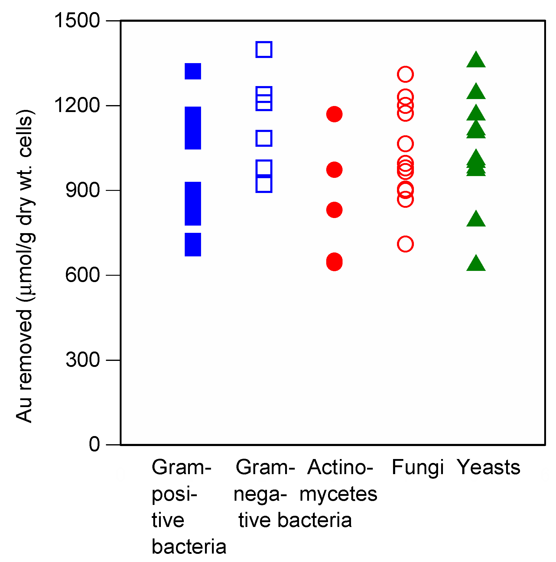
To determine the ability of different microbial species to remove a large quantity of gold (III), 48 microorganisms strains (5 actinomycetes, 19 bacteria, 13 fungi, and 11 yeasts) were screened. Gold (III) absorption was highest using P. maltophilia at pH 3.0 [9]. Therefore, we conducted our screening experiments using a solution with a pH adjusted to 3.0.
The ability of the microbial cells to remove gold significantly varied (Figure 1). Of the tested microorganisms, a high ability to remove gold was observed in all microorganisms. Nocardia erythropolis IAM1399 gram-positive bacterium, Eschelichia coli IAM1264, P. maltophilia IAM1554, and P. saccharophila IAM1504 gram-negative bacteria, Aspergillus niger IAM2534 and Chaetomium globosum IAM9272 and IAM9427 fungi, and Candida krusei, AHU3993 yeast removed gold at an amount over 1200 μmol/g dry cell weight in 72 h at 30 °C. Previous we found [9], gram-negative bacteria removed more gold (III) than gram-positive bacteria, actinomycetes, fungi, and yeasts, and the maximum amount of gold (III) removal was 360 µmol/g dry cell weight in 1 h at 30 °C. Therefore, many microorganisms can remove large quantities of gold (III) over longer time periods.
Because these results were time-dependent, we hypothesized that the reaction mechanisms may be different. Indeed the solution was almost colorless for 1 h, but changed to dark colors, including violet and dark green after longer time periods. Gold removal within 1 h likely occurred by biosorption, but longer time periods (72 h) caused the gold to reduce to a zero-valent gold.
Many positively-charged metal ions can be removed at the neutral pH using gram-positive bacteria and actinomycetes [10,11,12,13,14]. Negatively-charged gold (III) ions can be removed at an acidic pH using gram-negative bacteria [9] via a biosorption process. However, all metal ions tested can be removed in small amounts using yeasts and fungi [9,10,11,12,13,14]. Therefore, we investigated the removal of gold (III) using C. krusei, AHU3993 in detail, because this microorganism removed the largest amount of gold (III) in tested yeasts by biomineralization.
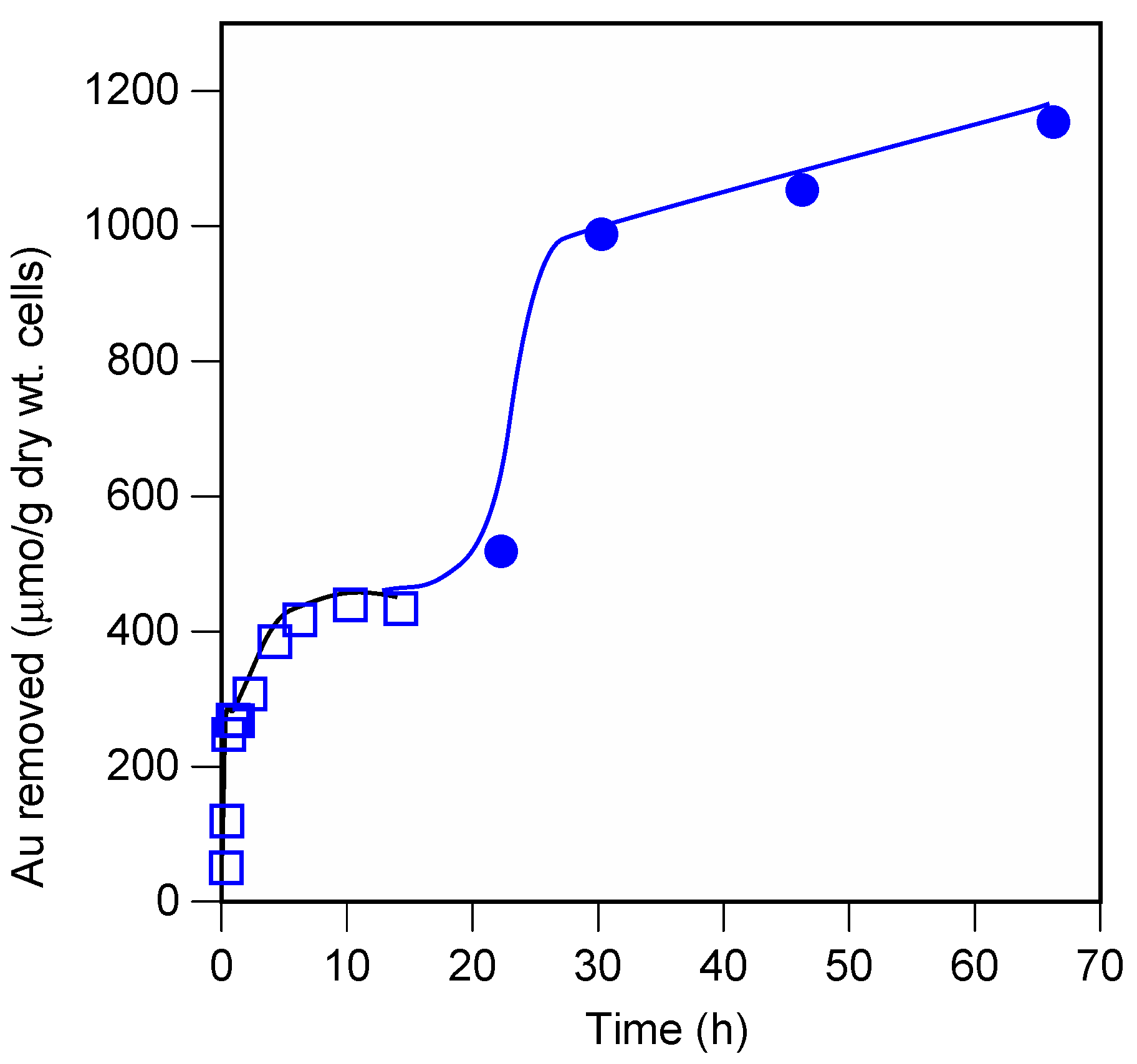
3.2. Time Required for Gold Removal Using C. krusei, AHU3993
C. krusei AHU3993 was examined at various times during the gold (III) removal process (Figure 2). The amount of gold removed increased with time. Importantly, gold removal reached two equilibria. The first equilibrium state was at approximately 10 h, and likely occurred by biosorption. At 22 h the amount of gold removed increased again, and the solution color became darker, indicating biomineralization. The amount of gold removed using C. krusei AHU3993 by biosorption was small [9], but the amount removed by biomineralization was much larger.
3.3. Effect of pH on the Removal of Gold (III)
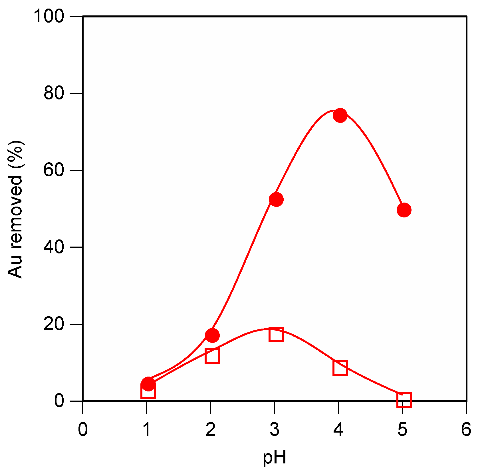
Gold (III) removal by C. krusei cells was markedly affected by pH (Figure 3). The maximum amount of gold removal occurred at pH 3 (1 h) or pH 4 (72 h).
These results suggest that longer time periods may change the reaction mechanism. The solution color was nearly colorless at 1 h. However, the color became dark (violet or green) during the 72 h period. Since the tetrachloroaurate ion has a negative charge, gold (III) can be effectively removed at pH 3 via biosorption [9]. Gold (III) can also be reduced to atomic gold (0) by the reductase activity of NADH [18] via biomineralization. Reduction proceeds by the equation.
2Au3+ + 3NADH→2Au + 3NAD+ + 3H+
The equilibrium in an acidic solution is driven to the left; suitable pH changed from 3 to 4.
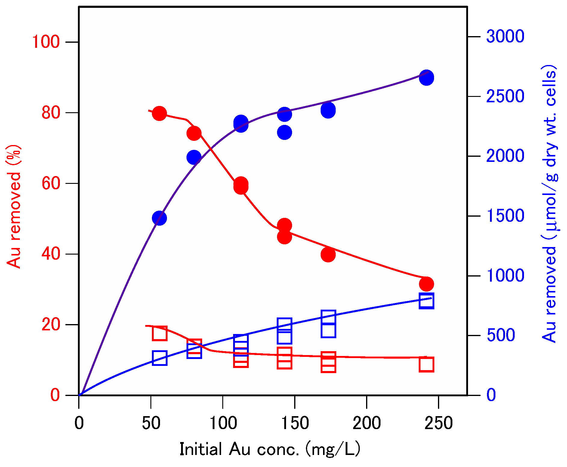
3.4. Effect of Cell Amount on Gold (III) Removal by C. krusei, AHU3993
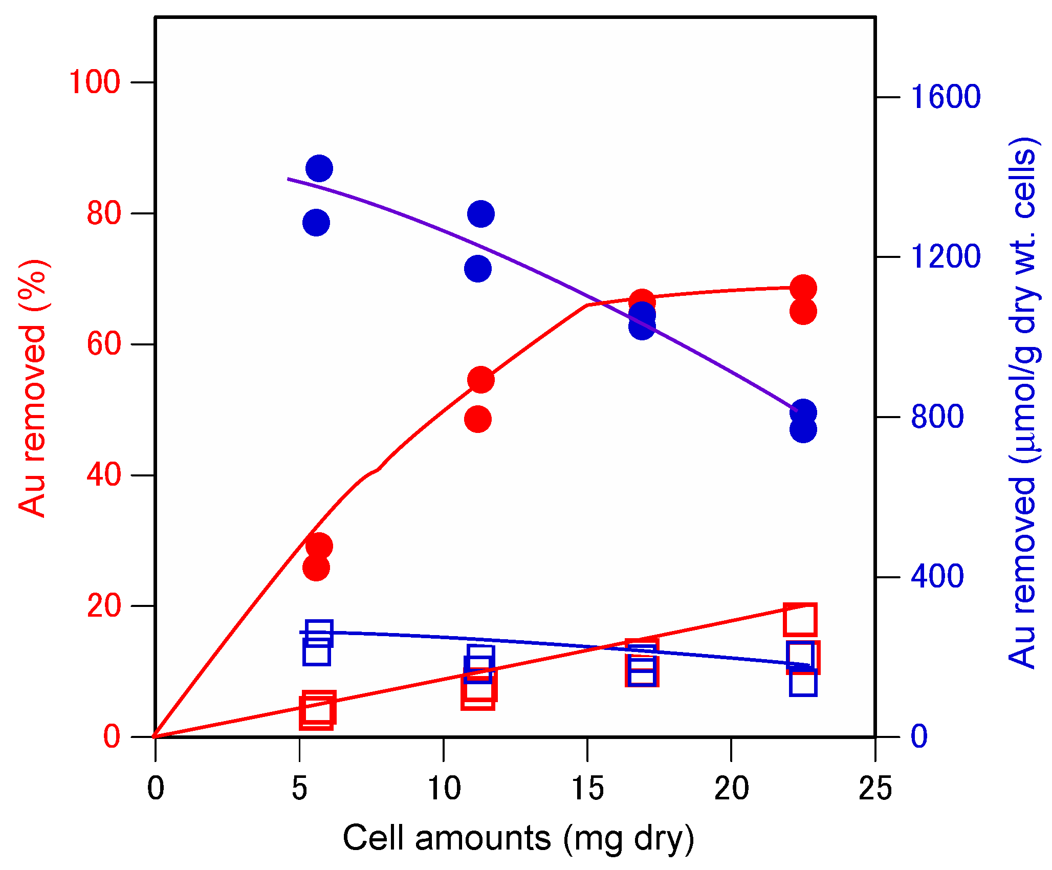
The relative amount of gold (III) removed (μmol/g dry weight cells) by C. krusei cells decreased with the increased cell amount (Figure 4). However, increasing the cell amount of C. krusei AHU3993 increased the total gold (III) removed. In 72 h approximately 1300 µmol gold/g dry weight cells were removed using 6 mg of the dry weight basis of C. krusei cells. Although the solution color did not change after the 1 h, the color changed to violet after 72 h.
3.5. Absorption Spectrometry Analysis of Gold Removal Using Varying Cell Amounts of C. krusei
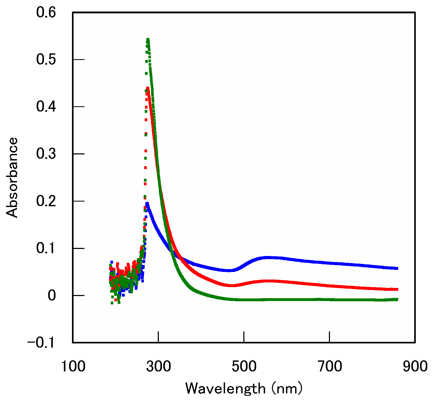
To distinguish between the biosorbed and the biomineralized gold, we analyzed the ionic gold (III) and colloidal atomic gold (III) by absorption spectrometry. The 300 nm absorbance peak decreased with an increasing cell amount of C. krusei, while, the broad peak at 500–550 nm increased (Figure 5). The 525 nm peak was identified as zero-valent gold [19] and the 500–650 nm peak was small and broad, because of the lower solubility of gold (0). Therefore, reduced gold was determined to be responsible by the cell amount. Accordingly, C. krusei caused both gold (III) biosorption and biomineralization during the 72 h period at 30 °C.
3.6. Effect of Gold (III) Concentration on Gold Removal by C. krusei
To determine the maximal gold (III) removal at pH 4.0, we examined the mechanism by which the gold (III) concentration affected gold removal by C. krusei cells. The amount of gold removed (µmol/g dry weight cells) by C. krusei cells increased with increased gold concentration, whereas the ratio of total amount of gold removed to gold concentration decreased (Figure 6). When the gold (III) concentration was 250 mg/L (1260 µM), 2600 µmol gold/gram dry cell weight was observed at pH 4.0.
3.7. Absorption Spectrometry Analysis of the Effect of Gold (III) Concentration on Gold Removal by C. krusei
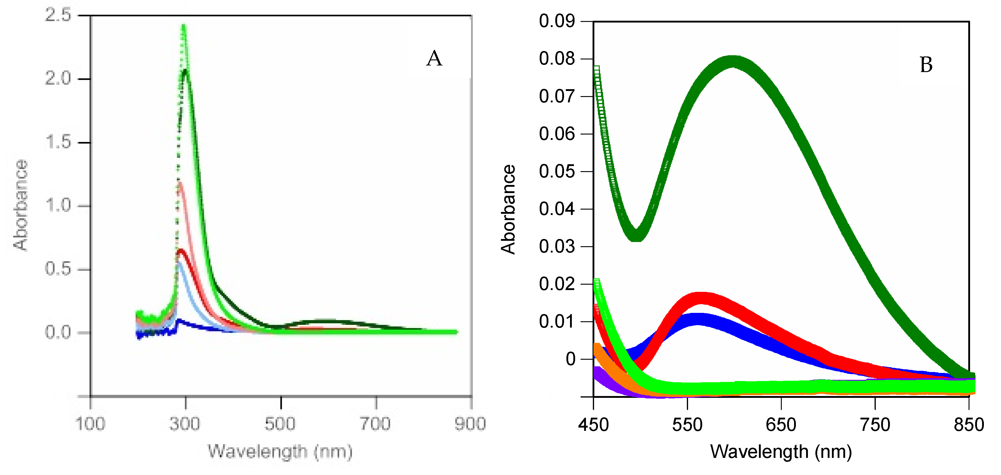
Absorption spectra were also measured to examine the effect of gold concentration on gold removal by C. krusei. The 300 nm absorbance peak and the 500–650 nm broad peak increased concomitantly with gold concentration (Figure 7). Therefore, reduced gold was found to be affected by the gold(III) concentration.
4. Conclusions
To optimize gold recovery, we first screened microorganisms for their ability to remove gold (III) from aqueous hydrogen tetrachloroaurate (III) solution (pH 3.0) after a 72 h incubation period at 30 °C. All the microorganisms tested removed gold from the solution. N. erythropolis IAM1399, gram-positive bacterium, E. coli IAM1264, P. maltophilia IAM1554, and P. saccharophila IAM1504, gram-negative bacteria, A. niger IAM2534, C. globosum IAM9272 and IAM9427, fungi, and C. krusei AHU3993, yeast removed gold at over 1200 µmol/g dry cell weight.
The effects of time, pH, cell amount, and gold concentration were analyzed by atomic absorption spectrometry. Absorption spectrometry analysis of the effect of cell amount and gold (III) concentration was also investigated. We observed that, small amounts of gold (III) were removed from the solution by biosorption over short time periods and large amounts of gold were reduced from gold (III) to zero-valent gold by biomineralization after 72 h by C. krusei AHU3993.
Author Contributions
Conceptualization, I.M. and T.T.; methodology, T.T.; software, T.T.; validation, T.T.; formal analysis, I.M and T.T.; investigation, I.M. and T.T.; resources, T.T.; data curation, I.M. and T.T.; writing—original draft preparation, T.T.; writing—review and editing, T.T.; visualization, T.T.; supervision, T.T.; project administration, T.T.; funding acquisition, T.T.; All authors have read and agreed to the published version of the manuscript.
Funding
This research received no external funding.
Conflicts of Interest
The authors declare no conflict of interest.
References
- Suhr, M.; Raff, J.; Pollmann, K. Au-Interaction of Slp1 Polymers and Monolayer from Lysinibacillus sphaericus JG-B53-QCM-D, ICP-MS and AFM as Tools for Biomolecule-metal Studies. J. Vis. Exp. 2016, 107, e53572. [Google Scholar] [CrossRef] [PubMed] [Green Version]
- Paez-Velez, C.; Rivas, R.E.; Dussan, J. Enhanced Gold Biosorption of Lysinibacillus sphaericus CBAM5 by Encapsulation of Bacteria in an Alginate Matrix. Metals 2019, 9, 818. [Google Scholar] [CrossRef] [Green Version]
- Gomes, N.C.M.; Camargos, E.R.S.; Dias, J.C.T.; Linardi, V.R. Gold and silver accumulation by Aspergillus niger from cyanide-containing solution obtained from the gold mining industry. World J. Microbiol. Biotechnol. 1998, 14, 149. [Google Scholar] [CrossRef]
- Matsumoto, M.; Nishimura, Y. Recovery by Aspergillus oryzae of gold from waste water from gold plating. Nippon Nougeikagakukaishi 1992, 66, 1765–1770. (In Japanese) [Google Scholar] [CrossRef]
- Pethkar, A.V.; Paknikar, K.M. Recovery of gold from solutions using Cladsporium cladosporioides biomass beads. J. Biotechnol. 1998, 63, 121–136. [Google Scholar] [CrossRef]
- Karamuchka, V.; Gadd, G.M. Interaction of Saccharomyces cerevisiae with gold: Toxicity and accumulation. BioMetals 1999, 12, 289–294. [Google Scholar] [CrossRef] [PubMed]
- Hosea, M.; Greene, B.; McPherson, R.; Henzl, M.; Alexander, M.D.; Darnall, D.W. Accumulation of elemental gold on the alga Chlorella vulgaris. Inorg. Chim. Acta 1986, 123, 161–165. [Google Scholar] [CrossRef]
- Kuyucak, N.; Volesky, B. Accumulation of gold by algal biosorbent. Biorecovery 1989, 1, 189–204. [Google Scholar]
- Tsuruta, T. Biosorption and recycling of gold using various microorganisms. J. Gen. Appl. Microbiol. 2004, 50, 221–228. [Google Scholar] [CrossRef] [PubMed] [Green Version]
- Tsuruta, T. Removal and recovery of lithium using various microorganisms. J. Biosci. Bioeng. 2005, 100, 562–566. [Google Scholar] [CrossRef] [PubMed]
- Tsuruta, T.; Umenai, D.; Hatano, T.; Hirajima, T.; Sasaki, K. Screening micro-organisms for cadmium absorption from aqueous solution and cadmium absorption properties of Arthrobacter nicotianae. Biosci. Biotechnol. Biochem. 2014, 78, 1791–1796. [Google Scholar] [CrossRef] [PubMed]
- Tsuruta, T. Removal and recovery of uranyl ion using various microorganisms. J. Biosci. Bioeng. 2002, 94, 23–28. [Google Scholar] [CrossRef]
- Tsuruta, T. Accumulation of thorium ion using various microorganisms. J. Gen. Appl. Microbiol. 2003, 49, 215–218. [Google Scholar] [CrossRef] [PubMed] [Green Version]
- Tsuruta, T. Accumulation of rare earth elements in various microorganisms. J. Rare Earths 2007, 25, 526–532. [Google Scholar] [CrossRef]
- Conn, E.E.; Stumpf, P.K.; Bruening, G.; Doi, R.H. Outlines of Biochemistry, 5th ed.; Wiley: New York, NY, USA, 1987; pp. 292–293. [Google Scholar]
- Fischer, W.; Ishizuka, I.; Landgraf, H.R.; Herrmann, J. Glycerophosphoryl diglucosyl diglyceride, a new phosphoglycolipid from Streptococcus. Biochim. Biophys. Acta 1973, 296, 527–545. [Google Scholar] [CrossRef]
- Fischer, W.; Landgraf, H.R.; Herrmann, J. Phosphatidyldiglucosyl diglyceride from Streptococci and its relationship to other polar lipids. Biochim. Biophys. Acta 1973, 306, 353–367. [Google Scholar] [CrossRef]
- Paul, R.J.; Schneckenburger, H. Oxygen concentration and the oxidation-reduction state of yeast: Determination of free/bound NADH and flavins by time-resolved spectroscopy. Sci. Nat. 1996, 83, 32–35. [Google Scholar] [CrossRef] [PubMed]
- Doremus, R.H. Optical properties of small gold particles. J. Chem. Phys. 1964, 40, 2389–2396. [Google Scholar] [CrossRef]
Figure 1.
Gold (III) removal from hydrogen tetrachloroaurate (III) solution using microorganisms after 72 h incubation.
Figure 1.
Gold (III) removal from hydrogen tetrachloroaurate (III) solution using microorganisms after 72 h incubation.

Figure 2.
The time course of gold removal using C. krusei AHU3993. Symbols: square (biosorption), circle: (biomineralization).
Figure 2.
The time course of gold removal using C. krusei AHU3993. Symbols: square (biosorption), circle: (biomineralization).

Figure 3.
Effect of pH on gold removal using C. krusei. Symbols, circle: contact 72 h (biomineralization), square symbols: 1 h (biosorption).
Figure 3.
Effect of pH on gold removal using C. krusei. Symbols, circle: contact 72 h (biomineralization), square symbols: 1 h (biosorption).

Figure 4.
Effect of the cell amounts of C. krusei AHU3993 used on gold removal. Red symbols: Au removed (%), blue symbols: Au removed (μmol/g dry weight cells), circle symbols, contact 72 h (biomineralization), square symbols 1 h (biosorption).
Figure 4.
Effect of the cell amounts of C. krusei AHU3993 used on gold removal. Red symbols: Au removed (%), blue symbols: Au removed (μmol/g dry weight cells), circle symbols, contact 72 h (biomineralization), square symbols 1 h (biosorption).

Figure 5.
Absorption spectrometry analysis of gold removal using varying C. krusei AHU3993 amount. The gold removal conditions were identical to that in Figure 4 (incubation for 72 h). Green line: the initial amount of gold (III); red line: incubation with 6mg dry weight basis cells, blue line: 22 mg dry weight basis cells.

Figure 6.
Effect of gold (III) concentration on gold removal by C. krusei AHU3993. Closed symbols: Au removed (%); open symbols: Au removed (μmol/g dry cell weight); circles: 72-h incubation (biomineralization); squares; 1-h incubation (biosorption).
Figure 6.
Effect of gold (III) concentration on gold removal by C. krusei AHU3993. Closed symbols: Au removed (%); open symbols: Au removed (μmol/g dry cell weight); circles: 72-h incubation (biomineralization); squares; 1-h incubation (biosorption).

Figure 7.
Absorption spectrometry analysis of the effect of gold (III) concentration on gold removal by C. krusei AHU3993. Gold removal conditions were identical to that in Figure 5. Symbols: Green line: 200 mg Au/L; red line: 100 mg Au/L; blue line: 50 mg Au/L. Light colors indicates gold (III) in the original solution, and each dark color indicates gold (III) levels after treatment. (A) Wavelength rom 200–870 nm, (B) Wavelength from 450–850 nm. Light blue line: initial 30 mg/L gold (III) solution; blue line: incubation with 30 mg/L gold (III) solution; light red line: initial 50 mg/L of gold (III) solution; red line: incubation with 50 mg/L gold (III) solution; light green line: initial 200 mg/L of gold (III) solution; green line: incubation with 200 mg/L gold (III) solution.

© 2020 by the authors. Licensee MDPI, Basel, Switzerland. This article is an open access article distributed under the terms and conditions of the Creative Commons Attribution (CC BY) license (http://creativecommons.org/licenses/by/4.0/).
Share and Cite
MDPI and ACS Style
Maeda, I.; Tsuruta, T. Microbial Gold Biosortion and Biomineralization from Aqueous HAuCl4 Solution. Minerals 2020, 10, 285. https://doi.org/10.3390/min10030285
AMA Style
Maeda I, Tsuruta T. Microbial Gold Biosortion and Biomineralization from Aqueous HAuCl4 Solution. Minerals. 2020; 10(3):285. https://doi.org/10.3390/min10030285
Chicago/Turabian StyleMaeda, Ichiro, and Takehiko Tsuruta. 2020. "Microbial Gold Biosortion and Biomineralization from Aqueous HAuCl4 Solution" Minerals 10, no. 3: 285. https://doi.org/10.3390/min10030285
Note that from the first issue of 2016, this journal uses article numbers instead of page numbers. See further details here.