Article

Dehydration of Diaspore and Goethite during Low-Temperature Heating as Criterion to Separate Unheated from Heated Rubies and Sapphires

1
Swiss Gemmological Institute SSEF, Aeschengraben 26, 4051 Basel, Switzerland
2
Department of Environmental Sciences, University Basel, 4056 Basel, Switzerland
3
Experimental Mineral Physics, Institute for Geochemistry and Petrology, Federal Institute of Technology, 8092 Zurich, Switzerland
*
Author to whom correspondence should be addressed.
Minerals 2023, 13(12), 1557; https://doi.org/10.3390/min13121557
Submission received: 26 September 2023 / Revised: 1 December 2023 / Accepted: 16 December 2023 / Published: 18 December 2023
(This article belongs to the Special Issue Gem Characterisation)

Abstract

:
Gem-quality rubies and sapphires are often commercially heat treated at about 800 °C or higher to enhance their color and clarity, and hence quality. For this study, selected corundum samples containing diaspore and goethite inclusions were heated step-by-step to a maximum of 1000 °C with the aim of monitoring the dehydration and phase transformation of these oxyhydroxides to corundum and hematite during heating. Based on our experiments and in agreement with the literature, the dehydration of diaspore in corundum occurs between 525 and 550 °C, whereas goethite transforms to hematite between 300 and 325 °C. As both diaspore and goethite may be present as inclusions in rubies, sapphires, and other corundum varieties (e.g., pink sapphires, padparadscha), these dehydration reactions and phase transformations can be considered important criteria to separate unheated from heated stones, specifically in cases in which other methods (e.g., microscopy, FTIR) are unsuccessful.

1. Introduction

Since historical times, it has been known that the visual appearance of certain gemstones may be enhanced by heat treatment [1] and references therein. For gem-quality corundum Al2O3, various heat treatment processes with or without additives have been developed, specifically in the last few decades. They are widely applied in the gem trade (e.g., in cutting/manufacturing and trading hubs in Sri Lanka, Thailand, and India) to enhance the color and additionally—in some cases—the transparency and stability of corundum. By using such heat treatments, it is possible to modify corundum of lower quality into visually beautiful stones of better color and clarity and consequently guarantee a steady supply of gems to the international market.
Heat treatment of ruby and sapphire varieties is usually carried out at temperatures ranging from about 800 to 1800 °C. Historically by using a simple and artisanal blow-pipe (e.g., in Sri Lanka) reaching temperatures of about 1200 °C [2], heating has evolved into a multitude of treatment options, which are often carried out under controlled conditions (e.g., atmosphere) in electric muffle furnaces [2,3,4,5]. Heating of corundum at higher temperatures (>1200–1350 °C) may have a considerable impact on its internal features and color. Notably, local discoid expansion cracks may develop around (transformed) solid and fluid inclusions. In addition, tiny rutile (TiO2) needles commonly present in corundum as so-called “silk” may start to (partially) dissolve at temperatures > 1400 °C. In contrast to this, a heat treatment process at about 800 °C to 1200 °C is generally known in the trade as “low-temperature” heating. It usually results in a slight (but desirable) to distinct (e.g., Fe-rich metamorphic sapphires) shift of color and may leave inclusions visually unaltered [6,7]. As such, the detection of heat treatment in corundum today relies not only on “classic” microscopic observations but also on infrared spectroscopy (FTIR) and Raman micro-spectroscopy.
The presence and arrangement of OH-Ti4+ related bands in the mid-infrared spectral range [8,9,10,11,12,13,14] of corundum of metamorphic origin (not applicable for basaltic sapphires) and the peak width (FWHM) of the main Raman peak (at about 1010 cm−1) of tiny zircon inclusions in corundum (mainly pink sapphires from Ilakaka, Madagascar) are considered important criteria to separate unheated corundum from heated stones [6,15,16]. However, both mentioned analytical approaches have certain limitations, i.e., hydroxide-related bands in infrared spectra are not always present in heated rubies and sapphires [17,18], and the FWHM of the main Raman peak of zircon inclusions in unheated and heated corundum may show considerable overlap [6,16]. Additionally, the Raman bandwidth of zircon inclusions is strongly dependent on the concentration of radioactive trace elements, crystallinity (metamictisation), and finally, geological and geographic origin where the corundum formed [19,20,21]. As shown in a recent study [7], further inclusions (e.g., apatite, spinel) may show a broadening of Raman peaks as a consequence of heating, and as such, these inclusions may assist in certain cases in detecting heat treatment of corundum.
It has been known and documented for many decades that the two isostructural oxyhydroxides goethite α-FeO(OH) and diaspore α-AlO(OH) transform upon heating into the anhydrous oxides hematite α-Fe2O3 and corundum α-Al2O3 (coupled with a release of water H2O) [22,23,24,25,26,27,28,29]. The pseudomorphous character of the dehydration phase transformation is due to the fact that there is no major change in the relative positions of the remaining oxygen atoms of the oxides compared with the originally present oxyhydroxides goethite and diaspore.
Although the temperature at which the phase transformation of these oxyhydroxides into hematite and corundum occurs is to some extent related to the duration of heating (minutes vs. days), pressure, and the size of the crystallites, goethite generally dehydrates to hematite at about 350 °C [26], whereas for diaspore this dehydration occurs at higher temperatures at about 550 °C [30]. XRD studies on the dehydration of goethite and diaspore suggest the formation of intermediate structural states during their phase transformation to hematite and corundum [26,27,30,31,32,33,34].
Raman spectroscopy has proven to be a very useful analytical method to document these dehydration phase transformations. In contrast to crystal structure analyses by XRD (X-ray powder diffraction), Raman spectroscopy has the advantage of being a non-destructive technique (NDT), requiring quasi-no sample preparation, and with the capability to analyze structural changes in situ during heating experiments [35,36,37,38]. However, as the focused laser beam used for Raman analyses may locally affect oxyhydroxides (e.g., goethite) considerably by degradation and dehydration effects [35,39], it is necessary to carefully keep laser power at low levels to avoid artifacts and misinterpretation.
The dehydration of goethite as a marker for a gemstone heat treatment has been known in the gemmological literature for quite some time [40,41,42,43]. The focus of this study is to present detailed data of in situ heating experiments not only for the goethite–hematite phase transformation but also for the dehydration of diaspore. Using real cases, this study also demonstrates the potential of this analytical approach to separate unheated from heated rubies and sapphires, even in cases in which other methods (e.g., FTIR, microscopy) were unsuccessful.

2. Materials and Methods

To study the dehydration of diaspore and goethite as inclusions in ruby and sapphires, we selected three unheated rough ruby samples and one faceted sapphire. The rubies were cut and polished into small flat discs perpendicular to the optic axis, containing either diaspore or goethite as inclusions. Two of these samples (rubies 120553_B and 85933_C3) originated from the economically important ruby deposit in the Montepuez area in northern Mozambique and were supplied by Gemfields Ltd., London, UK. The two other samples (ruby 126993_6 and sapphire 106424_21) reportedly originated from the Mogok area in northern Myanmar, known since historical times as a source of exceptional rubies and other gemstones and were supplied by a Burmese gemstone trader in Bangkok (SilkenEast Ltd.; Bangkok, Thailand). Their initial treatment status (unheated) and reported origin were analytically confirmed prior to the experiments. In addition, a small crystal fragment of gem-quality diaspore from the Muğla Province in western Turkey was included in our heating experiments (see Table 1).
Two different experimental setups were chosen for this study (see Table 2): (a) a Linkam TS-1500 heating stage, which was fixed directly to the Raman sample stage and which allows Raman analyses on exactly the same analytical spot during heating experiments, and (b) an electrical muffle furnace (Nabertherm LHT 18) similar to those commonly used for commercial heat treatment of gemstones. In both cases, heating was performed step-by-step, reaching a maximum temperature of 1000 °C in one experiment. Raman spectra were recorded after each heating step only after cooling to room temperature (about 25 °C) again.
Although it would be possible to analyze a sample at peak temperature in situ with the Linkam heating stage, the very strong Cr-related luminescence of the ruby samples at elevated temperatures when using a green laser (514 or 532 nm) made it necessary to cool the samples after each heating step. Both setups come with a controller unit, which allows for time-controlled heating (ramping up) and cooling (ramping down) of the sample. The temperature ramping rate was pre-selected at 80 °C/min for the Linkam stage and 50 °C/min for the electric furnace. The temperature was kept stable at each heating step for a defined time (4 min with the Linkam stage and 1 h with the electric furnace) and, after each heating step, subsequently cooled to room temperature for Raman analyses.
We used a Renishaw confocal Raman microscope (InViaTM) with a Peltier-cooled CCD detector (1024 × 256 pixels), resulting in a maximum spectral resolution of about 1.5 cm−1. The system is coupled with a Leica stereo microscope and a set of objectives (10× to 50×). The Raman spectra were collected using an argon-ion laser emitting at 514.5 nm. The laser beam was focused on the samples and inclusions using a 50× long-range objective, resulting in a spot size of ca. 2 µm. The laser power for analyzing the ruby surface and diaspore was set at a standard of 35 mW (100% laser emission in our setup, measured on the sample surface). For goethite analyses, the laser power was kept below 3 mW at the sample (10% laser) to avoid laser-induced degradation and dehydration effects during analysis [33,37]. The Raman spectra of all our samples are accumulated from 5 to 7 spectral scans at 15 s exposure time on the same analytical spot. All spectra were baseline corrected (WireTM software) using a built-in cubic-spline integration baseline function.
Additionally, all samples were analyzed using FTIR spectroscopy (Nicolet iS50 equipped with a DLaTGS detector using a diffuse reflectance accessory with 128 scans and 4 cm−1 resolution in the range from 400–6000 cm−1) before and after the heating experiments. The two Mozambique rubies (samples 85933_C3 and 120553_B) and the ruby from Myanmar initially showed very broad goethite and diaspore bands in their FTIR spectra, respectively, which disappeared after heating. The sapphire (sample 106424_21) showed no diaspore bands before heating. Importantly, the samples showed no features that would have allowed a straightforward heat treatment detection after the experiments (no 3232 cm−1 peak), except for the sample 123993_C (ruby with goethite), in which a small peak at 3232 cm−1 peak developed as a result of the heating experiment.

3. Experiments and Results

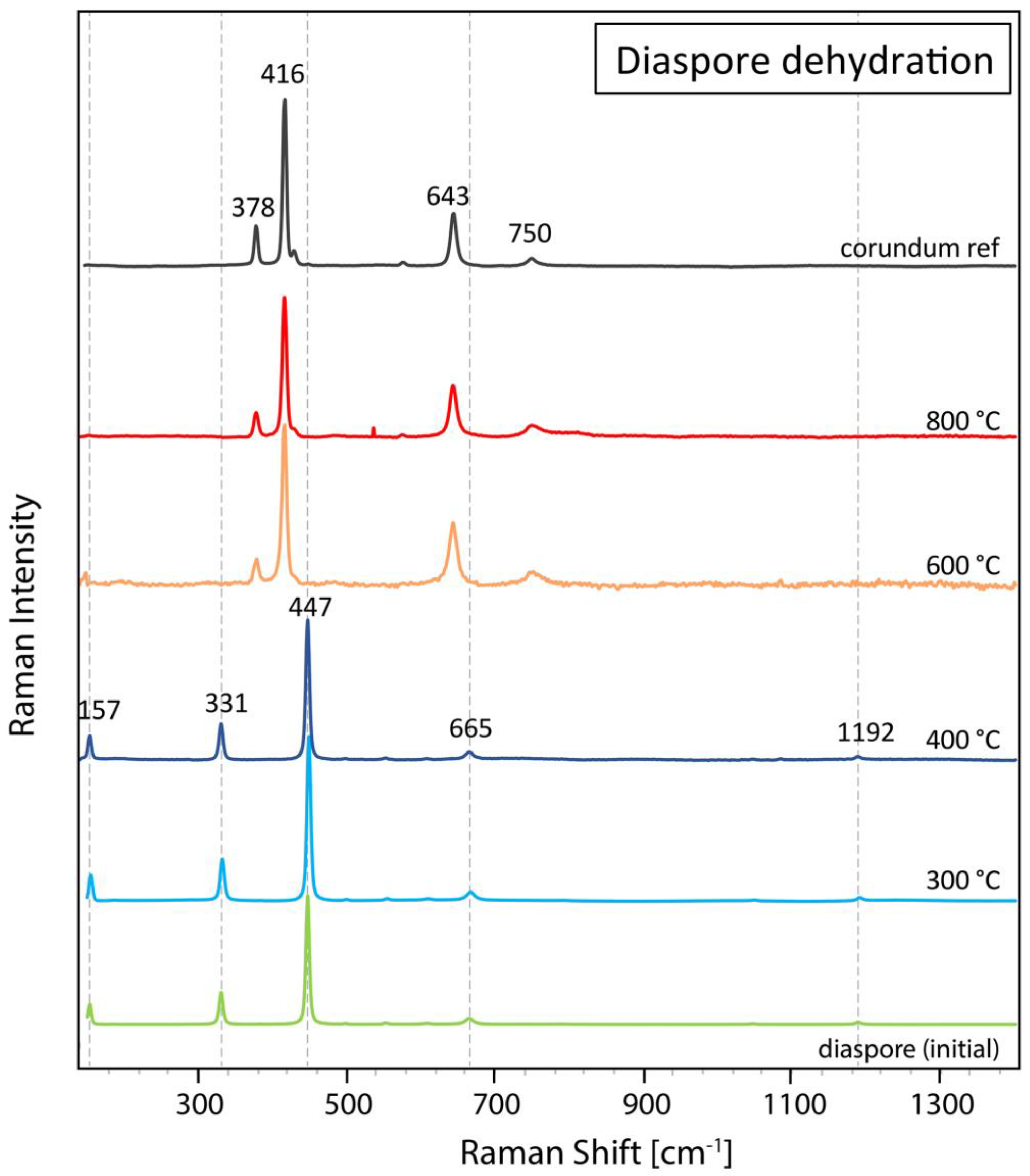
3.1. Heating of Diaspore in Ruby and Sapphires

For the first experiment, a flat polished ruby from the Mogok valley (Myanmar, sample 126993_6) containing a cluster of colorless diaspore inclusions (>2 mm) at just the surface (Figure 1) was heated at increasing temperature steps using the Linkam heating stage. For Raman analyses a defined position within the diaspore was chosen. Starting at 200 °C, the sample was subsequently heated in 25 °C steps until 600 °C, with a final heating step at 700 °C. At each step, the temperature was kept stable for 4 min. Raman spectra of a defined position within the diaspore inclusion were registered only after cooling each time back to room temperature. The aim of this dehydration experiment was to monitor the temperature range of the phase transformation from diaspore to corundum as closely as possible.
Up to 500 °C, Raman spectra reveal only peaks related to diaspore with the main peak at 447 cm−1 and adjacent side bands. At 525 °C, a diaspore spectrum is still visible, but for the first time, with a small peak at 416 cm−1 indicating the beginning of the phase transformation to corundum (see Table 3). At 550 °C, however, the Raman spectra only show peaks corresponding to corundum, but no diaspore peaks anymore, thus indicating that the dehydration and phase transformation from diaspore to corundum has occurred (Figure 2). As a consequence of this phase transformation, the colorless diaspore cluster has become whitish (see Figure 1). Further heating up to 700 °C does not have much further effect on the Raman spectrum. The absence of the 643 cm−1 peak in the heated ruby sample compared with the reference corundum is an orientation effect well-known in anisotropic crystal structures.
To verify that the phase transformation of diaspore to corundum is not influenced by the size of the inclusion, we selected a sapphire (106424_21) from Myanmar containing a tiny diaspore needle (few microns) within a fluid inclusion of a healed fissure (Figure 3a,b). Using the Linkam heating stage, this tiny inclusion was analyzed in five steps, starting from room temperature up to a maximum temperature of 700 °C (Figure 4).
The dehydration of the tiny diaspore inclusion (about 10 µm) occurred in a similar temperature range as the larger diaspore in ruby. At 600 °C and beyond, the diaspore has completely transformed into corundum. It is important to know that the presence of corundum peaks at various heights in the spectra below 500 °C in Figure 4 is related to the sapphire matrix, in which this fluid inclusion with the tiny diaspore is located. These peaks do not represent a gradually ongoing phase transformation process, as the diaspore remains stable up to 500 °C.
In addition to these two diaspore inclusions in ruby and sapphire, a transparent diaspore crystal fragment (sample 97003) was heated step-by-step to a maximum of 800 °C in an electric furnace to confirm the dehydration and phase transformation of diaspore to corundum during a so-called “low-temperature” heating treatment as commercially applied on ruby and other corundum varieties. For this experiment, the setup (electric furnace) and conditions were chosen in a manner similar to those used in the gem trade. Visually, this experiment transformed the colorless diaspore into a whitish corundum characterized by numerous tiny micro-fissures, which were induced by the heating and concurrent expulsion of water during the dehydration (Figure 5).
In total five Raman spectra were taken (before heating and after each heating step), always in the same crystallographic orientation and approximately the same position under the Raman microscope (Figure 6). As expected, the diaspore dehydration had fully succeeded after heating at 600 °C for 1 h, revealing now only Raman bands related to Al-O vibrational modes of corundum. By increasing the temperature further to 800 °C, the corundum spectrum became even more pronounced with a distinctly better peak/noise ratio.

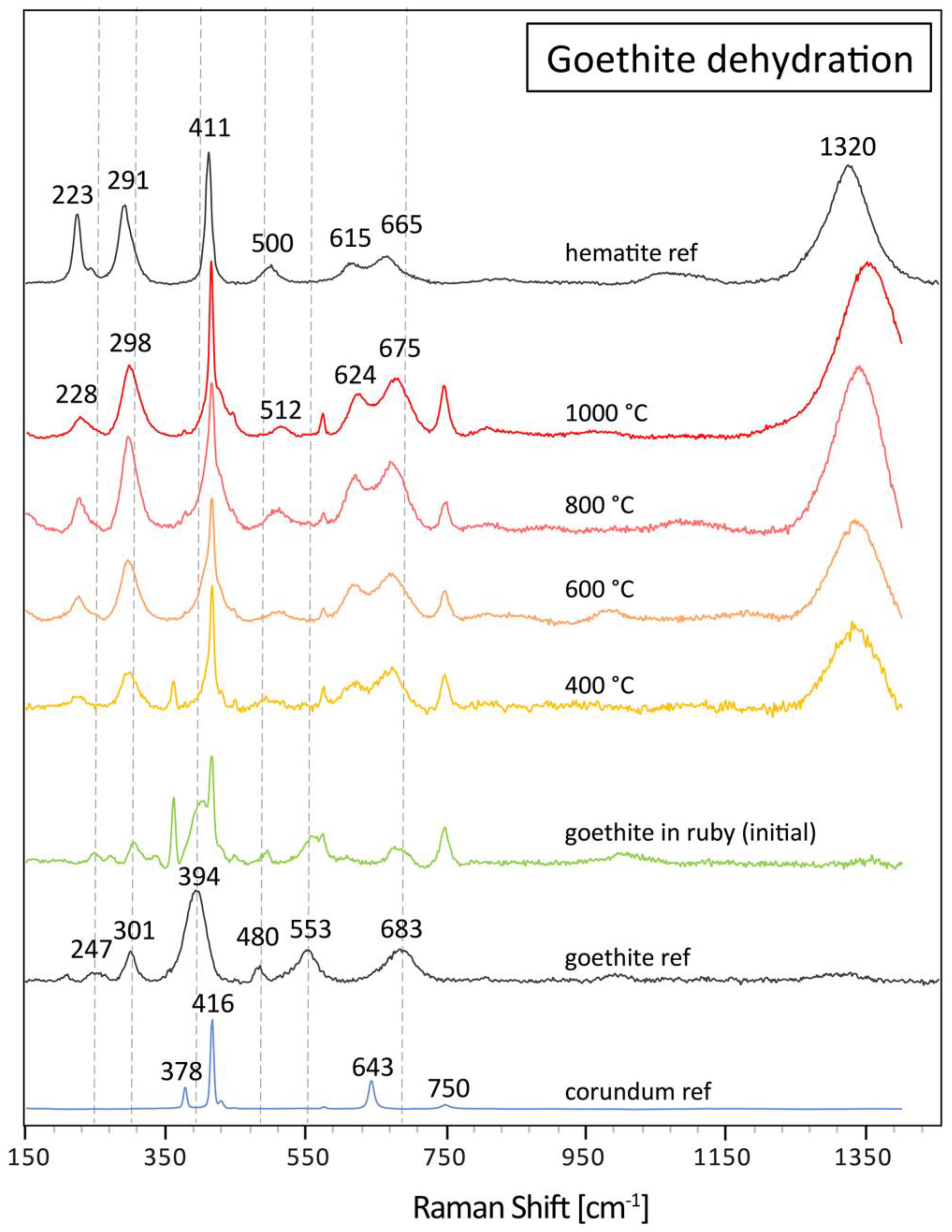
3.2. Heating of Goethite in Ruby
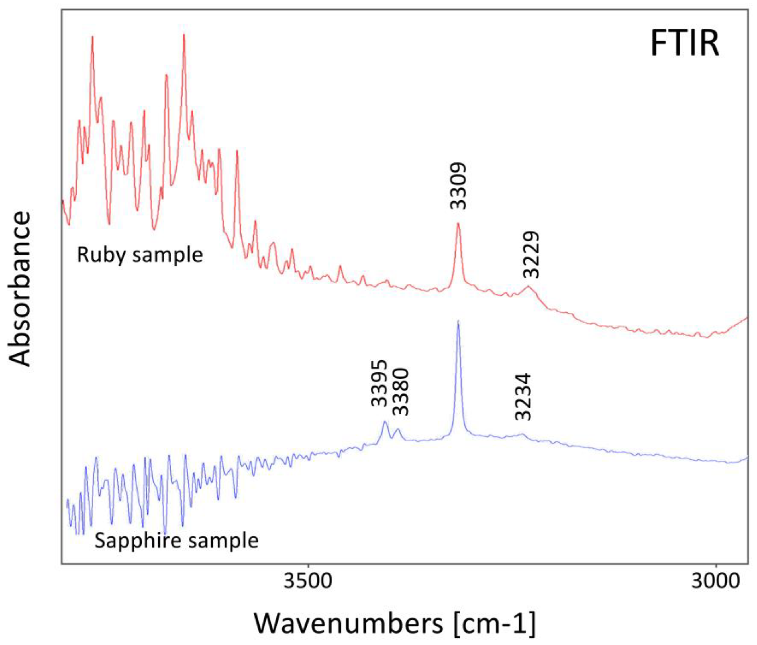
Similar to the heating experiment for diaspore dehydration, a flat polished ruby sample (85933_C3) containing orangey–brown goethite in fissures was heated repeatedly at increasing temperature steps using the Linkam heating stage. For Raman analyses a defined position within a goethite-bearing fissure was chosen. Starting at 150 °C, the sample was subsequently heated in 25 °C steps until 400 °C. At each step, the temperature was kept stable for 4 min. Raman spectra of a defined position within the goethite-containing fissure were registered only after cooling each time back to room temperature. The aim of this dehydration experiment was to monitor the temperature range of the goethite to hematite phase transformation when occurring as a thin film in a tiny fissure within the corundum as closely as possible (Figure 7).
Up to 300 °C, the goethite spectrum predominates, with small peak interferences with corundum peaks from the ruby host adjacent to the goethite-bearing fissure (Figure 8). At 325 °C, the Raman bands shift towards hematite positions. Namely, the main broad goethite band at about 400 cm−1 is transformed to a rather sharp peak (ideally 411 cm−1), coincidentally superposed by the main corundum peak at 416 cm−1, thus resulting in an inclined shoulder to the main corundum peak rather than a singular hematite peak as illustrated in the hematite reference spectrum. In addition, the smaller side peaks of goethite at 247, 301, and 683 cm−1 shift towards the hematite positions at 223, 291, and 665 cm−1. In addition, a distinct broad band at about 1320 cm−1 appears and develops further in size with increasing heating temperature. The weak and broad band at about 1320 cm−1 in the goethite spectra may be the result of small amounts of hematite in the unheated sample, but more likely is a result of local laser-induced degradation as it appeared during the accumulation of the five spectral scans for each Raman analysis. Only after heating above 325 °C does the phase transformation occur, resulting in the shift of bands and a strong band at about 1320 cm−1, which is characteristic of hematite.
To confirm that the dehydration and phase transformation of goethite to hematite occurs during heat treatment of rubies commonly adopted in the trade, we heated a second ruby from Mozambique (sample 120553_B) containing goethite in an electric furnace up to a maximum temperature of 1000 °C, and repeatedly took Raman spectra always at room temperature after each heating step (Figure 9).
Similar to the experiment with the Linkam stage, the dehydration of goethite to hematite is completed after heating to 400 °C for 1 h in the electric furnace. Further heating steps just increased the hematite peak intensities in the sample but with no further changes in peak positions. After heating in the electric furnace at a higher temperature, the Fe-staining in the fissures changes from an originally brownish–orange (goethite) to a brownish–red (hematite) color.

4. Discussion

The dehydration of the oxyhydroxides diaspore and goethite was observed in all five samples within the same temperature ranges (325 °C for goethite to hematite and 525 °C for diaspore to corundum, see Figure 10), regardless of the size of the inclusions and the experimental setup, i.e., a Linkam heating stage coupled to a Raman system for in situ analyses or an electric muffle furnace for heating at higher temperatures using similar setup and conditions as used in the gem trade for heat treatments. In both our setups, the accumulated heating time at which the phase transformations occurred was in total between 0.5 and 3 h, a rather short but realistic heat treatment duration when applied commercially on gem-quality rubies and other corundum varieties. Our findings are well in accordance with the extensive literature about goethite and diaspore dehydration using similar heating parameters [26,27,30,35,37,48,49].
Gem-quality rubies and other corundum varieties often contain microscopic inclusions, among them specifically diaspore and goethite. Diaspore is commonly found in corundum as a retrograde hydrothermal alteration product, e.g., as whitish acicular needles in ruby fissures or as tiny prismatic solids in fluid inclusions and negative inclusions [50]. Goethite is often present in corundum in fissures and (tiny) hollow channels as a pedogenic weathering product [43], introduced by Fe-enriched meteoric waters circulating within the gravels of secondary gem deposits. Another option is to find goethite as a solid inclusion due to the weathering and hydration of a former Fe-oxide inclusion in a gemstone. Although it is, in principle, possible to introduce goethite deliberately into fissures and cavities of gemstones, even after a heat treatment process, we have found no indication so far that such a filling process was applied, nor were our experiments successful in penetrating goethite powder deep into very fine fissures and hollow channels in corundum. For this, we diluted goethite in water and immersed two samples in the water for two hours before drying the samples in the air. In cavities at the surface, some Fe-hydroxide could be observed, but no Raman signal could be measured (only strong luminescence). In the small fissures and fine hollow channels, no goethite could be detected in the samples after our experiments.
To illustrate the direct application of the results of our study to separate unheated from heated corundum, we add two real cases: a ruby from Mozambique with a diaspore inclusion (in a negative crystal) and a sapphire from Sri Lanka with brownish goethite in a fissure. In both cases, it was readily possible to analyze these inclusions and to identify them as diaspore and goethite, respectively (Figure 11). Consequently, the presence of these two oxyhydroxides positively confirms that these ruby and sapphire samples were not heat treated. The two inclusion phases would have dehydrated during heating as commercially applied on corundum (usually >800 °C).
These two unheated examples are of specific interest, as they both show peaks (mainly at 3309 and at about 3230 cm−1) in their infrared spectra (Figure 12), which are similar to those encountered in heated corundum [8,9,10,11,12,18]. Due to this, these examples may be wrongly interpreted as heated. However, it is important to note that the broad peak at 3229 cm−1 in the ruby sample is not related to the 3232 cm−1 peak often observed after heat treatment. Similarly, the 3234 cm−1 peak analyzed in the sapphire sample is, in fact, forming a series with the peaks at 3395 and 3380 cm−1 and is unrelated to heating. This shows how relevant this study is for separating unheated from heated corundum, as the detection of diaspore and/or goethite inclusions may help to avoid misinterpretation of an unheated corundum as heated. A more detailed study about FTIR spectra of corundum regarding heat treatment detection is in preparation.
The results of our experiments confirm that phase transformations such as the described dehydration of diaspore to corundum and goethite to hematite occur at temperatures (about 325 °C for goethite and 525 °C for diaspore) much lower than any successful heat treatment on corundum (generally well above 800 °C) described in the gemmological literature [1,3,4,5,7,13,15,18,51,52] and known to the authors. Furthermore, these dehydration reactions cannot be suppressed or shifted towards higher temperatures by any heat treatment commonly applied on gemstones and proceed in a rather small temperature range and thus result in a quasi-sudden shift of the Raman spectrum from goethite to hematite and diaspore to corundum as soon as the dehydration reaction occurs.
We have not seen any influence of the size of the inclusion on dehydration temperature, and to our knowledge, such an effect has never been described in the literature. In our experiments, regardless of whether the oxyhydroxide inclusion was only a few microns (sapphire 106424_21) or several mm (ruby 126993_6) in size, the dehydration could be observed in the same temperature range well below commercially applied heat treatment of corundum. This is, in fact, also the advantage of this method, in our opinion, as the dehydration of diaspore (and goethite) is unrelated to the size or position of these inclusions within corundum.
Interestingly, a long duration of heating at low temperatures will only shift the dehydration to lower temperatures [26]. By using the Linkam heating stage, we actually used short heating times (4 min at maximum temperatures) to check if the dehydration is also occurring after very short heat treatment. This was proven; see, for example, the sapphire (sample 106424_21) with only a tiny diaspore in a fluid inclusion.
It is thus safe to say that both described dehydration phase transformations and, specifically, the presence of diaspore and goethite can be used in many cases as criteria to separate unheated from heated rubies or other corundum varieties. This is even valid for many other gemstones that are commercially heated, as long as the heating temperature is higher than the dehydration temperatures of goethite (about 325 °C) and diaspore (about 525 °C).
However, it is important to note that the absence of diaspore or goethite does not necessarily imply that ruby or other corundum variety (or any other gemstone) has been heat-treated. For example, it is possible to find naturally formed hematite in fissures and at the surface of corundum, specifically when the gemstone in its rough form is still partially coated with Fe-rich soil. In such cases, however, careful analyses of several Fe-rich pedogenic inclusions in the same gemstone will often reveal that hematite is present together with Fe-hydroxides, thus still proof that this stone was not commercially heat treated to enhance its visual appearance.

5. Conclusions

Diaspore and goethite are often found as inclusions in ruby and other corundum varieties and are readily identifiable using Raman micro-spectroscopy. For this Raman study, we have carried out heating experiments on gem-quality rubies (corundum) containing tiny inclusions of goethite and diaspore. For these experiments, we used a heating stage for in situ Raman analyses of these inclusions at different temperatures and an electric muffle furnace similar to those commercially used for gemstone treatment, e.g., in Sri Lanka and Thailand.
Our experiments reveal that regardless of the size of the inclusion, the dehydration of goethite to hematite occurs at about 325 °C, whereas the phase transformation of diaspore to corundum occurs at about 525 °C. These temperatures are well in accordance with the literature about the thermal transformation of both oxyhydroxides and their anhydrous oxides. Compared with this, commercial heat treatment of corundum is known to be carried out at temperatures above 800 °C, thus much higher than the described dehydration reactions. Consequently, experiments reveal that the presence of one of these two oxyhydroxides is a very safe criterion to positively confirm that a ruby or other corundum variety has not been heat treated to enhance its visual appearance. This is even true in cases in which other analytical methods, such as FTIR spectroscopy or Raman analyses on the width of the main Raman peak of zircon inclusions (FWHM1010) within corundum are not conclusive for the separation of unheated and heated rubies and other corundum varieties.

Author Contributions

All authors were involved in designing the experiments. M.S.K., P.L. and W.Z. collected and prepared the samples, including gemological characterization. M.S.K., G.S. and J.B. conducted heating experiments. M.S.K. performed data analysis and prepared the manuscript. All authors contributed to the figures and the manuscript. M.S.K., P.L. and W.Z. conceived of and supervised the project. All authors have read and agreed to the published version of the manuscript.

Funding

This research received no external funding.

Data Availability Statement

The data underlying the results in this paper are not publicly available at this time but may be obtained from the corresponding author upon request.

Acknowledgments

The authors would like to thank Sean Gilbertson and Elena Basaglia from Gemfields Ltd. for supplying unheated ruby samples from Mozambique. The Burmese ruby sample for this study was kindly donated by Miemie Htin Tut, Silkeneast Ltd., Bangkok. The authors would like to further thank SSEF staff, namely Walter A. Balmer, Alexander Klumb, Michael Rytz, Laurent Cartier, Hao Wang, Markus Wälle, and Carole Lachavanne for fruitful discussions and comments.

Conflicts of Interest

The authors declare no conflict of interest.

References

  1. Hughes, R.W.; Manorotkul, W.; Hughes, E.B. Ruby & Sapphire: A Gemologist’s Guide; RWH Publishing/Lotus Publishing: Bangkok, Thailand, 2017; p. 886. [Google Scholar]
  2. Notari, F.; Hainschwang, T.; Caplan, C.; Ho, K. The heat treatment of corundum at moderate temperature. InColor Mag. 2019, 42, 76–85. [Google Scholar]
  3. Nassau, K. Heat treating ruby and sapphires: Technical aspects. Gems Gemol. 1981, 17, 121–131. [Google Scholar] [CrossRef]
  4. Themelis, T. The Heat Treatment of Ruby and Sapphire; Publisher Gemlab Inc.: Wheatland, PA, USA, 1992. [Google Scholar]
  5. GIT. A brief history of gem corundum heat treatment in Thailand. InColor Mag. 2019, 68–74, Spring issue. [Google Scholar]
  6. Karampelas, S.; Hennebois, U.; Mevellec, J.-Y.; Pardieu, V.; Delaunay, A.; Fritsch, E. Pink to purple sapphires from Ilakaka, Madagascar: Insights to separate unheated from heated samples. Minerals 2023, 13, 704. [Google Scholar] [CrossRef]
  7. Hughes, E.B.; Vertriest, W. A Canary in the ruby mine: Low-temperature heat treatment experiments on ruby. Gems Gemol. 2022, 58, 400–423. [Google Scholar] [CrossRef]
  8. Smith, C.P. A contribution to understanding the infrared spectra of rubies from Mong Hsu, Myanmar. J. Gemmol. 1995, 24, 321–335. [Google Scholar] [CrossRef]
  9. Smith, C.P. Infrared spectra of gem corundum. Gems Gemol. 2006, 42, 92–93. [Google Scholar]
  10. Beran, A.; Rossman, G.R. OH in naturally occurring corundum. Eur. J. Mineral. 2006, 18, 441–447. [Google Scholar] [CrossRef]
  11. Krzemnicki, M.S. New Research by SSEF Studies Methods for Detecting Low-Temperature Heated Rubies from Mozambique. SSEF Press Release, 2018. Available online: https://www.ssef.ch/detecting-low-temperature-heated-rubies-from-mozambique/ (accessed on 1 July 2023).
  12. Saeseaw, S.; Khowpong, C.; Vertriest, W. Low-temperature heat treatment of pink sapphires from Ilakaka, Madagascar. Gems Gemol. 2020, 56, 448–457. [Google Scholar] [CrossRef]
  13. Balmer, W.; Leelawatanasuk, T.; Atichat, W.; Wathanakul, P.; Somboon, C. Update on FTIR characteristics of heated and unheated yellow sapphire. In Proceedings of the GIT Conference, Bangkok, Thailand, 6–9 December 2006; Volume 6–7, p. 91. [Google Scholar]
  14. Balmer, W.A. Petrology, Geochemistry, and Gemological Characteritics of Marble-Hosted Ruby Deposits of the Morogoro Region, Tanzania. Unpublished. Ph.D. Thesis, Depart. of Geology, Faculty of Science, Chulalongkorn University, Bangkok, Thailand, 2011; p. 185p. [Google Scholar]
  15. Wang, W.; Scarratt, K.; Emmett, J.L.; Breeding, C.M.; Douthit, T.R. The effects of heat treatment on zircon inclusions in Madagascar sapphires. Gems Gemol. 2006, 42, 134–150. [Google Scholar] [CrossRef]
  16. Krzemnicki, M.S.; Lefèvre, P.; Zhou, W.; Wang, H.A.O. Zircon inclusions in unheated pink sapphires from Ilakaka, Madagascar: A Raman spectroscopic study. In Proceedings of the International Gemmological Conference, Online, 20–21 November 2021. [Google Scholar]
  17. Saeseaw, S.; Kongsomart, B.; Atikarnsakul, U.; Khowpong, C.; Vertriest, W.; Soonthorntantikul, W. Update on “Low-Temperature” Heat Treatment of Mozambican Ruby: A Focus on Inclusions and Ftir Spectroscopy. GIA Lab Report. 2018. Available online: https://www.gia.edu/doc/low_HT_Moz_report.pdf (accessed on 1 July 2023).
  18. Pardieu, V.; Saeseaw, S.; Detroyat, S.; Raynaud, V.; Sangsawong, S.; Bhusrisom, T.; Engniwat, S.; Muyal, J. GIA Lab Reports on Low-Temperature Heat Treatment of Mozambique Ruby. GIA Lab Report, 2015. Available online: https://www.gia.edu/gia-news-research-low-temperature-heat-treatment-mozambique-ruby (accessed on 25 September 2023).
  19. Nasdala, L.; Irmer, G.; Wolf, D. The degree of metamictization in zircon: A Raman spectroscopic study. Eur. J. Mineral. 1995, 7, 471–478. [Google Scholar] [CrossRef]
  20. Nasdala, L.; Wenzel, M.; Vavra, G.; Irmer, G.; Wenzel, T.; Kober, B. Metamictisation of natural zircon: Accumulation versus thermal annealing of radioactivity-induced damage. Contrib. Mineral. Petrol. 2001, 141, 125–144. [Google Scholar] [CrossRef]
  21. Xu, W.; Krzemnicki, M.S. Raman spectroscopic investigation of zircon in gem-quality sapphire: Application in origin determination. J. Raman Spectrosc. 2021, 52, 1011–1021. [Google Scholar] [CrossRef]
  22. Deflandre, M.M. La structure cristalline du diaspore. Bull. Minéralogie 1932, 55, 140–165. [Google Scholar] [CrossRef]
  23. Goldsztaub, M.S. Etude de quelques derives de l’oxyde ferrique (FeOOH, FeO2Na, FeOCl); determination de leurs structures. Bull. De Minéralogie 1935, 58, 6–76. [Google Scholar]
  24. Rooksby, H.P. X-ray Identification and Crystal Structure of Clay Minerals; Brindley, G.W., Ed.; Mineralogical Society: London, UK, 1951; p. 250. [Google Scholar]
  25. Ervin, G., Jr. Structural interpretation of the diaspore–corundum and boehmite–a-Al2O3 transitions. Acta Crystallogr. 1952, 5, 103–108. [Google Scholar] [CrossRef]
  26. De Faria, L.J.; Gay, P. Disordered structural states in the dehydration of goethite and diaspore. Mineral. Mag. 1962, 33, 37–41. [Google Scholar]
  27. De Faria, L.J. Dehydration of goethite and diaspore. Z. Krist. 1963, 119, 176–203. [Google Scholar] [CrossRef]
  28. Ruan, H.D.; Frost, R.L.; Kloprogge, J.T.; Duong, L. Infrared spectroscopy of goethite dehydroxylation: III. FT-IR microscopy of in situ study of the thermal transformation of goethite to hematite. Spectrochim. Acta Part A Mol. Biomol. Spectrosc. 2002, 58, 967–981. [Google Scholar] [CrossRef]
  29. Kloprogge, J.T.; Ruan, H.D.; Frost, R.L. Thermal decomposition of bauxite minerals: Infrared emission spectroscopy of gibbsite, boehmite and diaspore. J. Mater. Sci. 2002, 37, 1121–1129. [Google Scholar] [CrossRef]
  30. Iwai, S.-I.; Yamamoto, H.; Morikawa, H.; Isobel, M. Topotactic thermal-transformation of diaspore to corundum. Mineral. J. 1973, 7, 137–158. [Google Scholar] [CrossRef]
  31. Carim, A.H.; Rohrer, G.S.; Dando, N.R.; Tzeng, S.-Y.; Rohrer, C.L.; Perrotta, A.J. Conversion of diaspore to corundum: A new alpha-alumina transformation sequence. J. Am. Ceram. Soc. 1997, 80, 2677–2680. [Google Scholar] [CrossRef]
  32. Pomiès, M.P.; Morin, G.; Vigneuad, C. XRD study of the goethite-hematite transformation: Application to the identification of heated prehistoric pigments. Eur. J. Solid State Inorg. Chem. 1998, 35, 9–25. [Google Scholar] [CrossRef]
  33. Grevel, K.D.; Burchard, M.; Fasshauer, D.W. Pressure-volume-temperature behavior of diaspore and corundum: An in-situ X-ray diffraction study comparing different pressure media. J. Geophys. Res. Solid Earth 2000, 105, 27877. [Google Scholar] [CrossRef]
  34. Löffler, L.; Mader, W. Transformation mechanism of the dehydration of diaspore. J. Am. Ceram. Soc. 2003, 86, 534–540. [Google Scholar] [CrossRef]
  35. De Faria, D.L.A.; Venâncio Silva, S.; de Oliveira, M.T. Raman microspectroscopy of some iron oxides and oxyhydroxides. J. Raman Spectrosc. 1997, 28, 873–878. [Google Scholar] [CrossRef]
  36. De Faria, D.L.A.; Lopes, F.N. Heated goethite and natural hematite: Can Raman spectroscopy be used to differentiate them? Vib. Spectrosc. 2007, 45, 117–121. [Google Scholar] [CrossRef]
  37. Gialanella, S.; Girardi, F.; Ischia, G.; Lonardelli, I.; Mattarelli, M.; Montagna, M. On the goethite to hematite phase transformation. J. Therm. Anal. Calorim. 2010, 102, 867–873. [Google Scholar] [CrossRef]
  38. Canımoglu, A.; Garcia-Guinea, J.; Correcher, V.; Karabulut, Y.; Tuncer, Y.; Can, N. Luminescent, structural, and thermal properties of the unusual “Anatolian” diaspore (zultanite) from Turkey. Spectrosc. Lett. 2014, 47, 292–300. [Google Scholar] [CrossRef]
  39. Hanesch, M. Raman spectroscopy of iron oxides and (oxy)hydroxides at low laser power and possible applications in environmental magnetic studies. Geophys. J. Int. 2009, 177, 941–948. [Google Scholar] [CrossRef]
  40. Koivula, J.I. Goethite inclusion alteration during the heat conversion of amethyst to citrine. Aust. Gemmol. 1987, 16, 271–272. [Google Scholar]
  41. Kammerling, R.C.; Koivula, J.I. Thermal alteration of Inclusions in “rutilated” topaz. Gems Gemol. 1989, 25, 165–167. [Google Scholar] [CrossRef]
  42. Koivula, J.I. Useful visual clue indicating corundum heat treatment. Gems Gemol. 2013, 49, 160–161. [Google Scholar] [CrossRef]
  43. Sripoonjan, T.; Wanthanachaisaeng, B.; Leelawatanasuk, T. Phase transformation of epigenetic iron staining: Indication of low-temperature heat treatment in Mozambique ruby. J. Gemmol. 2016, 35, 156–161. [Google Scholar] [CrossRef]
  44. Porto, S.P.S.; Krishnan, R.S. Raman Effect of Corundum. J. Chem. Phys. 1967, 47, 1009–1012. [Google Scholar] [CrossRef]
  45. Delattre, S.; Balan, E.; Lazzeri, M.; Blanchard, M.; Guillaumet, M.; Beyssac, O.; Haussühl, E.; Winkler, B.; Salie, E.K.H.; Calas, G. Experimental and theoretical study of the vibrational properties of diaspore (α-AlOOH). Phys. Chem. Miner. 2012, 39, 93–102. [Google Scholar] [CrossRef]
  46. Abrashev, M.V.; Ivanov, V.G.; Stefanov, B.S.; Todorov, N.D.; Rosell, J.; Skumrye, V. Raman spectroscopy of alpha-FeOOH (goethite) near antiferromagnetic to paramagnetic phase transition. J. Appl. Phys. 2020, 127, 205108. [Google Scholar] [CrossRef]
  47. Beattie, I.R.; Gibson, T.R. The single crystal Raman spectra of nearly opaque materials. Iron (III) oxide and chromium (III) oxide. J. Chem. Soc. 1970, A 6, 980–986. [Google Scholar] [CrossRef]
  48. Frost, R.L.; Kloprogge, J.T.; Russell, S.C.; Szetu, J. Dehydroxylation and the vibrational spectroscopy of aluminium (oxo)hydroxides using infrared emission spectroscopy. Part III: Diaspore. Appl. Spectrosc. 1999, 53, 829–835. [Google Scholar] [CrossRef]
  49. Liu, H.; Chen, T.; Zou, X.; Qing, C.; Frost, R. Thermal treatment of natural goethite: Thermal transformation and physical properties. Thermochim. Acta 2013, 568, 115–121. [Google Scholar] [CrossRef]
  50. Schmetzer, K.; Medenbach, O. Examination of three-phase inclusions in colorless, yellow, and blue sapphires from Sri Lanka. Gems Gemol. 1988, 24, 107–111. [Google Scholar] [CrossRef]
  51. Abraham, J.S.D. Heat treating corundum: The Bangkok operation. Gems Gemol. 1982, 18, 79–82. [Google Scholar] [CrossRef]
  52. Emmett, J.L.; Douthit, T.R. Heat treating the sapphires of Rock Creek, Montana. Gems Gemol. 1993, 29, 250–272. [Google Scholar] [CrossRef]
Figure 1. Diaspore aggregate in ruby (sample 126993_6) before (left) and after (right) heating to 500 °C. Image width 2.2 mm.
Minerals 13 01557 g001
Figure 2. Raman spectra of diaspore in the ruby sample (126993_6) heated up to a maximum temperature of 700 °C in the heating stage. The dehydration of diaspore to corundum occurs at about 525 °C. For comparison, a corundum reference spectrum is indicated at the top. The dotted vertical lines indicate the main diaspore Raman peaks. The spectra all have been baseline subtracted and vertically displaced for clarity.
Minerals 13 01557 g002
Figure 3. (a) Sapphire (sample 106424_21) exhibiting a naturally healed fissure. The yellow rectangle indicates the location of the micrograph on the right. Image width approx. 3 mm. (b) Tiny fluid tube within this healed fissure containing a diaspore needle of approx. 10 µm before the heating experiment. Image width 80 µm.
Minerals 13 01557 g003
Figure 4. Raman spectra of the tiny diaspore inclusion (about 10µm) present in a fluid inclusion in sapphire (sample 106424_21). The spectra were registered on the same spot during heating with the heating stage up to a maximum temperature of 700 °C. The dehydration of diaspore to corundum occurs above 500 °C. The dotted vertical lines indicate the main diaspore Raman peaks. The spectra all have been baseline subtracted and vertically displaced for clarity.
Minerals 13 01557 g004
Figure 5. The colorless diaspore fragment (sample 97003) before heating (left), transformed to whitish corundum with tiny micro-fissures after dehydration (right). Image width 10 mm.
Minerals 13 01557 g005
Figure 6. Raman spectra of diaspore crystal fragment (sample 97003) heated up to a maximum temperature of 800 °C in the electric furnace. The dotted vertical lines indicate the main diaspore Raman peaks. The spectra all have been baseline subtracted and vertically displaced for clarity purposes.
Minerals 13 01557 g006
Figure 7. Ruby from Mozambique (sample 85933_C3) with orange–brown goethite in a fissure. Image width 4 mm.
Minerals 13 01557 g007
Figure 8. Raman spectra of goethite in a fissure in ruby (sample 85933_C3) heated with the heating stage up to a maximum temperature of 400 °C. The dehydration of goethite to hematite occurs at about 325 °C. The dotted vertical lines indicate the main goethite Raman peaks. The spectra all have been baseline subtracted and vertically displaced for the sake of clarity.
Minerals 13 01557 g008
Figure 9. Raman spectra of goethite in a fissure in ruby (sample 120553_B) heated up to a maximum temperature of 1000 °C in an electric furnace. The dehydration of goethite to hematite occurs at about 325 °C. The dotted vertical lines indicate the main goethite Raman peaks. The spectra all have been baseline subtracted and vertically stacked for the sake of clarity.
Minerals 13 01557 g009
Figure 10. Comparison of the dehydration of diaspore to corundum and goethite to hematite in our five samples and experiments. Regardless of the size of the inclusions and the experimental setup (heating stage or electric muffle furnace), all diaspores and goethite each transformed in the same temperature range (about 525 °C for diaspore; about 325 °C for goethite).
Minerals 13 01557 g010
Figure 11. Raman spectra of goethite in sapphire fissure and diaspore in a ruby inclusion. The presence of these two hydroxides positively confirms that this sapphire and ruby of gem quality are unheated. The spectra all have been baseline subtracted and vertically displaced for clarity purposes. The peaks marked with asterisk * are related to corundum.
Minerals 13 01557 g011
Figure 12. Infrared spectra of the ruby and the sapphire sample are presented in Figure 11.
Minerals 13 01557 g012
Table 1. The samples heated and analyzed for this study, their identification, shape, color), geographic origin, and heating experiment parameters (system, maximum temperature, color after heating).
Sample IDWeight
(ct)
ShapeColourOriginHeatingMax T °CColour
after Heating
97003Minerals 13 01557 i001Diaspore0.40flat
fragment
colourlessMuğla Prov., TurkeyElectric furnace800whitish
126993_6Minerals 13 01557 i002Ruby with diaspore0.19polished
slab
redMogok, MyanmarHeating
stage
700no change
106424_21Minerals 13 01557 i003Sapphire with diaspore1.03facetedblueMogok, MyanmarHeating
stage
700no change
85933_C3Minerals 13 01557 i004Ruby with goethite0.52polished
slab
redMontepuez, MozambiqueHeating
stage
400no change
120553_BMinerals 13 01557 i005Ruby with goethite1.31polished
slab
redMontepuez, MozambiqueElectric furnace1000no change
Table 2. Summary of the heating experiment setups used for this study.
Samples 126993_6 (ruby with diaspore), 106424_21 (sapphire with diaspore), and 85933_C3 (ruby with goethite) were heated in air to different temperatures using a heating stage (Linkam TS-1500) fixed to the Raman sample stage.
  •
Step-wise heating up to a max. temperature 700 °C
  •
On each temperature step, the temperature was kept stable for 4 min
  •
Ramping up of temperature 80 °C per minute
  •
Raman analyses at room-temperature after each heating step
Samples 97003 (diaspore) and 120553_B (ruby with goethite) were heated in air to different temperatures using an electric muffle furnace (Nabertherm LHT 18).
  •
Step-wise heating up to a max. temperature 1000 °C
  •
On each temperature step, the temperature was kept stable for 1 h
  •
Ramping up of temperature 50 °C per minute
  •
Raman analyses at room-temperature after each heating step
Table 3. Position (Raman shift cm−1) of the main Raman peaks of diaspore, corundum, goethite, and hematite. All Raman bands are referenced by the RRUFF database (https://rruff.info/, accessed 25 September 2023). For the interpretation of the Raman-related vibrations in corundum, diaspore, goethite, and hematite, see the literature [44,45,46,47].
DiasporeCorundumGoethiteHematite
157 119 *
247223
331 301291
378
447416394–400411
500 480500
553615
665643683665
792750
1192 13201320
* not detected with our analytical setup; in bold the strongest Raman peak of each mineral phase.
Disclaimer/Publisher’s Note: The statements, opinions and data contained in all publications are solely those of the individual author(s) and contributor(s) and not of MDPI and/or the editor(s). MDPI and/or the editor(s) disclaim responsibility for any injury to people or property resulting from any ideas, methods, instructions or products referred to in the content.

